qemme cornea replacement by: qurat-ul-ain ali, edward sam, maksura alam, mieko kanai and estefany...
TRANSCRIPT

QEMME Cornea Replacement
By: Qurat-ul-ain Ali, Edward Sam, Maksura Alam, Mieko Kanai and
Estefany Condo

Keratoplasty v. Keratoprosthesis
Figure 1: (Left) Eye abnormalities due to mustard gas. (Right) Eye after simultaneous keratolimbal allograft and lamellar keratoplasty.
Source for Figure 1: Jafarinasab M R et al. Am J Ophthalmol; 152:925–932. 2011.
KERATOPLASTY

Keratoprosthesis
Problem Solution
Sources:Figure 1 LearnNursing. What to do in case of Burns (http://learnnursing.blogspot.com/2012/07/burns.html) Accessed 12/08/2012Figure 2 Small tear in contact lens (https://www.healthtap.com/#topics/small-tear-in-contact-lens) Accessed 12/08/2012Figure 3 EyeAtlas. (http://www.eyeatlas.com/box/232.htm) Accessed 12/08/2012Figure 4 Gomaa A et al. Clinical and Experimental Ophthalmology; 38: 211–224. 2010.Figure 5 Gomaa A et al. Clinical and Experimental Ophthalmology; 38: 211–224. 2010.Figure 6 Aquavella JV MD, et al. Am J Ophthalmol. 140:1032–1038. 2005.
Fig 1: Damage due to alkali burn
Fig 2: Damage due to corneal ulcer
Fig. 3: Damage due to Herpes (Dendritic keratitis)
Fig 5: Alphacor
Fig 6: Boston K-pro.
Fig 4: OOKP (tooth in eye procedure)

Source: Tan DTH et al.Ophthalmology. 115:503–510. 2008
Figure a: 34 year old amn attacked with acid. Figure b: Men with left eye after Osteo-odonto keratoprosthesis(OOKP)

Cornea Anatomy and Physiology
• 1/6 of eyeball surface.• Dimensions: 11.7 mm
horizontally by 10.6 mm vertically (anterior)
• Other 5/6 of the eyeball’s surface Sclera.
• Union between the cornea and the sclera is referred as the Sulcus Sclerae or Limbus
Figure 1, Layers of the Cornea,http://www.teenhelp.org/forums/f38-current-events-debates/t44683-organ-donation-consent-assumed-consent/ accessed October 29, 2012

Cornea Anatomy and Physiology1. Superficial cells age and their
desmosomes lose their attachment tone and end up lost in the tear film.
2. interwoven collagen fibrils3. 90% of the corneal thickness.
Transparency due to uniform spacing of collagen fibrils
4. 10 μm thick. Separates easily.5. Contains numerous
mitochondria, prominent endoplasmic reticulum, and a Golgi apparatus
Figure 2,Anatomy of the Eye,http://www.ophthobook.com/videos/anatomy-of-the-eye-video, accessed October 29, 2012

Proposed Device
Device Functions:• Refraction (n=1.33) and
focusing.• Source of Oxygen
Description:• Material: PHEMMA
hydrogel and collagen• Dimensions: 10 nm
diameter by 300μm thickness.• Purpose: Replace
corneas damaged by alkali (NaOH) burns.

Keratoplasty
• Reconstructive surgery• Donor tissue
http://oceanophthalmology.com/index.php?page=cornea

Keratoprosthesis (K-Pro)
• Keratoplasty is not a universal option
• Ideal K-Pro would “restore corneal clarity, integrate with host tissues and withstand a hostile ocular surface environment” [Snell et al, Clin. Anat. Of the Eye, 132-230, 1998]
• 3 of the most used devices:– AlphaCor™– Boston K-Pro– Osteo-Odonto K-Pro

AlphaCor™
• Composed of the hydrogel polymer poly(2-hydroxyethyl methacrylate) (PHEMA)
• ‘Core-and-Skirt’ design
http://lasermed.com.tr/alpha_dosyalar/frame.htm

Boston K-Pro
• Biosynthetic• Composed of poly
(methyl methacrylate) (PMMA) and donor cornea
• Features a front plate, a donated corneal button, and a titanium-ring back plate
Snell et al, Clin. Anatomy of the Eye, 132-230, 1998
http://www.centreforsight.com/Procedures/artificial-cornea.aspx

Osteo-Odonto K-Pro
• Composed of PMMA and host tissue—tooth and oral mucosa
• 3-month duration• Cosmetic prosthesis is
often attached
(Falcinelli et al, 123:1319-1329, 2005)

Experimental Design• Obtain approval from institutional animal care
and use committee • Population of rabbits will be determined by
power analysis• Three groups: one control and two
experimental• Rabbits’ corneas will be burned with alkali
solution (NaOH)1
1. Hackett et al., Invest Ophthamol Vis Sci., 52: 651-7, 2011.

Experimental Design
• Control with no treatment• Group that receives collagen as a treatment• Group that receives hydrogel polymer poly(2-
hydroxyethyl methacrylate) (PHEMA) as treatment1
1. Myung et al., Biotech. Progress, 24:735-741, 2008.

Experimental Design• Polypropene molds will turn
the collagen and PHEMA into suitable forms1
• Endothelial cells will be taken from the Descemet’s membrane
• Cells will be placed into culture media to create more
Figure 1. Descemet’s membrane is between the stroma and endothelium.2
1. Proulx et al. Exp Eye Res. 95(1):68-75, 2012. 2. Figure 2. Anatomy of the Eye,http
://www.ophthobook.com/videos/anatomy-of-the-eye-video, accessed October 29, 2012.
Culture media and growth factors.

Experimental Design
• Slit-lamp biomicroscopy will be used to take photographs of the affected area1
• Immunohistological staining will be used to ascertain other changes in morphology
• 895 Micro-Bionix Test System will be used to measure tensile strength of the implants
1. Hackett et al., Invest Ophthamol Vis Sci., 52: 651-7, 2011.

Experimental DesignFigure 2. Slit-lamp microscope used to take pictures of the affected area in the cornea.2
Figure 3. Diagram of the increasing stress a 895 Micro-Bionix Test System puts on the implants.1
1. Crabb et al.; Tissue Engineering, 12: 1565-1575, 2006.
2. Eol surplus. Microscopes and Illuminators. http://eolsurplus.com/images/Zeiss125Right.jpg

Sample size determination

• The data for the sample size determination was used from the article “Biomechanical and Microstructural characteristics of a Collagen Film-based Corneal Stroma Equivalent” written by Crabb RAB et al. (2006).
• The purpose of this study is to characterize the microstructure and biomechanical properties of collagen film as function of time in culture for a single-film stromal equivalent.
(Crab et al., Tissue Engineering 2006, 12.6)

• Collagen films are hydrated in culture media over weeks.
• The average mean and standard deviation of Ultimate tensile strengths (UTS) were estimated from their graphs into Table.
Group Control ExperimentalMean & Standard deviation
0.43 ± 0.11 0.36 ± 0.11
(Crab et al., Tissue Engineering 2006, 12.6)

• Two-sample t test calculator shown in Figure was used from a websitehttp://www.stat.uiowa.edu/~rlenth/Power/
• The sample size was determined to be 40 to obtain 80% of power (α=0.05).

Statistical Analysis
-Sample size of 40/group-α= 0.05-Parameters to be tested -amount of infection -amount of transparency - amount of strain on cornea

Amount of Infections in the Corneas
• Clinical check every 3 months postoperative for the next 18 months for a total of 6 clinical checks in total.
• Measure: The % total infections over the 18 months
• Statistical Tests: A one way ANOVA (F-test, with P ≤ 0.05), followed by a multiple comparison test (Holm t-test)

Table 1: Number of Infections recorded per clinical check for each group over 18 months postoperative
Group # of infection by 3rd month
# of infection by 6th month
# of infection by 9th month
# of infection by 12th month
# of infection by 15th month
# of infection by 18th month
% total infection
Collagencorneal
PHEMACorneal
Control

Amount of Corneal Transparency
• Transparency will be assessed once 1 month postoperative and then every 3 months along with the clinical checks for the next 18 months.
• Transparency ranked on a scale of 0-3: where 0 = most transparent, and 3= total
obscuration of the pupil• Method taken from: Crabb RA, Chau EP, Evans
MC, Barocas VH, Hubel A, (2006) Biomechanical and Microstructural Characteristics of a Collagen Film-based Corneal Stroma Equivalent. Tissue Engineering1. 12, pg(1566-1571)
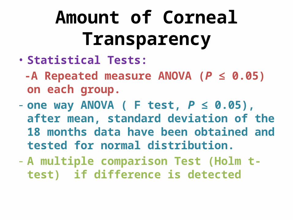
Amount of Corneal Transparency
• Statistical Tests: -A Repeated measure ANOVA (P ≤ 0.05) on each
group.- one way ANOVA ( F test, P ≤ 0.05), after mean,
standard deviation of the 18 months data have been obtained and tested for normal distribution.
- A multiple comparison Test (Holm t-test) if difference is detected

Amount of Strain of Cornea
• Create an Apparent stress (Mpa) vs. strain curve• Measure - The Ultimate Tensile Strength (UTS): Peak of curve - Modulus: Slope of the curve• Statistical Tests: -A two way ANOVA (two factors: UTS and Modulus) - A multiple comparison test (Fisher PLSD test, P ≤ 0.05 ) • Method taken from: Crabb RA, Chau EP, Evans MC, Barocas VH, Hubel A, (2006) Biomechanical and
Microstructural Characteristics of a Collagen Film-based Corneal Stroma Equivalent. Tissue Engineering1. 12, pg(1566-1571)