quality assurence and accountability in health: an experience from gujarat - dr j l meena
TRANSCRIPT
IIndian Journal of Community & Family Medicine | Vol. 1 | Issue 01 | Jan-June 2015
ISSN 2395 -2113 (Print)

Indian Journal of Community & Family Medicine | Vol. 1 | Issue 01 | Jan-June 2015
DisclaimerThe information and opinions presented in the Journal reflects the views of authors and not of the Journal or its Editorial Board or the Publisher. Publication does not constitute endorsement by the journal. Neither the Indian Journal of Community & Family Medicine nor its publishers nor any one else involved in creating, producing or delivering the Indian Journal of Community & Family Medicine or material contained therein, assumes any liability or responsibility for the accuracy, completeness, or the usefulness of any information provided in the Indian Journal of Community & Family Medicine, nor shall they be liable for any direct, indirect, incidental, special, consequential or punitive damage arising out of the use of Indian Journal of Community & Family Medicine. Indian Journal of Community & Family Medicine, nor its printer and distributor, nor any other party involved in the preparation of material contained in the Indian Journal of Community & Family Medicine represents or warrants that the information contained herein is in every respect accurate or complete, and they are not responsible for any errors or omissions or for the results obtained from the use of such material. Readers are encouraged to confirm the information contained herein with the other sources.
Editorial Office
Department of Community and Family Medicine All India Institute of Medical Sciences, Bhubaneswar, Odisha-751019
Under The Aegis ofMinistry of Health & Family Welfare
Government of India
Design & Printed by: Siva Prasad Patra (Binducreative, 09438668784)

Indian Journal of Community & Family Medicine | Vol. 1 | Issue 01 | Jan-June 2015
Editorial Advisers International
Hayato Uchida JapanJaideep Sood New ZealandDerek Hellenberg South AfricaRaj Bhopal UKAmit Arora UKSatish K Gupta UNICEFRichard Cash USAErich B Schneider USAKlea D. Bertakis USANeena Raina WHO
National
A K Mahapatra OdishaRajesh Kumar ChandigarhRakesh Sehgal ChandigarhB S Chavan ChandigarhC S Pandav DelhiS K Rasania DelhiSunil Gomber DelhiAdarsh Kumar DelhiPraveen Vashist DelhiJ L Meena GujaratD V Bala GujaratSunil Kumar GujaratGeeta Gathwala HaryanaS R Mazta Himachal PradeshPrahalad Kumar KarnatakaG Gururaj KarnatakaS Chhabbra MaharashtraB S Garg MaharashtraTh. Achouba Singh ManipurAnil Phukan MeghalayaGautam Roy PuducherryTejinder Singh PunjabA Muruganathan Tamil NaduSanjay Mehendale Tamil NaduKalpagam Polasa TelanganaV K Srivastava Uttar PradeshC M Pandey Uttar PradeshI S Gambhir Uttar PradeshC P Mishra Uttar PradeshG K Pandey West Bengal
Vol. 1, Issue 1, January-June 2015
Indian Journal of Community & Family Medicine
Editor- In - Chief Vikas Bhatia
PatronA K Mahapatra
EditorSonu H Subba
Editorial Board MembersSurekha Kishore Uttarakhand
Manisha Ruikar Chattisgarh
Sanjay K Rai Delhi
Arun M Kokane Madhya pradesh
Amit Dias Goa
Sitanshu S Kar Puducherry
P R Mohapatra Odisha
Swagata Tripathy Odisha
Subhashree Mahapatra France
Parvathy Nair USA
Shilpa Bhardwaj USA
Editorial TeamNeeraj Agarwal Deputy Editor
Pankaja R Raghav Deputy Editor
Abhiruchi Galhotra Associate Editor
Binod Kumar Patro Associate Editor
Surya Bali Associate Editor
Preetam B Mahajan Assistant Editor
Pankaj Bhardwaj Assistant Editor
Swayam Pragyan Parida Assistant Editor

Indian Journal of Community & Family Medicine | Vol. 1 | Issue 01 | Jan-June 2015
Dr. Vikas Bhatia is Dean of All India Institute of Medical Sciences, Bhubaneswar since August, 2012 which has been established by Ministry of Health & Family Welfare (MoH&FW), Govt. of India under Act of Parliament. He is also Professor and Head, Deptt. of Community and Family Medicine at AIIMS, Bhubaneswar and was entrusted with responsibility to start this Journal by MoH&FW, GOI.
He has experience of over 27 years in public health and has also worked as a family physician. During the mission to UNICEF for over 3 years as National Professional Officer/Health Officer, he made significant contribution with Govt. of Uttar Pradesh in Immunization, creating a network of Health & Nutrition Resource, Japanese vaccination drive, establishing SNCU, NRC, scaling up IMNCI, capacity building and others to strengthen maternal, child survival and development activities.
Dr. Bhatia has been awarded and honoured by UNICEF, MoH&FW, GOI and other organizations. With over 75 publications, authoring and technical advisor of 4 books and contributing 2 chapters, publishing 32 project reports/document, he has made enormous contributions in academics & public health.
Dr Sonu Hangma Subba is working as an Additional Professor in the Department of Community & Family Medicine at AIIMS Bhubaneswar. She has done her MBBS, MD and DNB from Lady Hardinge Medical College, Delhi. She is a GSMC-FAIMER fellow 2011 and had completed her PGD in Family Medicine from CMC Vellore. Her experience includes two and a half years in the polio eradication programme of the WHO and Govt of India. She has more than 30 publications and her interests are in medical education, epidemiology, non-communicable diseases and mental health.
Editorial Team & Board Members
Dr Vikas BhatiaEditor- In - Chief, IJCFM
Dr Sonu H Subba Editor, IJCFM

Indian Journal of Community & Family Medicine | Vol. 1 | Issue 01 | Jan-June 2015
Dr. Neeraj AgarwalDeputy Editor, IJCFMProfessor & HeadDeptt of Community and Family MedAll India Institute of Medical Sciences Patna, Bihar
Dr. Abhiruchi GalhotraAssociate Editor, IJCFMAdditional Professor Deptt of Community and Family MedAll India Institute of Medical Sciences-Raipur, Chhattisgarh
Dr. Surya BaliAssociate Editor, IJCFMAssociate Professor Deptt of Community and Family MedAll India Institute of Medical Sciences-Bhopal, Madhya Pradesh.
Dr. Pankaj BhardwajAssistant Editor Assistant ProfessorDeptt of Community and Family MedAll India Institute of Medical Sciences, Jodhpur, Rajasthan
Dr. Surekha KishoreProfessor & Head Deptt of Community and Family MedAll India Institute of Medical Sciences, Rishikesh, Uttaranchal
Dr. Pankaja Ravi RaghavDeputy Editor, IJCFMProfessor & Head Deptt of Community and Family MedAll India Institute of Medical Sciences-Jodhpur, Rajasthan
Dr. Binod Kumar PatroAssociate Editor, IJCFMAssociate Professor Deptt of Community and Family MedAll India Institute of Medical Sciences-Bhubaneswar, Odisha
Dr. Preetam B MahajanAssistant Editor Assistant ProfessorDeptt of Community and Family MedAll India Institute of Medical Sciences-Bhubaneswar, Odisha
Dr. Swayam Pragyan Parida Assistant Editor Assistant ProfessorDeptt of Community and Family MedAll India Institute of Medical Sciences, Bhubaneswar, Odisha
Dr. Manisha RuikarProfessor & Head Deptt of Community and Family MedAll India Institute of Medical Sciences, Raipur, Chhattisgarh
Editorial Team & Board Members

Indian Journal of Community & Family Medicine | Vol. 1 | Issue 01 | Jan-June 2015
Dr. Sanjay K. RaiAdditional Professor Centre for Community Medicine All India Institute of Medical Sciences, New Delhi.
Dr. Amit Dias Assistant ProfessorDepartment of Preventive & Social MedicineGoa Medical College, Goa
Dr. Prashant R Mohapatra Professor and Head Department of Pulmonary MedicineAll India Institute of Medical Sciences-Bhubaneswar, Odisha
Dr. Subhashree MahapatraResearch Scientist, Biotechnology Paris, France
Dr. Shilpa BhardwajDirector, Preventive Medicine, Clinical Assistant Professor of Internal Medicine.Moses H Cone Memorial Hospital, Greensboro, North Carolina, USA.
Dr. Arun M Kokane Additional Professor Deptt of Community and Family MedAll India Institute of Medical Sciences-Bhopal, Madhya Pradesh
Dr. Sitanshu S Kar Associate Professor Deptt of Preventive & Social MedicineJIPMER, Puducherry
Dr. Swagata TripathyAssistant Professor, Dept. of Trauma & Emergency Med, All India Institute of Medical Sciences, Bhubaneswar, Odisha
Dr. Parvathy NairClinical fellowChild and Adolescent Psychiatry Johns Hopkins Hospitals, Baltimore, Maryland, U.S.A
Editorial Team & Board Members

Indian Journal of Community & Family Medicine | Vol. 1 | Issue 01 | Jan-June 2015
Dr. Hayato UchidaProfessor of Public Health, Growth and Aging,School of Human Science and Environment, University of Hyogo, Japan
Dr. Derek Hellenberg Associate Professor and HeadDivision of Family Medicine, University of Cape TownSouth Africa
Dr. Amit Arora Consultant Physician and GeriatricianUniversity Hospital of North Staffordshire, U. K
Dr. Richard Cash Visiting Professor (Public Health) and Advisor in Global Health, PHFI, Delhi & Department of Global Health and Population, Harvard School of Public Health, Boston, U.S.A
Dr. Klea D. BertakisProfessor and ChairDeptt of Community and Family MedFounding Director, Center for Healthcare Policy and ResearchUniversity of California, DavisSacramento, CA, USA
Dr. Jaideep Sood Consultant Respiratory Physician Waitemata District Health Board (DHB) Auckland, New Zealand
Dr. Raj BhopalBruce and John Usher Professor of Public Health, Edinburgh University,Scotland, UK
Dr. Satish Kumar Gupta Health Specialist, UNICEF India Country Office, New Delhi
Eric B. SchneiderAssistant Professor, Surgery, Johns Hopkins School of Medicine& Assistant Professor, Epidemiology, Johns Hopkins Bloomberg School of Public Health, Baltimore, U.S.A
Dr. Neena Raina Regional Adviser, Child and Adolescent HealthWorld Health Organization - SEARONew Delhi
Editorial AdvisersInternational
Indian Journal of Community & Family Medicine | Vol. 1 | Issue 01 | Jan-June 2015
Dr. Sanjeev Kumar Rasania Professor & HeadDept. of Community Medicine,VM Medical College & Safdarjung Hospital, New Delhi
Dr. B S Chavan Professor & Head,Department Of Psychiatry, GMCH, Chandigarh
Dr. Adarsh Kumar Additional Professor Department of Forensic Medicine & Toxicology All India Institute of Medical Sciences, New Delhi
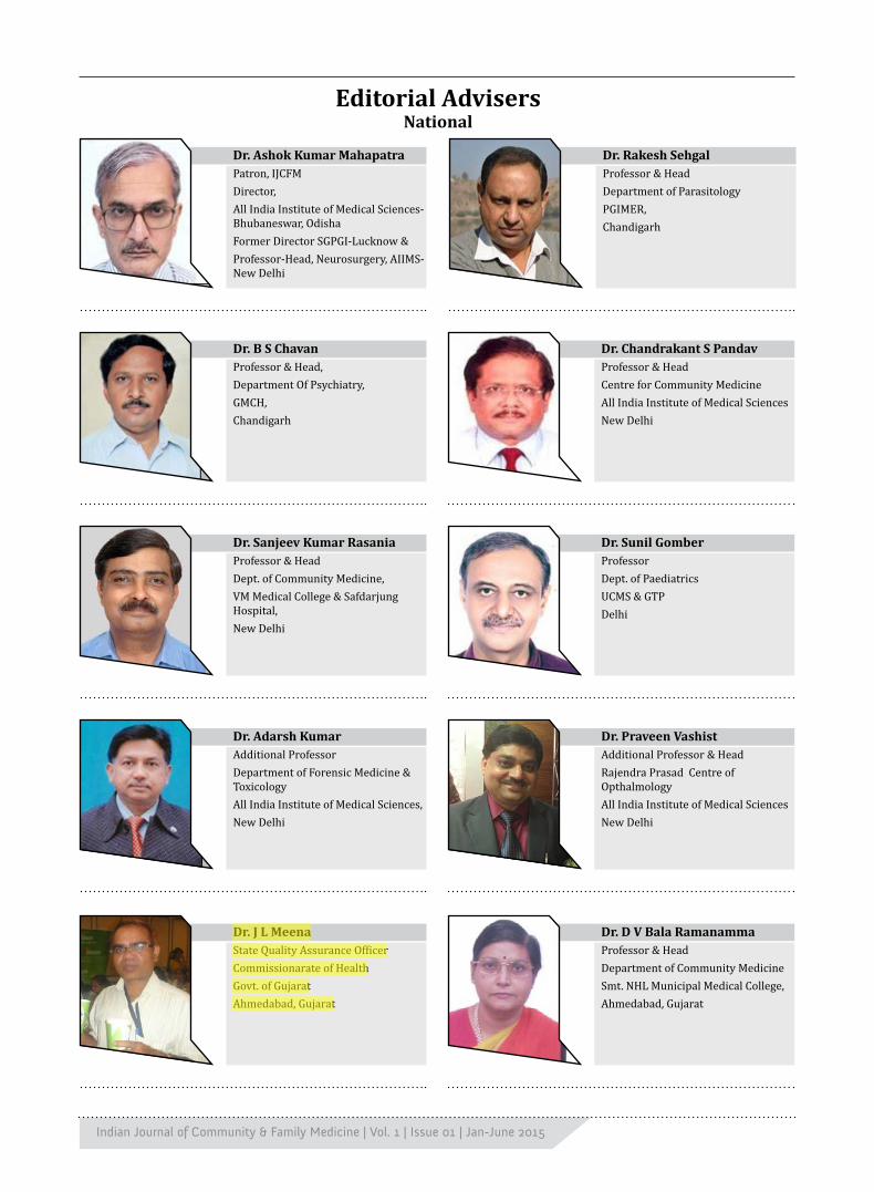
Dr. J L Meena State Quality Assurance OfficerCommissionarate of HealthGovt. of Gujarat Ahmedabad, Gujarat
Dr. Sunil Gomber Professor Dept. of PaediatricsUCMS & GTPDelhi
Dr. Chandrakant S Pandav Professor & HeadCentre for Community MedicineAll India Institute of Medical SciencesNew Delhi
Dr. Praveen Vashist Additional Professor & HeadRajendra Prasad Centre of OpthalmologyAll India Institute of Medical SciencesNew Delhi
Dr. D V Bala RamanammaProfessor & Head Department of Community MedicineSmt. NHL Municipal Medical College,Ahmedabad, Gujarat
Editorial AdvisersNational
Dr. Ashok Kumar MahapatraPatron, IJCFMDirector, All India Institute of Medical Sciences-Bhubaneswar, OdishaFormer Director SGPGI-Lucknow &Professor-Head, Neurosurgery, AIIMS-New Delhi
Dr. Rakesh Sehgal Professor & HeadDepartment of ParasitologyPGIMER, Chandigarh

Indian Journal of Community & Family Medicine | Vol. 1 | Issue 01 | Jan-June 2015
Editorial AdvisersNational
Dr. Sunil Kumar Director In-charge, National Institute of Occupational Health,Ahmedabad, Gujarat
Dr. G Gururaj Professor and Head Department of Epidemiology, National Institute of Mental Health and Neuro Sciences, Bangalore, Karnataka
Dr. S R Matza Professor and Head Dept. of Community Medicine, Indira Gandhi Medical College, Shimla, Himachal Pradesh
Dr. B S Garg Director, Dr. Sushila Nayar School of Public Health & Director- Professor of Community Medicine, Sevagram, Former Dean, MGIMSWardha, Maharashtra
Dr. Anil C Phukan MS, Professor & HeadDepartment of Microbiology,NEIGRIHMS, Shillong, Meghalaya
Dr. Geeta Gathwala Professor and Head Neonatal Services, Department of Pediatrics, Post Graduate Institute of Medical Sciences, Rohtak, Haryana
Dr. Shakuntala Chhabbra Director-Professor Dept. of Obstetrics & Gynaecology, MGIMS, Sewagram, Wardha, Maharashtra
Dr. Prahalad Kumar DirectorNational Tuberculosis InstituteBangalore, Karnataka
Dr. Th. Achouba Singh Professor & HeadDepartment of Community MedicineRegional Institute of Medical Science, Manipur
Dr. Gautam Roy Professor & Head Deptt of Preventive and Social Med,JIPMER, Puducherry
Indian Journal of Community & Family Medicine | Vol. 1 | Issue 01 | Jan-June 2015
Dr. Sanjay M. MehendaleDirector and Scientist G, National Institute of Epidemiology, Indian Council of Medical Research, Chennai, Tamil Nadu
Dr. V K Srivastava Professor & HeadDeptt. of Community MedicineHind Institute Medical SciencesBarabanki, UttarpradeshRegional Councillor International Epidemiological Association South East Asia Region
Dr. I S Gambhir Professor & HeadDepartment of GeriatricsInstitute of Medical Sciences, Banaras Hindu University, Uttar Pradesh
Dr. G K Pandey DirectorAll India Institute of Hygiene & Public HealthKolkata, West Bengal.
Dr. Tejinder Singh Professor of Pediatrics and Medical Education, Vice Principal, Christian Medical College,
Ludhiana, Punjab.
Dr. A. MuruganathanAdjunct Professor, the TN Dr MGR Medical universityPresident, Association of Physicians of India (2013-2014)Chennai, Tamilnadu
Dr. C M Pandey Professor & Head,Department of Biostatistics and Health Informatics, SGPGI Lucknow., Uttar Pradesh
Dr. C P Mishra Professor & HeadDepartment of Community Medicine,Institute of Medical Sciences, Banaras Hindu University, Varanasi, Uttar Pradesh
Dr. Kalpagam PolasaDirector In-charge, National Institute of Nutrition,Hyderabad, Telangana
Editorial AdvisersNational

Indian Journal of Community & Family Medicine | Vol. 1 | Issue 01 | Jan-June 2015
CONTENT
01 A new journey to reach community as one family with one goal V Bhatia
03 Research, publication and their relevance AK Mahapatra, BK Patro
06 Community Medicine, Family Medicine, and Public Health: The way forward R Kumar
09 Quality assurance & accountability in health; an experience from Gujarat JL Meena
13 Yellow fever vaccination: should India have more centres? V Bhatia, SP Parida
17 Neonatal care in India G Gathwala
23 ECG in coronary artery disease - basics M Chenniappan
32 Community screening for preventing cancer cervix G Dorairajan
36 Handling medicolegal cases at sub-district level A Kumar
40 Prevalence of selective non-communicable diseases and their risk factors among postmenopausal women residing in slum areas of Bhubaneswar city, Odisha SC Das, BC Das, Irfana, S Ray, Surendra, Prabhu
45 Outbreak of waterborne hepatitis E, Pune, Maharashtra, India, 2013 S Dhayarkar, M Chadha, A Tripathy, S Jadhav, N Deshmukh, S Mehendale
51 Out come of pregnancy among HIV infected women in a tertiary care center, Hubli, Karnataka, India AL Kumar, DD Bant, S Sheethal
55 A case of leptospirosis reported from metropolitan city of central India D Pandey, S Saroshe, S Dixit, C Jain
58 Medical education reforms in independent India T Singh, Anshu
62 Introduction of four new vaccines: boon to the Universal Immunization Program (UIP) in India SK Gupta
65 Influenza A(H1N1) in India – an update S Puri, V Saxena, R Singh
69 Depression among medical students in Goa S Rai, A Dias
73 An ANM’s success story from Bihar SH Subba, SS Ray
74 Effectiveness of learning intervention on basic life support and cardio pulmonary resuscitation among school children in a village of West Bengal A Dasgupta, S Banerjee, S Goswami, N Karmakar, A Das, S Das
Editorial
Perspective
Review Article
CME
Original Article
ShortCommunication
Case Report
Commentary
Update
Student/Resident Corner
Success Story
Letter to Editor

Indian Journal of Community & Family Medicine | Vol. 1 | Issue 01 | Jan-June 2015
1Indian Journal of Community & Family Medicine | Vol. 1 | Issue 01 | Jan-June 2015
First of all my heartfelt good wishes to all contributors, authors, advisory board members, team members and supporters in bringing out the Inaugural issue of Indian Journal of Community and Family Medicine. It is my deep desire that every effort must be made to live upto the expectations and maintain high standards of the journal. Thank You Team IJCFM.
While writing an editorial for the first issue, I thought to share the ideas and processes involved in making of the journal, in sharing the views about the future issues that are likely to come and how the medical fraternity is likely to be benefitted.
Ministry of Health & Family Welfare (MOH&FW), Government of India established six new All India Institute of Medical Sciences (AIIMS) under the Pradhan Mantri Swasthya Suraksha Yojna (PMSSY) by the Act of Parliament with the aim of correcting regional imbalances in quality tertiary level healthcare in the country and attaining self-sufficiency in graduate and postgraduate medical education and training. Therefore, six new AIIMS institutions became operational in Bhubaneswar (Odisha), Jodhpur (Rajasthan), Bhopal (Madhya Pradesh), Raipur (Chattisgarh), Rishikesh (Uttaranchal) & Patna (Bihar) in August 2012 with the beginning of MBBS course.1
Among various departments planned in these institutes, Department of Community and Family Medicine was also created to impart teaching to medical and nursing students as well as providing medical and health care to the community. Training of medical students at graduate and postgraduate level must inculcate the desired skills so that patients can be diagnosed and given appropriate medical care. In order to reduce the burden on specialists and ensuring the quality care close to doorsteps of community, doctors must be appropriately trained with availability of infrastructure. Deptt. of Community and Family Medicine have to play the important role. It is also expected that residents and faculty members must undertake research
focusing on certain diseases which are highly prevalent in geographical or are relevant in geographical locations of their operation.
Different nomenclatures of the department in various medical colleges and institutes in India such as Preventive & Social Medicine (P&SM), Community Medicine and Public health are being used until the recommendation of the Medical Council of India (MCI) instructed to use Community Medicine. Here at six new AIIMS, the department has been named as Community Medicine and Family Medicine. Does changing the name or bringing a new nomenclature is enough to bring the desired change in the medical education which finally has to address the challenges and medical needs of the community? Can’t we ensure to provide appropriate training and impart skills to develop family physician at graduate, postgraduate level and even at faculty level to match the health care needs of the community? Family Medicine came to be recognized as a medical specialty in India only in 1990s.2 Certain institutes in India such as AIIMS Delhi & MGIMS, Sevagram have a strong training program of family medicine and clinical exposure component in the departments of Community Medicine. Many other medical colleges/ institutes are also imparting quality medical care through their urban and rural health centers.
In view of the above dilemma, AIIMS, Bhubaneswar organized a two days workshop in January 2013 with active participation of faculty members from newly established AIIMS to deliberate on curriculum and giving a direction to the departments of Community Medicine and Family Medicine. Among important suggestions at the workshop, it was agreed to strengthen the family medicine component in all these newly established AIIMS. It was also deliberated that a scientific journal in Community and Family Medicine can be started to disseminate the knowledge and research among different Institutes of National Importance (INIs) in this specialty.
EDITORIAL
Address for Correspondence:Dr Vikas Bhatia, Dean, Prof & Head, Deptt. of Community and Family Medicine, All India Institute of Medical Sciences, Bhubaneswar- 751019, Odisha, India, Email : [email protected], [email protected]
A new journey to reach community as one family with one goal
Vikas Bhatia, Editor-in-chief, IJCFMDean, Prof & Head, Deptt. of Community and Family Medicine, All India Institute of Medical Sciences, Bhubaneswar- 751019, Odisha, India.

2Indian Journal of Community & Family Medicine | Vol. 1 | Issue 01 | Jan-June 2015
Efforts were further intensified to bring uniformity and under leadership of PMSSY, MOHFW, GOI, two days “Conclave on Community and Family Medicine in Institutes of National Importance with special emphasis on new AIIMS” was organized at NIHFW, New Delhi in December 2013. Directors of the Institutes and Invited experts from reputed Institutes focused on six areas and task forces were constituted. One of the key area was “Faculty development, including focus areas of research”. Twelve recommendations were made and one important recommendation was to publish a medical, scientific journal by the name of “Indian Journal of Community & Family Medicine” and AIIMS, Bhubaneswar was given the responsibility to initiate the process.3 Recommendations of the conclave on CFM were submitted and were approved by MoH&FW, GOI.
While discussing about the need of such a journal various factors and reasons were considered such as :
• To improve the knowledge and skills of family physicians among the young doctors and faculty of Community medicine.
• To enable the doctors working both in government and private sectors particularly in remote areas to upgrade their knowledge in public health and clinical areas. Many such doctors are not able to undertake full time courses. This journal will fill some gaps and can guide them.
• Journal will give a platform for sharing the research conducted in different INIs and other reputed medical institutes in the country which will encourage residents and faculty to publish quality research in areas of public health, clinical subjects, medical education etc. conducted at INIs. MCI and INIs require minimum number of publications to be made by each faculty member.4 Particularly, resident doctors keen to consider faculty position as their career can also take this as an opportunity.
• Majority of journals in India are charging processing and publication charges which at times is a discouraging factor. Potential authors can submit their research, give opinions & views and share success stories and good practices without any thought of being charged any money. Readers will be highly benefitted as the journal will be freely accessible on the websites. Efforts are also being made to create an APP for the journal, so that potential readers can access various articles on their mobiles, computers and tablets at any place in the country and abroad.
National Health Policy, 2002 also highlighted acute shortage of family medicine specialist in India, leading to
increased number of post graduate seats in “public health” or “family medicine” in the last few years.5
Thus, objectives of Indian Journal of Community and Family Medicine are:
1. To promulgate high quality research carried out in the institutes of national importance.
2. To provide a platform for disseminating information, ideas and innovative developments in the field of Family Medicine and Community Medicine.
3. To serve as an important and reliable source of information for the health professionals, decision makers as well as the general population.
4. To build a strong scientific base for both clinical and public health practices and policies.
Efforts have been made to bring highly qualified and reputed professionals in areas of their expertise in epidemiology, public health, infectious diseases, geriatrics, non-communicable disease, medical education, mental health, nutrition, occupational health, medicine, pediatrics, obstetrics & gynecology, emergency medicine, respiratory medicine, family medicine and many more to contribute and advice for the journal. Such reputed medical professionals of premier institutes from various states in India and continents around the world have been kind enough to work under one umbrella as one Family and agreed to share their expert advice to Community of doctors so that the people in the country can benefit to live a healthy life through quality Medical care as envisaged under new draft health policy document of Govt. Of India, 2015 with a commitment to universal coverage.6
This journal will provide a good opportunity for the researchers, academicians and family physicians to enhance their scientific knowledge. We all have one goal to be good physicians and give our best to the community and create a world free of pain, sorrows and diseases. Let’s play our role. Let’s commit to walk together on a new journey.
References1. The All India Institute of Medical Sciences (Amendment) Act,
2012, Pub. L.No. 37 of 2012. (Sep 12, 2012)2. Abraham S. Practicing and teaching family medicine in India.
Family Medicine 2007; 39(9): 671-2.3. National Institute of Health & family Welfare (NIHFW),
Government of India. Conclave on Community and Family Medicine in Institutes of National Importance with special emphasis on new AIIMS. Dec 2013.
4. Medical Council of India. Minimum qualification for teachers in medical institutions regulations, 1998 (amended up to Nov, 2010).
5. National Health policy 2002. Available from: http://mohfw.nic.in/np2002.htm.
6. National Health Policy 2015 Draft. Ministry of Health & Family Welfare, Government of India, New Delhi, December, 2014. Available at: http://www.mohfw.nic.in/showfile.php?lid=3014

3Indian Journal of Community & Family Medicine | Vol. 1 | Issue 01 | Jan-June 2015
PERSPECTIVE
Research, publication and their relevance
AK Mahapatra1, Binod Kumar Patro2
1 Director AIIMS, 2 Associate Professor, Department of Community & Family Medicine, All India Institute of Medical Sciences Bhubaneswar – 751019, Odisha, India.
Address for Correspondence: Dr AK Mahapatra, Director, All India Institute of Medical Sciences, Bhubaneswar – 751019, Odisha, India. Email: [email protected], Phone: +91-674-2467000
Research is an integral part of an academic career. In India, academic positions are evaluated by activities in three broad domains namely teaching, clinical care and research. Research domain gets high weightage in research institutions and institutes of national importance such as AIIMS, PGIMER, JIPMER and few other reputed institutions
Evaluation of teaching and clinical care domains are straight forward. Clinical care is being evaluated mostly by age old parameters such as no of clinics held, no of patients seen, no of operations/procedures performed etc. Although the indicators are straight forward it only captures quantitative information. No efforts are taken to capture qualitative information in clinical domains. In recent times, measures such as no of innovations/patents, no of new surgical/medical/diagnostic procedures developed are incorporated in annual performance appraisal report (APAR). Similarly evaluation of teaching domain involves looking at the quantitative information. Usually information such as total no. of classes/seminars/ tutorials taken, no. of MD/MS/PhD students enrolled, no of continued medical education (CME) credit points accumulated are assessed.
Research domain of academicians are evaluated by similar quantitative indicators such as no. of research projects completed/ ongoing, no. of funded research projects/ non-funded research projects, no. of publications in peer-reviewed journals, chapters in textbook, books and published.
In addition to the quantitative information, quality of research is evaluated on the quality of publication in scientific journals. The quality of journal publication are evaluated on certain screening tests such as peer-review, indexing etc. Publication in scientific journals passes the litmus test if the journal is peer-reviewed and indexed in standard medical/scientific database such as pubmed/ scopus/ovid and others.
Further, each journal competes with other to occupy the top space by being unique, most read, most cited and simply making the most impact. The journal Impact Factor is the single most important bibliometric indicator for its impact, which has its strengths and criticisms. The impact factor of a journal is the average number of citations received per article published in that journal, during the two preceding years. A journal’s impact factor is based on two elements: the numerator, which is the number of citations in the current year compared to items published in the previous two years, and the denominator, which is the number of substantive articles and reviews published in the same two years 1. Substantive articles usually include articles, reviews, proceedings, or notes but not editorials or letters-to-the-editor. For example, if a journal has an impact factor of 10 in 2015, then its papers published in 2013 and 2014 received 10 citations each on average in 2015. The impact
Table 1. Top International and National Medical Journals according to Impact Factor
International Journals
Impact Factor(2013)
Indian Journals Impact Factor(2013)
CA: A Cancer Journal for Clinicians
153.46 Indian Journal of Medical Research
2.06
New England Journal of Medicine
51.66 Indian Journal of Dermatology, Venereology and Leprology
1.21
Nature Reviews Genetics
41.06 Indian Journal of Cancer
1.13
Lancet 39.06 Indian Journal of Experimental Biology
1.20
Nature 38.58 Indian Paediatrics 1.04
Invited Article

4Indian Journal of Community & Family Medicine | Vol. 1 | Issue 01 | Jan-June 2015
factor of journals remains to be a contentious issue and has drawn criticisms from the scientific community regarding its use to evaluate research studies. It can be manipulated by changing editorial policies such as increasing the number of review articles or reducing the number of original articles, ahead of print publication (online first) or by encouraging authors for self citation2. Impact factor of individual journals are published time to time from the official website of Thomson Reuters. The top medical journals according to impact factors for the year of 2013 are presented in Table 1.
Individual researchers are evaluated on derived indicators such as average impact factor of the publications, average impact factor of top five/twenty publication, average impact factor of most recent five/twenty publications. However the above mentioned derived indicators can only compare contemporary academicians. Individual research outputs evaluated based upon general indicators such as number of publications do not weigh the impact of publications.
The h-index was proposed in the year 2005, by J.E. Hirsch, to overcome the shortcomings of the Impact Factor in evaluating the impact of an individual researcher’s research output. The h-index attempts to measure both the volume and impact of the published study of a scientist or scholar. Index h, is defined as the number of articles with ≥ h citations. A researcher has an index h if the h of his or her N p articles have been cited at least h number of times and the remaining (N p - h) articles have been cited ≤ h times. An h-index of 10 translates to a minimum of 10 research publications of a researcher having been cited at least 10 times. The h-index grows as the citations accumulate over a period of time, and thus, it depends on the ‘academic age’ of a researcher. Hirsch originally suggested for faculty of research universities that h ~ 12 might be a typical value for advancement to associate professor, and h ~ 18 might be typical value for advancement to full professor3.
The h-index can be manually calculated using citation databases. Subscription base databases, such as, Scopus and Web of Knowledge, provide automatic calculation of the h-index. Each database is likely to produce a different h value for the same researcher owing to different subscriptions (coverage of journals).
The h-index has its own share of limitations and criticisms. One of the major limitations of the h index is its insensitiveness to the small number studies, which are highly cited. Similarly, it does not take account of the large number articles, which have less than h citations. As the h-index is dependent on the number of publications, a junior researcher with a relatively short research career is likely to have a disappointingly low h-index despite
his innovations. The h-index also does not account for the positioning of the researchers in the list of authors 4. Similarly people working and publishing about rare topic shall be less citations and low h-index. The limitations of h-index are addressed with new indices such as h core index, i10 index, m index etc.
h core index measures the number of items that contribute to the h-index, should be used along with h-index to make more meaningful inference. Another similar indicator which is used alongside of h-index is i10-index which refers to total number of publications with at least 10 citations. The m-Index is the H-index divided by the number of years that a scientist has been active. Strengths and weaknesses of different bibliographic indicators are placed in Table 2.
Table 2. Strengths and Weaknesses of different bibliographic indicators
Indicator Strength WeaknessNo of Publication
Measures Quantity Silent of quality
Total no of Citations
Measure total impact
Influenced by small number of good publications
Citations per article
Good for comparison of researchers across age group
Penalizes high citations and rewards less citations.
Impact Factor Assigned to the Journal not to the author
Impact factor can be manipulated by editorial policies.
h Index Measure both Quantity and Quality
Penalizes high citationsInsensitive to the position of the author
h Core Index Rewards best articles
May not reflect true output for young researcher
i 10 index 10 is an arbitrary number
Young researcher may not find favourable indices
m index Combined measure of h index and years of active research
Disadvantage for infrequent publication from researcher.
In summary, a research is considered to be complete if it has translated into one or often more than one publication in a peer-reviewed indexed journal which has been cited by people working on the same topic. Bibliometric indicators are in place to help in evaluating individuals for coveted positions and career progression. Due attention should be provided on the strengths and weaknesses of each bibliometric indicators for use in academic set up. Decisions should be based on a combination of indicators rather than a single indicator.
Mahapatra AK: Research, Publication and their relevance

5Indian Journal of Community & Family Medicine | Vol. 1 | Issue 01 | Jan-June 2015
References1. Garfield E. The history and meaning of the journal impact factor.
JAMA 2006;295:90-3.2. Seglen PO. Why the impact factor of journals should not be used
for evaluating research. BMJ 1997;314:498-502.
3. Hirsch JE. An index to quantify an individual’s scientific research output. Proc Natl Acad Sci U S A 2005;102:16569-72.
4. Patro BK, Aggarwal AK. How honest is the h-index in measuring individual research output?. J Postgrad Med 2011;57:264-5.
Mahapatra AK: Research, Publication and their relevance
Mahatma Gandhi communicated a quintessential message to the nation through his efforts to educate people around him about cleanliness. He wished to see a “Clean India” where people work hand in hand to make the country clean. To work seriously towards this vision of Gandhiji, Prime Minister Shri Narendra Modi -launched it on October 2, 2014 and asked people from all walks of life to help in successful implementation of this mission. Swachh Bharat Abhiyaan- exhorts people to devote 100 hours every year towards the cause of cleanliness. (http://india.gov.in/spotlight/swachh-bharat-abhiyaan-ek-kadam-swachhata-ki-ore)

6Indian Journal of Community & Family Medicine | Vol. 1 | Issue 01 | Jan-June 2015
Invited Article
PERSPECTIVE
Community Medicine, Family Medicine, and Public Health: The way forward
Rajesh KumarProfessor of Community Medicine & Head, School of Public Health, Post Graduate Institute of Medical Education and Research, Chandigarh, India.
Address for Correspondence: Dr. Rajesh Kumar, Professor of Community Medicine & Head, School of Public Health, Post Graduate Institute of Medical Education and Research, Chandigarh 160012. Email: [email protected].
Traditionally, medicine - the art of healing - had focused primarily on the diagnosis and treatment of sick individuals using medications and surgical procedures. However, advancements in the science and technology, during the last few centuries, have not only transformed it into the ‘science and art’ of restoring health but its scope has also been expanded to prevention & control of diseases and promotion of health. And over the years, unprecedented growth has also occurred in its specialties and super-specialties which now cover prevention, diagnosis, and treatment at cellular, organ, individual, family, community, society, and public level.
Historically, Public Health, Social Medicine, Preventive Medicine, Community Medicine, and Family Medicine have developed as distinct branches of medicine at various time periods to address the issues of health and disease more comprehensively. One of the common theme that runs through these branches is that disease and death (except in old age) is an abnormality which can be prevented by human agency.1 The agent (such as biological, physical, chemical, socio-economical), host (such as susceptibility, vulnerability, life styles), and the environment (such as air, water, food, shelter)– the Epidemiological Triad –provide a sound basis for understanding the process of disease causation, and the ‘Three Levels of Prevention’ form the core principles for their practice,2 that is, (i) Primary Prevention of disease through specific protection and health promotion, (ii) Secondary Prevention of disability and deaths through screening, early diagnosis and prompt treatment, and (iii) Tertiary Prevention of limiting the disability through rehabilitation. However, these specialties also have distinct features which vary according to the socio-political contexts in which these have evolved.
In India, Public Health and Family Medicine are emerging with a distinct non-clinical and clinical focus respectively. These two streams are the core components of Community Medicine which also incorporates the tenets of Social Medicine and Preventive Medicine. Establishment of
Public Health Schools and Community & Family Medicine Departments in several Institutes of National Importance (INI) has led to some contentious arguments. A closer look at the historical events is necessary to understand these developments.
In the British India, army and civil medical services had developed by early 19th century and in 1835 a Medical College was established in Bengal, but sanitary commissions were appointed during the second half of 19th century to coordinate and advise on the sanitary work, vital statistics, and vaccinations. Public Health Act was promulgated in Madras Presidency in 1939. Government of India had appointed Sanitary Commissioners in center and provinces.3 Medical Officer of Health were also appointed in Municipalities to carry out the sanitary and public health activities while Civil Surgeons were responsible for medical relief at civil dispensaries and hospitals. The Director General of Indian Medical Service, and the Commissioner of Public Health administered these two distinct services in British India. The All India Institute of Hygiene and Public Health was established in Kolkata in 1932 to augment the teaching and training capacity in public health and Diploma in Public Health (DPH) course was started in India. The study of hygiene was also introduced in the curriculum of Medical Colleges. However, in 1946, the landmark Bhore Committee Report recommended the integration of medical relief and sanitary/public health work at all levels of health administration.4 Later, in 1978, World Health Organization also advocated for the comprehensive approach in Primary Health Care.5
In pursuance of the Bhore Committee Report, the Director General of Health Services was assigned the responsibility for both medical and public health affairs. Consequently Medical Council of India mandated all medical colleges to introduce the specialty of Social and Preventive Medicine so that all physicians could become competent in carrying out both medical and public health work.6 Later, the specialty of Community Medicine was established to

7Indian Journal of Community & Family Medicine | Vol. 1 | Issue 01 | Jan-June 2015
Kumar R: Community Medicine, Family Medicine, and Public Health: The Way Forward
train community physicians who could deliver promotive, preventive, curative and rehabilitative services in the health centers. However, specialist positions were not sanctioned for a Community Physician at the Community Health Center (CHC). Although specialist positions were sanctioned in CHC for a Physician, Surgeon, Pediatrician and Obstetrician; however, these specialist positions largely remain vacant.
In theory, the integrated approach appears to be an efficient way of dealing with both medical and public health problems particularly in view of the shortage of medical professionals, but in practical terms this approach led to a decline in the public health skills and infrastructure, as most of the doctors considered medical/clinical function as their primary responsibility and public health work which was mostly non-clinical administrative in nature was left to the subordinate staff such as Sanitary Inspectors. In the absence of a public health cadre and lack of specific positions for Public Health Officers and Community Physicians in the health services, Public Health/ Social & Preventive Medicine/Community Medicine specialties generally remained restricted to academic activity.
With the declining trend of communicable disease epidemics, which used to rage during the colonial period, the rapid rise in population was perceived to be a bigger problem. Hence, the priority shifted from public health to population control. The remaining public health staff at the grass root level, i.e., Sanitary Inspectors and Vaccinators, etc. were converted into Health Assistants and Multi-Purpose Health Workers to involve them fully into family planning, immunization and other national programs. This thorough neglect of sanitation and public health attracted the attention of the nation when the resurgence of plague occurred in 1994. To reverse this situation Government of India highlighted in the National Health Policy 2002 that a large number of specialists should be trained in Public Health.7 World Health Organization also emphasized in Calcutta Declaration (1999), the need for development of Public Health as a distinct discipline.8
In view of the emerging challenges of public health, Post-Graduate Institute of Medical Education and Research(PGIMER) Chandigarh established a School of Public Health in 2004 and pioneered a multidisciplinary two-year Master of Public Health (MPH) program in India. After nearly a decade of successful training of public health professionals at Masters’ level, a three-year Bachelor of Public Health (BPH) course has also been planned. Several other institutions such as Jawaharlal Institute of Postgraduate Medical Education & Research (JIPMER) have also set up School of Public Health and MPH programs. These courses could provide necessary human resources for designing a Public Health Service.9 The Union Ministry of Health & Family Welfare has also supported
the establishment of a public health cadre in the National Health Policy 2015 Draft.10 The separation of public health and medical functions has also been advocated recently.11
In the integrated health administration, even medical functions have been compromised as General Duty Medical Officers and Specialist Doctors have to spend lot of their time away from the Health Centers/Hospitals for carrying out non-clinical administrative duties, hence, they are not able to provide quality medical care. A single Specialist (Physician/ Surgeon/ Pediatrician/ Obstetrician) at the CHC is not able to ensure regular delivery of specialist services, hence, multi-tasking is required at CHC level which can be met by deployment of about half a dozen Family Physicians rather than one specialist in each of the major specialties.
Family Physicians can play an important role in providing affordable Universal Health Care to people. A family doctor provides primary and continuing care to the entire family within the communities and coordinates comprehensive health care services with other specialists, as and when needed. Hence, there is a need to train doctors in Family Medicine. The new All India Institutes of Medical Sciences (AIIMS) have set up Department of Community & Family Medicine (CFM) which is expected to establish Family Medicine Residency Program in due course. A two-year Diploma in Family Medicine is already available in Christian Medical College Vellore and National Board of Examination has also approved Family Medicine Program in several private hospitals. The Family Medicine training includes management of emergencies, treatment of various medical, surgical, pediatric and obstetric problems, that is, the care of entire family in its environment, appropriate referrals, and follow up. According to the National Health Policy document, there is an acute shortage of specialists in Family Medicine.
In the current scenario, when India is facing double burden of communicable and non-communicable diseases, people are looking forward to have the ‘Right to Health and Healthcare’. A whole-of-government approach is needed to address the social determinants of health and provision of universal healthcare. Public Health needs to adopt socio-ecological approaches rather than depending only on bio-medical or techno-managerial methods and it should open itself to allied professionals. In addition to keeping a focus on social determinants, public health should also be concerned with the issues related to access to healthcare. On the other hand Medicine should adopt a more comprehensive approach by nurturing Family Medicine.
The focus of Community Medicine should be comprehensive Primary Health Care, and Community Physicians should also forge partnerships with Family Physicians and Public Health Officers to press for a comprehensive Public
8Indian Journal of Community & Family Medicine | Vol. 1 | Issue 01 | Jan-June 2015
References1. Terris M. Evolution of Public Health and Preventive Medicine
in the United States of America. American J Public Health. 1975;65:161-169.
2. Leavell HR, Clark EG. Preventive Medicine for the Doctor in His Community - An Epidemiologic Approach. New York: McGraw-Hill; 1958
3. Mushtaq MU. Public Health in British India: A Brief Account of the History of Medical Services and Disease Prevention in Colonial India. Indian J Community Med. 2009;34:6-14.
4. Report of the Health Survey and Development Committee. Calcutta: Government of India Press; 1946.http://www.nhp.gov.in/sites/default/files/pdf/Bhore_Committee_Report_VOL-1.pdf
5. Declaration of Alma-Ata International Conference on Primary Health Care, Alma-Ata, USSR, 6-12 September, 1978. http://www.who.int/publications/almaata_declaration_en.pdf
Kumar R: Community Medicine, Family Medicine, and Public Health: The Way Forward
Health Act which should incorporate ‘Right to Health and Healthcare’ and suitable organizational structure at center, state, district and sub-district level to manage both the sub-systems, that is, Public Health Administration and Medical Care Administration in a balanced manner. Few states such as Tamil Nadu provide a template for development of administrative structures for Public Health and Medical Care. Once sufficient health infrastructure for Public Health
and Medical Care is created, higher level of government investments, which are expected to flow to health sector according to the National Health Policy 2015 Draft10,can be utilized more efficiently, and it will be possible to achieve the goal of Universal Health Care in the faceable future.
6. Thakur HP, Pandit DD, Subramanian P. History of Preventive and Social Medicine in India. J Postgrad Med. 2001;47:283
7. National Health Policy-2002. Ministry of Health and Family Welfare, Government of India, New Delhi. http://mohfw.nic.in/showfile.php?lid=2325
8. World Health Organization. Public Health in South-East Asia in 21st Century. Report and the Recommendations of the Regional Conference, Calcutta, 22-24 November, 1999. http://whqlibdoc.who.int/searo/2000/SEA_HSD_235.pdf
9. Kumar R. Human Resources for Public Health Service. Indian J Community Med. 2007;32:1-2.
10. National Health Policy 2015 Draft. Ministry of Health & Family Welfare, Government of India, New Delhi, December, 2014.http://www.mohfw.nic.in/showfile.php?lid=3014
11. Ahmed F. An Alternative Model of Health Delivery System to Improve Public Health in India. Indian J Public Health. 2014;58:261-266.
9Indian Journal of Community & Family Medicine | Vol. 1 | Issue 01 | Jan-June 2015
Introduction
Gujarat is the first state in India to initiate quality improvements in the public healthcare facilities. The quality improvement aims to attain the vision of creating the network of finest public health care institutions in providing preventive, promotive, curative and rehabilitative health care services with the state of art technology, easy accessibility, affordability and quality. Gujarat is also the only state to set up the State & District Quality Assurance cell to bring productivity and effectiveness in health care delivery system. State Quality Improvement Programme of DoHFW, Government of Gujarat (GoG) proposes to develop and institutionalize the use of the field based, practical and feasible indicators in quality assessment transforming the existing supervision practices into a more standardized and structured process. A pool of health care professionals in the public health sector trained in the implementation of health care quality standards has been developed for the same. The basic principal is that any sustainable change in terms of institutionalization of Quality Assurance (QA) will come from within the system and not from outside. It is hoped that interventions from demand side (for example, community and individuals demanding better services) will also put pressure on the system to deliver quality services which will in turn give impetus for investing in QA.
The core objective of the Quality Assurance Cell (QAC) are to facilitate the improvement of systems and processes of service delivery in the healthcare facilities as per the standard technical protocol to meet the laid down standards (e.g. IPHS/ MCI/GOI guidelines) as appropriate; to establish & develop quality management systems at the hospital level, leading to enhancement in service quality and leading to Quality certifications by the Quality assurance cell; to implement & monitor quality of MCH services at health; and to undertake such other GOI / State initiatives entrusted with the QAC from time to time (e.g. Maternal Death Review, Mother Child Tracking System etc.).
Strategies
Target Population of State Quality Improvement Programme is rural and urban population covered by Public Healthcare Facilities. The Quality Improvement is brought out in stepwise process of quality assessment to identify the gaps in performance of healthcare facilities followed by quality improvement to reduce the identified performance gaps. Multipronged approach & step- wise strategies are used by State Quality Improvement Programme of Gujarat for Quality Improvement in the above facilities. The following processes are undertaken to attain the envisaged objectives.
Citizen Charter
Citizens charters were placed at accredited healthcare institutes (including Medical Colleges, District Hospitals, CHCs & PHCs) clearly displaying Vision, Mission & Objective elements, patient rights and responsibilities, services available, proper grievance redressal policies to handle the staff and patient grievance. Complaint boxes & books were made available. A committee also has been formed at all the levels to handle the grievance of the patient and staff; Employee satisfaction survey were done regularly. All statutory requirements were being fulfilled.
Adoption of the newer quality standards & national guidelines
Different available quality standards such as National Quality Standard for PHC, CHC & District Hospitals of Ministry of Health & Family Welfare (MoH&FW), Government of India, Indian Public Health Standards (IPHS), National Accreditation Board of Hospitals & Health care Providers (NABH), National Accreditation Board for testing & Calibration Laboratories (NABL) were adopted. As per the Operational guidelines as per National Health Standard for Quality Assurance state quality assurance committee and district Assurance committee, were put in place. One Monitoring officer at
PERSPECTIVE
Quality assurance & accountability in health:An experience from Gujarat
Jeetu Lal MeenaState Quality Assurance Medical Officer, Commissionarate of Health, Govt. of Gujarat, Gandhinagar-382010, India.
Address for Correspondence: Dr Jeetu Lal Meena, State Quality Assurance Medical Officer, Commissionerate of Health, Medical Services and Medical Education, Govt. Gujarat, Gandhinagar-382010. Email: [email protected] Received: 27.11.2014, Accepted: 15.12.2014

10Indian Journal of Community & Family Medicine | Vol. 1 | Issue 01 | Jan-June 2015
state and district level and accreditation fees as per the guidelines proposed. Implementation of Kaizen (originally Japanese management concept for gradual, continuous improvement) & 5 S in all healthcare facilities including administrative offices to ensure workplace safety, efficiency, cleanliness, increase quality and Create Quality Culture in the state are being followed.
Earmarking additional budget
Extra budgets (about Rs. 237 crores for 5 Years) as per requirement of NABH / NABL sanctioned from 13th finance commission, GoG and NHM.
Patient Safety
Mother & Family Friendly Hospital Initiatives where quality medical care is offered following evidence based protocols and check lists with special focus on women and children in a conducive hospital environment for beneficiaries to stay comfortably and service providers to practice the skills
Bio-Medical Waste Management
Essential equipments and infrastructure facilities in each health facility such as adequate no. of colour coded Biomedicalwaste (BMW) containers and bags as per BMW Guide line (Red, Yellow, Blue and Black); puncture proof Containers for sharps; mutilators (needle / syringe cutters); calibrated weighing machine for BMW; personal protected equipments like gloves, caps, masks, aprons & gum-boots etc; 1% fresh sodium hypochlorite or bleaching powder solution; BMW record register; mercury spill management kit; blood spill management kit; post exposure prophylaxis kit; BMW storage rooms with lock & key; different forms & formats (Needle Stick Injury & Annual Report etc.) were provided at all health facilities. Bio-medical waste was segregated and stored as per colour coded system by health facility followed by collection, transportation, treatment and final disposal by Centralized Biomedicalwaste Treatment Facility (CBWTF) as per MoU for the entire State.
Gujarat Pollution Control Board (GPCB) is the regulatory authority for the implementation of Bio Medical Waste (Management and Handling) Rules, 1998 for Gujarat State and is inspecting health facility as per their schedule and issuing notices for the non compliance of BMW Rules.
All the health care staff were trained regarding the BMW Rules
Radiation Safety
The state tied-up with Atomic Energy Regulatory Board (AERB), Mumbai to set up Directorate of Radiation Safety under AD (ME) to enforce regulations and guidelines
stipulated under rules 29, 30, 31 and 32 of the Radiation Protection Rules 1971. A Governing Body called the Radiation Safety Council was constituted by the Atomic Energy Regulatory Board to review periodically the working of the directorate. Only equipment types approved by the Atomic Energy Regulatory Board to be installed in the new installations & the old and unsafe equipment not satisfying minimum radiological safety requirements to be phased out. The layout of the installations was to be carried out under the guidance of specialists in radiological protection to ensure that all radiological safety requirements are satisfied in every installation from the very beginning. Similarly, training programme for technologists and radiologists were to be organized to ensure that they are informed about the safe practices.
Fire Safety
Detailed activity checklist were placed for full fire safety measure in all the government run hospitals. Fire safety committee was formed at all heath care facilities. Regular mock drills were conducted at individual health facilities.
Cleanliness Drives
Mahatma Gandhi Swachhta Mission was institutionalized. The mission had clearly laid responsibilities & timeline for implementation.
Sustainability
Quality assurance officers were appointed at State level, District level; Assistant Hospital Administrator (AHA) at each facility level. At facility level, Designated NABH Coordinators & NABL Directors were placed. New positions of approximately 2000 of clinicians, paramedical and other posts sanctioned by Government of Gujarat as
Table 1. Stakeholders Involvement in State Quality Improvement Programme (SQIP)
Stakeholders Type of Involvement
Staff Skill mapping & motivation
Patients Feedback & grievance redressal system linked to corrective actions
Panchayat Raj Institutes Members
Periodic quality checks & feedbacks
Third Parties Outsourcing for Hospital Waste & Human Resource (HR) Management
Quality Council of India (QCI)
Memorandum of Understanding (MOU) signed with Quality Council of India (QCI) on 7th July, 2007
Meena JL: Quality Assurance & Accountability in Health: An Experience from Gujarat
per requirement were recruited and placed. State level review committee was formed under the Chairmanship of Commissioner of Health, Medical Service & Medical

11Indian Journal of Community & Family Medicine | Vol. 1 | Issue 01 | Jan-June 2015
Table 2. Achievements & Trainings organized under SQIP
Indicator/Name of Scheme/ Component Performance (2013-14)
NABH/NABL Accredited Healthcare Facilities
29(DH-2; Medical college blood bank-5; Medical college laboratories-5; Mental hospitals-2; PHCs-12; CHC-1; NABL Food & Drug Laboratories-2
Family Friendly Hospital 28Total amount received by scrape disposal Rs. 405,75,382Total facilities having the fire safety facilities with regular mock drill 941Total facilities having GPCB certificate 1522Cleanliness drive in our healthcare institutes 1522Instrument & Equipment Audit 1522Status of certificate being obtained from AERB 19
Training No. of ParticipantsNABH lead assessor training for administrators, clinicians & staff nurses. 290
NABL Internal audit for administrators, clinicians & staff nurses. 238
QIP (BMW, Kaizen, HOPE, Radiation Safety) Training 3655
CQI Champions Training 3540
Training of trainers for BMW management 1531
Post Graduation Certificate Course in Quality Management and Accreditation of Health care Organization (PGQM & AHO) for Additional Directors, MS, CDMO, Clinicians & DQAMOs
212
ACLS & BLS Training 3-4 MOs & 3-4 Staff nurse in each institute for ACLS & Whole staff for BLS
Training on Codes like blue, red, pink Whole staff
Others: Disaster management (Fire & Non Fire emergencies); Sentinel events, new Infection control practices, new born care training, post mortem
Education and State Quality Assurance Officer as Member secretary. Similarly, District level review committee was formed under the chairmanship of collector of the district, district quality assurance officer as member secretary.
Stakeholders Involvement
Important stakeholders were involved in the whole process to sustain the quality improvement process. The activities undertaken to involve the different stakeholders are presented in Table 1.
Achievements
Various achievements under Stakeholders Involvement in State Quality Improvement Programme (SQIP) are highlighted in Table 2. Some facility specific changes due to Quality Improvement Initiatives in infrastructure & arrangements (Figure 1), electric safety (Figure 2), Rehabilitation of patients (Figure 3) are depicted here as an example of changes brought about by quality improvement programme.
The Programme has received many recognition in the form
of Awards from National/ International organizations including appreciation awarded by QCI to DoHFW, FICCI Health care Excellence Award to Dist Hospital Gandhinagar, PHC Gadboriad, Community Health Centre Bardoli, Super Specialist Spine Institute Ahmedabad.,Operational excellence award for Quality Improvement Programme etc.
Challenges
There are various bottlenecks in sustaining quality improvement efforts such as, huge gaps in human resource [24,054 out of 47,030 (51%) sanctioned posts are vacant across different health facilities in Gujarat]; lack of real time monitoring system for quality indicators; and financial crunches [125.76 out of 330.33 (38%) Lakh Rs. were sanctioned in NHM PIP 2014-15 for Quality Improvement].
Conclusion
The ultimate aim of GoG is to get all the 172 facilities (Medical Colleges, DH, Blood Banks, Laboratories, PHC & CHC) across the state to be compliant to national
Meena JL: Quality Assurance & Accountability in Health: An Experience from Gujarat

12Indian Journal of Community & Family Medicine | Vol. 1 | Issue 01 | Jan-June 2015
quality standards in phased manner. Presently, lessons learned from State Quality Improvement Programme for health facilities (5S Implementation) is being applied to administrative section of health deptt. as well in other 54 Deptts. of GoG.
Figure 1. Improvement in Infrastructure Figure 2. Electrical Safety
Figure 3. Rehabilitation of the patients at Mental Hospital, Ahmedabad
Meena JL: Quality Assurance & Accountability in Health: An Experience from Gujarat
Suggested Readings1. Indian Public Health Standards. www.nrhm.gov.in/nhm/nrhm.
guidelines/indian_public_health_standards.html.2. National Accreditation Board for Hospitals. www.nabh.com3. Organising for improvement.Kaizen Institute. www.kaizen.com4. 5-S Implementation Manual. www.lean.org/fusetalk/forum/
attachments.

13Indian Journal of Community & Family Medicine | Vol. 1 | Issue 01 | Jan-June 2015
Abstract
Yellow fever is endemic in rural areas of West Africa & South America and has never been reported in India. Except for the causative organism, all other factors responsible for transmission do exist in India. Case fatality rate is very high and there is no cure for Yellow Fever. Prevention through vaccination is the only available option to decrease morbidity & mortality. As per guidelines established by MoHFW, vaccination is mandatory for all travellers moving to or coming from YF endemic countries. Due to increase in international travel, a larger number of people from various parts of India require Yellow Fever vaccination and the demand will continue to increase in future. In India, there are only 27 locations in 10 states and 2 UTs which have the facility to vaccinate for Yellow Fever. People of many states have to cover a large distance for the vaccination. There is an urgent need to increase the number of sites. With the operationalization of various institutes under MoH&FW, there is an opportunity to create the network, closer to the potential travellers.
Key words: Yellow Fever, Vaccination, Facilities
Address for Correspondence: Dr. Vikas Bhatia, Professor & Head, Department of Community and Family Medicine, All India Institute of Medical Sciences-Bhubaneswar 751019, Odisha, Email: [email protected]
PERSPECTIVE
Yellow fever vaccination: should India have more centres?
Vikas Bhatia1, Swayam Pragyan Parida2
1Professor and Head, 2 Assistant Professor, Department of Community and Family Medicine, All India Institute of Medical Sciences-Bhubaneswar, Odisha.
Yellow fever is an acute viral haemorrhagic fever affecting humans and non-human primates and transmitted by Aedes & Haemagogus mosquitoes. The illness due to Yellow Fever (YF)is characterized by hepatic, renal, myocardial involvement; haemorrhage and high mortality. Majority of persons affected with Yellow Fever have no symptoms or mild illness. Out of all symptomatic, 15 - 20% persons progress to severe form with fever, jaundice, bleeding, shock & multi-organ failure. In 20-50% of persons with severe disease, YF is fatal. Treatment of Yellow Fever is completely symptomatic and prevention through vaccination is the only available option to decrease morbidity & mortality. Yellow Fever is endemic in Africa & South America, but it has the capability to cause widespread epidemic. Hence Yellow Fever is included in the list of notifiable diseases under International Health Regulation and is considered as a public health emergency of International concern.1,2
Epidemiology of Yellow Fever
It is endemic in rural areas of West Africa & South America with frequent outbreaks in East & Central Africa. Ethiopia has reported a large outbreak between 1960 to 1962 with 1,00,000 cases. Nigeria too reports many cases. In South America, Peru & Bolivia reported highest number of cases. Various studies indicate that YF is largely under-reported
due to poor surveillance in affected rural areas. As per WHO estimate worldwide 2,00,000 cases and 30, 000 deaths occur annually due to Yellow Fever. Out of all cases 87% are reported from Sub-Saharan Africa and the rest are from South America.
Gradually an increasing trend is being observed with frequent epidemics in a number of countries and there is expansion of endemic zone to newer urban territories too. The risk of epidemics looms mainly over densely populated poor urban areas where widespread A. aegypti breeding places are found. Owing to this trend, Yellow Fever is being considered as a Re-emerging disease of Public Health Importance.
A. aegypti is the most important vector responsible for disease transmission in urban areas because of its adaptability to human domestic environment and preferential day biting nature. Though transmission has never been seen in Asia & Australia, but because of high vector density and a large susceptible population, these areas are at risk of importation & the onset of an epidemic. Countries like India & Australia strictly require proof of Yellow Fever vaccination from travellers moving to or arriving from Yellow Fever endemic regions.1-3
Received: 20.01.2015, Accepted: 21.03.2015

14Indian Journal of Community & Family Medicine | Vol. 1 | Issue 01 | Jan-June 2015
Yellow Fever vaccine
Validity of Yellow Fever vaccine starts from the 10th day of receiving the vaccine and lasts for 10 years from the day of vaccination. Hence persons from non-endemic areas need a booster every 10 years. Passengers travelling to YF endemic countries are advised to take vaccination 10 days before arrival in those countries. Passengers arriving in India from other endemic countries within 10 days of YF vaccination will be quarantined for 6 days from the date & time of departure from the endemic regions. Yellow Fever vaccine was produced by CRI Kasauli. Since 2013, the vaccine production in India has stopped & Govt. of India is procuring YF Vaccine through WHO and supplying it to the vaccination centres as per the demand placed.(Fig1a)3,4
Table 1. Showing characteristics of YF vaccine
Type Live attenuated viral vaccine derived from 17 D strain
Nature Freeze dried lyophilized vaccine, diluted with NS
Storage Between 2-80 c
Schedule One dose for all persons aged 9 months and above
Booster Every 10 years for people living in non endemic regions
Dose Each constitutes 0.5 ml
Route & site Intra muscular /Subcutaneous, in deltoid or antero-lateral aspect of thigh
Efficacy More than 90% after 10 days
Protection Remains for 20-35 years & probably lifelong
C o n t ra i n d i c a -tions
Immunodeficiency states, HIV with CD4 count < 200, serious egg allergy, pregnancy & hypersensitivity to previous dose, age less than 6 months
Precautions Age 6-8 months, age above 60 years, pregnancy & breast feeding
Adverse events Anaphylaxis, encephalitis, neurological involvement, organ involvement, acute visceral disease
Recommendation for vaccination
Almost 50 countries in Africa & South America have included Yellow Fever vaccine in their National Immunization Schedule. Mass vaccination program used to be conducted in those countries during outbreak. Apart from that, travellers moving to or living in areas of YF infection should be vaccinated. Vaccination is also indicated for persons travelling to countries that do not officially report, but lie in the vicinity of yellow fever endemic countries. A traveller’s risk for acquiring YF is dependent upon factors such as immunization status, location of travel, occupational & recreational activities while travelling, local rate of YF virus transmission & duration of travel.5
Vaccination requirement before International travel are as follows
1. Yellow fever vaccine: Different manufacturers produce YF vaccines, but for certification of vaccination only WHO approved vaccines are considered.
2. Authorized vaccination centre: This is vital for administration of vaccine. Department of Health of respective countries has the authority to designate centres for YF vaccination.
3. Proof of vaccination: Vaccinee should produce an International certificate of vaccination that has been completed, signed and validated with the centre’s stamp at the time of administration (Fig1b).
As per WHO guidelines & International Health Regulations, any failure to complete any part of the certificate will render the YF vaccination certificate to be invalid. Only the original certificate of Yellow Fever vaccination is accepted as authentic & clearance is given during International travel.6-8
Yellow Fever in India
Yellow fever cases are not detected in India but factors are conducive because of the presence of abundant A. aegypti vector and a large susceptible population. Govt. of India has been maintaining strict vigil on international movement
Figure 1a. Yellow fever vaccine Figure1b. International certificate of vaccination
Bhatia V : Yellow fever vaccination: Should India have more centres?

15Indian Journal of Community & Family Medicine | Vol. 1 | Issue 01 | Jan-June 2015
of passengers to prevent entry of Yellow Fever virus. All passengers coming to or going from India to Yellow Fever endemic countries should have a valid International Yellow Fever card. The Directorate General of health Services & MoHFW have set up 27 vaccination centres all over the country (figure 3).9
Due to increase in international travel, more people in India requires Yellow Fever vaccination and the demand will continue to increase. People from states without the vaccination centres have to travel a long distance which may go up to hundreds of Kilometres or at times 2 to 3 days of travel time is needed to get vaccinated in approved centres located in other states. The vaccination days are fixed in all those centres. If a person misses fixed immunization days, that creates a lot of inconvenience to travellers coming from far off places. Rs. 300 is charged for a single dose of vaccination per passenger which includes cost towards vaccine, syringes, etc.3
Table 2. Showcasing states with & without YF vaccination sites
States with vaccination sites (No.)
States without authorized site
Delhi (6) J & K Arunachal Pradesh
West Bengal (3) Rajasthan Manipur
Andhra Pradesh (1) Uttaranchal Meghalaya
Kerala (1) Haryana Mizoram
Gujarat (5) Chandigarh Nagaland
Maharashtra (3) Bihar Sikkim
Goa (2) Jharkhand Tripura
Karnataka (1) Odisha Assam
Himachal Pradesh (1) Chhattisgarh Dadra & Nagar Haveli
Uttar Pradesh (1) Madhya Pradesh
Pondicherry
Telangana (1) Daman & Diu Lakshadweep
Tamil Nadu (2) A & N islands
A person from the North-Eastern region spends almost 3-4 days in travel to get vaccinated in the nearest centre in Kolkata, West Bengal. A person from J&K has to reach Chandigarh initially & then has to go to Kasauli, Himachal Pradesh for vaccination. A large region of India, comprising of Madhya Pradesh, Bihar, Chhattisgarh and Uttaranchal has no authorized centre. This leads to loss of valuable man-hours to get a single shot of Yellow Fever vaccine.
Most of the vaccination sites are established more or less over the coastal belt of India near major ports. This is insufficient in present times and more centres are required and ideal target should be to establish at least one centre in every state to provide hassle free services.
Reasons for establishing more centres are
1. International travel has increased manifold. Along with increased travel, more number of international airport & ports have come up.
2. Many Institutes have been created under MoH&FW. This provides more facilities and expert manpower to provide yellow fever vaccine related services.
3. Monitoring: there is expansion of YF endemic areas due to urbanization & industrialization. All determinants except YFV are present in the entire country. It requires a strict vigil on international travel and provision of Yellow Fever vaccine.
4. A potent vaccine is available.5. The established centres are not evenly distributed
throughout the country which creates a lot of inconvenience in respect to money, time & hardship to the travellers.
Figure 2. Map of India showing Yellow Fever Vaccination sites in India_________________________________________________
N.B.The dots denote areas where authorized vaccination centres are present. The figure in parentheses show number of approved sites in a state.
The basic prerequisites for establishing authorized centres5
1. Space and infrastructure for vaccination centre: This includes space for waiting area, injection room, space for cold chain & vaccine storage, observation room etc.
2. Equipment: Ice Lined Refrigerators are required for storing vaccines & diluents at 2-8 degrees centigrade.
Bhatia V : Yellow fever vaccination: Should India have more centres?

16Indian Journal of Community & Family Medicine | Vol. 1 | Issue 01 | Jan-June 2015
References 1. Yellow Fever. World Health Organization. Available from
http://www.who.int/csr/disease/yellowfev/en/ [accessed on 11/2/15]
2. Yellow Fever Vaccine for Adults. Mathai D, Vasanthan AG. Adult Immuization 2014.The association of Physicians of India.
3. MoHFW Guidelines for Yellow Fever vaccination. Available from http://www.mohfw.nic.in/showfile.php?lid=2783. [Accessed on 6/2/15]
4. Yellow fever vaccine & recommendations. Available from http://www.cdc.gov/yellowfever/vaccine/index.html [accessed on 6/2/15]
5. Requirement for use of a new international certificate for vaccination or prophylaxis for Yellow Fever vaccine.CDC MMWR weekly. Jan 4, 2008/56(51);1345-1346. Available from http://www.cdc.gov/mmwr/preview/mmwrhtml/mm5651a4.htm
6. World Health Organization. International health regulations (2005) second edition. Geneva, Switzerland: World Health Organization; 2005.Available at http://whqlibdoc.who.int/publications/2008/9789241580410_eng.pdf.
7. Health Regulations. Airport Authority of India. Available from http://www.airportsindia.org.in/immigration/health.jsp[accessed on 6/2/15]
8. MoHFW, GoI. Advisory for passengers coming and returning to India from Yellow Fever endemic countries. Available from http://mohfw.nic.in/WriteReadData/l892s/9642270354Advisory.pdf [accessed on 6/2/15]
9. Bureau of Immigration. Health Regulations. Available from http://boi.gov.in/content/health-regulation.[accessed on 6/2/15]
Bhatia V : Yellow fever vaccination: Should India have more centres?
3. Trained Manpower: Doctors, Staff Nurse support staff.
In last 3 years, six new All India Institute of Medical Sciences have been established by Govt. of India by an Act of Parliament with the objective to provide quality health services. These Institutions are situated in states where
there are no centres for Yellow Fever vaccination. In view of increased tourism and availability of more Institutes of National Importance, MoHFW, GoI may consider creating more centres in the interest of its citizens and it will further help to keep a strict vigil on entry of Yellow Fever into India.

17Indian Journal of Community & Family Medicine | Vol. 1 | Issue 01 | Jan-June 2015
Abstract
The last decade has witnessed momentous changes in the Neonatal health scenario in India. Newborn Health care has received unprecedented attention and resources. It is the current focus of Central and state governments and the various funding agencies. And, like never before, there is an opportunity for the champions of newborn health to take their agenda forward . India contributes to one-fifth of global live births and more than a quarter of neonatal deaths. About two-thirds of infant deaths and half of under-five child deaths are during the neonatal period. The neonatal mortality rate has reduced over the years but the decline is at a much slower pace as compared to deaths in the older infant groups. Among neonatal deaths, the rate of decline in early NMR is much lower than that of late NMR. The Millennium Development Goal-4 (MDG-4), which stipulates a two-thirds reduction in under-five deaths by 2015, obviously cannot be achieved without ensuring a substantial reduction in the neonatal mortality rate (NMR). This article reviews the current status of Neonatal Care in India.
Key words : Neonatal care, India, Neonatal mortality, Neonatal morbidity, Special newborn care unit
Address for Correspondence:Dr Geeta Gathwala, 6J/8, Medical Campus, Pt. Bhagwat Dayal Sharma PGIMS, Rohtak-124001, E-mail : [email protected]
Neonatal and child mortality rates in India
The last decade has witnessed momentous changes in the Neonatal health scenario in India. Newborn Health care has received unprecedented attention and resources. It is the current focus of Central and state governments and the various funding agencies. And, like never before, there is an opportunity for the champions of newborn health to take their agenda forward .
India contributes to one-fifth of global live births and more than a quarter of neonatal deaths (Fig 1). About two-thirds of infant deaths and half of under-five child deaths are during the neonatal period. The neonatal mortality rate has reduced over the years but the decline is at a much slower pace as compared to deaths in the older infant groups. Among neonatal deaths, the rate of decline in early NMR is much lower than that of late NMR.
The Millennium Development Goal-4 (MDG-4), which stipulates a two-thirds reduction in under-five deaths by 2015, obviously cannot be achieved without ensuring a substantial reduction in the neonatal mortality rate (NMR).
Table 1 provides the most recent estimates (2012) of the child and neonatal mortality rates in the country. With the current NMR of 29 per 1,000live births,2 about 70 percent of infant deaths and more than half of under-five child deaths in the country fall in the neonatal period (Figure 2). The early neonatal mortality rate (ENMR)- deaths in the first week of life - is 23 per 1,000 live births.2 This implies that the first week alone accounts for about 45 percent of total under-five child deaths.
Invited Article
REVIEW ARTICLE
Neonatal care in IndiaGeeta Gathwala
Professor and Head, Department of Pediatrics and Neonatal Services, Pt. Bhagwat Dayal Sharma Post Graduate Institute of Medical Sciences, Rohtak, Haryana, India
Table 1. Current Child and Neonatal Mortality Rates (2012)Under-five child mortality rate (U5MR) 52Infant mortality rate (IMR) 42Neonatal mortality rate (NMR) 29Early neonatal mortality rate (ENMR) 23Late neonatal mortality rate (LNMR) 6
Source: SRS Statistical Report, 20122
Note: Rates expressed per 1000 live births

18Indian Journal of Community & Family Medicine | Vol. 1 | Issue 01 | Jan-June 2015
Figure 3. Trends of NMR and IMR
Figure 4. Rural-urban NMR of India
Trends and projections of neonatal mortality
The NMR has declined from 44 per 1000 live births in 2000 to 29 per 1000 live births in 2012, but the rate of decline has been slow and lags behind that of infant mortality rate (IMR; deaths in the first year of life) (Figure 3). The average annual rate of reduction (AARR) of NMR was only modest, at around 3.5 percent in this period (Figure3). The rate of reduction was less than that of IMR during the same period (4.0 percent annual reduction). Among neonatal deaths, the rate of decline in the ENMR was much lower than in the late NMR - AARR of 2.8 percent and 5.8 percent respectively .
Neonatal mortality: Rural-urban, poor-rich differences
There are important rural-urban and socioeconomic differentials in the NMR. The NMR in rural areas is twice that in urban areas (33 vs. 16 per 1,000 live births, respectively Fig 4).2 Similarly, an analysis of the NFHS-3 (2005-2006) data shows that the NMR among the poorest 20 percent of the population is more than double that of the NMR of the richest 20 percent.3
Causes of neonatal mortality
A systematic analysis of global, regional, and national causes of child mortality in 2012 identified prematurity and infections to be the major causes of neonatal deaths in India.4 The review, which included data from the Million Death Study from India 5, found perinatal asphyxia and malformations to be the other two significant causes of neonatal mortality (Figure 5). These findings are similar to the overall global pattern.4
Timing of neonatal deaths
A pooled analysis of the data on the timing of neonatal deaths indicates that about three-fourths of all neonatal
Source: Child mortality report, 20131
Gathwala G: Neonatal Care in India
Figure 1. India’s contribution to global burden of neonatal deaths
Figure 2. Neonatal deaths as a percentage of infant and under-five deaths
Source: SRS Statistical Reports (2000-2012)
deaths occur in the first week of life( Fig 6). The first 24 hours account for more than one-third (36.9 percent) of the deaths that occur in the entire neonatal period.
Neonatal morbidities
Neonatal morbidities constitute a huge burden to the health system and society in general. The SEARCH study by Bang et al. (1995-96) provided a detailed insight into the burden of common morbidities in rural community settings.6 The most common morbidities identified in this study were breastfeeding problems (25.6 percent),
Infant Deaths Under - 5 Deaths
Rural Urban

19Indian Journal of Community & Family Medicine | Vol. 1 | Issue 01 | Jan-June 2015
umbilical sepsis (19.8 percent), neonatal sepsis/ pneumonia (18.0 percent) and hypothermia (17.0 percent).
Low Birth Weight (LBW) / preterm birth
Nearly 30% of neonates in India - 7.5 million - are born with a low birth weight (<2500 g).7 This accounts for 42% of the global burden, the largest for any country. About 60% of the LBW infants are born at term, but subjected to intrauterine growth restriction, while the remaining 40% are born preterm.8 LBW infants are also predisposed to a variety of adult onset diseases in later life because of the anomalous programming of affected fetuses.
Neonatal sepsis
Burden of neonatal sepsis in India is enormous - hospital-based studies suggest an incidence of 30 per 1000 live births,8 while community-based studies indicate an incidence of 2.7% to 17% of all live births.9 Nearly one-fifth of neonates with sepsis die in the hospital; the figure rises to 50% for those with culture-proven sepsis. They are also at a high risk of major neuro-developmental disabilities at a later age.
Perinatal asphyxia
Perinatal asphyxia not only leads to neonatal deaths but also accounts for a significant proportion of still-births. The reported incidence varies from 2% to 16.2% in community-based studies,10 with the case fatality rates ranging from 38.5% to 74%.
Coverage of key interventions during antenatal, natal and postneonatal periods
Figure 7 depicts the coverage of key interventions in antenatal, natal, and postnatal periods, and during infancy (Coverage Evaluation Survey, 2009).11 Only a quarter of pregnant women have full antenatal check-up, i.e., 3 or more antenatal checkups, at least 1 tetanus toxoid injection and at least 100 (>100) iron and folic acid tablets. Seventy
Figure 5. Causes of neonatal deaths
three percent have institutional deliveries, and more than three - fourths are attended by a skilled birth attendant during delivery. Only one third of neonates are breastfed within one hour after of birth.11 Less than half of the infants received 3 postnatal visits from health care providers in the first 10 days of life. Exclusive breastfeeding rate reduces to about one third by six months of age.
Determinants of Newborn Health In India
The mother’s status is central to neonatal health. Health, nutrition, education and empowerment of women are long term goals which can improve neonatal outcomes substantially.
Social and economic factors can negatively affect neonatal health through proximate determinants, particularly poverty, rural location, social groups, and gender inequity.There is a significant variation in neonatal mortality among states and across different districts within states..12, 13 Coverage of maternal and newborn services varies considerably within states and across districts; marginalized communities and families belonging to scheduled castes and tribes have lower access to services.Women’s education plays an important role in improving neonatal health outcomes.14,15 Childbirth before the maternal age of 20 years is associated with a high newborn mortality.A birth interval of less than 24 months increases the risk of neonatal mortality substantially; thus, family planning has huge neonatal survival benefits.16,17Care seeking for female newborns is often compromised as compared to that for males (Fig 8).
National Programmes For Neonatal Health
With the launch of the National Rural Health Mission (NRHM) in 2005, newborn healthcare has received unprecedented attention and resources.
The country launched several new initiatives to boost maternal-neonatal care under NRHM. The Janani Suraksha Yojana (JSY) provides for cash incentive for childbirth in facilities.18 The Facility – Based Newborn Care (FBNC)
Gathwala G: Neonatal Care in India
Figure 6. Distribution of deaths (week-wise)
Source: Liu L 2012 4
Others
Malformations
Birthasphyxia
Preterm
Sepsis
Pneumonia
Infection33%
Diarrhea

20Indian Journal of Community & Family Medicine | Vol. 1 | Issue 01 | Jan-June 2015
refers to clinical services provided by skilled personnel at health facilities round the clock. 19, 20 It aims to build a three-tier system of neonatal care encompassing the Primary Health Centers (PHCs), the Community Health Centers (CHCs) and the District Hospitals.
Community based interventions have been shown to improve neonatal outcomes.21-23 The Home-based Newborn Care (HBNC) programme envisages home visiting by the Accredited Social Health Activists (ASHAs) for neonatal examination, sickness detection and family counselling. Data from 1,475 live births showed that HBNC resulted in a 51% decline in NMR.24 The Navjat Shishu Suraksha Karyakram (NSSK) aims at provision of essential newborn care including resuscitation in facilities at all delivery points. The Janani Shishu Suraksha Karyakram (JSSK) goes a step further and entitles mothers and infants to free transportation, care, medicines, and diagnostics in all public facilities.
As a part of the recently instituted Rashtriya Bal Suraksha Karyakram (RBSK), the focus has been placed on screening infants for selected birth defects and developmental delays.
Checklist based interventions aid management of complex or neglected tasks and have been shown to improve performance of health systems. Supervision and monitoring of health services in a pilot study from Karnataka have been observed to markedly improve the delivery of health care practices.25
The health system development has seen a transformational change in these years in increasing access of health services to the poor. More than 9,00,000 village level ASHA workers, 33,000 nurses, 14,000 paramedics, 8,000 doctors and 3,000 specialists have been inducted; more than 20,000 facilities have been constructed; and over 20,000 ambulances deployed. Medical graduate seats have increased by 55 per cent and postgraduate seats by 75 per cent. More than 500 000 village health
Source: Coverage Evaluation Survey 2009 19
Figure 7. Coverage of key interventions in antenatal, natal, and postneonatal periods
Source: SNCU online data: MP, Maharashtra and Haryana, 2013 40
Figure 8. More male admissions to SNCUs resulting from gender bias in care seeking
and sanitation committees have been formed to catalyse community participation.
The country is presently saving additional 440 000 neonatal lives annually compared to the year 2000. The JSY has ensured that an additional 12 to 13 million women are now delivering in government facilities each year with institutional delivery rate touching 80%. More than 0.6 million newborn babies, mostly from poor families, are receiving care in the newly created Special Newborn Care Units (SNCUs) in the district hospitals across the nation each year.
The national government is committed to a National Health Assurance Mission that ensures accessible, affordable and effective universal healthcare. Maternal, newborn and child healthcare would be at the core of this Mission. We need to make sure that no woman, no neonate and no child should be left behind as the country surges ahead in development and welfare. Inequity, whether geographic, or of class or gender, is unacceptable. Removing inequities would require addressing the key social determinants, mounting a behaviour change movement on scale and ensuring access to quality services by every mother, every baby in every part of the country. The goal of averting every preventable newborn death would require not only further acceleration of present initiatives, but also implementation of new approaches and programmatic innovations.
Facility-Based Newborn care
Facility-based newborn care (FBNC) refers to clinical services provided by skilled personnel at health facilities round-the-clock.19,20 Facility-based newborn care (FBNC) in the public health system got a boost under NRHM with a nationwide creation of Newborn care corners (NBCC) at every point of child birth, Newborn stabilization Units (NBSUs) at community Health centres, and special Newborn care Units (SNCUs) at district hospitals. The number of SNCUs, NBSUs and NBCCs has grown at a
Gathwala G: Neonatal Care in India
21Indian Journal of Community & Family Medicine | Vol. 1 | Issue 01 | Jan-June 2015
very rapid pace, however the quality of care is variable. Guidelines and protocols on FBNC are currently in place.26 The linkages of SNCUs with NBSUs and NBCCs are weak.NBSUs have not received the required attention and remain a weak link in most districts. Shortage of doctors and absence of mechanisms for timely repair of equipment are common challenges. Establishment of sustainable quality assurance systems for FBNC in all states is needed to ensure high standards of care.
Newborn Health in Urban India
National health survey data show that overall neonatal mortality rates are lower in urban than in rural areas, but the figures mask substantial urban inequalities.The most recent estimates for 2011- 2012, based on modified methods, put 14 percent of urban people below a poverty line of Rs 1,000 per person per month compared with 26 percent of rural people, at a poverty line of Rs 816.27, 28
Within the National Health Mission, the National Urban Health Mission provides an opportunity for strategic thinking and actions to improve urban newborn health. The reproductive, Maternal, Newborn and child Health + adolescent initiative adds to this an opportunity to integrate healthcare over the life cycle. A number of pilot initiatives have begun, including private-public partnerships, community action models, demand-side financing and insurance, but evaluation has been limited. We need to develop models of newborn care for urban populations with focus on the poor within the NUHM framework. Convergence mechanisms of public, private, and third sector institutions whose work affects urban newborn health: water and sanitation, urban planning, transport and building etc need to be developed, as all have a role. We need to review and redefine the roles of urban local bodies in public health including newborn health.
Perinatal and referral transport
A review of the association between duration of inter-facility transport and perinatal mortality shows that neonates transported for a long duration had higher
odds/rates of death than those transported for a short duration.29 The nation had no functional model of either emergency response systems or assured transport for pregnant women and sick newborns when NRHM was launched in 2005. Today, we have the National ambulance service that has two sub categories: dial 102 to cater to the needs of pregnant women and children; and dial 108 catering to the patients of critical care, trauma, and accident victims.
Way Forward
Newborns should not only survive, but also thrive and attain the highest developmental potential. This mandates that we track and take care of every child beyond survival for growth, nutrition, health and development. Towards this goal governmental action in a special mission mode is the need of the hour. Bhakoo and Kumar in a recent review of the Neonatal care situation in India suggested that neonatal care be delivered as a continuum through an integrated district based model run by empowered District Coordination Committees with a smooth flow of referral and back referrals between different levels of care. The prioritisation and planning should be based on local data, needs and geopolitical scenario rather than a single National plan , which can provide a broad guideline.30
The XII Plan goal of IMR of 25 by 2017 would require NMR to be around 18 per 1000 live births.The Every Newborn Action Plan (ENAP) that has been recently endorsed by the World Health Assembly calls for an NMR of less than 10 per 1000 live births by 2035 in all countries. This is in line with the imperatives of the goal of U5MR of 20 or less by 2035 set in the ‘Committing to Child Survival: A promise renewed’ initiative that India co- hosts. India has the potential to achieve the 2035 goals much earlier. But translating such aspirations into deliverables would require long term view, executional brilliance, intersectoral synergy and above all political resolve. The India Newborn Action Plan (INAP), launched by the Government of India in September 2014, indeed incorporates these very principles. A step in the right direction, the Plan effectively sets the stage for an acceleration in neonatal survival and health.
Gathwala G: Neonatal Care in India
References1. UNICEF, WHO, The World Bank, and United Nations. Levels &
Trends in Child Mortality. Report 2013 - Estimates Developed by the UN Inter-agency Group for Child Mortality Estimation. New york: 2013
2. Registrar General of India. Sample Registration System (SRS) statistical report 2012. New Delhi: 2013.
3. Indian Institute of Population Studies. National Family Health Survey (NFHS-3) 2005-06. Mumbai: 2007.
4. Liu L, Johnson HL, Cousens S et al. Global, regional, and national causes of child mortality: an updated systematic analysis for 2010 with time trends since 2000. Lancet 2012;379:2151-61.
5. Bassani DG, Kumar R, Awasthi S et al. Causes of neonatal and child mortality in India: a nationally representative mortality
survey. Lancet 2010;376:1853-60.6. Bang AT, Reddy HM, Baitule SB et al. The incidence of
morbidities in a cohort of neonates in rural Gadchiroli, India: seasonal and temporal variation and a hypothesis about prevention. J Perinatol 2005;25Suppl 1:S18-28.
7. Lee ACC, Katz J, Blencowe H et al. National and regional estimates of term and preterm babies born small for gestational age in 138 low-income and middle-income countries in 2010. Lancet Glob Health 2013; 1: e26–36.
8. National Neonatal Perinatal Database. Report for the year 2002–03. Available at: http://www.newbornwhocc.org/pdf/ nnpd_report_2002-03.PDF (accessed June 15, 2014).
9. Bang AT, Bang RA, Baitule S et al. Burden of morbidities and the
22Indian Journal of Community & Family Medicine | Vol. 1 | Issue 01 | Jan-June 2015
Gathwala G: Neonatal Care in India
unmet need for health care in rural neonates - a prospective observational study in Gadchiroli, India. Indian Pediatr. 2001;38:952-65.
10. Daga SR, Daga AS, Dighole RV et al. Rural neonatal care: Dahanu experience. Indian Pediatr. 1992 Feb;29(2):189- 93.
11. United Nations Children’s Fund. Coverage Evaluation Survey 2009. New Delhi: 2009.
12. Garde M, Sabina N. Inequalities in child survival: looking at wealth and other socio-economic disparities in developing countries. UK: Save the Children:2010.
13. Registrar General of India. Sample registration system (SRS) statistical report 2011. New Delhi: 2011.
14. Agha S, Carton TW. Determinants of institutional delivery in rural Jhang, Pakistan. International journal for equity in health 2011;10(1):1-12.
15. Navaneetham K, Dharmalingam A. Utilization of maternal healthcare services in southern India. Social science & medicine 2002;55(10):1849-1869.
16. Ogunlesi TA, Ogunlesi FB. Family socio-demographic factors and maternal obstetric factors influencing appropriate health-care seeking behaviours for newborn jaundice in Sagamu, Nigeria. Mater Child Health J 2012;16(3):677-684.
17. Upadhyay RP, et al. High neonatal mortality rates in rural India: what options to explore? ISRN Pediatrics 2012;2012:10.
18. MoHFW. Jananisurakshayojna, Guidelines for implementation. New Delhi: MOHFW, Government of india.
19. Darmstadt Gl, Bhutta Za, Cousens S et al. Evidence-Based, cost-effective interventions: how many newborn babies can we save? lancet 2005; 365(9463): 977-988.
20. Neogi S B, Malhotra S, Zodpey S et al. Does facility- based newborn care improve neonatal outcomes? a review of evidence. Indian pediatrics. 2012; 49(8): 651- 658.
21. Nair N, Tripathy P, Prost A et al. Improving newborn survival in low-income countries: community-based approaches and lessons from South Asia. PLOS Med 2010; 7(4): e1000246.
22. Bhutta ZA, Darmstadt G, Hasan BS et al. Community-based interventions for improving perinatal and neonatal health outcomes in developing countries: a review of the evidence. Pediatrics 2005; 115 Suppl2: 519-617.
23. Lassi ZS, Haider BA, Bhutta ZA. Community-based intervention packages for reducing maternal and neonatal morbidity and mortality and improving neonatal outcomes. Cochrane Database Syst Rev 2010;11.
24. Mavalankar DV, Raman P. ANKUR project: a case study of replication of home based newborn care. Centre for Management Health Services.Ahmedabad: Indian Institute of Management .
25. Spector JM, Agrawal P, Kodkany B et al. Improving quality of care for maternal and newborn health: prospective pilot study of the WHO safe childbirth checklist programme.Plos one. 2012; 7(5): e35151.
26. Facility based newborn care: operational Guideline. MOHFW; Government of India; 2011.
27. Planning Commission Report of the expert group to review the methodology for estimation of poverty. New delhi: Government of india; 2009.
28. Planning commission. Press note on poverty estimates, 2011-2012. 2013, New delhi: Government of India.
29. Mori R Fujimura M, Shiraishi J et al. . Duration of inter facility neonatal transport and neonatal mortality: systematic review and cohort study. Pediatrics international 2007; 49(4): 452-458.
30. Bhakoo ON, Kumar P. Current Challenges and Future Prospects of Neonatal Care in India. Indian Journal of Pediatrics 2013; 80: 39-49.
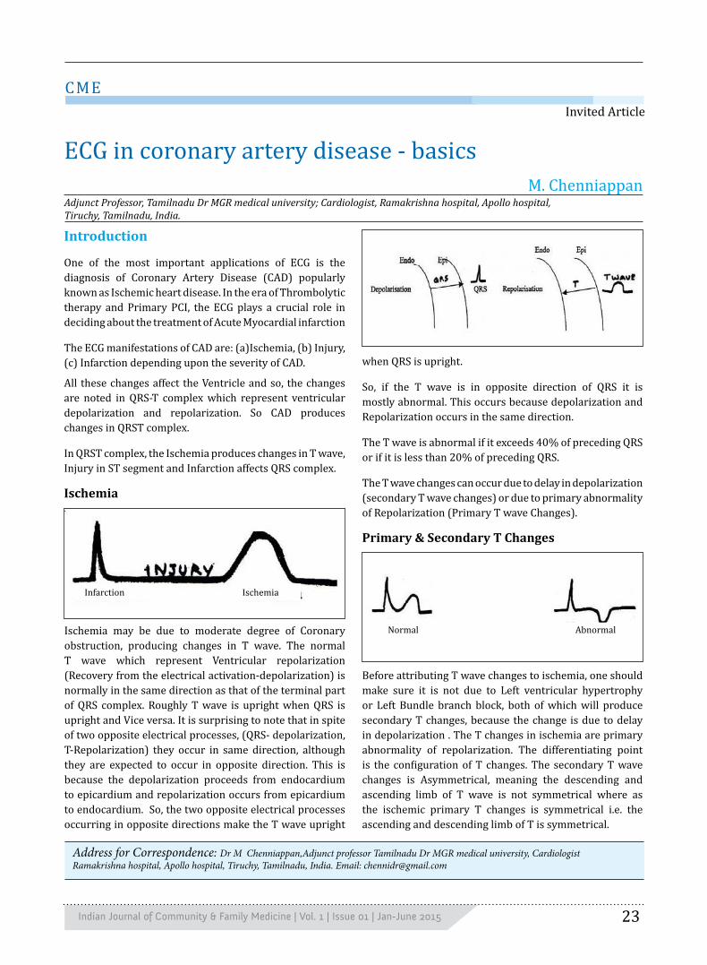
23Indian Journal of Community & Family Medicine | Vol. 1 | Issue 01 | Jan-June 2015
Introduction
One of the most important applications of ECG is the diagnosis of Coronary Artery Disease (CAD) popularly known as Ischemic heart disease. In the era of Thrombolytic therapy and Primary PCI, the ECG plays a crucial role in deciding about the treatment of Acute Myocardial infarction
The ECG manifestations of CAD are: (a)Ischemia, (b) Injury, (c) Infarction depending upon the severity of CAD.
All these changes affect the Ventricle and so, the changes are noted in QRS-T complex which represent ventricular depolarization and repolarization. So CAD produces changes in QRST complex.
In QRST complex, the Ischemia produces changes in T wave, Injury in ST segment and Infarction affects QRS complex.
Ischemia
when QRS is upright.
So, if the T wave is in opposite direction of QRS it is mostly abnormal. This occurs because depolarization and Repolarization occurs in the same direction.
The T wave is abnormal if it exceeds 40% of preceding QRS or if it is less than 20% of preceding QRS.
The T wave changes can occur due to delay in depolarization (secondary T wave changes) or due to primary abnormality of Repolarization (Primary T wave Changes).
Primary & Secondary T Changes
Address for Correspondence: Dr M Chenniappan,Adjunct professor Tamilnadu Dr MGR medical university, CardiologistRamakrishna hospital, Apollo hospital, Tiruchy, Tamilnadu, India. Email: [email protected]
Invited Article
CME
ECG in coronary artery disease - basicsM. Chenniappan
Adjunct Professor, Tamilnadu Dr MGR medical university; Cardiologist, Ramakrishna hospital, Apollo hospital, Tiruchy, Tamilnadu, India.
Ischemia may be due to moderate degree of Coronary obstruction, producing changes in T wave. The normal T wave which represent Ventricular repolarization (Recovery from the electrical activation-depolarization) is normally in the same direction as that of the terminal part of QRS complex. Roughly T wave is upright when QRS is upright and Vice versa. It is surprising to note that in spite of two opposite electrical processes, (QRS- depolarization, T-Repolarization) they occur in same direction, although they are expected to occur in opposite direction. This is because the depolarization proceeds from endocardium to epicardium and repolarization occurs from epicardium to endocardium. So, the two opposite electrical processes occurring in opposite directions make the T wave upright
Before attributing T wave changes to ischemia, one should make sure it is not due to Left ventricular hypertrophy or Left Bundle branch block, both of which will produce secondary T changes, because the change is due to delay in depolarization . The T changes in ischemia are primary abnormality of repolarization. The differentiating point is the configuration of T changes. The secondary T wave changes is Asymmetrical, meaning the descending and ascending limb of T wave is not symmetrical where as the ischemic primary T changes is symmetrical i.e. the ascending and descending limb of T is symmetrical.
AbnormalNormal
Infarction Ischemia

24Indian Journal of Community & Family Medicine | Vol. 1 | Issue 01 | Jan-June 2015
Chenniappan M: ECG In Coronary Artery Disease - Basics
ECG Changes Of Ischemia:
The ischemic T changes depend upon the location of ischemia. The ischemia may be at subendocardial region or sub epicardial region.
The T wave vector will go away from the area of insult. If the ischemia is in a subendocardial region, the T wave vector will go towards the subepicardial region, and the lead over this area will record Tall T wave; so subendocardial ischemia will produce Tall T waves. This is most often due to Acute Coronary Syndrome. (ACS)
Fig 1: ECG showing secondary ST T changes due to LVH
If the ischemia is in the subepicardial region, the T wave vector will go towards subendocardial region i.e. away from the electrode which will record negative T wave or T inversion (Symmetrical).
T wave changes may be due to secondary and primary. The ischemia produces primary T changes. In ischemic changes T waves are symmetrical.
The subendocardial ischemia produces Tall T waves and subepicardial ischemia produces symmetrical T inversion.
Endo Epi
Sub endocardialIschemia
Electrode
Tall T
Depolarisation
Repolarisation
EpiEndo
SubepicardialIschemia
Electrode
SymmetricalT inversion
Wide/ Tall QRS asymmetricalSecondary T changes
Symmetrical T changePrimary/ Ischemic

25Indian Journal of Community & Family Medicine | Vol. 1 | Issue 01 | Jan-June 2015
Fig 2: ECG showing Tall T waves due to subendocardial ischemia.
The T wave changed can be due to acute, sub-acute or chronic presentation of coronary artery disease.
Having already seen the ECG signs of ischemia, let us now see the changes produced by injury on the ST segment.
Injury
In contrast to ischemia which is more often chronic, injury is usually a manifestation of an acute event, indicating an emergency situation, needing an early energetic intervention. It is usually a manifestation of either acute unstable angina or Acute Myocardial infarction. Injury represents either acute critical obstruction or acute total obstruction of a major epicardial coronary artery. Injury produces changes in ST segment.
ST Segment Changes:
ST segment is the area which is between the QRS
Chenniappan M: ECG In Coronary Artery Disease - Basics
complex and the beginning of T wave. It is normally very difficult to say where it begins and where it ends because its junction with QRS complex and T wave is so smooth.
The injury produces changes of either ST elevation or ST depression depending upon the site of injury. The earliest sign of injury is horizontality of ST segment, where the junction between the ST segment with QRS and T wave becomes acute, resulting in ST segment hugging the baseline for more than 0.12 sec.
Normal ST Segment Gentle upslope

26Indian Journal of Community & Family Medicine | Vol. 1 | Issue 01 | Jan-June 2015
If the patient has classical angina, and the ECG is showing this horizontal ST segment, one should immediately suspect CAD, although there is no significant elevation or depression of ST segment or T changes.
Fig 3: ECG showing symmetrical deep T inversion due to sub epicardial ischemia.
ST Elevation – Epicardial Injury
Just like ischemia, injury can also happen either in sub epicardial area or subendocardial area. The sub epicardial injury will produce ST segment elevation as the ST vector will travel towards the site of insult i.e. towards the electrode. ST segment elevation represents acute total occlusion due to red thrombus which is rich in fibrin. So early intervention with either Thrombolytic therapy or Primary PCI is indicated.
Majority of acute myocardial infarction will start as sub epicardial injury and then progresses to necrosis or infarction.
In the era of thrombolytic therapy and Primary PCI one should correctly diagnose type of injury and plan appropriate management.
ST segment depression - Sub endocardial injury.
If the injury is happening at subendocardial region, the ST vector will travel towards the site of insult, to the subendocardial region, i.e. away from the electrode resulting in ST segment depression.
There are varieties of ST segment depression; not all ST segment depression represents injury.
The classical ST segment depression of sub
Chenniappan M: ECG In Coronary Artery Disease - Basics
Normal ST segment
Horizontal ST segment Earliest sign of injury

27Indian Journal of Community & Family Medicine | Vol. 1 | Issue 01 | Jan-June 2015
endocardial injury usually represents acute unstable angina or non ST elevation MI due to critical occlusion due to white thrombus. As the thrombus is not totally occluding and not rich in fibrin, thrombolytic therapy is not given when patient presents with acute severe chest pain and ST segment depression.
Infarction:
In contrast to ischemia, injury, the infarction changes produced in QRS complex are usually irreversible. So, in the treatment of CAD one should aim at preventing infarction or necrosis by energetically and effectively treating ischemia and injury.
Q wave:
The infarction alters QRS changes by producing
Figure 4. ECG showing ST elevation due to sub epicardial injury.
Chenniappan M: ECG In Coronary Artery Disease - Basics
Q wave in ECG. The Q wave is the first negative deflection in the QRS, meaning a downward wave below the baseline. One should be also aware, that the R wave is first positive deflection of the QRS, meaning upward wave above the baseline. The S wave is the second negative deflection of the QRS, i.e., second downward deflection following R wave. The Q wave usually represents a necrotic myocardium, which is incapable of producing electrical activation or depolarization.
As you are already aware, there are certain leads (V5, V6) can have a q wave, due to septal depolarization, which occurs from Left to Right.
One should not mistake this q wave for infarction. So pathological Q wave, which represents myocardial necrosis, has to satisfy certain criteria:

28Indian Journal of Community & Family Medicine | Vol. 1 | Issue 01 | Jan-June 2015
Figure 5. ECG showing ST segment depression due to sub endocardial injury.
Chenniappan M: ECG In Coronary Artery Disease - Basics
Endo Epi
ST depression
ST vector
Cavity
Injury
Types of ST depression:
Upsloping(Phusiological) (injury)
Horizonal Sagging(injury)
downsloping(LVH/LBBB sometime injury)
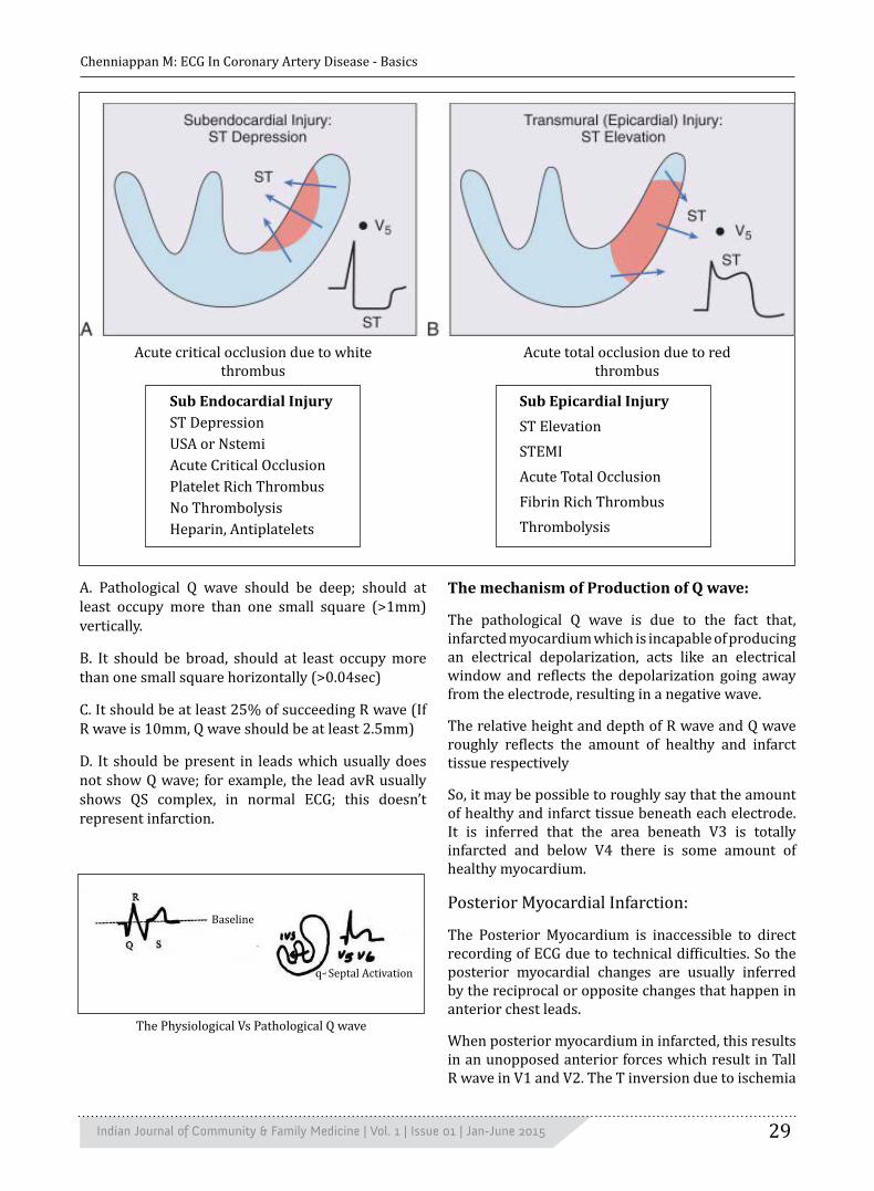
29Indian Journal of Community & Family Medicine | Vol. 1 | Issue 01 | Jan-June 2015
Acute total occlusion due to red thrombus
Acute critical occlusion due to white thrombus
Sub Endocardial InjuryST DepressionUSA or NstemiAcute Critical OcclusionPlatelet Rich ThrombusNo ThrombolysisHeparin, Antiplatelets
Sub Epicardial InjuryST ElevationSTEMIAcute Total OcclusionFibrin Rich ThrombusThrombolysis
The Physiological Vs Pathological Q wave
Chenniappan M: ECG In Coronary Artery Disease - Basics
Baseline
q- Septal Activation
A. Pathological Q wave should be deep; should at least occupy more than one small square (>1mm) vertically.
B. It should be broad, should at least occupy more than one small square horizontally (>0.04sec)
C. It should be at least 25% of succeeding R wave (If R wave is 10mm, Q wave should be at least 2.5mm)
D. It should be present in leads which usually does not show Q wave; for example, the lead avR usually shows QS complex, in normal ECG; this doesn’t represent infarction.
The mechanism of Production of Q wave:
The pathological Q wave is due to the fact that, infarcted myocardium which is incapable of producing an electrical depolarization, acts like an electrical window and reflects the depolarization going away from the electrode, resulting in a negative wave.
The relative height and depth of R wave and Q wave roughly reflects the amount of healthy and infarct tissue respectively
So, it may be possible to roughly say that the amount of healthy and infarct tissue beneath each electrode. It is inferred that the area beneath V3 is totally infarcted and below V4 there is some amount of healthy myocardium.
Posterior Myocardial Infarction:
The Posterior Myocardium is inaccessible to direct recording of ECG due to technical difficulties. So the posterior myocardial changes are usually inferred by the reciprocal or opposite changes that happen in anterior chest leads.
When posterior myocardium in infarcted, this results in an unopposed anterior forces which result in Tall R wave in V1 and V2. The T inversion due to ischemia

30Indian Journal of Community & Family Medicine | Vol. 1 | Issue 01 | Jan-June 2015
The small letter q represents physiological and big letter Q represent pathological.
Figure 6. ECG showing Pathological Q in Inferior Leads
Chenniappan M: ECG In Coronary Artery Disease - Basics
WIDE DEEPINFARCT (NO DEPOLARISATION)
ELECTRICAL WINDOW
Physiological, leads V5, V6<1mm, <0.04 sec.
Pathological, leads other than avR<1mm, <0.04 sec.
Narrow Not Deep
Deep Q, Small rMore infarct, less healthy tissue beneaththe area of electrode
Small Q, Tall RMore Healthy tissue less infarctarea beneath that electrode.
Only Q wave, Only infqrct tissue, No healthy area
For example, if V3 shows Q only
and V4 shows Qr.
in posterior wall results in upright T wave in V1V2. This posterior MI usually accompanies inferior MI or Inferolateral MI.
The leads V1, V2 shows the reciprocal changes of Q wave and inverted T wave, of the posterior wall in the form of Tall R and upright T wave.
So, one should always checkup leads V1,V2, for Tall R waves in the presence of inferior MI to rule out Posterior wall MI.
To sum up infarct changes:
A.Infarction produces Q wave
B.Q wave represents myocardial infarction or necrosis which is usually irreversible.
C.One should differentiate between physiological and pathological Q wave.
D.Q wave is produced by electrically inert myocardium which reflects the distant depolarization going away from the electrode.
E.The relative depth of Q and R waves reflects the amount of infarct and healthy tissue respectively
beneath that electrode.
Posterior MI is inferred by the reciprocal changes in anterior chest leads in the form of Tall R in V1, V2

31Indian Journal of Community & Family Medicine | Vol. 1 | Issue 01 | Jan-June 2015
Figure 7. ECG showing Tall R wave with upright T wave in V1 representing Posterior MI.
INFARCTION
QRS Complex
Q WavePathological Vs
Irreversible
AlwaysChronic
ISCHEMIA
T Wave
SubepicardialSub Endocardial
DD: Sec. T ChangeHyperkalemia
Acute,subacute or
chronic
INJURY
ST Segment
SubendocardialSubepicardial
Thrombolyse or Not
Always Acute
CADSuggested Readings
1. Lecture series in Electrocardiography by Dr.Chenniappan
2. ECG course - An interactive multimedia tutorial by Dr. Chenniappan and Dr. Gandhimathinathan
Summary Box
Chenniappan M: ECG In Coronary Artery Disease - Basics

32Indian Journal of Community & Family Medicine | Vol. 1 | Issue 01 | Jan-June 2015
Introduction
The incidence of cervical cancer in India is alarmingly high at 26.2 per lakh women It is very high in the south of India and is the leading cancer among women in reproductive age group in our country. 1 There are around 32 deaths for every 100 new cases detected.2 There were over 5 lakh new cases diagnosed in India in 2012.3 Further as many as 5-13 million women have precancerous lesions. 4
Cervix is an organ which is easily accessible for inspection. Most of the cancers of the cervix arise in the transformation zone (area between the old and new squamo-columnar junction). It is easily amenable for testing by cytology and other means. Screening programs can detect precancerous lesions. Precancerous disease may take 5-10 years to become cancerous giving plenty of opportunity and time to intervene and prevent the occurrence of cancer. Precancerous lesions are easily and completely treatable. In spite of this we have not been able to reduce the burden of cancer cervix in our country. Even today the new cases diagnosed are at advanced stage. The fact that the highest age specific incidence rate of 98.2 per 100,000 for cancer cervix was seen in the 60-64 yr age group 5 reflects a lost opportunity of prevention by screening and early detection of these cases.
Screening would not only reduce the cancer burden, but also reduce the burden of advanced stages in the long run. Thus there is urgent need to bring universal screening for cancer cervix in women at the community level.
Methods of ScreeningThe screening tests include the following
1. Conventional cytology i.e. the cervical (Pap) smear test2. Fluid sampling technique with automated thin layer preparation (liquid based cytology) 3. Human Papilloma Virus (HPV) testing 4. Polar probe 5. Laser induced fluorescence 6. Visual inspection of cervix after application of Lugol’s
iodine (VILI) or acetic acid (VIA). 7. Speculoscopy 8. Cervicography
Pap Smear has been the conventional method. It is easy to carry out with minimal prior training. However if not properly taken or immediately fixed there can be difficulty in getting representative cells in the slide and the interpretation. It has a low sensitivity anywhere from 37 to 87 % and a high specificity of 86-100 %.6 So it is only moderately accurate. However western countries have reduced the burden of cancer cervix by this conventional cytological screening carried out widely in the population and it continues to be the time tested highly specific screening test.
Fluid sampling technique with automated thin layer preparation (liquid based cytology) is more likely to yield representative cells with lesser contamination with blood etc. The sample can also be used for papilloma virus test. But high cost and need for advanced infrastructure and technology is a limiting factor with this method.
Association of human papilloma virus as the etiological agent for cancer cervix is well accepted. HPV 16 and 18 are the most common strains (70%) among cervical cancer patients.7 HPV DNA testing in the cervical secretions is an important strategy and is recommended for mass application where the resources permit.8 However, it is very expensive and so cannot be advocated for mass screening program in low resource setting like ours.
In a country like ours inability to visit the hospital due to loss of daily wage, lack of awareness about the need for screening and social taboos in discussing matters related to sexual and gynecological problems is the biggest bottle neck in hospital based screening. A village based low tech-nology, objective; one time test with immediate result that can be carried out by the peripheral health workers in the village itself at a time convenient to the target population is likely to be the most effective in preventing invasive cancer cervix on the long run.
Invited Article
CME
Community screening for cancer cervix preventionGowri Dorairajan
Professor, Dept. of Obstetrics and Gynaecology, JIPMER, Puducherry, India.
Address for Correspondence:Dr Gowri Dorairajan, Professor, Deptt. of Obstetrics & Gynaecology, JIPMER, Puducherry. India. Email: [email protected]

33Indian Journal of Community & Family Medicine | Vol. 1 | Issue 01 | Jan-June 2015
Visual inspection with acetic acid (VIA) and visual inspection with Lugols Iodine (VILI) have been found to be feasible, safe and acceptable in early detection of preinvasive lesions.9 Pooled randomized control trial has shown VIA to be an effective method in preventing cancer cervix.10 In a study conducted in Uttar Pradesh state of India, 4148 women completed the screening by cytology and VIA and VILLI. Screen positivity rates by Paps (ASCUS and above), VIA and VILI were 2.6%, 9.7% and 13.5% respectively. The screen positive cases were subject to colposcopy and biopsy. Sensitivity and specificity of detecting the CIN III+ lesions were 87.5 and 98.8% for Pap, 50.0% and 96.7% for VIA and 50.0% and 95.7% for VILI respectively.11
With the revised strategy by the Government of India regarding screening for cancer cervix at community level, the visual inspection with acetic acid and Logol’s iodine is being carried out widely at the primary health centre level. However lot of findings are misinterpreted and false alarms create anxiety and increase referrals to the tertiary care hospital. In this regard one must strictly understand and adhere to the categorization and the interpretation as laid down by the International agency for Research on Cancer. 12, 13
VIA findings
I. VIA findings are categorized as negative when any of the following findings were observed:
• No acetowhite lesions, or faint, ill-defined, bluish white or doubtful lesions• Acetowhitening on cervical polyps or on nabothian cysts• Dot or streak-like acetowhitening on the cervix• White line-like prominent squamocolumnar junction (SCJ) after application of acetic acid• Angular or geographic acetowhite lesions far away from the SCJ or the external os, if SCJ was not visible (satellite lesions)
II. VIA are categorized as positive when any of the following were observed:
• Well-defined, opaque, acetowhite lesions touching the SCJ or the external os, if SCJ was not visible• A large circumferential acetowhite lesion surrounding the external os• Pre-existing wart or leukoplakia turning intensely white after application of acetic acid• Ulcero-proliferative growth turning densely acetowhite after application of acetic acid
VILI findings
I. VILI should be reported as negative when any of the following is observed:
• Normal cervix where squamous epithelium turned mahogany brown or black and when the SCJ is visible, the columnar epithelium beyond it did not change color.• Scattered or diffuse ill-defined or patchy, non- or partial iodine uptake areas in the transformation zone or all over the cervix, not restricted to the transformation zone.
• Thin, yellow, non iodine uptake areas with angular, or digitating margins, resembling geographical areas, distant from the SCJ.
II. VILI are categorized as positive when any of the following were observed:
• Well-defined dense, thick, bright, mustard or saffron yellow, iodine non-uptake areas in the transformation zone, touching the SCJ.• Circumferential yellow non-iodine uptake area around the external os• Ulcero-proliferative growth, turning yellow after the application of iodine solution.
Setting up a clinic for screening
It is possible to set up screening clinics at community or primary health centre level. It requires an examination table with focus lamp for good lighting and sterile gloves for carrying out the procedure in a room with privacy. The equipments needed are as below (Figure1)
A. Sterile Cuscos vaginal speculumB. Ayer’s Spatula (disposable or reusable wooden ones
autoclaved)C. Glass slide (numbered for identification).D. 95% ethyl alcohol in a Koplik Jar to hold and fix the
smeared slides.E. Swab on a holder.F. 3-5% freshly prepared acetic acid
Figure1. Equipments for setting up screening clinic
Dorairajan G: Community Screening for cancer cervix prevention

34Indian Journal of Community & Family Medicine | Vol. 1 | Issue 01 | Jan-June 2015
G. Lugol’s iodineH. Cervical Punch Biopsy forups
Besides these, there should be forms to fill up patient details and books for entering the slide number and pt identifying details to prevent mix up of slides and to keep track of the reports. The slides are transported to the cyto -pathology lab where it is stained and examined. The cytological screening requires support of a cytopathology lab and a referral centre for abnormal reports and for further evaluation with colposcopy.
Procedure for Screening
Whom to screen
Screen all sexually active women from 35 to 55 years of age in the community.14
Pap Smear test: This test is a surface cytology from the cervix. The patient is put in dorso lithotomy position. Cuscos speculum is inserted and the valves of the speculum is opened (Figure2), to visualise the cervix satisfactorily. The longer end of the spatula rests at the external os and it is rotated by 360o around the external os to take representative cells from the transformation zone (Figure3). The spatula should be smeared on numbered or coded slide and transferred to the jar of ethyl alcohol immediately for fixing it. The slides have to be fixed for at least half an hour before they can be transported to a facility with cytopathologists and technicians to prepare and stain the slides.
nodular infiltrating hard growth, then biopsy of the same should be done with the cervical punch biopsy forceps (H in figure1). If biopsy procedure is not possible or lab support is not available then the women should be informed and immediately referred to a centre for a biopsy without delay. After obtaining cytology one should perform the visual inspection 1 minute after application of 3-5% freshly prepared acetic acid on the cervix with a swab on holder and thereafter with Lugol’s iodine(Schiller’s test).(interpret findings as per IACR guidelines detailed before).
Interpretation
If the visual inspection tests are highly abnormal then the patients can be referred for colposcopy even without waiting for the Pap smear reports or they can be treated with cryotherapy.
The Pap smear is reported according to the Bethesda System. The smears reported as inflammation usually do not require antibiotics unless specific infections like Candida, Trichomonas etc are reported.
Koilocytic changes reported in the Pap smear or biopsies signify Human Papilloma Virus induced changes and such women need to be closely followed up to see whether they clear their infection or it gets converted to mild dysplasia.
The women whose Pap Smear is reported as low grade squamous intra epithelial lesions ( LGSI)( CIN I or mild dysplasia as per old nomenclature) need to be referred to a set up for colposcopy. If the colposcopy biopsy also confirms mild dysplasia then the patient can be simply followed up with Pap smear repeated after 3 months. If the Pap smear is reported as HGSIL (CIN II or III or moderate/ severe dysplasia of the old nomenclature) then colposcopy is mandatory. If the diagnosis of colposcopic biopsy is confirmed as HGSIL, the woman needs treatment either with locally ablative procedures like cryo therapy or electro cautery or laser ablation or excision procedures. Such women after treatment need to be followed up with Pap smear after 6 weeks of the procedure.15
Human papilloma virus detection in the cervical mucus is recommended in these patients with abnormal Pap smear and or abnormal VIA, to decide how aggressive the treatment and follow of these cases should be.
WHO guidelines recommend that VIA followed by treatment with cryo and/or large loop excision of the transformation zone is better option than cytology and colposcopy, where the expertise for these is a constraint.8
In a large study that included 58000 women from India and Africa, VIA was found to have high sensitivity of 80-83% and specificity of around 84-85% for CIN II and III
Dos and Don’ts: the smear should not be taken during periods or active cervicovaginal infections. One should avoid the use of antiseptics to clean the vagina before taking Pap smear. Overnight abstinence from sexual intercourse should be preferred. The smear is taken with an Ayers spatula as described above. The spatula should be immediately smeared on a labelled slide to prevent drying artefacts.
If the speculum examination reveals a proliferative cauliflower like growth or an ulcerative growth or a
Figure2. Cusco’s Speculum opened
Dorairajan G: Community Screening for cancer cervix prevention

35Indian Journal of Community & Family Medicine | Vol. 1 | Issue 01 | Jan-June 2015
lesions. 16 VILI was found to have 10% higher sensitivity and specificity. Pap smear was found to have low specificity of 57% and high specificity of 93%. Thus combining the various methods may be more accurate as screening strategies to prevent cancer cervix.
Conclusion
Uterine cervix is easily accessible for examination and subjecting to the various screening methods. The screening procedures can be easily performed with minimal training. The equipments and instruments needed for carrying out screening are low cost and can be easily set up in a village also. One must adhere to the clearly laid out guidelines for the interpretation of the screening results. Good networking with a cytopathologist and a centre for referring for colposcopy and treatment will make the program self sufficient.
Figure3. Diagram to show the use of Ayre’s spatula for taking cytology.
References1. Nandakumar A, Gupta P. C, Gangadharan P, Visweswara R. N.
and Parkin D. M. Geographic pathology revisited: Development of an atlas of cancer in India. International Journal of Cancer 2005; 116: 740–754.
2. Institute for Health Metrics and Evaluation. The challenge ahead: Progress in breast and cervical cancer. Institute of Health Metrics and Evaluation; 2011. Available at http://www.healthmetricsandevaluation.org/publications/policy-report/challenge-ahead-progress-and-setbacks-breast-and-cervical-cancer.
3. Ferlay J, Soerjomataram I, Ervik M, Dikshit R, Eser S, Mathers C et al. GLOBOCAN 2012 v1.0, Cancer Incidence and Mortality Worldwide: IARC Cancer Base No. 11 [Internet]. Lyon, France.
4. International Agency for Research on Cancer, 2013, Available at: http://globocan.iarc.fr
5. Bishop A, Sherris TJ. Cervical dysplasia treatment: key issues for developing countries. Bull Pan Am Health Organ 1996; 30:378-86.
6. Nandakumar A, Ramnath T, Chaturvedi M. The magnitude of cancer cervix in India. Indian J Med Res. 2009; 130:219-221.
7. Nanda KA, McCrory DC, Myers ER, Bastian LA, Hasselblad V, Hickey JD et al. Accuracy of the Papanicolaou test in screening for and follow-up of cervical cytologic abnormalities: a systematic review. Ann Intern Med. 2000; 132(10):810-9.
8. de Sanjose S, Quint WG, Alemany L.et al. Human papillomavirus genotype attribution in invasive cervical cancer: a retrospective cross-sectional worldwide study. Lancet Oncol. 2010; 11:1048–1056. doi: 10.1016/S1470-2045(10)70230-8.
9. WHO guidelines for screening and treatment of precancerous lesions for cervical cancer prevention.
Geneva. 2013: 29-33. Available at: http://apps.who.int/iris/bitstream/10665/94830/1 /9789241548694_eng.pdf
10. Sankaranarayanan R, Shyamalakumary B, Wesley R, Sreedevi Amma N, Parkin DM & Nair MK. Visual inspection with acetic acid in the early detection of cervical cancer and precursors. International Journal of Cancer1999; 80: 161–163.
11. Sankaranarayanan R, Esmy PO, Rajkumar R, Muwonge R, Swaminathan R, Shanthakumari S et al. Effect of visual screening on cervical cancer incidence and mortality in Tamil Nadu, India: a cluster-randomised trial. Lancet. 2007 ; 370(9585):398-406.
12. Satyanarayana L, Asthana S, Bhambani S, Sodhani P, Gupta S. A comparative study of cervical cancer screening methods in a rural community setting of North India. Indian J Cancer 2014;51:124-8.
13. Sankaranarayanan R, Ramani S and Wesley A. Practical manual on visual screening for cervical neoplasia. IARC technical publication. No.41 IARC press.
14. Sankaranarayanan R, Basu P, Wesley RS et al. Accuracy of visual screening for cervical neoplasia: Results from an IARC multicentre study in India and Africa. International Journal of Cancer. 2004; 110: 907–913.
15. World Health Organization. Progress in Reproductive Health Research. Geneva: World Health Organization; 2004. 65.
16. Miller AB, Nazeer S, Fonn S, Brandup-Lukanow A, Rehman R, Cronje H et al. Report on consensus conference on cervical cancer screening and management. Int J Cancer. 2000; 86(3):440-7.
17. Arbyn M, Sankaranarayanan R, Muwonge R, Keita N, Dolo A, Mbalawa CG et al. Pooled analysis of the accuracy of five cervical cancer screening tests assessed in eleven studies in Africa and India.. Int J Cancer. 2008 ;123(1):153-60.
Dorairajan G: Community Screening for cancer cervix prevention

36Indian Journal of Community & Family Medicine | Vol. 1 | Issue 01 | Jan-June 2015
In India almost 80-85% of total medicolegal workload is taken care of by MBBS doctors whether it is conducting postmortem examination in the mortuary or examining a victim in the casualty/emergency of the district hospital/CHC/PHC. Medicolegal duty is a compulsory duty for all the doctors and is a part and parcel of the medical practice as well. Presently the subject of Forensic Medicine is being taught during 2nd Professional of MBBS and that too having limited number of teaching hours. By the time a medical student passes MBBS at the end of 4½ years and undergoes internship training he practically forgets whatever little he had learnt during that period. It has been observed that when these doctors are put on medicolegal duty they find it extremely difficult to handle the medicolegal cases. As a result at times because of poor knowledge not only the outcome of case is affected in a court of law but they also land into trouble. Whenever any case comes to hospital, the
doctor has to perform two duties simultaneously- medical (for treating/saving the life of the person) & medicolegal (towards legal system). Medical duties of course are first and foremost and supersede medicolegal duties.
What is a Medicolegal Case (MLC)?
MLC is defined as “any case of injury or ailment where, the attending doctor after history-taking and clinical examination considers that investigations by law enforcement agencies are warranted to ascertain circumstances and fix responsibility regarding the said injury or ailment according to the law”.
Any case can be converted to MLC at any stage with emergence of fresh allegations or new facts even if at first stance it has been entered as a non-medicolegal case.
1. Who is responsible to label a case as MLC? (authority)
The decision to label a case as MLC should be based on sound professional judgment, after detailed history taking and thorough clinical examination and can be done by any of the following personnel.
(a) Emergency Medical Officer(b) Casualty Medical Officer (c) Medical Officer In-charge of Emergency(d) Duty Medical Officer (e) Resident Duty Officer(f) Medical Officer In-charge of ward who is attending
to the case2. Situations/Cases which are to be labeled as MLCs
The following list is just a guiding one considering common occurrence and not exclusive so doctors must use their professional judgment to decide any other case as MLC which is not covered in the provided list. (Table 1)
3. General Guidelines for dealing with MLCs:
I. It is the duty of the examining doctor to record the findings in a meticulous manner. They should
Invited Article
CME
Handling medicolegal cases at sub-district LevelAdarsh Kumar
Additional Professor, Dept. of Forensic Medicine & Toxicology, All India Institute of Medical Sciences, New Delhi, India.
Address for Correspondence: Dr. Adarsh Kumar, Additional Professor, Dept. of Forensic Medicine & Toxicology, All India Institute of Medical Sciences, New Delhi. Email: [email protected]
Assault by any meansAccidents like Road Traffic Accidents (RTA), industrial accidents etc
Cases of trauma with suspicion of foul play
Burns and Scalds
Electrocution Chemical injuries
Fire Arm Injuries Custodial torture
Sexual Offences Undiagnosed coma
Cases of asphyxia due to hanging, strangulation, drowning, suffocation
Attempted suicide by any means
Suspected Poisoning Alcohol Intoxication
Death due to Snake Bite or Animal Bite
Drug overdose & Drug abuse
Death over operation theatre table
Criminal abortions
Brought Dead cases to the Casualty/ Emergency
Deaths occurring within 24 hours of hospitalization without having any provisional diagnosis
Table 1. Common Medico-legal conditions
37Indian Journal of Community & Family Medicine | Vol. 1 | Issue 01 | Jan-June 2015
approach the task with due care and attention.
II. All the Medico legal records (MLRs) should be written in legible handwriting avoiding using abbreviations. Writing must be legible so that it can be understood and readable by all concerned and need not be an artistic one. He may use capital letters when writing the name, address etc. He should use simple terms instead of complicated medical terms as far as possible.
III. Alleged history of incidence in brief should be recorded. Instead of just writing Road Traffic Accidents (RTA)assault etc, a brief history of what has happened should be recorded, at least with reference to the manner of infliction and time of occurrence. When the injured person is unconscious or otherwise unable to give exact details of what has happened, the version of the person accompanying the injured person should be recorded, specifying the name of person giving the history.
IV. Particulars of person who accompanied/brought the alleged victim should be recorded. If brought by PCR/police; name, belt no, police station should be recorded.
V. The injuries should be serially numbered, described well in detail mentioning the type, size, shape, placement and site with distance from anatomical landmarks. Abbreviations like Ab for abrasion, LW for lacerated wound, TS for traumatic swelling, # for fracture, KUO-kept under observation should not be used since they are liable to be interpreted by different persons in different manner. CLW which is frequently used is not a scientific entity ,should be avoided. Since it connotes contradictory meaning ie. Whether it is Cut (sharp weapon) or lacerated wound (blunt) or clean lacerated wound.
VI. The examining doctor should record their findings in good detail which will not only justify the opinions expressed by them but also enable experts to review the report in the foreseeable future allowing them to draw viable conclusions with regard to the injuries examined.
VII. Examining doctors should express an opinion regarding the nature of injuries examined in all cases, after due investigations wherever required. In case of X-ray reports, the same may be endorsed on original MLR itself but incorporating in office copy also. The date of giving the subsequent opinion with proper signature must be mentioned on MLR.
VIII. It is not justified to withhold the opinion regarding
nature of injuries i.e. whether simple / grievous hurt, with the idea of observing the outcome of the injuries by reexamining the injuries after a considerable lapse of time for healing of the wound before giving a final opinion.
IX. In case of unpredictable future course of the injuries, a provisional opinion is to be mentioned with the provison that it is liable to change in light of the future course of events of the injuries.
X. In case any exhibits/evidentiary material are preserved they should be properly documented, labeled and sealed before handing over to the investigation authorities so as to maintain the ‘Chain of custody’.
XI. At the end of MLR, the doctor should put his signature, date and write his full name in capital letters with State Medical Council Registration Number. A personalized stamp should be prepared for every doctor dealing with MLRs; which should be kept in safe custody.
XII. Proper receipt should be recorded in the MLR clearly mentioning the name of the personnel receiving the MLR and exhibits. The Officer receiving the MLR should put his signature with date, write his name, designation, belt number and police station on the space provided at the bottom end of the office copy of the MLR or on its back side.
XIII. In case the patient is referred for further care it should be mentioned in the MLR. In this case if examining doctor is unable to express opinion with regard to the nature of hurt due to lack of investigations (like X-rays); a clear cut advisory must be given to the Investigating officer in writing that it is the duty of the Investigating Officer (I.O.) to get a fresh MLC made at the referred hospital.
XIV. In such cases wherein a patient undergoes treatment in different hospitals and the hospital which recorded the initial MLC report has no control over documents and records of a different hospital; it is the primary responsibility of the Investigating authorities to pursue a medico-legal case in order to procure an opinion from the concerned doctor who has prepared first MLR.
4. Few things to remember:
(a) Primary Duty of Doctor is to save the life of the patient in cases of emergencies; so resuscitation and stabilization of the patient takes precedence over medicolegal formalities. The consent for treatment is implied in all emergencies.
Kumar A: Handling MLC at Sub-District level

38Indian Journal of Community & Family Medicine | Vol. 1 | Issue 01 | Jan-June 2015
(b) Emergency medical care should be administered to all cases brought to any hospital. After the initial stabilization, the patient may be transferred to other hospital, and if necessary by service ambulance.
(c) All hospitals should maintain a MLC register and the MLC should be initiated and documented in the register.
(d) Medicolegal documents should be prepared in duplicate, with utmost care giving all necessary details, preferably written with a ball-point pen and avoiding overwriting. If any overwriting or correction is made, it should be authenticated with the full signature and stamp of the Medical Officer (MO).
(e) The Duty Constable/nearest Police Post attached to hospital should be immediately informed as and when a MLC is registered or admitted. The particulars of the patient and a short summary of the case should be mentioned in the despatch book. This is as per provisions Under Section 39 of Criminal Procedure Code. Any failure to communicate the MLC may invite prosecution under Sections 176 and/or 202 of IPC.
(f) Medicolegal documents are to be considered as confidential records and should be stored under safe custody to avoid tampering. Medical records must be thorough, complete and should document each and every significant event in the course of care of the patient. All the documents including case sheets, X-rays and investigation reports should be preserved meticulously in the medical record section for a minimum period of 5 years and handed over to the concerned authorities (Police Investigating Officer / Court / Court of Inquiry) as and when required. MLRs are however are to be preserved for a minimum 10 years as per DGHS guidelines.
(g) All evidences collected should be mentioned in medicolegal documents to establish the chain of custody in a court of law subsequently. Samples and specimens collected for medicolegal purposes should be properly sealed, labeled and handed over to the concerned investigating officer after taking due receipt. All the articles should be preserved in sealed packets/envelopes with taking special precautions as follows:
(i) Clothing worn by the patient showing evidence of injury such as tears, bullet holes, cuts, blood stains etc. In case of multiple tears, cuts or holes etc., each piece of evidence should be encircled and numbered with matching description in the MLC report and case sheet.
(ii) Bullets recovered from a body should be marked by etching an initial or a mark on the bottom before preservation.
The list of articles/samples to be preserved in different situations is mentioned under Table 2.
(h) In case of discharge / transfer /absconding/death of such a case in the hospital, again the police should be informed.
(i) Dying Declaration- In cases where the patient wishes to make a dying declaration, the area magistrate should be intimated. If the Magistrate is unable to come and record a statement or where the MO feels that he might not be able to reach the patient in time, the MO may record the dying declaration himself in presence of two independent witnesses whose signatures are also to be affixed in the declaration. The MO will certify the soundness of mind of the person before making the dying declaration and after recording it.
(j) Fees-Usually fees should not be charged by government doctor preparing MLCs however practice differs from state to state. Many states have specified fees for making MLCs so whatever prescribed should be followed.
(k) The duplicate copy should be issued to the injured person or to those authorized by him or to those who are lawfully entitled to receive it on his behalf upon a written requisition for the same. When the patient is referred to a higher center for further or expert management, the duplicate copy may be issued immediately along with the referral letter. In situations where the patient is severely injured or unconscious and is unable to receive the wound certificate copy himself or to authorize someone to receive it and when there are no persons lawfully entitled to receive the copy on the injured person’s behalf, the doctor need not issue the same to the available bystanders. In such situations the doctor should incorporate the relevant points in the MLR in the referral letter.
5. Precautions to be taken while dealing MLCs
(I) One should not rely on memory while writing reports or during recording of evidence in a court of law.
(II) Consent should be both informed and written. The subject should write his name and put his signature in continuation to consent written by him. When the subject is illiterate, the doctor should write the sentence of informed consent and should read over the same to the subject. Then the subject should sign or put his thumb impression. Whenever a person not under arrest refuses consent, he should be asked to write the informed refusal and sign the same.
(III) Physical examination of a person without his consent is assault except in situations specified by the Law. Hence consent should be obtained before conducting any medico-
Kumar A: Handling MLC at Sub-District level
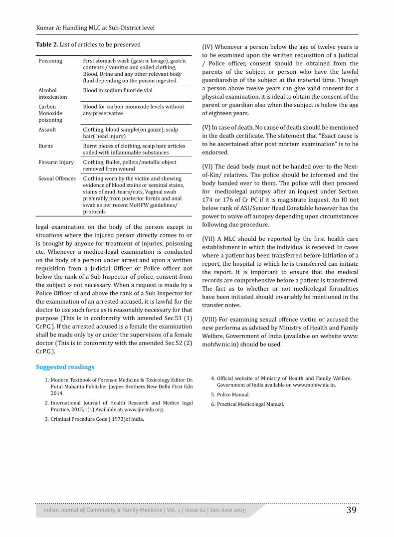
39Indian Journal of Community & Family Medicine | Vol. 1 | Issue 01 | Jan-June 2015
legal examination on the body of the person except in situations where the injured person directly comes to or is brought by anyone for treatment of injuries, poisoning etc. Whenever a medico-legal examination is conducted on the body of a person under arrest and upon a written requisition from a Judicial Officer or Police officer not below the rank of a Sub Inspector of police, consent from the subject is not necessary. When a request is made by a Police Officer of and above the rank of a Sub Inspector for the examination of an arrested accused, it is lawful for the doctor to use such force as is reasonably necessary for that purpose (This is in conformity with amended Sec.53 (1) Cr.P.C.). If the arrested accused is a female the examination shall be made only by or under the supervision of a female doctor (This is in conformity with the amended Sec.52 (2) Cr.P.C.).
(IV) Whenever a person below the age of twelve years is to be examined upon the written requisition of a Judicial / Police officer, consent should be obtained from the parents of the subject or person who have the lawful guardianship of the subject at the material time. Though a person above twelve years can give valid consent for a physical examination, it is ideal to obtain the consent of the parent or guardian also when the subject is below the age of eighteen years.
(V) In case of death, No cause of death should be mentioned in the death certificate. The statement that “Exact cause is to be ascertained after post mortem examination” is to be endorsed.
(VI) The dead body must not be handed over to the Next-of-Kin/ relatives. The police should be informed and the body handed over to them. The police will then proceed for medicolegal autopsy after an inquest under Section 174 or 176 of Cr PC if it is magistrate inquest. An IO not below rank of ASI/Senior Head Constable however has the power to waive off autopsy depending upon circumstances following due procedure.
(VII) A MLC should be reported by the first health care establishment in which the individual is received. In cases where a patient has been transferred before initiation of a report, the hospital to which he is transferred can initiate the report. It is important to ensure that the medical records are comprehensive before a patient is transferred. The fact as to whether or not medicolegal formalities have been initiated should invariably be mentioned in the transfer notes.
(VIII) For examining sexual offence victim or accused the new performa as advised by Ministry of Health and Family Welfare, Government of India (available on website www.mohfw.nic.in) should be used.
Table 2. List of articles to be preserved
Poisoning First stomach wash (gastric lavage), gastric contents / vomitus and soiled clothing, Blood, Urine and any other relevant body fluid depending on the poison ingested.
Alcohol intoxication
Blood in sodium fluoride vial
Carbon Monoxide poisoning
Blood for carbon-monoxide levels without any preservative
Assault Clothing, blood sample(on gauze), scalp hair( head injury)
Burns Burnt pieces of clothing, scalp hair, articles soiled with inflammable substances
Firearm Injury Clothing, Bullet, pellets/metallic object removed from wound
Sexual Offences Clothing worn by the victim and showing evidence of blood stains or seminal stains, stains of mud, tears/cuts, Vaginal swab preferably from posterior fornix and anal swab as per recent MoHFW guidelines/protocols
Suggested readings
1. Modern Textbook of Forensic Medicine & Toxicology Editor Dr. Putul Mahanta Publisher Jaypee Brothers New Delhi First Edn 2014.
2. International Journal of Health Research and Medico legal Practice, 2015;1(1) Available at: www.ijhrmlp.org.
3. Criminal Procedure Code ( 1973)of India.
Kumar A: Handling MLC at Sub-District level
4. Official website of Ministry of Health and Family Welfare, Government of India available on www.mohfw.nic.in.
5. Police Manual.
6. Practical Medicolegal Manual.

40Indian Journal of Community & Family Medicine | Vol. 1 | Issue 01 | Jan-June 2015
Abstract
Background: Non-communicable diseases (NCDs) represent the biggest threat to women’s health worldwide. Deaths of women from NCDs during their most productive years (40-60 years) can result in tragedy for families and catastrophic expenditure. Therefore, this present study was undertaken to estimate the prevalence of selected NCDs & risk factors in post-menopausal women residing in different slums of Bhubaneswar city and to assess their current health status after attaining menopause. Methods: This cross- sectional study was conducted among 240 post-menopausal women residing in slums of Bhubaneswar city, Odisha. For assessment of risk factors for NCDs, B.M.I estimation was done; blood pressure was measured according to standard guidelines and depression and anxiety was measured by clinical psychologist using Hamilton Depression Rating Scale (HDRS) and Hamilton Anxiety Rating Scale (HAM-A). A predesigned and pretested questionnaire was administered to assess various post-menopausal related symptoms. Results: Overall, 70.5% of women were overweight, 17.8% of subjects were hypertensive and pre-hypertensive (57.2%). Prevalence of depression and anxiety was found to be 22% and 27.9%. Addiction to beetle chewing was found among 40% of subjects. The most common complaints of postmenopausal women were irritability (84.1%), muscle or joint pain (74.1%), palpitation (65.4%) hot flushes (60%) and night sweats (54.1%). Conclusion: This study reveals that NCDs and their risk factors are common among post-menopausal women and there is a need for timely screening and treatment for those conditions.
Keywords: Postmenopausal women, NCDs, risk factors , slums
Introduction
Menopause is the time of a woman’s life when reproductive capacity stops. It is a condition faced by every woman in her later period of life and its associated effect can hamper the quality of life.1 According to Indian menopause society research there are about 65 million Indian women over the age of 45 who silently bear their health problems.
About 53% of deaths are due to Non-communicable diseases.2 Despite global efforts for preventing NCDs, it accounted for around 20% of all diseases in Odisha.3 Menopause itself is one of the major factors, which can lead to NCD.4 Lower level of estrogens put them at increased risk for a number of health conditions. Almost, 59% of rural women in Odisha are suffering due to NCDs.3
Reproductive and Child Health-II and National Health Mission only address women in the reproductive age group.5 Postmenopausal women cross the boundaries of reproduction but do not fall in elderly group therefore do not receive attention of either group. Factors that affect age at menopause may have important clinical implications because early menopause is associated with an increased risk of cardiovascular disease and osteoporosis, whereas delayed menopause has been associated with increased risk of breast cancer and endometrial cancer.6
India is experiencing a rising burden of NCDs causing significant morbidity and mortality, both in urban and rural population, with considerable loss in potentially productive years (age 35-64years) of life.7 Since, there is no published literature on NCDs and their risk factors
ORIGINAL ARTICLE
Prevalence of selective non-communicable diseases and their risk factors among post-menopausal women residing in slum areas of Bhubaneswar city, Odisha
Sai C Das1, B C Das2, Irfana3, Shalini Ray4, Surendra5, Prabhu6
1,3-5 Post Graduate students, Department of Community Medicine,2 Director, School of Medicine, Kalinga Institute of Medical Sciences, Bhubaneswar, Odisha, India. 6 Clinical Psychologist, Utkal University, Bhubaneswar, Odisha, India
Address for Correspondence: Dr Sai C Das, Department of Community Medicine, Kalinga Institute of Medical Sciences, KIIT Road, Chandaka Industrial Estate, Patia, Bhubaneswar, Odisha. Email: [email protected] Received: 29.11.2014, Accepted: 11.01.2015

41Indian Journal of Community & Family Medicine | Vol. 1 | Issue 01 | Jan-June 2015
among post- menopausal women from slum areas of Odisha, the present study was conducted to find same and to assess the current health status of women after attaining menopause in the slums of Bhubaneswar city.
Methodology
This cross-sectional study was conducted in different slums of Bhubaneswar among post- menopausal women. Sample size was calculated after conducting a pilot study on 20 women in one of the randomly selected slums to find the prevalence of one or more menopausal symptom. With this prevalence of 80%, allowable error of 10%, 95% confidence level, and design effect of 2, minimum sample size required was 200. Assuming 20% of non-response rate, the final sample size was 240. This study was carried out during January 2013 to September 2013. Ethical approval was sought from institutional ethics committee of Kalinga Institute of Medical Science, Bhubaneswar. A written informed consent was obtained prior to the inclusion of the study subjects. For illiterate participants, study purpose was clearly explained in local language, oral consent was taken followed by thumb impression.
Post-menopausal women below the age of 60 years residing in the study area from last 6 months and who had attained natural menopause were considered for the study purpose, whereas non- volunteers, women above 60years and those having any known health disease or on any form of hormonal therapy were excluded from the study.
Multi stage sampling was adopted to select the study population. Bhubaneswar Municipal Corporation is divided into 5 geographical zones i.e. North, East, Central, West and South, out of which 3 zones were selected using simple random sampling (Lottery method). From each zone, one ward and then two slums per ward were selected randomly. Each slum was considered as a study cluster. Total of 6 slums were selected. To arrive at sample size of 240, sample was to be drawn from each slum, which was calculated according to proportional allocation i.e. proportion of slum population in the zone to the total slum population in the city.
A quick sketch map was prepared for each selected slum depicting external boundaries, internal boundaries, and all lines representing streets, roads. One central point was selected in each slum (e.g. Market, temples or the Central Square, etc.) taking help from a local health worker. Standing at the centre a particular direction was randomly chosen, and in the same direction subsequent households, starting from first household in the left side was studied until the desired sample was obtained from each slum.
Blood pressure was measured in sitting position
according to standard guidelines. Average of the three readings five minutes apart was taken. If any-one reading was abnormal, one another reading was taken after ten minutes of rest. After measurement, hypertension was diagnosed as per JNC 7 criteria or if the subject was on BP-lowering medication(s). 8
Women with severe symptoms were referred to tertiary care hospital and those with mild symptoms were given advice at primary care level.
The study instrument consisted of pretested, self-designed, semi-structured, oral interview based schedule. The pretesting was done in one of the randomly chosen slum. After analysis of the results from pre-testing, changes were made in the study instrument. The components of questionnaire were included socio-demographic profile, age at menopause, socio-economic status and the diseases. The menopausal symptoms reported by the study subjects were taken for a recall period of last 3 months from the date of interview. Menopausal symptoms assessed in the study were divided into six categories: Vasomotor, Vaginal, psychosomatic, psychological, Sexual, urinary complaints and “others”. For assessing the socio-economic status, B.G Prasad’s social classification for the year 2013 was used.9 Hamilton Depression Rating Scale (HDRS)
and Hamilton Anxiety Rating Scale (HAM-A) were administered by a trained psychologist assess problems of anxiety and depression. Anthropometric measurements like weight was recorded in kilograms using weighing machine (Model: NOVA BS 120), height was recorded in centimetres to the nearest 0.1 cm using a Stadiometer and body mass index (BMI) was calculated. Blood pressure was recorded using a mercury sphygmomanometer (Model: Diamond BP Apparatus with Normal cuff).
Data was entered into an excel spreadsheet and double-checked for errors. Data was analyzed using Microsoft excel. The descriptive statistics were calculated for the background variables and for the prevalence of the selected menopausal symptoms.
Operational definition of overweight and obesity were BMI of ≥ 25 and ≥ 30 respectively and of hypertension was as per the Joint National Committee (JNC VII) criterion, a systolic (SBP) ≥140 mmHg or a diastolic blood pressure (DBP) ≥90 mmHg.8,10
Results
The age range for the subjects was 45-60 years. It was observed that 95 (39.5%) of the postmenopausal women were in the age group of 51-55 years. The educational level showed that 146 (60.8%) women were illiterate and only 2.8% were graduate and above. Occupation of 195 women
Das SC: Risk factors & NCDs in post-menopausal women in slum of Odisha
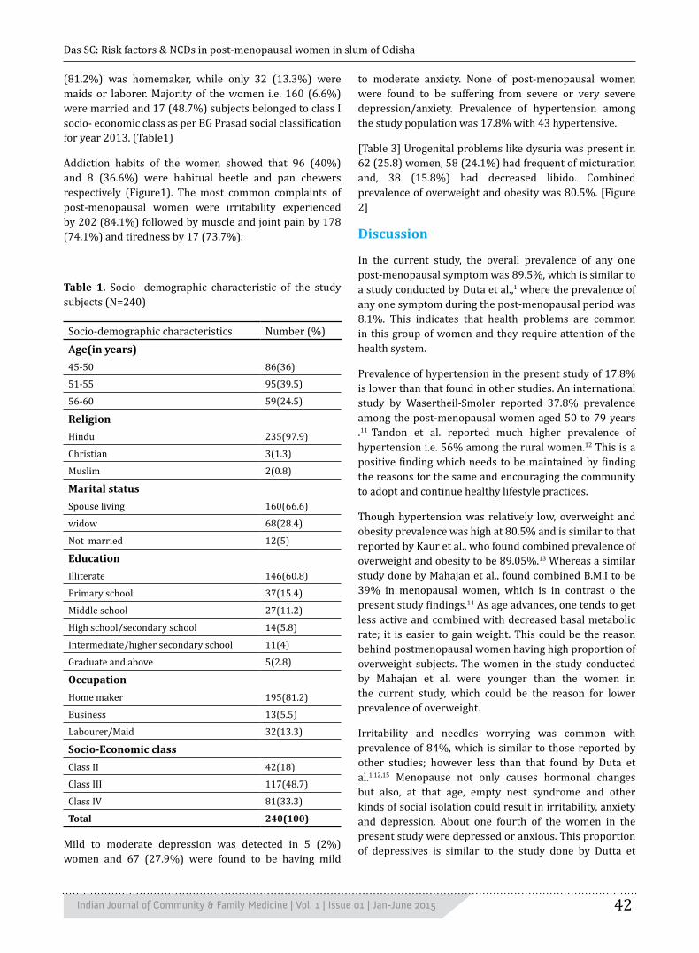
42Indian Journal of Community & Family Medicine | Vol. 1 | Issue 01 | Jan-June 2015
(81.2%) was homemaker, while only 32 (13.3%) were maids or laborer. Majority of the women i.e. 160 (6.6%) were married and 17 (48.7%) subjects belonged to class I socio- economic class as per BG Prasad social classification for year 2013. (Table1)
Addiction habits of the women showed that 96 (40%) and 8 (36.6%) were habitual beetle and pan chewers respectively (Figure1). The most common complaints of post-menopausal women were irritability experienced by 202 (84.1%) followed by muscle and joint pain by 178 (74.1%) and tiredness by 17 (73.7%).
Table 1. Socio- demographic characteristic of the study subjects (N=240)
Socio-demographic characteristics Number (%)Age(in years)45-50 86(36)
51-55 95(39.5)
56-60 59(24.5)
ReligionHindu 235(97.9)
Christian 3(1.3)
Muslim 2(0.8)
Marital statusSpouse living 160(66.6)
widow 68(28.4)
Not married 12(5)
EducationIlliterate 146(60.8)
Primary school 37(15.4)
Middle school 27(11.2)
High school/secondary school 14(5.8)
Intermediate/higher secondary school 11(4)
Graduate and above 5(2.8)
OccupationHome maker 195(81.2)
Business 13(5.5)
Labourer/Maid 32(13.3)
Socio-Economic classClass II 42(18)
Class III 117(48.7)
Class IV 81(33.3)
Total 240(100)
Mild to moderate depression was detected in 5 (2%) women and 67 (27.9%) were found to be having mild
to moderate anxiety. None of post-menopausal women were found to be suffering from severe or very severe depression/anxiety. Prevalence of hypertension among the study population was 17.8% with 43 hypertensive.
[Table 3] Urogenital problems like dysuria was present in 62 (25.8) women, 58 (24.1%) had frequent of micturation and, 38 (15.8%) had decreased libido. Combined prevalence of overweight and obesity was 80.5%. [Figure 2]
Discussion
In the current study, the overall prevalence of any one post-menopausal symptom was 89.5%, which is similar to a study conducted by Duta et al.,1 where the prevalence of any one symptom during the post-menopausal period was 8.1%. This indicates that health problems are common in this group of women and they require attention of the health system.
Prevalence of hypertension in the present study of 17.8% is lower than that found in other studies. An international study by Wasertheil-Smoler reported 37.8% prevalence among the post-menopausal women aged 50 to 79 years .11 Tandon et al. reported much higher prevalence of hypertension i.e. 56% among the rural women.12 This is a positive finding which needs to be maintained by finding the reasons for the same and encouraging the community to adopt and continue healthy lifestyle practices.
Though hypertension was relatively low, overweight and obesity prevalence was high at 80.5% and is similar to that reported by Kaur et al., who found combined prevalence of overweight and obesity to be 89.05%.13 Whereas a similar study done by Mahajan et al., found combined B.M.I to be 39% in menopausal women, which is in contrast o the present study findings.14 As age advances, one tends to get less active and combined with decreased basal metabolic rate; it is easier to gain weight. This could be the reason behind postmenopausal women having high proportion of overweight subjects. The women in the study conducted by Mahajan et al. were younger than the women in the current study, which could be the reason for lower prevalence of overweight.
Irritability and needles worrying was common with prevalence of 84%, which is similar to those reported by other studies; however less than that found by Duta et al.1,12,15 Menopause not only causes hormonal changes but also, at that age, empty nest syndrome and other kinds of social isolation could result in irritability, anxiety and depression. About one fourth of the women in the present study were depressed or anxious. This proportion of depressives is similar to the study done by Dutta et
Das SC: Risk factors & NCDs in post-menopausal women in slum of Odisha

43Indian Journal of Community & Family Medicine | Vol. 1 | Issue 01 | Jan-June 2015
Table 2. Distribution of postmenopausal women according to their symptoms (N=240)
Symptoms N(%)Vasomotor symptoms
Hot flushes 145(60)
Night sweats 135(54.1)
Vaginal symptoms
Uterine prolapsed 38(15.8)
Vaginal dryness 42(17.5)
Psychosomatic symptoms
Sleep disturbances 155(64.5)
Muscle and Joint pain 178(74.1)
Breast pain 8(3.3)
Irritability 202(84.1)
Tiredness 177(73.7)
Head reeling/Dizziness 57(23.7)
Headache 107(44.5)
Palpitation 157(65.4)
Constipation 13(5.4)
Pain abdomen 15(6.2)
Hyperacidity 166(69.1)
Psychological symptoms
Depression** 55(22)
Forgetfulness 17(7.0)
Anxiety** 67(27.9)
Urinary symptoms
Urinary incontinence 58(24.1)
Dysuria 62(25.8)
Sexual complaints
Decreased libido 38(15.8)
Other complains
Visual problems (excluding cataract) 17(7.0)
Hearing defect 31(12.9)
Dental problems 66(27.5)
Respiratory related complains 37(15.4)
Table 3. Distribution of study subjects according to blood pressure status (N=240)
Blood pressure No. %Normal 60 25
Pre hypertension 137 57.2
Hypertension stage I 20 8.3
Hypertension stage II 23 9.5
Total 240 100
Figure 2. Distribution of sample according to their B.M.I (kg/m2)
Das SC: Risk factors & NCDs in post-menopausal women in slum of Odisha
Figure 1. Distribution of study participants according to substance abuse (N=240)
al., who reported a prevalence of depression of 24.7% but their population had higher proportion of anxiety at 35.4%.1 Vijayalakshmi et al. too reported a much higher level of anxiety at 6.6%.15 However, the proportion of women suffering from depression in the present study was less as compared to a study done by Mahajan et al. who found prevalence of depressive mood to be 40%.14 In the current study, more than half of the women who were widows or single suffered from depression as compared to those who were married. This corroborates with the well known phenomenon that married people have better mental health.
44Indian Journal of Community & Family Medicine | Vol. 1 | Issue 01 | Jan-June 2015
This indicates that along with physical health, mental health of women in postmenopausal group should also be attended to.
Another most prominent complaint among post-menopausal women and expected due to the wear and tear associated with age was orthopedic problems, which was reported by almost three fourth of women. Almost similar finding was reported in a study, which was done by Mahajan et al., where prevalence of joint pains was found to be 69.6%.14 However, in a study, done by Dutta et al., only 20% of post-menopausal women had joint pains, which could be explained by younger women in their population with nine tenth of them below 55 years.1 Since all the information related to post-menopausal symptoms are self-reported by respondents, response bias cannot be
ruled out. Due to shyness of women, there was difficulty in eliciting history related to sexual complaints.
Conclusion
The present study was probably the first of its kind in Odisha to assess the NCDs and their risk factors in post-menopausal women, along with other problems they experience due to hormonal change. This study showed high prevalence of NCDs like overweight & obesity and orthopedic problems along with anxiety and depression. It shows that the health care workers should be sensitized to screen post-menopausal women for various symptoms. The policy makers should also take into consideration this group of women have multiple health problems but do not get adequate atention as they neither come in the reproductive group nor the elderly.
References1. Dutta R, Dcruze L, Anuradha R, Rao S, Rashmi MR. A Population
Based Study on the Menopausal Symptoms in a Rural Area of Tamil Nadu. J Clin Diagn Res 2012;4:597-601.
2. National Summit on Noncommunicable Diseases Universal Coverage and Noncommunicable Diseases: New Delhi Call for Action: New Delhi, India. 23-24 August 2011 Jointly Organized by Ministry of Health and Family Welfare, Government of India and WHO Country Office for India. Indian journal of community medicine: official publication of Indian Association of Preventive & Social Medicine. 2011; 36(Suppl 1):S81-3.Epub 2012/05/26
3. Binnendijk E, Koren R, Dror DM. Can the rural poor in India afford to treat non-communicable diseases?. Trop Med Int Health 2012; 17(11), 1376-1385.
4. Sivasankaran S, Thankappan K. Life course prevention of non-communicable diseases. Indian J Med Res 2013; 137: 874-877.
5. Madhukumar S, Gaikwad V, Sudeepa D. A community based study on perceptions about menopausal symptoms and quality of life of post-menopausal women in Bangalore rural. Int J Health Sci Res 2012; 2:49-56.
6. Hidayet NM, Sharaf SA, Aref SR, Tawfik TA, Moubarak I. Correlates of age at natural menopause: A community-based study in Alexandria. East. Mediterr. Health J 1999; 5:307–19
7. K. Park, Park’s Textbook of Preventive and Social Medicine, M/SBanarasidas Bhanot, Jabalpur, India, 22nd edition, 2013.
8. Chobanian AV, Bakris GL, Black HR, Cushman WC, Green LA,
Izzo J, et al. The Seventh report of the joint national committee on prevention, detection, evaluation, and treatment of high blood pressure. JAMA 2003; 289: 2560-2572 [Accessed on 2013 July 13]
9. Sharma R. Revision of Prasad’s social classification and provision of an online tool for real- time updating. South Asian J Cancer 2013; 2(3):157
10. World Health Organization (2000) Obesity: Preventing and managing the global epidemic. WHO Technical Report Series 894, Report of a WHO Expert Committee, WHO, Geneva
11. Sylvia Wassertheil-Smoller. Hypertension and Its Treatment in Postmenopausal Women: Baseline Data from the Women’s Health Initiative. American Heart Association 2000;36:780-789
12. Tandon V, Mahajan A, Sharma S, Sharma A. Prevalence of cardiovascular risk factors in postmenopausal women: A rural study. Journal Midlife Health 2010;1(1):26–29
13. Khokhar KK, Kaur G, Sidhu S. Prevalence of Obesity in Working Premenopausal and Postmenopausal Women of Jalandhar District, Punjab. J Hum Ecol 2005;18(1):49-51
14. Mahajan N, Aggarwal M, Bagga A. Health issues of menopausal women in North India. Journal of mid-life Health 2012;3(2):84–87.
15. Vijayalakshmi S, Chandrababu R, Victoria L. Menopausal transition among Northern Indian women. Nitte University Journal of Health Science 2013; 3(2):73-78.
Das SC: Risk factors & NCDs in post-menopausal women in slum of Odisha

45Indian Journal of Community & Family Medicine | Vol. 1 | Issue 01 | Jan-June 2015
Abstract
Introduction: Following the report of clustering of jaundice cases from Kolvan village in Pune district, Maharashtra on 16th June, 2013, we investigated the outbreak with objectives to confirm the diagnosis, to describe it and to make recommendations for the control of outbreak. Methods: We defined a suspected case of acute hepatitis as an individual presenting with acute onset of yellow eyes or dark urine with or without fever and other constitutional symptoms in a resident of Kolvan village from 28 May 2013 onwards. Following house to house survey, we prepared a line list of suspected cases. We prepared epi-curve and spot maps of cases and determined the attack rates. We tested blood and water samples and conducted environmental survey. Results: Overall attack rate was 3.3% (n=37); highest in Gavthan area of Kolvan village (14.5%) and in the age group of41-60 years (4.9%). It was more in males (4.3%) than females (2.3%). Hepatitis E IgM antibody was found in 16/ 20 (80%) case patients. Water samples from river, common water source (well) and water storage tanks confirmed presence of coliform bacteria suggesting contamination of drinking water source with faecal matter through newly constructed trench between a well and the river. Conclusions: The continuing common source outbreak occurred possibly due to drinking of faecally contaminated water from the trench. We recommended closure of the trench, temporary alternate water supply arrangement and regular chlorination of drinking water source.We focused on community education regarding safe drinking water and improving sanitation in the village.
Key words: Outbreak, Hepatitis E, Pune
Introduction
Hepatitis E virus (HEV) is known to be the commonest cause of acute infectious hepatitis in developing countries having sub-optimal sanitation and problems with safe water supply.1 The virus commonly spreads through feco-oral route although zoonotic route is also reported.1-3 It usually causes a self limiting disease but in rare cases deaths are noted following development of fulminant liver failure.4, 5 The reported mortality rate with HEV is 0.4-4.0% with possible higher rates seen among pregnant women where it may reach up to 20%.6,7 Globally, over three million cases of Hepatitis E are reported every year with around 57,000 hepatitis E related deaths.8 Large hepatitis
E epidemics have been reported in Central and South-East Asia, North and West Africa and in Mexico while sporadic cases are reported in developed countries.6, 7, 9-12
HEV has been reported as a cause of many hepatitis outbreaks in India and is considered as a major public health problem. India reported the world’s first outbreak of Hepatitis E (29,300 cases) in New Delhi in 1955 and subsequently in 1991 the largest epidemic of viral hepatitis E (79,000 cases) was reported from Kanpur city of India.13,14 HEV infection is responsible for 30–70% of the cases of acute and sporadic hepatitis in India.15
Cases of jaundice cases were reported in Health Sub Centre of Kolvan village in Pune district of Maharashtra from 28th
ORIGINAL ARTICLE
Outbreak of waterborne hepatitis E, Pune, Maharashtra, India, 2013
Sampada Dhayarkar1, Mandeep Chadha2, Anuradha Tripathy2, Sanjeev K Jadhav3, Nandkumar Deshmukh3, Sanjay Mehendale4
1National AIDS Research Institute, 73, G Block, MIDC, Bhosari, Pune 411 026, Maharashtra, India2National Institute of Virology, 20-A, Dr Babasaheb Ambedkar Road, Pune 411001, Maharashtra, India3Directorate of Health Services, Pune, Maharashtra, India4National Institute of Epidemiology, Second Main Road, Tamil Nadu Housing Board, Ayapakkam, Near Ambattur, Chennai 600 077, Tamil Nadu, India
Address for Correspondence: Sampada Dhayarkar, National AIDS Research Institute, 73, G Block, MIDC, Bhosari, Pune 411 026, Maharashtra, India. Email: [email protected] Received: 30/12/2014, Accepted: 12/02/2015

46Indian Journal of Community & Family Medicine | Vol. 1 | Issue 01 | Jan-June 2015
May 2013. The increase in cases of jaundice was notified to the District Health Officer on 16th June 2013 and three blood specimens were sent for confirmation of diagnosis. We initiated the investigation on 17th June 2013 with the following objectives: to confirm the outbreak, to confirm the diagnosis, to describe the outbreak in terms of time, place and person, and to formulate recommendations for the control of the outbreak.
Methods
To confirm the existence of the outbreak, we reviewed Integrated Disease Surveillance Project (IDSP) reports of Kolvan village for occurrence of any cases of jaundice in the past five years. We also verified data about any migration or recent change in surveillance activity with the health officials. We had preliminary discussions with villagers and staff from the sub centre about commonly reported symptoms by the affected hepatitis patients from 28th May 2013 onwards and about the water supply system of the village.
Based on the symptoms reported, we defined a suspected case of acute hepatitis case patient as an individual with acute onset of yellow eyes or dark urine with or without fever, fatigue, loss of appetite, loose motions, abdominal pain, clay coloured stools, nausea, vomiting, joint pain, pruritus in a resident of Kolvan village from 28th May 2013 onwards.16 A confirmed case of hepatitis E was defined as a suspected Hepatitis E with positive IgM antibody test or any asymptomatic person with positive IgM antibody test. The investigation team included staff from health system inclusive of clinician, nurse and health visitors. We searched for jaundice cases actively through door to door survey. Information regarding age, gender, date of onset of symptoms, duration of illness, treatment, hospitalization, outcome of illness for every case patient and pregnancy status in case of females were collected on data abstraction form. We also collected information on history of travel from case patients and source of drinking water.
Outbreak was described by date of onset of symptoms using Epi curve and geographical distribution of cases was mapped. Area wise incidence as well as attack rates by age and gender were calculated. We analyzed the symptom profile of all case patients and calculated proportions. Population data available at the sub centre was used as denominator. Blood samples for HEV IgM antibody testing were collected from case patients from the line list and suspected patients (contacts of case patients) who were willing for blood collection and sent them to National Institute of Virology, Pune for confirmation.
We collected water samples from Walki river trench (a trench that was built between the river and the well which was reported to be the commonest water source for the village), old well (old water source which was not in use since one year but was being re-used following reporting of the cases), new tank (a water tank currently being used to store water before supply to the households), Bore well 1, Bore well 2 and school bore well 3 as well as water samples from 16 households for chemical & microbiological testing at Public Health Laboratory. Data was analyzed using Microsoft Excel 2007 and Epi Info version 3.5.4.
Results
Review of records indicated that there was no jaundice case reported since 2008 until the present outbreak. One outbreak of Hepatitis E (n=64) was reported in the nearby area (approximately 6 km from Kolvan) in 2009. There was no change in surveillance system or population characteristics or movement. Testing of the laboratory samples confirmed the diagnosis of hepatitis E outbreak.
Total population of Kolvan village was 1120 with 564 males (50%) and 556 females (50%).Total 37 cases were reported between 28th May 2013 and 23rd August 2013. First case, reported on 28th May 2013, was a male who gave history of travel to many nearby villages due to his occupation (selling bangles) but could not remember eating or drinking water at any house with a known jaundice patient. Epidemiologic curve by week of onset of symptoms indicated single peak in the first week of July the 13 and cases subsiding after that (Figure 1). The last two cases were reported on 23rd August 2013. Overall attack rate was 3.3%. Cases were more concentrated in Kolvan Gavthan area with an attack rate of 14.5% which was more than other areas. The attack rate was 4.3% and 2.3% in males and females respectively and the difference was not statistically significant (p=0.07). The attack rate was 4.9% in the age-group of 41-60 years, 3.4% in the age-group of 20-40 years and 3.0% in the age-group of 0-19 years. Proportion of males among the case patients was 65% while age group of 20-40 years contributed 43%. There were no reported deaths. Dark or yellowish discoloration of urine (n=29, 81%), yellowish sclera (n=23, 64%), fever
Table 1. Age and gender wise attack rate of acute Hepatitis E, Kolvan village, Pune, Maharashtra, India, 2013
Demographic Characteris-
tics Category Population
No of case
patients
Attack rate per 100
Total 1120 37 3.3
Sex Male 564 24 4.3
Female 556 13 2.3
Age group 0-19 267 8 3.0
20-40 475 16 3.4
41-60 263 13 4.9
60+ 115 0 0.0
Dhayarkar S: Outbreak of Hepatitis E in Pune, India 2013
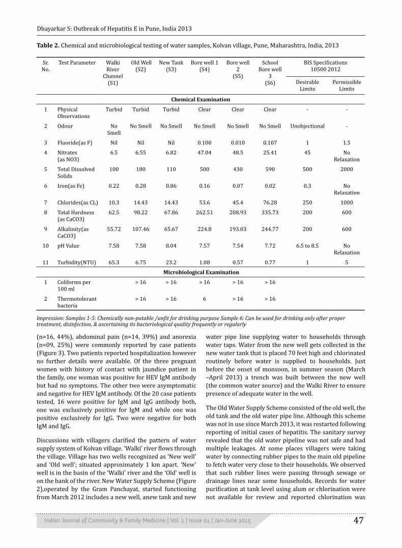
47Indian Journal of Community & Family Medicine | Vol. 1 | Issue 01 | Jan-June 2015
(n=16, 44%), abdominal pain (n=14, 39%) and anorexia (n=09, 25%) were commonly reported by case patients (Figure 3). Two patients reported hospitalization however no further details were available. Of the three pregnant women with history of contact with jaundice patient in the family, one woman was positive for HEV IgM antibody but had no symptoms. The other two were asymptomatic and negative for HEV IgM antibody. Of the 20 case patients tested, 16 were positive for IgM and IgG antibody both, one was exclusively positive for IgM and while one was positive exclusively for IgG. Two were negative for both IgM and IgG.
Discussions with villagers clarified the pattern of water supply system of Kolvan village. ‘Walki’ river flows through the village. Village has two wells recognized as ‘New well’ and ‘Old well’; situated approximately 1 km apart. ‘New’ well is in the basin of the ‘Walki’ river and the ‘Old’ well is on the bank of the river. New Water Supply Scheme (Figure 2),operated by the Gram Panchayat, started functioning from March 2012 includes a new well, anew tank and new
water pipe line supplying water to households through water taps. Water from the new well gets collected in the new water tank that is placed 70 feet high and chlorinated routinely before water is supplied to households. Just before the onset of monsoon, in summer season (March –April 2013) a trench was built between the new well (the common water source) and the Walki River to ensure presence of adequate water in the well.
The Old Water Supply Scheme consisted of the old well, the old tank and the old water pipe line. Although this scheme was not in use since March 2013, it was restarted following reporting of initial cases of hepatitis. The sanitary survey revealed that the old water pipeline was not safe and had multiple leakages. At some places villagers were taking water by connecting rubber pipes to the main old pipeline to fetch water very close to their households. We observed that such rubber lines were passing through sewage or drainage lines near some households. Records for water purification at tank level using alum or chlorination were not available for review and reported chlorination was
Table 2. Chemical and microbiological testing of water samples, Kolvan village, Pune, Maharashtra, India, 2013
Sr. No.
Test Parameter
Walki River
Channel(S1)
Old Well (S2)
New Tank(S3)
Bore well 1(S4)
Bore well 2
(S5)
School Bore well
3(S6)
BIS Specifications10500 2012
Desirable Limits
Permissible Limits
Chemical Examination
1 Physical Observations
Turbid Turbid Turbid Clear Clear Clear - -
2 Odour No Smell
No Smell No Smell No Smell No Smell No Smell Unobjectional -
3 Fluoride(as F) Nil Nil Nil 0.100 0.010 0.107 1 1.5
4 Nitrates (as NO3)
6.5 6.55 6.82 47.04 48.5 25.41 45 No Relaxation
5 Total Dissolved Solids
100 180 110 500 430 590 500 2000
6 Iron(as Fe) 0.22 0.28 0.86 0.16 0.07 0.02 0.3 No Relaxation
7 Chlorides(as CL) 10.3 14.43 14.43 53.6 45.4 76.28 250 1000
8 Total Hardness (as CaCO3)
62.5 98.22 67.86 262.51 208.93 335.73 200 600
9 Alkalinity(as CaCO3)
55.72 107.46 65.67 224.8 193.03 244.77 200 600
10 pH Value 7.58 7.58 8.04 7.57 7.54 7.72 6.5 to 8.5 No Relaxation
11 Turbidity(NTU) 65.3 6.75 23.2 1.08 0.57 0.77 1 5
Microbiological Examination
1 Coliforms per 100 ml
> 16 > 16 > 16 > 16 > 16
2 Thermotolerant bacteria
> 16 > 16 6 > 16 > 16
Impression: Samples 1-5: Chemically non-potable /unfit for drinking purpose Sample 6: Can be used for drinking only after proper treatment, disinfection, & ascertaining its bacteriological quality frequently or regularly
Dhayarkar S: Outbreak of Hepatitis E in Pune, India 2013

48Indian Journal of Community & Family Medicine | Vol. 1 | Issue 01 | Jan-June 2015
irregular prior to clustering of jaundice cases.
Water source for all the case patients was found to be new well which is a part of the New Water Supply Scheme. Ten out of 37 cases reported not having toilet facility in their houses. The villages reported that many residents followed practice of open air defecation and used the bank of Walki River for defecation. Water is released from Mulshi dam in Walki River every 15 days.
Water samples from the trench, the old well and the new tank were turbid. Coliform and thermo tolerant bacteria were present in the samples from the old well, the new tank; bore wells 1, 2 as well as the school bore well 3. Except the school bore well, all water sources and water samples were reported as chemically non potable for drinking. Water from the school bore well 3, was recommended for drinking only after proper treatment and disinfection if no other water source was available (Table 1).
Based on discussions with the villagers and staff of the sub centre, clinical history, findings of the environmental survey and water sample reports we generated the hypothesis that Hepatitis E outbreak occurred in Kolvan village due to drinking of water contaminated with faecal matter. We hypothesized that the river water could have been contaminated with faecal matter when water was released from dam, flushing banks of the river. This contaminated water could have mixed with the water in the new well water through the trench created between river and new well leading to contamination of the
common source of water used by the villagers.
“S” and “P” forms are formats designed under IDSP for weekly reporting of jaundice cases from sub centre (SC) and primary health centre (PHC) respectively. Review of reporting of this surveillance activity indicated that the reporting of cases was done for only few cases on “S” forms from the sub centre while there was no reporting on “P” forms.
Discussion
We confirmed the outbreak of hepatitis E in Kolvan village of Pune district between 28 May 2013 and 23 August 2013, which probably occurred following faecal contamination of common water supply of the village and drinking of contaminated water. Time distribution of cases indicates a point source common vehicle outbreak. There was no report of mortality or any other adverse event even among pregnant women. The outbreak revealed the need of ensuring safe water supply, improvement in environmental sanitation, and promoting behavior change in the community regarding hygienic practices and use of toilets.
Outbreaks of hepatitis E are traditionally linked to inadequate safe water supply and improper drainage system.17, 18 The attack rates in different age groups were higher than these reported in a similar outbreak from western Maharashtra.17 Although the epidemiological features of hepatitis E outbreak reports difference in attack rates in males and females19, our study showed no
Figure 1. Distribution of acute hepatitis E cases by week of onset of symptoms, Kolvan village, Pune,
Maharashtra, India, 2013
Dhayarkar S: Outbreak of Hepatitis E in Pune, India 2013

49Indian Journal of Community & Family Medicine | Vol. 1 | Issue 01 | Jan-June 2015
Figure 3. Symptom profile of hepatitis E cases during hep-atitis E outbreak, Kolvan village, Pune, Maharashtra, India, 2013 (N=36)*
difference in attack rates in males than females possibly indicating equal exposure and risk for both the sexes. Symptoms, and self limiting nature of the disease was consistent with documented natural history of disease.6,
8, 20 Majority of the reported hepatitis outbreaks have shown unimodal peaks although multiple peaks have been reported in a few outbreaks.21-25 Our findings were consistent with the reported outbreaks with one single peak suggestive of point source, common vehicle outbreak.
The outbreak was more concentrated in the densely populated area of the village. Lack of awareness of hygienic practices, inadequacy of toilets or reluctance to use toilets despite availability might be associated with spread of
Figure 2. Distribution of acute Hepatitis E cases by area and water system in Kolvan village, Pune, Maharashtra, India, 2013
the epidemic. Although construction of new water supply scheme was relatively recent, the sanitary survey showed that water taps were constructed at low level. Many needed repairs and they needed to be relocated to such heights not to be contaminated by flowing water. Our study emphasized the need for training of health care staff on early identification of outbreaks and prompt investigation to control the epidemic.
One of the limitations of this study was the inability to pinpoint the source of infection as the suspected water source was being used by almost all the households. In this scenario of universal exposure, we could not conduct an analytical study to test the hypothesis that outbreak could have occurred due to contamination of water from a single source or from multiple sources. We also could not rule out other sources of infection like zoonotic and food borne hepatitis.
We recommended continued surveillance of cases for few weeks following end of the outbreak; using alternate safe water supply until the new water supply system was safe and scrupulous chlorination of alternate safe water before supply. In the community, we recommended use of boiled water for drinking and use of medichlor tablets in households for disinfection. We suggested examination of pipelines for possible leakages after restarting of Gram Panchayat supplied water.
Measures were initiated for controlling the epidemic. The trench between river & new well was closed immediately. Water supply from both water supply systems was stopped
*One laboratory confirmed hepatitis case was asymptomatic hence excluded in symptom profile
Dhayarkar S: Outbreak of Hepatitis E in Pune, India 2013

50Indian Journal of Community & Family Medicine | Vol. 1 | Issue 01 | Jan-June 2015
and arrangement for alternative source of safe water was made. The bore well near the school was identified as an alternate source for drinking water. Water from the bore well was collected in specially placed tanks and supplied to village after super chlorination. Case search and symptomatic management of cases were continued. Hospitalization was advised for seriously ill cases or pregnant women. We promoted advocacy regarding of safe drinking water and sanitation in the village.
For prevention of recurrence we recommended re-laying of new pipeline for avoiding contamination at tap level and repair of old pipelines. We also recommended ensuring regular chlorination of water before supply as outbreaks of HEV have been reported to be successfully controlled by chlorination of water and local or external supervision for chlorination activity and ascertaining bacteriological quality frequently to ensure water potability.7,19 Behavioural change takes time and hence continuous focus on education among villagers regarding use of toilets and
following hygienic practices were also recommended. We recommend examination of water sources and pipelines for possible contamination and leakages prior to onset of rainy season to avoid future outbreaks of water borne diseases.
Acknowledgments
We thank Dr Karanjkar, Taluka Health Officer, Mulshi, Dr Garud, Medical Officer Male Primary Health Care, Dr Birade, Kolvan Sub Centre, Mr Shinkar, Zilla Parishad, Mr Wanwale, Multi Purpose Worker, and Kolvan Sub Centre for their support in data collection.
Funding
This study work was supported by intramural funding available for Masters of Public Health (MPH) programme at National Institute of Epidemiology, Chennai, Tamil Nadu, India.
References 1. Meng XJ. Zoonotic and foodborne transmission of hepatitis E
virus. Semin Liver Dis. 2013; 33(1): 41-9.2. Yugo DM, Meng XJ. Hepatitis E virus: foodborne, waterborne
and zoonotic transmission. Int J Environ Res Public Health. 2013; 10(10): 4507-33.
3. Tei S, Kitajima N, Takahashi K, Mishiro S. Zoonotic transmission of hepatitis E virus from deer to human beings. Lancet. 2003; 362(9381): 371-3.
4. Teshale EH, Howard C, Grytdal S. A large outbreak of hepatitis E in northern Uganda. Emerg Infect Dis. 2010; 16: 126-9.
5. Sharapov MB, Favorov MO, Yashina TL, Brown MS, Onischenko GG, Margolis HS, et al. Acute viral hepatitis morbidity and mortality associated with hepatitis E virus infection: Uzbekistan surveillance data. BMC infectious diseases. 2009; 9(1): 35.
6. World Health Organization. Hepatitis E. In: Department of Communicable Disease Surveillance and Response. [Available at www.who.int/csr/disease/hepatitis/HepatitisE_whocdscredc2001_12.pdf].
7. Delia B, Guthmann JP, Hilde K, NuhaH, Mercedes T, Iza C, et al. High Mortality Associated with an Outbreak of Hepatitis E among Displaced Persons in Darfur, Sudan. CID. 2006; 42(12): 1679-84.
8. World Health Organization. Hepatitis E Fact sheets. July 2013 [Available from: http://www.who.int/mediacentre/factsheets/fs280/en/#]
9. Corwin AL, Khiem HB, Clayson ET, Pham KS, Vo TT, Vu TY. A waterborne outbreak of hepatitis E virus transmission in southwestern Vietnam. Am J Trop Med Hyg. 1996; 54(6): 559-62.
10. Tsega E, Krawczynski K, BG H, Nordenfelt E, Negusse Y, Alemu W, et al. Outbreak of acute hepatitis E virus infection among military personnel in northern Ethiopia. J Med Virol. 1991; 34(4): 232-6.
11. Albetkova A, Drobeniuc J, Yashina T, Musabaev E, Robertson B, Nainan O, et al. Characterization of hepatitis E virus from outbreak and sporadic cases in Turkmenistan. J Med Virol. 2007; 79(11): 1696-702.
12. Khan A, Tanaka Y, Kurbanov F, Elkady A, Abbas Z, Azam Z. Investigating an outbreak of acute viral hepatitis caused by hepatitis E virus variants in Karachi, South Pakistan. J Med
Virol. 2011; 83(4): 622-9.13. Teshale EH, Hu DJ. Hepatitis E: Epidemiology and prevention.
World journal of hepatology. 2011; 3(12): 285.14. Naik SR, Aggarwal R, Salunke PN, Mehrotra NN. A large
waterborne viral hepatitis E epidemic in Kanpur, India. Bulletin of the World Health Organization, . 1992; 70(5): 597-604.
15. Acharya S, Madan K, Dattagupta S, Panda S. Viral hepatitis in India. The National medical journal of India. 2005; 19(4): 203-17.
16. IDSP. Case definitions. In: IDSP, editor.17. Gurav YK, Kakade SV, Kakade RV, Kadam YR, Durgawale
PM. A study of Hepatitis E outbreak rural area of Western Maharashtra. IJCM, 2007; 32(9): 182-4.
18. Vyas S, xParikh S, Kapoor R, Patel V, Solanki A. Investigation of an epidemic of hepatitis in Ahmedabad city National Journal of Community Medicine 2010; 1(1): 27-9.
19. Rosario V, Lalit N, Jeyaram I, Reddy PK, Sarkar R, Eapen CE, et al. Investigation of an epidemic of Hepatitis E in Nellore in South India. Tropical Medicine & International Health. 2010; 15(11): 1333-9.
20. Centre for Disease Control. Hepatitis E Yellow Book _ Travelers’ Health _ CDC; 2014.
21. Labrique AB, Thomas DL, Stoszek SK, Nelson KE. Hepatitis E: an emerging infectious disease. Epidemiologic reviews. 1999; 21(2): 162-79.
22. World Health Organisation. Hepatitis E. Geneva: World Health Organisation; 2012.
23. Singh V, Raje M, Nain C, Singh K. Routes of transmission in the hepatitis E epidemic of Saharanpur. Tropical gastroenterology: official journal of the Digestive Diseases Foundation. 1997; 19(3): 107-9.
24. Teshale EH, Grytdal SP, Howard C, Barry V, Kamili S, Drobeniuc J, et al. Evidence of person-to-person transmission of hepatitis E virus during a large outbreak in Northern Uganda. Clinical infectious diseases. 2010; 50(7): 1006-10.
25. Swain SK, Baral P, Hutin YJ, Rao TV, Murhekar M, Gupte MD. A hepatitis E outbreak caused by a temporary interruption in a municipal water treatment system, Baripada, Orissa, India, 2004. Transactions of the Royal Society of Tropical Medicine and Hygiene. 2010; 104(1): 66-9.
Dhayarkar S: Outbreak of Hepatitis E in Pune, India 2013

51Indian Journal of Community & Family Medicine | Vol. 1 | Issue 01 | Jan-June 2015
Abstract
Background: Human Immunodeficiency virus (HIV) infection in pregnancy has become the most common medical complication of pregnancy in some countries. Reports have shown that HIV infection during pregnancy can have adverse effects on both the mother and the infant. As of 2007, 90% of HIV infected children got the virus through MTCT. Hence, this study was planned to assess the outcome of pregnancy among HIV infected women. Methodology: A case series study was carried out on all HIV positive pregnant women on ART who delivered in KIMS hospital, Hubli during the six months of study period. Results: Of the total 25 HIV positive women, 16 (64%)were primi-gravida and 6 (24%) had obstetric history of G2P1L1A0. Seventeen (68%) women had full time normal vaginal delivery, 3 (12%) had a preterm and 3 (12%) delivered by emergency LSCS. Pregnancy outcome showed that 19 (76%) delivered a full term baby, 3 (12%) preterm baby, 1 (4%) had a stillbirth, 1 (4%) encountered intra uterine death of foetus and 1 (4%) underwent medical termination of pregnancy. Conclusion: Majority of HIV infected pregnant women delivered full term normal vaginal delivery of near normal birth weight without any birth defects. It indicates that antiretroviral therapy of pregnant HIV infected women is beneficial and should be emphasized.
Keywords: HIV, Pregnancy, Outcome.
Introduction
Human Immunodeficiency Virus infection in pregnancy has become the most common medical complication of pregnancy in some countries. Women are particularly susceptible to HIV infection for both biological and socio-cultural reasons.1 Acquired Immunodeficiency syndrome (AIDS) has become a great challenge to public health.
According to World Health Organization (WHO) in 2013, about two thirds of all pregnant women living with HIV in low- and middle-income countries, close to one million women received antiretroviral (ARV) drugs that prevent mother-to- child transmission.2 According to national HIV Sentinel Surveillance (HSS) 2012-2013, the overall HIV prevalence among ANC attendees continued to be low at 0.35% in the country, with an overall declining trend at the national level.3 Yet, it was estimated that around 38,000 HIV positive pregnant women needed Prevention of Parent to Child Transmission (PPTCT) services in 2011.4
Transmission of HIV from a mother to child can be in-
utero, during labor and delivery or during breastfeeding the infant. Studies have shown that 60-70% of the transmission occurs during delivery, 20-30% through breastfeeding and <10% in-utero.5 The HIV-positive women have been found to be relatively younger and more likely to have maternal and fetal adverse effects.6 323 women using highly active antiretroviral therapy (HAART) found 2.6 times increased risk of preterm delivery with exposure to HAART with a protease inhibitor (PI) and 1.8 times increased risk associated with HAART without a PI. US studies found an increased very low birth weight rate associated with HIV infection.7 Adverse outcomes like birth defects were not common and was equal with exposure in the first trimester and after first trimester.8 The prevalence of congenital anomaly (CA) after first trimester antiretroviral therapy (ART) was found to be similar to that of second and third trimester ART.9
Maternal characteristics and co-morbidities do influence pregnancy outcomes, even under optimal ART. Reports on pregnancy outcome in HIV infected women from North
SHORT COMMUNICATION
Outcome of pregnancy among HIV infected women in a tertiary care centre, Hubli, Karnataka, India
Anil Kumar L1, Bant Dattatreya D2, Sheethal S3
1Postgraduate Student, 2Professor and HOD, Department of Community Medicine, KIMS, Hubli. 3 Postgraduate Student, Department of Microbiology, KIMS, Hubli, Karnataka, India.
Address for Correspondence: Dr Anil KL, Department of Community Medicine, KIMS, Hubli. Email: [email protected]: 29.11.2014, Accepted: 11.01.2015
52Indian Journal of Community & Family Medicine | Vol. 1 | Issue 01 | Jan-June 2015
Karnataka are limited. Therefore, the present study was taken to assess the pregnancy outcome in HIV infected women delivering in KIMS hospital, Hubli.
Methodology
A descriptive study was conducted with case series of HIV-positive pregnant women who delivered in Karnataka Institute of Medical Sciences (KIMS) hospital, Hubli, Karnataka, India during the period of 6 months from April 2014 to September 2014.
A pilot study was done with a representative sample and a final structured proforma was prepared through which data was collected. Information was collected from each mother-infant pair for a period of 48 hours after childbirth, including specific socioeconomic characteristics, clinical and biological features about pregnancy, childbirth and postnatal period using a semi-structured questionnaire. For twins, twin pair were included in the study. Clinical factors included mode of delivery, type of ARV treatment and any existent co-morbid conditions. Biological factors included hemoglobin percentage, CD4 count, smoking, alcohol intake and any drug consumption during pregnancy. During labour, two drugs Efavirenz and Zidovudine were given. The postpartum intervention was given as per the national programme.
Adverse pregnancy outcome was defined as occurrence of any of the following conditions: pre-term delivery, cervical shortening, premature rupture of membranes (PROM), spontaneous abortion, fetal anomalies, intrauterine growth retardation, intrauterine death (IUD), low birth weight, still birth, perinatal death and neonatal death. Explanatory variables included were maternal cigarette smoking, drug use, co-infections (hepatitis B, hepatitis C and syphilis), ART regimen, CD4+ cell count and maternal age.
The study was approved by the Institutional Scientific and Ethical Committee of Karnataka Institute of Medical Sciences, Hubli. Respective higher authorities’ permission has been obtained before starting the study like District AIDS prevention and control officer (DAPCO) Officer, Dharwad, The Nodal Medical officer, ART Centre, KIMS, Hubli and The Senior Medical Officer, ART Centre, KIMS, Hubli. Individual consent was obtained before interviewing the pregnant women.
All data was entered into MS Excel and subsequently analyzed using SPSS Vn. 17. Descriptive analysis were done for baseline values and chi-square test wherever applicable.
Results
A total of 25 women delivered during the study period and their age showed that 17 (68%) of them belonged to
20-25 years, 4 (16%) in 26-30 years age group and rest in 31-35 years age group each. Religion of 16 (64%) women was Hindu and 5 (20%) were Muslims. All of them were married and 16 (64%) subjects were residing in rural areas in comparison to 9 (36%) from the urban areas. Eight (32%) females were illiterate, 7 (28%) each were schooled up to primary and secondary education and only a mere 3 (13%) had attained pre-university education. None belonged to Class I socio economic status group. (Table 1)
Parity of the women showed that 16 (64%) were primi-gravid women and 6 (24%) had obstetric score of G2P1L1A0. Three (12%) cases had < 3 antenatal visits, 8 (32%) women had got 5 antenatal visits done and one had directly presented in labour. All women had received appropriate TT injections and Iron and Folic Acid tablets recommended during pregnancy. Even then, anaemia was present in 6 (24%) women, 2 (8%) had previous baby deaths, 3 (12%) had previous LSCS and 1 (4%) was an elderly primi. Nine (36%) women were detected to be HIV positive before current pregnancy and 16 (64%) were detected during their current pregnancy. In the study group, 17 (68%) women had full term vaginal delivery, 3 (12%) had a pre-term vaginal delivery, 3 (12%) were taken up for emergency LSCS, 1 (4%) underwent elective LSCS and 1 (4%) underwent MTP. Pregnancy outcome showed that 19 (76%) delivered a full term baby, 3 (12%) delivered a preterm baby and one each had stillbirth, intra uterine death of fetus and medical termination of pregnancy. Three (12%) women delivered newborns with birth weight < 1.5 kg, 6 (24%) delivered 1.5-2.49 kg baby and 8 (32%) gave birth to 2.5 kg baby. None of them had the habit of smoking or consumption of alcohol and none were suffering from any co-infections (hepatitis B, hepatitis C and syphilis). All the patients were diagnosed to have HIV-1 infection and started on TNF+LMY+EFV (Tenofavir+Lamivudine+Efavirenz) drug regimen with a mean CD4 count of 598+ 238 and hemoglobin percentage of 9.20+ 1.25. CD4 counts > 500/ mm3 was found in 16 (64%) cases, 350-499/ mm3 in 7 (28%) cases and each (4%) had counts of 200-349/ mm3 and 200/ mm3. (Table2)
Discussion
Infections like HIV could have substantial adverse effect on the pregnancy outcomes, but timely diagnosis and treatment with ART could bring improve the outcomes. The findings from the study showed that 12% had preterm delivery and 1% had resulted in stillbirth. Varied outcomes were recorded from other researchers from different geographical areas. A study done in Thailand by Areechokchai et al. recorded the incidence of pre-term delivery at 10.7% and Jennifer et al. in Nigeria found that there was no significant association between maternal HIV-status and the risk of adverse pregnancy outcomes
Anil Kumar L: Pregnancy outcome among HIV infected women

53Indian Journal of Community & Family Medicine | Vol. 1 | Issue 01 | Jan-June 2015
such as abortions, stillbirths and infant death after birth.5 Boer et al. in Netherlands found that the increase in pre-term delivery in HIV infected women was mainly confined to those women who used HAART for longer duration of time, therefore the finding could be attributed to more advanced HIV infection.7 A recent study in Brazil found that prematurity was not independently associated with HIV infection.10 The outcome found in the present study is better than those seen in studies in other parts of the world where the proportion of preterm was as high as 20%.11, 12, 13 There were other studies that showed similar result as the present study, of no adverse outcome among newborns of HIV positive mothers. This indicates the need for continued attention to birth outcomes in HIV positive women, as there seems to be no consistency in the findings of various studies and it has important bearing on child health.
The present study appreciated 17(68%) full term vaginal delivery, which is similar to other studies.7,14 The European collaborative study documented geographical variation in mode of deliveries in the HAART era with an increasing proportion of vaginal deliveries, mainly in the U.K, Belgium and Netherland.15 The mode of delivery was attributed to the availability of infrastructure for surgical intervention,
Anil Kumar L: Pregnancy outcome among HIV infected women
Table 1. Showing distribution according to socio demographic profile.
Age n (25) %
20-25 17 68
26-30 4 16
31-35 4 16
Religion
Hindu 16 64
Muslims 5 20
Christian 1 4
Others 3 12
Residence
Urban 9 36
Rural 16 64
Total 25 100
Educational status
Illiterate 8 32
Primary 7 28
Secondary 7 28
Pre-university 3 12
Socio-economic status
Class II 1 4
Class III 6 24
Class IV 15 60
Class V 3 12
Table 2. Showing obstetric profile of HIV infected pregnant women
Obstetric Index n (25) %
Primi gravid 16 64
G2P1L1A0 6 24
G5P4L4A0 1 4
G2P1L0A0D1 2 8
No of ANC
< 3 3 12
5 8 32
6 9 36
7 4 16
Direct in labour 1 4
High risk detected
Anaemia 6 24
Previous baby death 2 8
Previous LSCS 3 12
Elderly 1 4
NIL 13 54
Mode of delivery
FTVD 17 68
PTVD 3 12
Emergency LSCS 3 12
Elective LSCS 1 4
MTP 1 4
Outcome of delivery
Full term baby 19 76
Preterm baby 3 12
Stillbirth 1 4
IUD 1 4
MTP 1 4
Birth weight of Newborn (kg)
< 1.5 3 12
1.5 – 2.49 6 24
2.5 8 32
2.6 – 3.5 3 12
> 3.5 1 4
Diagnosis of HIV
Before current pregnancy 9 36
During current pregnancy 16 64
CD4 count
> 500/ mm3 16 64
350 − 499/ mm3 7 28
200 −349/ mm3 1 4
< 200/ mm3 1 4

54Indian Journal of Community & Family Medicine | Vol. 1 | Issue 01 | Jan-June 2015
Anil Kumar L: Pregnancy outcome among HIV infected women
Reference1. HIV in pregnancy: A review, Joint United Nations Programme
on HIV/AIDS (UNAIDS), 1998.2. World Health Organization. HIV Reporting Global Update on
Health Sector Response to HIV, WHO: Geneva, 2014.3. Annual Report 2013-14, Department of AIDS Control, Ministry
of Health & Family Welfare, GOI.4. Infection control and waste management plan for National
AIDS Control Support Project (NACSP) 2012-2017, National AIDS Control Organization, Ministry of Health and Family Welfare, Government of India, 2012. Available at:http://www.naco.gov.in/upload/NACSP.pdf [last cited on 20/10/2014]
5. Jennifer VT, Valentine Okoye, Francis Donkene and Dr Summit Kar Comparing Pregnancy Outcomes in HIV Infected and HIV-Uninfected Women in Adamawa State, Nigeria: a registry-based study. International Conference on Medical, Biological and Pharmaceutical Sciences, Pattaya, 2011: 502-510.
6. Priya Thayaparan, Mohanarathi K, Thambiah B. Adverse pregnancy outcomes in HIV positive women: A study from a District General Hospital in the UK. International Symp on HIV and Emerging Infect Diseases (ISHEID), Marseille, France, 2012:23-25.
7. Boer K, Nellen JF, Patel D, Timmermans S, Tempelman C, Wibaut M, et al. The AmRo study: pregnancy outcome in HIV-1-infected women under effective highly active antiretroviral therapy and a policy of vaginal delivery. Br J Obstet Gynaecol. 2007; 114:148–55.
8. Heather Watts, Sharon Huang, Mary C, Kathleen AK et al. Birth defects among a cohort of infants born to HIV-infected women on antiretroviral medication. J Pernat Med, 2011; 39(2); 163-170.
9. Esau CJ, Guilherme AC, Margot RK, Laura FH et al. Maternal Antiretroviral Use during Pregnancy and Infant Congenital Anomalies: The NISDI Perinatal Study. J Acquir Immune Defic Syndr, 2010; 53(2):176-185.
10. T.P. da Costa, M.C. Leal, J.C. Mota, E.S. Machado and E. Costa et al. Comparison of pregnancy characteristics and outcomes between HIV-infected and HIV-non-infected women in Brazil. AIDS Care, 2013; 25(6):686-690.
11. Karoline AP, Olav L, Tracy R.G and Louise V et al. Pregnancy and delivery outcomes of HIV infected women in Switzerland. J. Perinat. Med, 2010;38:353–358.
12. Stratton P, Tuomala RE, Abboud R, Rodriguez E et al. Obstetric and newborn outcomes in a cohort of HIV infected pregnant women: a report on women and infant transmission study. J Acquir Immu Defic Syndr Hum Retroviral, 1999;20(2):179-86
13. Denise N, Azucena B, María L, Anna B et.al. Impact of maternal human immunodeficiency virus infection on birth outcomes and infant survival in rural Mozambique. Am. J. Trop. Med. Hyg, 2009; 80(5):870–876
14. Darin A, Chureeratana B, Benjaluck P, Punnee P, and Wirach M. Pregnancy Outcomes among HIV-Infected Women Undergoing Antiretroviral Therapy. The Open AIDS Journal, 2009;3:8-13.
15. European Collaborative Study. Mode of delivery in HIV-infected pregnant women and prevention of mother-to-child transmission: changing practices in Western Europe. HIV Med. 2010;11:368–378.
16. Anbarasi S, Sonali S, Gautam Roy and Subitha L. Experiences of HIV Positive Mothers from Rural South India during Intra-Natal Period. J of Clin and Diagn Research, 2013 Oct, Vol- 7(10): 2203-2206.
17. Maurice B, Jean de DB, John M and Claire P. Socio-economic, clinical and biological risk factors for mother-to-child transmission of HIV-1 in Muhima health centre (Rwanda): a prospective cohort study. Arch of Pub Health 2013, 71-74.
18. Lily K, Inam C, Chewe L and Nigel R. Opportunities for Africa’s Newborns: Prevention of mother-to-child transmission of HIV/AIDS programmes. WHO, chapter III:113-126. Available at:http://www.who.int/pmnch/media/publications/aonsectionIII_7.pdf [last cited on 21/10/2014]
doctors risk assessment approach and patient preference.
The birth weight of newborns in the present study corroborates with the studies done in other parts of the world.5,7,11,13 This reflects that birth weight depends on the duration and severity of HIV infection. Age distribution of the present study was similar to those found by Boer et al. found the median age was 29 years.7 The study done in Thailand by Areechokchai et al. showed median 27 years. Anbarasi et al also recorded that majority of their subjects were less than 30 years of age.16 The involvement of this age group women would mean that the problem would continue to affect their entire reproductive lives should they choose to bear more children. Therefore timely screening and treatment would be vital to curtail the adverse effects.
The present study relating to the high risk factors, recorded 6 (24%) pregnant women having anemia which is less than that observed by Naniche (51.3%).13 This could be due to early detection and timely intervention, which will positively affect the outcome. Regular antenatal check-ups of had been undergone by 96% of women and they had their ANC check-ups done by a doctor and all of them received IFA tablets and TT injections. This was similar
to the study which suggests that there is improvement in health care delivery services regards to mother and child care issues.16
Due to advancement in the antiretroviral therapy, HIV positive people can live healthier lives with almost normal life expectancies. This reinforces the desire in HIV positive women to have their own children and plan their pregnancies. The scaling up of effective interventions for the prevention of HIV transmission from mother to child (PPTCT) is still limited because of inadequate access to antenatal and postnatal services particularly in the developing countries. 17,18 The present study was limited by small sample size, no comparison group with HIV negative women and lack of information on HIV viral load.
Conclusion
Majority of HIV infected pregnant women delivered FTNVD of near normal birth weight newborn without any birth defects pointing that antiretroviral therapy for treatment of a pregnant HIV infected women is beneficial which should be emphasized. Equal importance to maximize early and regular ANC visits has to be included, which would further ensure early detection & treatment of all high risk conditions including HIV.

55Indian Journal of Community & Family Medicine | Vol. 1 | Issue 01 | Jan-June 2015
AbstractLeptospirosis is an emerging public health problem in India which affects both human and animals. A 62 years old male, resident of rural area presented in out patient department of a private super specialty hospital with complaints of fever with chills, pain in abdomen and difficulty in breathing. His initial investigation showed plasmodium vivax positive, acute cholecystitis, elevated liver enzyme and thrombocytopenia. He was unresponsive for intravenous antibiotics. Next day he developed per rectal bleeding. After negative finding of dengue testing, diagnostic test for leptospirosis was applied, which was positive. This case of leptospirosis should serve to alert health-care providers and the general public regarding the clinical and public health importance of this severe, sometimes fatal, disease.
Keywords: Leptospirosis, Public Health, Epidemiology
Introduction
Leptospirosis is a common zoonosis worldwide that affects mammals, including human beings.1-3 It is an emerging public health problem in India and other developing countries3,4 and considered an occupational hazard of agricultural workers, sewage workers, veterinarians etc.5 In the recent past, the incidence has been showing sudden upsurges in Kerala, Tamil Nadu, Maharashtra, Gujarat, Delhi, North East, Karnataka, Bihar, North India, Pondicherry.6,7 WHO estimates the incidence of leptospirosis between 0.1–1 cases/100,000 population/year in temperate, non-endemic areas and between 10–100 cases/100,000 population/year in humid, tropical and endemic areas.6
Case Report
A 62 years old male, resident of rural area of Dewas district, goat-herd by profession, presented in an evening OPD of a private super specialty hospital of Indore district with complaints of fever with chills, pain in abdomen and body ache for more than 10 days. He also had difficulty in breathing and diarrhea for the last 2 days. Fever was moderate and of sudden onset in nature. Pain in abdomen was mainly located in epigastrium and right hypochondrium region. It was non-radiating and not
associated with any aggravating or relieving factor. He didnot have any history of haemtemesis, melena, weight loss, diabetes mellitus, hypertension or tuberculosis.
On general examination, he was conscious, well oriented, with blood pressure of 100/70 mm of Hg in left arm in supine position, pulse rate 96/minute, respiratory rate was 22/minute, febrile with body temperature of 100oC, grade 3 dehydration and icteric. The lung was clear on auscultation, abdomen was soft and tender.
Routine blood examination revealed, that his hemoglobin was 11.8 gm/dl, white blood cell 8200 cell/mm3, platelet count was 86000/mm3, erythrocytes were normocytic and normocromic, neutrophil count was within normal range. Total bilirubin was found elevated (4.97mg/dl) with direct 2.73mg/dl and indirect 2.24mg/dl, alkaline phosphatase (ALP) of 142 U/lit, aspartate aminotransferase (AST) of 224 U/lit, alanine aminotransferase (ALT) of 254 U/lit, prothombin time of 18 seconds, International normalized ratio(INR )1.6 and creatine kinase of 94 U/lit. Electrolytes like sodium and potassium were within normal range. The urine analysis showed clear fluid with few pus cells but no hematuria and proteinuria. Chest radiograph and ECG were normal. Abdominal ultrasonography showed, calculi in gall bladder (Cholelithiasis) and thick cystic wall (cholecystic changes) which was suggestive of acute
CASE REPORT
A case of leptospirosis reported from metropolitan city of central India
Dhruvendra Pandey1, Satish Saroshe2, Sanjay Dixit3, Chakresh Jain4
1,4Resident, 2Assistant Professor, 3Professor& Head, Department of Community Medicine, MGM Medical College, Indore, Madhya Pradesh, India.
Address for Correspondence: Dr. Dhruvendra Pandey, Resident, Department of Community Medicine, MGM Medical College, Indore (Madhya Pradesh), E mail: [email protected], Mob No. 08269075295 Received: 15.11.2014, Accepted: 28.01.2015

56Indian Journal of Community & Family Medicine | Vol. 1 | Issue 01 | Jan-June 2015
cholecystitis. Patient was admitted in general ward with diagnosis of acute cholecystitis with jaundice. Treatment for acute cholecystitis was started with IV fluid and IV antibiotics, Metronidazole, Clindamycin, Levofloxacin, Ceftriaxone.
Next day patient had two episodes of per rectal bleeding and patient was shifted immediately to Intensive care unit and 2 units of Fresh Frozen Plasma and one unit of packed cell RBC were transfused. Platelet transfusion could not be done as the facility was not available at the center. On serological examination, malaria was positive on rapid diagnostic kit especially for plasmodium vivax and Dengue was found negative. After this, diagnosis was changed to acute cholecystitis with vivax malaria. Treatment for malaria i.e. artesunate and primaquine was started and G6PD was advised before initiating primaquine.
Despite of all this complaints and therapeutic interventions patient had persistant rise of temperature with chills. His platelet count was 86,000cells/mm3, electrolytes were normal; Hemoglobin was 9.6gm/dl. Considering the fact that patient was a goat herd by profession, he was advised to have IgM Leptospirosis test done. On fifth day of his admission, he was diagnosed as leptospirosis positive, IgM leptospira 11.6 (The test procedure was performed according to the protocol provided along with the kit. The results were interpreted according to manufacturer’s instructions, i.e. values, < 9 ELISA units were considered negative, 9–11equivocal, and > 11 positive). Doxycycline 100mg BD was added to the regimen. His platelet count started increasing to 1.2 lakh and bilirubin started decreasing. Patient started improving and became afebrile and was discharged with proper advice of regular follow up. Unfortunately, MAT (Microscopic Agglutination Study) testing was not applied due to nonavailibility of test facilities at the treating health center.
Patient did not have past and family history of leptospirosis infection. None of previously reported case of leptospirosis was found in published literature from central India especially Indore division and vicinity. Patient was a goat-herd by profession and frequently goes to forest and hilly area along with his goat. He lives in kuchha house in the vicinity of goats. Hand pump situated at 50 meter distance was the main source of drinking water. Sanitary latrine was present. Family members had poor personal hygiene. Patient had history of recent visit to his native place Banswara situated in Rajasthan.
Discussion
Leptospirosis had been suspected in India since the early part of the 20th century. The first report of bacteriologically confirmed and common-source outbreak of leptospirosis was reported from the Andaman Islands in 1929.8 In India, the disease is more commonly associated with
natural disasters, especially during the monsoon period. A multi-centric study in India showed that leptospirosis accounts for about 12.7% of cases of acute febrile illness responsible for attendance at hospitals.9 Peak incidence of the disease is found during August and September.2 In the present case, the patient came to hospital in the month of August and September. Leptospira organisms require humid weather for their survival. Rodents and domestic animals (i.e., cattle and dogs) harbor leptospires and shed the bacteria in urine; they may disseminate the organism to rain water and drinking water sources.2 The sero-prevalence is highest among cows (40.3%) followed by buffaloes (37.0%) and goats (36.3%).10 Humans are accidental host, getting infection either by direct contact with infected urine or tissues through injured skin or nasal or mouth mucosa or eyes or by indirect way (endemic outbreaks) through contact with contaminated stagnant water, dirt, or food.9
The fatality rate in severe leptospirosis may be as high as 20%.5 Hematological manifestations are common in leptospirosis and are usually manifested as thrombocytopenia which is a significant predictor of the development of acute respiratory failure, and is currently the main cause of death in this disease.11 Tantitanawat and Tanjatham12 found that platelet counts 100,000/mm3 were an independent risk factor for death in leptospirosis. Hepatic dysfunction in severe leptospirosis can be seen and conjugated serum bilirubin levels may increase to above 80 mg/dl, accompanied by modest elevations in transaminases, which rarely exceed 200 U/lit.13 Routine laboratory testing is non diagnostic but may show mild increase in aminotransferases, and increased serum bilirubin and ALP.13
Isolation of the organism by culture of clinical specimens (blood, CSF, urine) during the first 7 to 10 days of the illness is considered the gold standard of diagnosis. However, this method is difficult, requires longer than 16 weeks. The majority of leptospirosis cases are diagnosed by serologic testing of which MAT and IgM ELISA are most common .13
The diagnosis of leptospirosis requires a high degree of clinical suspicion because the disease’s numerous manifestations can mimic other tropical infections and easily confused with other febrile diseases or other nonspecific febrile illnesses, as well as noninfectious diseases such as small vessel vasculitides, systemic lupus erythematosus, or even malignancies.13
The current choices of treatment for mild leptospirosis include oral doxycycline and amoxicillin and for fulminant leptospirosis include parenteral high-dose penicillin G and broad-spectrum third generation cephalosporin (cefotaxime and ceftriaxone).14, 15 Role of penicillin is still doubtful as per one report.16 However, metanalysis has
Pandey D et al. : A Case Report of Leptospirosis Reported From Metropolitan City of Central India

57Indian Journal of Community & Family Medicine | Vol. 1 | Issue 01 | Jan-June 2015
shown ceftriaxone and penicillin to be equally effective in treatment of severe leptospirosis.17 Selection of proper antibiotics does not seem to impact morbidity especially in severe leptospirosis.18
In the present case, the diagnosis of leptospirosis was not initially considered because potential risk factors were not identified at the outset. It is not common to think about leptospirosis by health care providers. However during the course of the treatment the treating physicians realised that the the patient was goat-herd by occupation and leptospirosis went for serological testing using IgM ELISA. The present case report realizes the importance of social history and for that matter proper history taking is
vital. With the advent of modern sophisticated equipments and diagnostics we are laying less importance of proper history taking. Had the history taking been thoroughly comprehensive the diagnosis of leptospirosis could have been made much earlier and unnecessary morbidity of the patient could have been reduced in the course.
Both neglected and under-reported, there is scant epidemiological data on leptospirosis therefore masking the true scope of disease prevalence and making reliable morbidity and mortality statistics difficult to ascertain.19 Based on the observations made, there is an urgent need in strengthening of surveillance system, orientation & training of health professional for this disease.
Pandey D et al. : A Case Report of Leptospirosis Reported From Metropolitan City of Central India
References:1. World Health Organization. Human Leptospirosis: Guidance
for Diagnosis, Surveillance and Control; WHO: Geneva, Switzerland, 2003.
2. Chaudhry R, Premlatha M, Mohanty S, Dhawan S, Singh K, Dey AB. Emerging Leptospirosis, North India December 2002. Emerging Infectious Diseases. Vol. 8, No. 12, 1526-1527.
3. Sehgal SC. Leptospirosis in the horizon. Natl Med J India 2000;13: 228–30.
4. World Health Organization (WHO). Leptospirosis in India – report of the investigation of a post-cyclone outbreak in Orissa, November 1999. Wkly Epidemiol Rec 2000; 27: 217–223.
5. Levett PN. Leptospirosis. Clin Microbiol Rev 2001; 14 (2): 296–326.
6. Sethi S, Sharma N, Kakkar N, Taneja J, Chatterjee SS, et al. (2010). Increasing Trends of Leptospirosis in Northern India: A Clinico-Epidemiological Study. PLoS Negl Trop Dis 4(1): e579.
7. A. DircioMontes Sergio, E. Gonz´alez Figueroa, Verdalet Guzm´anMar´ıa Saadia, Soler Huerta Elizabeth, Rivas S´anchez Beatriz, M. Altuzar Aguilar V´ıctor, and J. Navarrete Espinosa. Leptospirosis Prevalence in Patients with Initial Diagnosis of Dengue. Journal of Tropical Medicine Volume 2012, Article ID 519701.
8. P. Vijayachari, Ahmed N, Sugunan A. P., Ghousunnissa S, Rao K. R, et. al. Use of Fluorescent Amplified Fragment Length Polymorphism for Molecular Epidemiology of Leptospirosis in India. Journal Of Clinical Microbiology, Aug. 2004, Vol. 42, No. 8.3575–3580.
9. Parasuraman Basker, Pichai Kannan, Karumana Gounder Kolandaswamy. Study on the Prevalence of Leptospirosis among Fever Cases Reported from Private Clinics in the Urban areas of Villupuram District, Tamil Nadu, India. Osong Public Health Res Perspect 2014 5(1), 54-67
10. Sharma S., Vijayachari P., Sugunans A. P., Sehgal C. Leptospiral carrier state and seroprevalence among animal population – a cross-sectional sample survey in Andaman and Nicobar Islands. Epidemiol. Infect. (2003), 131, 985–989
11. Elizabeth F. Daher, Geraldo B. Silva Junior, Charles O. Silveira et. al. Factors associated with thrombocytopenia in severe leptospirosis (Weil’s disease). CLINICS 2014;69(2):106-110
12. Tantitanawat S, Tanjatham S. Prognostic factors associated with severe leptospirosis. J Med Assoc Thai. 2003;86(10):925-31.
13. PS Singh, V Tiwari. Acute fulminent leptospirosis with multi-organ failure: Weil’s disease. JIACM 2012; 13(4): 330-3
14. Raptis L, Pappas G, Akritidis N: Use of ceftriaxone in patients with severe leptospirosis. Int J Antimicrob Agents 2006; 28(3): 259–261.
15. Suputtamongkol Y, Niwattayakul K, Suttinont C, et al. An open, randomized, controlled trial of penicillin, doxycycline, and cefotaxime for patients with severe leptospirosis. Clin Infect Dis 2004; 39(10): 1417-24.
16. Panaphut T, Domrongkitchaiporn S, Vibhagool A, Thinkamrop B, Susaengrat W. Ceftriaxone compared with sodium penicillin g for treatment of severe leptospirosis. Clin Infect Dis Off Publ Infect Dis Soc Am. 2003 Jun 15;36(12):1507–13.
17. Charan J, Saxena D, Mulla S, Yadav P. Antibiotics for the Treatment of Leptospirosis: Systematic Review and Meta-Analysis of Controlled Trials. Int J Prev Med. 2013 May;4(5):501–10.
18. Brett-Major DM, Coldren R. Antibiotics for leptospirosis. Cochrane Database Syst Rev. 2012;2:CD008264.
19. Bandara et al. Globalization of leptospirosis through travel and migration. Globalization and Health 2014;10:61.

58Indian Journal of Community & Family Medicine | Vol. 1 | Issue 01 | Jan-June 2015
Systems approach is a very important concept in education.1 In essence, it means that various components of any system are interdependent and any change in one is likely to produce a change – desirable or undesirable – in the other components.2 Although systems approach is included in almost all training related to educational technology, its application in actual practice has been rather limited.
It is interesting to look at medical education as a system. At the macro level, it is made up of society, regulatory bodies, government, medical schools, teachers and students. At the micro level, various smaller systems can be identified. 1,2 These could be a combination of teachers, students and curricula; or it could be the more conventional combination of educational objectives, teaching methods and assessment. Each one of these is important, dependent on others and individually capable of causing reactionary changes in other components of the system. For example, a change in the examination pattern will automatically change the way in which students learn and teachers teach. Yet, when planning or implementing educational reforms, we seem to forget this phenomenon.
Since the time of independence, many ‘reforms’ related to medical education have been implemented in India. 3,4 Most of these were initiated due to a perceived problem- be it the number of graduates produced by the country, the type of curriculum being followed, or the quality of the end products. Any mention of reforms is likely to evoke varied responses from different stakeholders. For some, attempts at initiating reforms will represent a progressive approach to keep medical education up to date. For others, the same reforms might appear to be desperate attempts to rescue medical education from the decline it is facing. None of these extreme stands, however, is the absolute truth. And it is this lack of clarity, which has resulted in too many interventions happening too soon- several of which resemble a Band-Aid type of stop-gap therapy, rather than a radical cure. This is of course, quite understandable – one cannot demolish a house in which one is living to build a new one; rather, one has to make small alterations
depending on one’s changing needs.
In this discourse, we will focus on three main components of the medical education system, viz. students, teachers and the curricula.
Student selection
Students form the backbone of any educational system. The well-known meta-analysis by Hattie suggested that 50% of the variance in student achievement can be attributed to the quality of students; another 30% is attributable to teachers and the remaining 20% is related to various other factors like method of teaching and quality of schools.5-6 Although this analysis pertained to school education, the results, to a large extent, can be extrapolated to higher education as well. The characteristics of students in terms of their epistemological beliefs, study habits, learning approaches and values have a large influence on the outcome of education. The way we select our students makes a large impact on the quality of our final product.
Our entrance examinations do not meet any of the acceptable criteria for selection. Although the argument is applicable at all levels, we will discuss the issue of selection at the postgraduate level, as the arguments are more apparent there. In our country, we are more impressed by objectivity rather than by the reliability of selection procedures. If objectivity was so impressive an attribute, then the rank order of students should have been the same in all entrance examinations. Unfortunately that does not happen and it is not unusual to see a student making it to the top 50 in one entrance test, but totally missing the mark in another. Selecting students on the basis of a single test is one of the worst approaches to student selection. The latest obsession with the single national eligibility and examination test (NEET) has further compounded the problem. A look at the Medical Council of India’s Vision 2015 document, suggests that the primary purpose of having an exit examination was to weed out ‘incompetent’ students. There are many flaws in this line of thought.7
Invited Article
COMMENTARY
Medical education reforms in independent India
Tejinder Singh1, Anshu2
1 Professor of Pediatrics and Medical Education, Director, CMCL-FAIMER Regional Institute Christian Medical College, Ludhiana, Punjab, India 2 Professor, Department of Pathology, MGIMS, Sevagram, Wardha, Maharashtra, India.
Address for Correspondence: Dr. Tejinder Singh, MD, DNB, MHPE, Professor of Pediatrics and Medical Education, Director, CMCL-FAIMER Regional Institute, Christian Medical College, Ludhiana, Email: [email protected]

59Indian Journal of Community & Family Medicine | Vol. 1 | Issue 01 | Jan-June 2015
Although not explicitly stated, the concept of NEET seems to have been largely based on the United States Medical Licensing Examination (USMLE). Both these examinations are conducted at the national level and the merit list for selection is made on the basis of the results of these examinations. However, the similarity ends there. Although the questions asked in NEET are not in public domain, it is logical to assume that when students are expected to solve 200 multiple choice questions (MCQs) in 180 minutes, these can only be simple recall type. The questions asked in other entrance examinations in India (like the All India PG entrance examination) are esoteric, generally test trivia, are often based on single case reports and are known to test recall only.8 In contrast, the questions asked in USMLE are application-oriented and contextual. Here 322-355 MCQs are to be answered in 8 hours.9 In addition, scores at USMLE do not override other measures of a student’s ability like her past performance, reference letters and personal interview.10 The total testing time for USMLE is more than 41 hours in contrast to our 3 hours.9-10 In addition, the exit examination in the USA is amply complemented by a robust system of ongoing in-training assessment, which helps to ensure that all those appearing for the exit examination have reached a desired level of competence in terms of clinical skills.9-10
A large volume of research has demonstrated that scores of an entrance examination alone are not a good predictor of future performance. A recent paper and probably the only one on this aspect from India- also documented lack of any correlation between performance in MBBS course and scores in the pre-medical tests.11 The only predictor of future performance was past performance.11 Somehow, we in India, don’t seem to believe in this concept. How else can one justify that the result of University examinations based on assessment by 56 examiners (14 subjects multiplied by 4 examiners each) is not trusted by anyone in the country!
Teachers
Teachers are the next important component of this system. Medical teachers in India wear multiple hats- including those of a teacher, a clinician and sometimes also an administrator. Several of them also engage in private practice. There is no system of ensuring that a person appointed as a medical teacher has the requisite teaching skills or at least acquires them after being appointed. In view of this issue, teacher training was initiated by the National Teacher Training Centers (NTTCs) and started at five medical schools in India.12 While the initiative was laudable, the intake capacity was limited for a country as large as ours. Cessation of central funding forced these NTTCs to either curtail their operations or shut shop. The Foundation for Advancement of International Medical Education and Research (FAIMER) also started its fellowship programs in 2005 with the establishment of three regional centers at Mumbai, Ludhiana and
Coimbatore.13 This is a longitudinal faculty development program, using a project based approach. The program has been shown to positively impact certain attributes of medical teachers.14-15 But clearly the numbers weren’t enough.
To fill in the void, the Medical Council of India initiated a nationwide faculty development program in 2009, through a network of 20 regional centers. The three-day program was meant to sensitize medical teachers to the basic concepts of educational methods. The program picked up after an initial lag, when the declaration forms for inspection included a question on having attended this program. Close to 20,000 teachers have been covered under this program.16 Being under the aegis of the Medical Council of India, the program seems to have good acceptability, though rumbles of discontent can be heard over social media sites and online groups of medical teachers. Recently, the Medical Council of India also introduced an advanced course in medical education to make the training more application oriented. This has been launched through a network of 10 Nodal centers.16 The initial response has been encouraging. It must be noted that there has been no program evaluation - at least nothing is available in the public domain - for both the MCI and the NTTC programs, and their impact on medical education in India is yet to be scientifically demonstrated.
The major limitation of these faculty development programs has been the lack of transfer of new knowledge and skills to the actual workplace. While we do not have any such study from India, global experience suggests that less than 20% of what is learnt is applied on the job. There are various reasons for this low figure. Our own research suggests that changing attitudes of the participants is as important as providing them new knowledge.17 Most of our training programs not only ignore the attitudinal aspect but also conduct training in conditions distant from back home, which make application less likely.
Curricula including assessment
Curricular changes have been few in India and we have been following the apprenticeship model of training all along. Many changes have been attempted in the educational methods though. Some of the important changes were included in 1997 regulations, which tried to bring in the concept of integrated teaching and increase the emphasis on skill training.18 These regulations also gave due importance to internal assessment, which was missing earlier. Unfortunately, neither of these regulations made any significant impact in the system. While integrated teaching was started at some centers as educational projects,19 internal assessment got mired in various controversies, mainly related to examiner bias.20 The latest entrant in this scenario is competency based medical education (CBME).
Singh T: Medical education reforms in independent India

60Indian Journal of Community & Family Medicine | Vol. 1 | Issue 01 | Jan-June 2015
CBME requires a great degree of assessment support. Assessment in a competency based curriculum has to be largely based on direct observation. It has to be ongoing rather than being limited to half yearly or yearly assessment moments.21
While the Vision 20157 and other related documents dealt in detail about the various aspects of CBME and its implementation, they unfortunately stopped short of proposing an assessment plan. CBME obviously cannot work with the traditional checklist based objectivized assessment. Just as internal assessment failed due to our obsession with objectivity, there is the lurking fear that half-hearted attempts at starting CBME without paying adequate attention to strengthening assessment simultaneously will flop.
As a nation, we strongly believe that all that is objective is reliable, and as long as objectivity is maintained everything else – including validity– becomes redundant. Nothing can be farther from the truth.22 Educational assessments resemble the judicial process to a large extent (not surprising, considering that many assessment experts were from legal backgrounds). A rule is set and then evidence is analyzed for the degree of conformity. The higher the stakes, the more is the need for a variety of evidences. In a traffic violation, the testimony of a traffic cop may be sufficient, but in the case of a homicide, even self-admission is not considered enough and has to be corroborated by other supporting evidence. Law enforcement agencies do not simply collect minimum evidence. They try to collect as much evidence as possible, commensurate with the degree of crime. The judge then analyses this evidence to come out with a considered opinion. There is scope for appeal and review. Depending the requirement, there is more than one judge or sometimes even a full bench. It is not surprising therefore, that the society accepts even death penalty based on the subjective assessment of a judge. Strangely
when it comes to student assessment, we prefer the one shot historical evidence; collect only minimal evidence required; interpret the results in a norm-based approach; provide no other opportunity to the student for review and treat every assessment point as decision making point. No wonder, no one trusts these results!
A number of studies have shown the importance of expert subjective judgments in almost every field of life.23 The world has come full circle, back to recognizing the importance of expert subjective assessment24 as a useful tool to promote learning.We need to be looking for reliable assessments much more than objective assessments,oras Vleuten et al say ‘objectified’ assessments.24 And we need to give due weightage to these assessments to predict future performance of students.
The common thread which runs through all the components of the educational system is the need to restore the place of teachers as experts, who teach and assess using their expertise and provide effective feedback based on direct observation of students’ performance. When we talk of reforms for medical education in India, building a strong assessment system should be the priority. Unless teachers value good assessment and spend sufficient time and effort on designing a robust assessment system appropriate for our needs, our educational system just cannot be good enough.
Change is imperative to improve a system, and medical education in India certainly needs to push its reforms agenda. However in this process, we need to pause and ponder if the change being mooted is necessary and backed by scientific evidence. It is better to be slow than to be sorry. Implementing change is another issue and needs as much of a scientific rigor. Let us hope that our past experiences will make us wiser and allow us to give a direction to medical education in India, which will produce physicians, who will improve the health status of the country.
Singh T: Medical education reforms in independent India
References1. UNESCO. A systems approach to teaching and learning
procedures: A guide for educators. 2nd ed. 1981, The UNESCO Press, Paris
2. Stewart A. Key issues in medical education: Implications of educational technology trends. Indian J Ped 1993;60 (6): 729-738
3. Sood R. Medical Education in India.Med Teach 2008; 30 (6):585-91.
4. Supe A, Burdick WP. Challenges and issues in medical education in India. Acad Med 2006; 81 (12): 1076-80.
5. Hattie J. Teachers make a difference. What is the research evidence? Paper presented at the Australian Council for Educational Research Annual Conference on Building TeacherQuality, Melbourne, October 2003;Retrieved from http://www.educationalleaders.govt.nz/content/download/501/3926/john_hattie.pdf . Accessed on 6 December 2014.
6. Hattie J. Visible learning: A synthesis of over 800 meta-analyses relating to achievement. 2009, 1st ed. Routledge, New York
7. Medical Council of India. Vision 2015 Document. Published in 2011. Retrieved from www.mciindia.org/tools/announcement/MCI_booklet.pdf
8. Anand AC. PG entrance for dummies (Are you looking for a postgraduate seat?) Natl Med J India 2011; 24: 38-42
9. United States Medical Licensing Examination website. www.usmle.org/ 2014. Accessed on 6 December 2014
10. Website of the Educational Commission for Foreign Medical Graduates (ECFMG). 2014. www.ecfmg.org/2014ib/application-for ust learning.html. Accessed on 6 December 2014
11. Gupta N, Nagpal G, Dhaliwal U. Student performance during the medical course: role of pre-admission eligibility and selection criteria. Natl Med J India 2013; 26 (4):223-6.
12. Srinivas DK, Adkoli BV. Faculty development in medical education in India: The need of the day. Al Ameen J Med Sci 2009; 2 (1): 6-13.
13. Burdick WP. Global faculty development: lessons learned from the Foundation for Advancement of International Medical

61Indian Journal of Community & Family Medicine | Vol. 1 | Issue 01 | Jan-June 2015
Singh T: Medical education reforms in independent India
Education and Research (FAIMER) initiatives. Acad Med 2014; 88 (8):1097-9.
14. Burdick WP,Morahan PS, Norcini JJ. Slowing the brain drain: FAIMER education programs. Med Teach 2006; 28 (7): 631-4
15. Singh T, de Grave W, Ganjiwale J, Supe A, Burdick WP, van der Vleuten C. Impact of a fellowship program for faculty development on the self-efficacy beliefs of health professions teachers: A longitudinal study. Med Teach 2013; 35 (5): 359-64.
16. Website of the Medical Council of India. http://www.mciindia.org. Accessed on 6 December 2014
17. Singh T, de Grave W, Ganjiwale J, Muijtjens A, van der Vleuten C. Paying attention to intention to transfer in faculty development using the theory of planned behavior. Am J EducRes 2014; 2 (6): 361-5
18. Regulations on Graduate Medical Education 1997. Medical Council of India website. Retrieved from http://www.mciindia.org/Rules-and Regulation/GME_REGULATIONS.pdf.Accessed on 6 December 2014
19. Ghosh S, Pandya HV. Implementation of integrated learning program in neurosciences during first year of traditional medical course: Perception of students and faculty. BMC MedEduc2008; 8: 44.
20. Singh T, Anshu. Internal assessment revisited. Natl Med J India 2009; 22(2): 82-4
21. Frank JR, Snell LS, Cate OT, Holmboe ES, Carraccio C, Swing SR et al. Competency based medical education: Theory to practice. Med Teach 2010; 32: 638- 45
22. Singh T. Student assessment: issues and dilemmas regarding objectivity. Natl Med J India 2012; 25 (5): 287-290
23. Singh T. Objectivity and reliability revisited. In: Principles of assessment in medical education. Singh T, Anshu (eds). 1st edition, 2012, Jaypee Brothers. New Delhi. pp 31-42
24. van der Vleuten CPM, Norman GR, De Graaf E. Pitfalls in the pursuit of objectivity: issues of reliability. Med Educ 1991; 25 (2 ):110 -8
Early DetectionIt’s Not Beyond Us
Join us on February 4thworldcancerday.org

62Indian Journal of Community & Family Medicine | Vol. 1 | Issue 01 | Jan-June 2015
Universal Immunization coverage is an important element of universal health coverage to achieve the Millennium Development Goals (MDGs) by 2015. By the end of 2015, immunization is expected to contribute in reducing an estimated 25% of child mortality, as per the goals set out in the Global Immunization Vision and Strategy (GIVS).1,2
Government of India decided to introduce four new vaccines into the country’s Universal Immunization Programme (UIP). These are Inactivated Polio Vaccine (IPV), Measles Rubella (MR) vaccine, Rota virus and vaccine against adult Japanese Encephalitis.3. The recommendations to introduce these new vaccines have been made after numerous scientific studies and comprehensive deliberations by the National Technical Advisory Group of India (NTAGI), the country’s apex scientific advisory body on immunization.
Vaccines have been a contentious issue in public health circles, with some urging caution in the choice of vaccines introduced while expanding the Universal Immunization Programme (UIP), from both a safety and economic perspective.
New & underutilized vaccines introduction (NUVI) will help the country accelerating its efforts towards reaching MDG 4, besides meeting global and national targets such as polio eradication and measles elimination. The long-term health benefits of immunization accrue to all MDGs, but specifically to MDG 4. Vaccines are also one of the most pro-poor and pro-woman interventions in development, thereby affecting MDGs 1 and 3.4
Including the recent introduction of the pentavalent vaccine, this decision is one of the most significant policy leaps in 30 years in public health, preventing at least one lakh infant deaths, deaths of adults in working age group and up to 10 lakhs hospitalizations each year. With these new vaccines, India’s UIP will now provide free vaccines against 13 life threatening diseases, to 27 million children annually, the largest birth cohort in the world.3
The introduction of new lifesaving vaccines will play a key
role in reducing the childhood and infant mortality and morbidity in the country. Although many of these vaccines are already available through private practitioners, the government will now ensure that the benefits of vaccination reach all sections of the society, regardless of social and economic status.4
Pentavalent Vaccine
There is enough evidence that suggest that Hib bacterial infection is a public health problem worldwide and especially in countries like India where pneumonia is a major cause of child death. There is available data to prove that introduction of Hib vaccines help in reducing the disease burden. More than 180 countries have introduced Hib vaccine as either standalone or in combination formulation in their national immunization programs 5
.
India introduced pentavalent vaccine in 2011 in Tamil Nadu and Kerala,6, 7 later expanded to 6 more states in 2012. The plan is to expand to 12 states and UTs in 2014 and entire country in 2015. Pentavalent vaccine combines 5 antigens (diphtheria, Pertussis, Tetanus, Hepatitis B and Hib) in one vaccine, therefore reduce number of pricks to the child and also storage and transport requirements
Pentavalent vaccine is expected to reduce Under 5 mortality by approximately 4%, averting 72,000 child deaths per year in India.8,9
Inactivated Polio Vaccine
India achieved polio free status in 2014, and was taken out of the list of endemic countries.10
It will be participating in polio end game strategic plan of 2013-201811,12 by introducing Inactivated Polio Vaccine (IPV) in routine immunisation (RI) in last quarter of 2015 and will switch from tOPV to bOPV both under RI and polio campaigns in a globally synchronized manner by early 2016.
It would be given as Injectable polio vaccine in a single dose at 14 weeks with DPT3. IPV will be in addition to oral polio vaccine.
UPDATE
Introduction of four new vaccines: Boon to the Universal Immunization Program (UIP) in India
Satish Kumar GuptaHealth Specialist at UNICEF India Country Office, New Delhi, India
Address for Correspondence: Dr. Satish Kumar Gupta, MD (Pediatrics), Health Specialist, UNICEF, 73, Lodi Estate, New Delhi 110003, India, Email: [email protected], Phone: +91-11-24690401
Invited Article

63Indian Journal of Community & Family Medicine | Vol. 1 | Issue 01 | Jan-June 2015
Rubella
Global Vaccine Action Plan reflects the commitment of 194 countries to eliminate measles by 2020.13 The world has also committed to 95% reduction of measles deaths by 2015. Measles vaccine is expected to avert an additional 13.4 million deaths this decade in the world’s poorest countries.14
Countries have been slower to introduce a rubella-containing vaccine and to date 130 countries have done so. Fifty nine countries till now including India have not yet introduced rubella-containing vaccine, representing about 6 of every 10 children born. Access to measles vaccine for one-year old children is an indicator for the Millennium Development Goal 4: to reduce under five deaths by two-thirds by 2015.14
Ensuring the commitment to eliminate Measles and control of Rubella by year 2020,15 Measles Rubella (MR) campaign
taken up as other important strategies for the control and elimination of rubella.
Adult JE vaccine
The strategy for Japanese Encephalitis (JE) vaccination is to conduct a one- time campaign targeting children from 1-15 years of age. After campaign, JE vaccination is included into routine immunization in that area. Initially, only one dose of JE vaccine was provided in routine immunization at the age of 16 to 24 months (with DPT/OPV booster). From April, 2013 onwards two doses for JE vaccination are provided under routine immunization, the first dose at 9 to12 months and second dose at 16 to 24 months of age. Out of 179 JE endemic districts in the country, 152 districts have been covered by vaccination campaign from 2006 to 2014. Further, a catch up round to cover children missed out during the campaign and routine immunization rounds has been carried out in 2014 for ten districts of Uttar Pradesh and eight districts of Bihar.16
In recent advancement, 20 districts from State of Assam, West Bengal & Uttar Pradesh have been identified to be taken up under adult JE vaccination (aged 15-70 years)
Rotavirus
The newest entrant in the UIP is the Rotavirus vaccine which will be introduced in phases, in parallel with evaluation of the results of post-marketing surveillance and pilot observational study of the recently licensed indigenous 116E strain vaccine. The recommendation is based on the huge burden of rotaviral diarrhea in the country, with estimated 11.37 million episodes requiring 3.27 million outpatient visits, 872,000 inpatient admissions and 78,000 deaths annually.17 Despite the reported low vaccine efficacy (55-60%) in developing countries, introduction of rotavirus vaccine is expected to avert the diarrhea-related morbidity and mortality significantly.
It is recommended to introduce Rotavirus vaccine, an oral vaccine, along with the first, second and third doses of DPT in the UIP in phased manner. It is also proposed to establish an expert committee to assess the progress in a few states and union territories on the basis of the disease burden and vaccine availability. .18
Acknowledgement: The author wish to acknowledge valuable input received from Dr. Gajendra Singh during drafting of the manuscript
Conflicts of interest: None
Pentavalent Vaccine Introduction & Scale up
India
Gupta S K: Introduction of four new vaccines: Boon to the Universal Immunization Program (UIP) in India
References1. Multi Year Strategic Plan 2013-17, Universal Immunization
Program , Ministry of Health & Family Welfare , Govt of India.2. WHO Strategic Plan 2010-15. Department of Immunization,
Vaccines and Biologicals. Available from:http://www.who.int/immunization/documents/IVB_SP_2010-15_final_Ver.pdf
3. Government of India Press release. Available from:http://pib.nic.in/newsite/PrintRelease.aspx?relid= 106055. [Accessed on Jan 13, 2015].
4. http://www.india-seminar.com/2012/631/631_interview_seth_berkley.htm
is planned targeting children 9 months to 15 years of age in a phased manner over a period of three years. Subsequently, the Rubella vaccine will be introduced as two doses of MR vaccine in the place of measles containing vaccine at 9-12 months and 16-24 months as per NTAGI recommendations. Surveillance for congenital rubella syndrome and sero-surveys for disease tracking has been

64Indian Journal of Community & Family Medicine | Vol. 1 | Issue 01 | Jan-June 2015
Gupta S K: Introduction of four new vaccines: Boon to the Universal Immunization Program (UIP) in India
5. Operational guidelines, pentavalent vaccine introduction, Ministry of Health & Family Welfare, Govt. of India 2014.
6. Government of India. Pentavalent vaccine. Guide for health workers. Ministry of Health and Family Welfare, Govt. of India, New Delhi, 2014.
7. Gupta SK, Sosler S, Lahariya C. Introduction of Haemophilus type b as liquid pentavalent vaccine (DTP+ HepB+Hib) vaccine in two states of India. Ind Pediatr 2012; 49: 707-09
8. Watt JP, Wolfson LJ, O’Brien KL, Henkel E, Deloria-knol M, McCall N, et al. Burden of disease caused by Haemophilus influenzae type b in children younger than 5 years: global estimates. Lancet 2009; 374: 903-11.
9. Subcommittee of NTAGI. NTAGI subcommittee recommendations on Haemophilus influenzae type b (Hib) vaccine introduction in India. Indian Pediatr 2009; 46: 945-54.
10. Government of India Press release: http://pib.nic.in/newsite/PrintRelease.aspx?relid=104700, [Accessed Jan 13, 2015].
11. Polio End Game Strategic Plan 2013-18. World health Organization, Geneva. Available from: http://www.polioeradication.org/Portals/0/Document/Resources/
StrategyWork/PEESP_EN_US.pdf12. http://www.who.int/immunization/diseases/poliomyelitis/
inactivated_polio_vaccine/about/en/13. Global Vaccine Action Plan 2011-2020, . Available
f ro m : h t t p : / / w w w. u n i c e f . o r g / i m m u n i z a t i o n / f i l e s /GlobalVaccineActionPlan.pdf
14. www.measlesrubellainitiative.org, [Accessed Jan 13, 2015]15. Available from: http://www.searo.who.int/entity/
immunization/measles_rubella_resolution_rc66/en/16. Government of India Press release: Available from:http://pib.
nic.in/newsite/PrintRelease.aspx?relid=113850 Accessed Jan 13, 2015
17. John J, Sarkar R, Muliyil J, Bhandari N, Maharaj K. Bhan MK, et al. Rotavirus gastroenteritis in India, 2011-2013: Revised estimates of disease burden and potential impact of vaccines. Vaccine. 2014;32: A5-9.
18. Government of India Press release. Available from:http://pib.nic.in/newsite/PrintRelease.aspx?relid=109282 [Accessed Jan 13, 2015]
Ministry of Health & Family Welfare launched “Mission Indradhanush” with the goal to achieve full immunization coverage for all children by 2020 through a Catch-Up campaign. The Mission Indradhanush, depicting seven colours of the rainbow, aims to cover all those children by 2020 who are either unvaccinated, or are partially vaccinated against seven vaccine preventable diseases which include diphtheria, whooping cough, tetanus, polio, tuberculosis, measles and hepatitis B.
Between 2009-2013 immunization coverage has increased from 61% to 65%, indicating only 1% increase in coverage every year. To accelerate the process of immunization by covering 5% and more children every year, the Mission Mode has been adopted to achieve target of full coverage by 2020.
Total 201 high focus districts in the country have been identified in the first phase which have nearly 50% of all unvaccinated or partially vaccinated children. These districts will be targeted by intensive efforts to improve the routine immunization coverage. Out of the 201 districts, 82 districts are in just four states of UP, Bihar, Madhya Pradesh and Rajasthan and nearly 25% of the unvaccinated or partially vaccinated children of India are in these 82 districts of 4 states.
Under Mission Indradhanush, four special vaccination campaigns will be conducted between January and June 2015 with intensive planning and monitoring of these campaigns. While 201 districts will be covered in the first phase, 297 will be targeted for the second phase in the year 2015.

65Indian Journal of Community & Family Medicine | Vol. 1 | Issue 01 | Jan-June 2015
UPDATE
Influenza A(H1N1) in India –an update
Sonia Puri1, Vartika Saxena2, Richa Singh3 1Associate Prof, Deptt. of Community Medicine, GMCH, Chandigarh, India, 2Additional Professor, 3Senior Resident, AIIMS Rishikesh, Uttarakhand, India.
Address for Correspondence: Dr. Sonia Puri, Associate Prof, Deptt Of Community Medicine, GMCH-32, Chandigarh, India. Email: [email protected]
Introduction
Influenza A (H1N1) pandemic 2009 virus has again put India on high alert. This strain of influenza virus was first detected in human in April, 2009 and it spread very quickly resulting in a pandemic the same year.1 Since the dawn of 20th century, four pandemics of influenza (1918- Spanish Influenza. 1957- Asian Flu, 1967-68- Hong Kong flu) have occurred. The 2009 pandemic is the first of 21st century caused by Influenza A (H1N1) virus.2 The pandemic was officially declared to be over in August, 2010 by World Health Organization.3 However, the virus is still in circulation and has been causing influenza cases every year just like seasonal flu virus with occasional spurt in number of cases. The outbreak occurred again in 2014 in various parts of the world but in India, the spurt in number of cases raised the level of concern among the authorities. In effect, influenza A (H1N1) pandemic 2009 virus has
Received: 30.01.2015, Accepted: 06.03.2015
affected India seriously with total number of confirmed cases crossing 20,000. The worst hit states are Rajasthan, Gujarat, Telangana and New Delhi. Other states have also joined the list. The total number of cases reported from December, 2014 till date (02.03.2015) in India is 21,412 with 1,158 reported deaths.4
Epidemiology- Influenza virus is an RNA virus (orthomyxovirus) with segmented RNA as genetic material. There are three types of Influenza viruses based on nucleo-protein and M capsid proteins, i.e A, B and C. Type A influenza virus has a unique property of affecting multiple species (birds, swine, humans) and exhibiting antigenic shifts and drifts. The influenza A virus which is currently a cause for concern in India is the 2009 pandemic virus and it has evolved by genetic re-assortment of swine, human and avian influenza viruses. Unlike other seasonal influenza viruses, it affects otherwise healthy young adults
Figure 1. No. of cases and deaths due to Influenza A (H1N1) in India since 2009
* Data till February 17, 2015 (Source: Office of the Director, Emergency Medical Relief, Directorate General of Health Services, Government of India, New Delhi.)

66Indian Journal of Community & Family Medicine | Vol. 1 | Issue 01 | Jan-June 2015
more. Till date, other subtypes of influenza type A virus are a combination of 17 H and 10 N antigen subtypes.5 There is no cross immunity among subtypes. Natural immunity against this disease after infection is also short-living. The transmission from human to human takes place by droplet infection and fomites. Dry and cold weather enhances the survival of virus outside human body. The incubation period ranges from 1-7 days and the disease can be transmitted from infected person to others during a period which extends from 1 day before to 7 days after onset of symptoms; may be longer in certain cases like children and those having severe disease.
Clinical features
Classically, the patient presents with influenza like illness which includes upper respiratory tract infection symptoms (runny nose, sore throat and cough), high grade fever, malaise, fatigue and headache. Gastrointestinal symptoms may also be there like vomiting and diarrhea. There may be a very rapid progression from upper to lower respiratory tract infection resulting in shortness of breath/dyspnoea, tachypnoea and hypoxia. In severe and complicated cases, central nervous system may be involved resulting in encephalitis. Patients having any other co-morbidity like COPD, asthma, chronic renal or hepatic failure, are likely to have an exacerbation of preexisting diseases and they are also prone to develop progressive disease.
For identification, management and reporting, the following case definitions have been given-
Case definition7
Suspected case: A person with acute febrile respiratory illness (fever>38°C) with onset within 7 days of close contact with a person who is a confirmed case of influenza A (H1N1) 2009 virus infection, or within seven days of travel to areas where there are one or more confirmed cases, or resides in a community where there are one or more confirmed cases.
Probable case: A person with an acute febrile respiratory illness who: (1) is positive for influenza A, but unsubtypable for H1 and H3 by influenza PCR or reagents used to detect seasonal influenza virus infection, or ; (2) is positive for influenza A by an influenza rapid test or an influenza immune-florescence assay and meets criteria for a suspected case, or; (3) individual with a clinically compatible illness who died of an unexplained acute respiratory illness who is considered to be epidemiologically linked to a probable or confirmed case.
Confirmed case: A person with an acute febrile respiratory illness with laboratory confirmed influenza A (H1N1) 2009 virus infection at WHO approved laboratory by one or more of the following tests:
a. Real time PCR
b. Viral culture
c. Four fold rise in influenza A (H1N1) virus specific neutralizing antibodies.
Diagnosis
Diagnosis is made by collecting nasopharyngeal swab, throat swab, nasal swab, wash or aspirates and isolating virus in them through RT-PCR, viral culture etc. Also, paired sera (14 days apart) should be tested for four fold antibody titer rise.
Treatment
Ministry of Health and Family Welfare, Government of India has issued revised guidelines on 11th of February, 2015 for categorizing and managing the patients presenting with flu like symptoms during the outbreak wherein it has been recommended that the patients be categorized as Category A, B and C based on the presenting symptoms and managed accordingly.8
Category A are the patients presenting with mild fever and cough/sore throat with or without bodyache, headache, vomiting and diarrhea should be categorized as category A and treated for these symptoms only. They should not be tested for H1N1 and should not be administered Oseltamivir. Instead, they should be followed and reassessed at 24-48 hours to see if they develop progressive disease.
Category B involves patients who in addition to Category A symptoms, present with high grade fever and severe sore throat or they have any high risk condition like children with predisposing risk factors, pregnancy, age> 65 years, lung disease, heart disease, liver disease, HIV, kidney disease, neurological disorders and cancer. They should not be tested for H1N1 but should be given oseltamivir drug and prescribed home isolation. They can be prescribed appropriate antibiotics for community acquired pneumonia.
Category C involves the patients who in addition to category A and B symptoms, have one or more signs of progressive disease or worsening of underlying chronic illness. Children with influenza like illness with symptoms like somnolence, high and persistent fever, inability to feed, difficult breathing are also categorized in this category. All these Category C patients should be tested for H1N1, immediately hospitalized and their treatment should be started with supportive therapy and oseltamivir drug.
The drug Oseltamivir is given to Category B and C patients as per the following dosing schedule.8
Puri S: Influenza A (H1N1) in India- an update

67Indian Journal of Community & Family Medicine | Vol. 1 | Issue 01 | Jan-June 2015
• < 15 kg weight- 30 mg BD for 5 days
• 15-23 kg - 45 mg BD for 5 days
• 24-<40 kg - 60 mg BD for 5 days
• >40 kg - 75 mg BD for 5 days
For infants,
• < 3 months - 12 mg BD for 5 days
• 3-5 months - 20 mg BD for 5 days
• 6-11 months - 25 mg BD for 5 days
Dose and duration may be altered according to clinical condition as per physician’s discretion. In view the rising number of cases and mortality, the Central Drugs Standard Control Organization(CDSCO) on 20th February 2015 relaxed the rule of oseltamivir sale in India. It has issued a list of pharmacies that will be allowed to sell oseltamivir in each state. Six states in the Western Zone, nine in the Eastern Zone and four in the South Zone will have multiple pharmacies that will stock and sell oseltamivir.9 The list of pharmacies can be accessed at the MoH&FW website.
Preventive strategies
The Government of India has taken certain steps in order to contain the spread of H1N1 epidemic. Since prompt identification, diagnosis and management of affected patients is necessary to reduce the brunt of attack of this disease, the central government is assisting the state governments in taking measures for the same.
Worst hit states like Rajasthan, Gujarat, Maharashtra, Telangana etc. have been armed with public health teams in order to assess and appraise the situation and provide assistance in management of outbreak.10 Surveillance has been enhanced under Integrated Disease Surveillance Programme(IDSP) for detecting cases as early as possible. Laboratories are being strengthened and support for the same is being provided by IDSP with its network of 12 labs for conducting test for H1N1, quality assurance, providing viral transport media and diagnostic reagents.10 Indian Council of Medical Research(ICMR) network of labs have also been involved in testing for the H1N1. National Centre for Disease Control(NCDC) is also providing diagnostic kits and transport media to various identified hospitals in affected states.10
Clinicians have been trained in risk categorization and management of cases. Government has procured various personal protective equipments, N-95 masks and oseltamivir drug and stocked it to be used in case of emergency.
Since this disease is spread by direct contact with a case or subclinical case, droplet infection and through contaminated fomites, maintenance of hygiene, prevention of droplet infection, simple measures like hand washing
are all effective in the prevention of spread of this disease. Such do’s and don’ts are being advertised in the country through various media to create awareness.
Latest drug/vaccine recommendations
Neuraminidase inhibitor (Oseltamivir) is the drug of choice for treatment as well as chemoprophylaxis. Chemoprophylaxis is indicated for10
• Health care workers coming in contact with suspected, probable or confirmed patients,
• Contacts with high risk usually, but in phase-5 epidemic, all contacts of suspected, probable or confirmed patients must be given prophylaxis.
• Mass chemoprophylaxis in a limited geographical area (5 km from the epicenter) can be taken up at the discretion of rapid Response Teams in consultation with State Health Department/MOHFW, Government of India.
Chemoprophylaxis should be provided till 10 days after last exposure (maximum 6 weeks period) in the same dosage as in treatment except for <3 months old who are not to receive unless in critical situation.
As far as vaccine is concerned, it is recommended that vaccine should contain all the circulating strains of influenza virus. For use in northern hemisphere in 2015-2016, the recommended strains include the following11:
• an A/California/7/2009 (H1N1)pdm09-like virus;
• an A/Switzerland/9715293/2013 (H3N2)-like virus;
• a B/Phuket/3073/2013-like virus.
It is recommended that quadrivalent vaccines containing two Influenza B viruses contain the above three viruses and a B/Brisbane/60/2008-like virus.
These strains have been decided by the WHO consultation and information meeting on the composition of influenza vaccine held on 25th-26th of February, 2015.
The vaccines are made available in live as well as killed form. Inactivated (killed) vaccines can be administered to people aged 6 months onwards, the dose being 15 microgram of HA antigen (0.5 ml) to those more than or equal to 3 years of age and 0.25 ml to those less than 3 years of age. Two doses one month apart are required for those not primed (i.e. children up to 6 years usually) and single dose suffices for others. Live vaccine (intra-nasal spray) also has similar strains but it cannot be given to children less than 2 years of age and individuals aged >49 years, immune-compromised and people suffering from other diseases.
The ideal time for vaccination is considered to be before the onset of season favourable for development of cases, i.e. before June in India (except Tamil Nadu where it is
Puri S: Influenza A (H1N1) in India- an update

68Indian Journal of Community & Family Medicine | Vol. 1 | Issue 01 | Jan-June 2015
advised to be given before the month of October). It takes at least 1-2 weeks for the development of immunity against the disease, maximum immunity is there after one month of vaccination and by 6-12 months, the immunity wanes off. So, annual vaccination is required.
Though some agencies like Advisory Committee on Immunization Practices in United States of America recommend routine immunization with influenza vaccine,
References1. The 2009 H1N1 Pandemic: Summary Highlights, April 2009- April
2010. Centre from Disease Control and Prevention. Accessed from url http://www.cdc.gov/h1n1flu/cdcresponse.htm (cited on 3 March 2015).
2. Edwin D. Kilbourne. Influenza Pandemics of the 20th Century. Emerging Infectious Diseases, January 2006; 12(1): 9-14. Accessed from url http://wwwnc.cdc.gov/eid/article/12/1/pdfs/05-1254.pdf (cited on 3 March 2015).
3. “WHO/Europe | WHO Director-General declares H1N1 pandemic over”. World Health Organization (WHO). 10 August 2010. Archived from the original on 25 May 2011. (cited on 3 March 2015).
4. Swine Flu death toll touches 1,158; more than 21,000 affected. News Nation. Accessed from url http://www.newsnation.in/article/71638-swine-flu-death-toll-touches-1158-21000-affected.html (cited on 3 March 2015).
5. Epidemiology of seasonal Influenza. Ministry of Health and Family welfare- Guidelines on Swine flu. Accessed from url http://mohfw.gov.in/showfile.php?lid=3069 (cited on 3 March 2015).
6. Pandemic Influenza (H1N1) 2009. CD Alert, Monthly news letter of National centre of Disease Control, Directorate General of Health Services, Government of India, August-September, 2009;13(2):1-8
7. Guidelines on Risk Categorization. Technical guidelines. Ministry of Health and Family welfare- Guidelines on
in India, due to resource constraints and other valid reasons, the Government has recommended immunization of at risk individuals only especially healthcare workers (including laboratory personnel’s, Rapid response team members, other people like drivers involved in transportation of patients).12 Indian Academy of Paediatrics recommends that vaccine should be administered to people suffering from other chronic diseases and children at high risk of developing severe disease also.
Swine flu. Accessed from url http://mohfw.gov.in/index1.php?lang=1&level=1&sublinkid=4987&lid=3070 (cited on 3 March 2015).
8. Clinical management Protocol and Infection Control Guidelines. Directorate General of Health Services, Ministry of Health and Family Welfare. Government of India. Accessed from url http://mohfw.nic.in/showfile.php?lid=1170 (cited on 3 March 2015).
9. What’s New. List of outlet having schedule X license authorized to sell oseltamivir formulation for swine flu (H1N1) N O T I C E. Central Drugs Standard Control Organization. (Available at: http://cdsco.nic.in/forms/list.aspx?lid=2039&Id=23 (cited on 28 Feb 2015).
10. Action taken by Government of India. Ministry of Health and Family welfare- Guidelines on Swine flu. Accessed from url http://mohfw.gov.in/showfile.php?lid=3080 (cited on 3 March 2015).
11. Recommended composition of influenza virus vaccines for use in the 2015-2016 northern hemisphere influenza season. WHO Influenza Programme. World Health Organization. Accessed from url: http:// www.who.int/influenza/vaccines/virus/recommendations/2015_16_north /en/ (cited on 3 March 2015).
12. Seasonal Influenza A (H1N1): Guidelines for Vaccination of Health Care Workers (Updated on 14th February 2015). Ministry of Health and Family Welfare, Directorate General of Health Services (Emergency Medical Relief). Accessed from url http://mohfw.gov.in/showfile.php?lid=3073 (cited on 3 March 2015).
Puri S: Influenza A (H1N1) in India- an update

69Indian Journal of Community & Family Medicine | Vol. 1 | Issue 01 | Jan-June 2015
Introduction
Depression is the most common pervasive disorder which impairs the quality of life. It is the leading cause of disability worldwide and the fourth leading cause of global disease burden. It is characterized by sadness, loss of interest or pleasure, feelings of guilt or low self-worth, disturbed sleep or appetite, feelings of tiredness and poor concentration.1 The Diagnostic and Statistical Manual (DSM) IV defines depression as a state in which 5 or more of the following symptoms are present in a two week period;having depressed mood throughout the day, marked diminished pleasure in most activities of the day, significant weight loss, insomnia, psychomotor agitation, excessive fatigue throughout the day, and feeling of worthlessness, diminished concentration and suicidal thoughts.2
According to the Medical Council of India, there are 381 medical colleges in the country which train around 50,078 medical students.3 Depression and professional
dissatisfaction has been found to be common among medical students.4 There are huge repercussions for ignoring and under-treating depression; such as suicide, which claims around 8 million lives annually.5 According to the World Health Organization, suicide in now rated as the top three causes of death in the age group of 15-29.6 There are several reports of students who have attempted suicide during their MBBS/PG courses in the past.7 In this context, it was decided to study the current magnitude of depression among Goa medical college students. Depression has been considered a normal phenomenon in the life of a medical student but now it is time to work on identifying and treating it.8
Material and Methods
This was a cross-sectional study carried out over a period of six months at Goa Medical College, the only teaching hospital in the State. Sample size was calculated allowing a 5% margin of error and 95% confidence interval and 30% prevalence based on previous studies. Sample
STUDENT/RESIDENT CORNER
Depression among medical students in Goa
Shreya Rai1, Amit Dias2
1 Final Year MBBS student, 2 Assistant Professor, Department of Preventive & Social Medicine, Goa Medical College, Bambolim, Panjim, Goa,India.
Address for Correspondence: Dr. Amit Dias, Assistant professor, Department of Preventive and Social Medicine, Goa Medical Col-lege, Bambolim, Panaji, Goa,India. E mail: [email protected]
Abstract
Background: Depression is the most common pervasive disorder and leading cause of disability and suicide worldwide. Medical curriculum could be demanding and stressful. There is limited data on stress and depression among medical students. This study was designed to find out the current status of depression among medical students and understand associated factors to help in taking early preventive and corrective measures. Methodology: This study was conducted over a period of 6 months among medical students at Goa Medical College who were selected using stratified random sampling. Data was collected using a structured performa and the General Health Questionnaire (GHQ12) which measures depression and anxiety. Open ended questions were also asked. The data was analyzed using appropriate statistical tests. Results: A total of 193 students participated in the study of whom 36% (71) were males and 64% (122) females. Thirty two students (16.5%) were found to be depressed. The mean GHQ total score was found to be 2.2 (95% CI, 1.8, 2.5). Females and interns were found to have higher GHQ scores. The students most commonly complained of being constantly under strain (37 %), inability to concentrate (30%) and lack of sleep (28%). Conclusion: Depression was quite common among medical students particularly females and interns which is comparable to western literature. There is a need for early detection and management in order to prevent co morbidities and reduce the risk of suicide.
Key words: Stress, Depression, Medical students
Received: 10.02.2015, Accepted: 27.02.2015

70Indian Journal of Community & Family Medicine | Vol. 1 | Issue 01 | Jan-June 2015
size was calculated to be 197 and we set a target of 200 students. All those who were above the age of 16 years, and gave consent for the study were included as these two were the inclusion criteria. Stratified random sampling method was used to get a representation of students from all the academic years. Forty students from each academic year across the five and half years were selected to get a total of 200. The exclusion criteria were age <16 years old and those who did not give informed consent. Data was collected using a structured questionnaire. The data covered background information about the participant and a detailed information regarding risk factors, symptoms of depression and any addiction. Basic anthropometric measurements were taken using standard procedures and equipments. Body Mass index (BMI) was calculated and a value of 25 kg/m2 and above was considered overweight. Stress was measured using the 12 item General Health Questionnaire (GHQ) which has been standardized and used in India for various epidemiological studies to measure depression and anxiety.9 A cut off score of 6 and above was considered as depressed. The project proposal was approved by the IRB at Goa Medical College. Informed consent was taken and strict confidentiality was maintained.
Results
Seven students, all belonging to the intern’s batch, did not give consent resulting in 193 participants enrolling for the study.
Of these, 36% (71) were males and 64% (122) were females. The total number of students who reported to have depression was 16.5 % (32).It was noted that depression (defined as a score of 6 or above on the GHQ) was present in 18.8% (23) females and 12.6% (9) males. The mean total GHQ score was 2.2 (95% CI 1.8, 2.5);among males it was 1.9 (95% CI 1.4, 2.5) and among females 2.4 (95% CI 1.9, 2.8). Percentage of depressed students among 1st MBBS was7% (3) , and interns 27%(9). The mean GHQ score was 1.7(95%CI 0.9, 2.4) among first year MBBS students and 2.2 (95%CI 1.4, 3.1) among interns. Even though the highest percentage of depressed students was in internship, the mean GHQ score was higher among final MBBS students. (Table1)
Goa Medical College has a 15% reservation for the All India Quota (AIQ). Out of the total number of students reporting with depression, 87 % (28) were students from the state of Goa and rest were students from All India Quota. Among students from this state 19% (28) were found to have depression and among those form the All India Quota 8% (4) were found to have depression. Mean GHQ score of students from Goa was 2.3 (95%CI 1.8, 2.7) and others was 1.8 (95% CI 1, 2.5).
Analysis of BMIfound that among those who were
depressed, 9.4% (3) had a BMI >25 kg/m2 while in those students who were not depressed, 8.7% had a BMI above 25 kg/m2 However the difference was not significant (χ2= 0.015, p= 0.901).
Addictions found in medical students were alcohol intake of 10% (20), smoking 7% (14), while one student (0.5%) reported other drug intake. In this study, 29% (58) of the students reported excessive sleeping, 22% (43) reported excessive fatigue, 14% (28) reported frequent aches and pains, 5% (11) had insomnia and 2 % (5) reported peptic ulcer. (Table 2)
Eighty eight percent (171) of the students regarded medical education to be stressful. Eighty percent (32) of the first year, 72% (28) of the second year, 95% (38) of the third year, 97 % (38) of the fourth year and 100% (33) of the interns found medical education to be stressful. Thus perceived stress of medical education increased with the academic year. We also noted their suggestions on how it could be decreased with the help of open ended questions. They provided several suggestions to deal with the situation; 49%(95) suggested to counsel students, 48%(94) to abolish exams, 46%(90) to have a grading system instead of marks, 35%(68) regular medical checkup and even increase in postgraduate seats and extracurricular activities.
Discussion
The percentage of students depressed in the current study was lower than that reported by a previous similar study.4
Depression was more commonamong females compared to males .
There is a wide variation in the GHQ scores of medical students from different parts of the world; British study revealed a mean score of 11.7, whereas it was one in Malaysia and 6.1 in a another state in India.10-12 Though it is not recommended to make a direct comparison between the mean scores across studies as the version of the instrument and its scoring could differ; however, one cannot ignore the vast disparities and one may need to look at the reasons for it. It may indirectly lead to interventional measures if one can find the reason for low scores. None the less, there is a need for a standard screening tool to be used across centers to make appropriate comparisons.
We found that the prevalence of depression increased with the academic year. A similar trend was also found in a study conducted in Mumbai, India, where stress significantly more among 2nd and 3rd year compared to first year MBBS students.13 However other studies have found that the first year was the period of maximum stress. 14,15) This could be due to the fact that they would have left their homes and had additional strain of going through the medical course. It was also observed that depression was more common
Rai S: Depression in medical students in Goa

71Indian Journal of Community & Family Medicine | Vol. 1 | Issue 01 | Jan-June 2015
among local students from Goa as compared to those who had come from outside the state, which was unexpected considering that nonlocal students were far from the comfort of their homes. Though the study revealed that a higher proportion of female students, interns, overweight students and students from this state had depression compared to others but the results were not statistically significant. Hence, interventions for early detection and prevention of depression should focus on the medical student community as a whole. This is especially so because the study reported that the symptoms of feeling constant strain, decreased concentration and losing sleep were common among the students. Similar symptoms were also found out in various studies from the High Income Countries. 16,17,18
Many medical courses are in a process of undergoing reforms in as per a report “Tomorrows Doctors” by the General Medical Council (United Kingdom) in order to develop a less stressful medical education system.19
. Many valuable suggestions like counseling of students, find an alternate system for evaluation like a grading system, regular medical checkup have come from the students themselves in the present study. Similar studies can be done in other institutes and their suggestions taken into account while formulating intervention measures where feasible.. The medical councils should also take cognizance of such suggestions. Introduction of exit exam in the end of final year, changes in the MBBS curriculum and common entrance test for postgraduate exams are policy decisions towards achieving this goal.20,21 There is a need for more programs by the medical fraternity and the government to reform the medical education system in the best possible way to reduce the stress it imposes on medical students.
Table 1. Risk factors of depression among medical students
Risk factor GHQ Mean (95%CI)
Depression present n (%)
No depression % (n)
P value
Sex Male 1.9 (1.4, 2.5) 9 (12%) 62 (88%) 0.402
Female 2.4 (1.9,2.8 ) 23 (18%) 99 (82%)
Residence From the state 2.3 (1.8,2.7) 28 (19%) 117(81%) 0.099
AIQ 1.8 (1,2.5) 4 (8%) 44 (92%)
Level 1st year 1.7 (0.9, 2.4) 3 (7%) 37 (93%)0.168
2nd year 1.7 (1,2.3) 4 (10%) 36 (90%)
3rd year 2.3 (1.5,3) 6 (15%) 34 (85%)
4th year 3.1 (2.3,3.8) 10 (25%) 30 (75%)
Intern 2.2 (1.4,3.1) 9 (27%) 26 (73%)
Table 2. Response to individual questions in the General Health Questionnaire 12.
GHQ item n (%) 95% CI of Propn
Constantly under strain 73 (37) 30.19-43.81
Not able to concentrate 59 (30) 23.53-36.47
Lost sleep 55 (28) 21.67-34.33
Not playing useful part 46 (23) 17.06-28.94
Feeling unhappy 37 (19) 13.47-24.53
Not able to enjoy daily activity 34 (17) 11.7-22.3
Losing confidence 34 (17) 11.7-22.3
Not able to make decision 26 (13) 8.26-17.74
Not able to face problems 24 (12) 7.42-16.58
Not able to overcome difficulty 23 (11) 6.59-15.41
Feeling worthless 17 (08) 4.17-11.83
Not happy 06 (12) 2.65-9.35
Conclusion
The study found that one out of every sixth student was depressed with depression more common among females, localites, heavier and senior students. With their complaints of feeling under constant pressure and sleeplessness, it is imperative that mental health of medical students be given adequate and timely attention by schools, medical councils and governments alike.
Acknowledgement
The authors would like to thank the ICMR for opportunity to work on this research as part of the STS project. Special thanks to the Dean, Goa Medical College and the ethics committee for their approval and inputs.References
1. Depression. Available at: http://www.who.int/topics/depression/en (Accessed on 2015-01-01)
2. Diagnostic and Statistical Manual 4. Available at: http://www.nlm.nih.gov/research/umls/sourcereleasedocs/current/
DSM4 (Accessed on 2015-01-01)3. List of Colleges Teaching MBBS. Available at: www.mciindia.org
“List of Colleges Teaching MBBS” (Retrieved 2014-06-29. )4. Sergio Baldassin, Tânia Correa de Toledo, Ferraz Alves 1, Arthur
Rai S: Depression in medical students in Goa

72Indian Journal of Community & Family Medicine | Vol. 1 | Issue 01 | Jan-June 2015
Rai S: Depression in medical students in Goa
Guerra de Andrade, Luiz Antonio, Nogueira Martins: The characteristics of depressive symptoms in medical students during medical education and training: A cross-sectional study.BMC medical education 2008, 8:60 doi: 10.1186/1472-6920-8-60
5. Steve Hyman et al. Mental Disorders in Disease Control Priorities in Developing Countries, 2d ed. Dean T. Jamison et al. (New York: Oxford University Press, 2006).
6. World Health Organization (2014) Preventing suicide: a Global Imperative
7. Jha MK,Mazumder A,Garg A:Suicide by medical students a disturbing trend: J Punjab Acad Forensic Med Toxicol 2014;14(1)
8. Sandra Salmans: Depression: questions you have - answers you need. Students’ Medical Society.isbn .1997
9. Goldberg DP,Blackwell B:psychiatric illness in general practice. A detailed study using a new method of case identification. British medical journal 1970, 1:439-443.
10. Firth J. Levels and sources of stress in medical students. Br Med J (Clin Res Ed) 1986; 292:1177–80.
11. Sherina MS, Rampal L, Kaneson N. Psychological stress among undergraduate medical students. Med J Malaysia. 2004;59:207–11.
12. Reang T, Bhattacharya H A Study to Assess the Emotional Disorders with Special Reference to Stress of Medical Students of Agartala Government Medical College and Govinda Ballabh Pant Hospital .Indian J Community Med. 2013 Oct-Dec; 38(4): 207–211
13. Supe AN. A study of stress in medical students at Seth G.S. Medical College. J Postgrad Med. 1998;44:1–6.
14. Mahajan AS. Stress in Medical Education: a global issue or Much Ado About Nothing specific? South-East Asian Journal of Medical Education. 2010;4:9–13.
15. Miller P.Mc C. The first year at medical school, some findings and students perceptions. Medical Education.1994;28:5–7.
16. Dahlin M, Nilsson C, Stotzer E, Runeson B. Mental distress, alcohol use and help-seeking among medical and business students: A cross-sectional comparative study. BMC Med Educ 2011;11:92.
17. Prinz P, Hertrich K, Hirschfelder U, de Zwaan M. Burnout, depression and depersonalisation- Psychological factors and coping strategies in dental and medical students. GMS Z Med Ausbild 2012;29.
18. Fan AP, Kosik RO, Su TP, Lee FY, Hou MC, Chen YA, et al. Factors associated with suicidal ideation in Taiwanese medical students. Med Teach 2011;33:256-7.
19. General Medical Council of United Kingdom: Tomorrows Doctors (2009)
20. Major changes in MBBS curriculum. Available at: http://www.thehindu.com/features/education/major-changes-in-mbbs-curriculum-proposed/article1582837.ece (Accessed on 2015-01-01)
21. National Eligibility cum Entrance Test. Available at: http://www.mciindia.org/NEET/NEETPG.aspx (Accessed on 2015-01-01)

73Indian Journal of Community & Family Medicine | Vol. 1 | Issue 01 | Jan-June 2015
Public Health System in India is arrayed with a large number of health workers of all categories and its progress towards MDGs can be attributed to each one of them who makes the system work. However; amidst them, few stand out for their commendable work, unabated dedication and enthusiasm and significant contribution. Such is the story of Kalpana Sinha, an Auxillary Nurse Midwife (ANM) posted at Primary Health Centre (PHC) Sampatchak, District Patna in Bihar.
“ANM Kalpana Sinha, with her passion and hard work has uplifted our PHC to the topmost position in the district - that too for more than 5 years in succession”, says Dr Sudha Sankar Ray, the PHC Medical Officer (MO) in-charge.
PHC Sampatchak was little known before 2007; however, since 2008, it has topped the list in Family Planning Programme for more than 5 years continuously - almost double to the 2nd placed PHC. If one thinks of population:achievement ratio - it stands three times the other PHCs. This extra-ordinary performance led the District Magistrate to award a “Letter of Appreciation” to the PHC MO & team in 2011.
This feat was achieved though services that was usual but carried out with unusual dedication and approach. The ANM herself noticed that many of the mothers who were delivering in the PHC were in their teens. She stressed upon
the importance of spacing and limiting child birth among them. She spearheaded the important service features like post partum IUCD insertion and round the clock services. Post partum tubectomy was promoted for ones who had completed their families along with strong motivation for non-scalpel vasectomy (NSV). She also stressed upon follow up of the service users and ensured their satisfaction. Since her arrival, number of NSVs rose from 2-3 in 2008-2010 to 38 and 27 in 2010-2011 and 2011-2012 respectively. Similarly, number of tubectomies rose from 1401 in 2008 to 1590 in 2011. In 2012-2013, family planning coverage was 187% and IUCD coverage was 111%. This reflects the diligence and the quality of services provided in the PHC as they are catering to women beyond their area.
Besides family planning, she has keen interest in immunization. Immunization coverage data of 2012-2013 shows that 31 other units in the district had average immunization coverage of around 20%, whereas in Sampatchak, all the children had been immunized. Her initiative led the PHC to adopt the “false P” concept from Pulse Polio Immunization for houses where the child were not immunized. This was followed by searching and providing all the children with routine immunization. The success of this initiative was revealed when several survey teams came to the area and failed to find a single unimmunized child.
MO in-charge, Dr Sudha Sankar Ray says that, “She is intelligent and well acquainted with the PHC work. The visiting teams have praised her. Kalpana Sinha may not be too polite, but her zeal, availability at all and odd times besides her hard work and rigorous effort has uplifted the PHC and we owe a lot to her.”
Every issue of Indian Journal of Community and Family Medicine will publish a success story and readers are requested to send their contribution. Success may be in public health or medical care. This will give opportunity to grass root level workers like Auxillary Nurse Midwives, Anganwadi workers, ASHAs, Paramedical staff to show case their achievements. However, the achievements of all levels of health care providers including medical and nursing fraternity will be considered.
Kalpana Sinha, an Auxillary Nurse Midwife (ANM) posted at Primary Health Centre Sampatchak, District Patna in Bihar
SUCCESS STORY
An ANM’s Success Story from Bihar
Sonu H Subba1, Sudha Sankar Ray2
1 Additional Professor, Department of Community & Family Medicine, AIIMS Bhubaneswar,Odisha, India. 2 Medical officer Incharge, Primary Health Centre Sampatchak, District Patna, Bihar ,India.
Address for Correspondence: Sonu H Subba, Additional Professor, Department of Community & Family Medicine, AIIMS, Bhubaneswar- 751019, Odisha, India. Email: [email protected] Received: 23.12.2014, Accepted: 30.01.2015

74Indian Journal of Community & Family Medicine | Vol. 1 | Issue 01 | Jan-June 2015
Sir,
Sudden death due to cardiac or non-cardiac causes is plagued by the fact that majority of them do not get adequate and timely medical aid. About 335,000 people every year die of coronary heart disease alone, without being hospitalized in America.1 No such country wide or zonal statistics on sudden cardiac death (SCD) are available in India. One study which estimated the proportion of SCD revealed that it contributed to 10.3% of overall mortality.2
Overall, about 80 percent of all sudden cardiac arrests happen at home, and almost 60 percent are witnessed helplessly by bystanders.3 Sudden collapse due to cardiac cause or any other cause demands immediate attention. However, it is not always possible to reach a health set- up within this period especially in our country where adequate health care is lacking. Death may be prevented if the sudden cardiac arrest victim receives effective cardiopulmonary resuscitation (CPR) and defibrillation within a few minutes after collapse from the bystanders improving the victim’s chance of survival.1,4
Therefore, if a lay man is trained in Basic Life Support (BLS) and CPR he may be able to rescue the person or delay an imminent catastrophe. With this understanding, many countries have training programmes on BLS and CPR for the general population. However, such a programme is lacking in developing countries like India. Hence, this was an attempt to see whether school students could be trained in BLS and CPR and if it would make significant change in their knowledge regarding the same.
We conducted an interventional study among students of standards 9th and 10th of Gobindapur Purnachandra Vidyayatan which is a Secondary School of Gobindapur
village in Singur, West Bengal. Gobindapur village was randomly selected from the 4 villages under Nasibpur Health Centre, Dearah, which is the rural field practice area of All India Institute of Hygiene and Public Health (AIIHPH), Kolkata.
LETTER TO EDITOR
Effectiveness of learning intervention on basic life support and cardio pulmonary resuscitation among school children in a village of West Bengal
Aparajita Dasgupta1, Saugat Banerjee2, Subrata Goswami3, Nabarun Karmakar2, Amiya Das2 Sulagna Das2
1Professor & HOD, Dept. of Preventive & Social Medicine, All India Institute of Hygiene & Public Health, 110- Chittaranjan Avenue, Kolkata-700073, India, 2JR, Department of PSM, All India Institute of Hygiene and Public Health, 3Course Director, ESI Institute of Pain Management, ESI (MB-Scheme), Department of Labor, Govt. of West Bengal, India.
Address for Correspondence: *Professor & HOD, Dept. of Preventive & Social Medicine, All India Institute of Hygiene & Public Health, 110- Chittaranjan Avenue, Kolkata-700073, India, E-mail:- [email protected] Received: 08.12.2014, Accepted: 11.01.2015
Table 1. Socio-demographic characteristics of the students
No. (%) No. (%)
Age Male Female <14 16(8.1) 25(12.6)
14-15 66(33.3) 67(33.3)
>=16 13(6.5) 11(5.6)
Total 95 (47.98) 103(52.02)
Religion Male Female Hindu 89 (44.94) 100 (50.50)
Muslim 6 (3.03) 3 (1.51)
Total 95 (47.98) 103(52.02)
Class Male Female 9th 46 (23.23) 67 (33.83)
10th 49 (24.74) 36 (18.18)
Total 95 (47.97) 103 (52.02)
Literacy status Fathers education
Mothers education
Illiterate 64 (32.3) 63 (31.8)
Up to class IV 54 (27.3) 26 (13.1)
Up to class X 67 (33.8) 103 (52)
Up to class XII 9 (4.5) 6 (3)
Above class XII 4 (2) 0
Total 198 198

75Indian Journal of Community & Family Medicine | Vol. 1 | Issue 01 | Jan-June 2015
Approval was also obtained from Institutional Ethics Committee of AIIHPH, Kolkata and due permissions were obtained from respective authorities. Participation of the students was purely voluntary after obtaining written consent from their parents and assuring confidentiality regarding the data that would be collected. Total 198 students of standards 9th and 10th were present on the day the intervention was carried out and all were willing to participate. Therefore, all of them were included in the study.
A pre-tested and validated questionnaire in Bengali language prepared by the researchers was used for this study. The questionnaire consisted of two parts; first part contained the socio-demographic factors and the second consisted of questions on the three domains to be tested, namely basic physiology, cardiac events management and cardiopulmonary resuscitation. This questionnaire was used for pre and post-test. During post-test, additional questions regarding feedback on how they liked the teaching session and their opinion regarding inclusion of CPR and BLS in their syllabus were also asked. Both the pre-test and post-test awareness were assessed on the same day of the visit. The internal consistency of the scale was assessed with Cronbach’s alpha, which was 0.636 for the scale.
The contents and teaching-learning methodology for the intervention were meticulously prepared, and all efforts were given to make it appropriate for the students. The presentations were lucid, simple, interesting with many variations like lectures, discussions, charts, photograph. Finally a 10 minutes video clipping was shown to display the steps of CPR. Following this all queries of the students were clarified by the researcher. Data was analysed using (SPSS) version 17. Paired t- test was done to assess the change in pre and post test scores.
Of the total 198 students, 103(52%) of them were girls and 95(48%) of them were boys. Age ranged from 11 to 19 years with mean age of 14. As for father’s education, 64(32.3%) were illiterate while majority of them (59.6%) had studied up to 4th standard. Many of the mothers had studied up to secondary level 103 (52%) while 63 (31.8%) were illiterate. (Table1)
There was significant difference between the pre and
posttest scores with improvement in the knowledge on BLS and CPR following the intervention session in all the domains. Overall pre intervention score ranged from 2-11 with mean of 6.47 and post intervention score ranged from 9-23 with mean score of 17.39. The maximum attainable score of 24 was attained by only 2 students during post-test. (Table2)
Feedback from the students showed that 97% of them opined that BLS should be included in their syllabus. Among them, 65.28% thought that it should be started from 8th or 9th standard and 84.3% students found the experience of learning the intervention extremely good and informative.
The current study shows that the untrained students have poor knowledge regarding life threatening events and their possible precautions and management regardless of their socio-demographic variability. Though, they performed better in the some questions even during pre-intervention since those questions dealt with human physiology which was part of their syllabus. The improvement between pre and post responses clearly demonstrate that if a properly planned and well-structured program could be introduced into their syllabus, there would be a significant boost in their knowledge and in future these young people would be instrumental in saving lives. Similar observations of improvement in knowledge and skills of BLS among school students have been found in studies done in other countries.5,6
After the programme, most of the students reported the experience to be useful and they strongly felt that BLS should be included in their Life Science syllabus preferably from standard 8th onwards. They also expressed their wish that such programmes should be conducted from time to time in their school with proper hands on experience.
Countries like USA routinely conducts certificate courses on BSL and CPR for their citizens and has installed AED in public places strategically so that any person having certificate on CPR could give the first dose of cardio version shock within 3 minutes of collapse.1,4 However, countries like India have not been as proactive in the matter. This study shows that it is not only useful to teach school children the BLS and CPR skills but they also retain well and could use their skills in emergencies when required.
Table 2. Assessment of Knowledge on CPR (Pre intervention and Post Intervention)
Domains Attainable scorerange
Pre intervention score Post intervention score Paired sample t-test*t- valueMean (SD) Range Mean (SD) Range
Basic physiology 0-7 3.77 (1.05) 1-6 5.45 (1.23) 2-7 15.19
Catastrophic event 0-12 1.84 (1.00) 0-6 6.62 (1.57) 2-11 31.82
Important issues of CPR 0-5 0 4.28 (0.84) 2-5 t-test not done here
Over all 0-24 6.47 (1.49) 2-11 17.39(3.17) 9-23 47.85
*all the t score were significant at p <0.001. at degree of freedom =197
Dasgupta A: Effectiveness of learning intervention on BLS & CPR in school children
76Indian Journal of Community & Family Medicine | Vol. 1 | Issue 01 | Jan-June 2015
References
1. American heart association. Sudden Deaths From Cardiac Arrest — Statistics:1-3 [Available at:www.senatormoore.com/news/archive/2010/.../AHA_Cardiac_Arrest.pdf;d.o.a.23-12-2013 [cited on ]
2. Rao BH, SastryBK,Chugh SS, Kalavakolanu S, Christopher J, Shangula D, Korabathina R, RajuPK.. Contribution of sudden cardiac death to total mortality in India - a population based study. Int J Cardiol. 2012 Jan 26;154(2):163-7.
3. Caffrey SL, Willoughby PJ, Pepe PE, Becker LB. Public use of automated external defibrillators. N Engl J Med. 2002;347:1242-1247.
4. Eisenberg MS, Mengert TJ. Primary care: cardiac resuscitation. NEJM 2001;344:1304-1313.
5. M. Connollya M, Tonera P, Connollyb D, McCluskeya DR, The ‘ABC for life’ programme—Teaching basic life support in schools. Resuscitation Feb 2007;72(2):270–279
6. Fleischhackl R, Nuernberger A, Sterz F, Schoenberg C, Urso T, Habart T, Mittlboeck M, Chandra-Strobos N. School children sufficiently apply life supporting first aid: a prospective investigation. Critical Care 2009, 13:R127
Dasgupta A: Effectiveness of learning intervention on BLS & CPR in school children
WORLD HEALTH DAY 201 5#safefood | www.who.int /whd / food-safety
From farm to plate, make food safe
HOW SAFE IS YOUR FOOD?

77Indian Journal of Community & Family Medicine | Vol. 1 | Issue 01 | Jan-June 2015
About the Journal and its scope
Indian Journal of Community and Family Medicine (IJCFM) envisaged during the Community and Family Medicine Conclave held in the National Institute of Health & Family Welfare, New Delhi in December 2013. Approved by the Ministry of Health & Family Welfare, Government of India, it reflects the commitment to promote research and improve health care.
Objectives of the journal
1. To promulgate high quality research carried out in the institutes of national importance.
2. To provide a platform for disseminating information, ideas and innovative developments in the field of Family Medicine and Community Medicine.
3. To serve as an important and reliable source of information for the health professionals, decision makers as well as the general population.
4. To build a strong scientific base for both clinical and public health practices and policies.
IJCFM will to cater to the needs of
1. Medical Officers at various levels of health care institutions2. Faculty members of medical colleges3. Policy makers at state and national level4. Functionaries of the National Health Mission5. Consultants in hospitals and institutions6. Researchers in academic and other institutions7. Junior and Senior Residents 8. Non-governmental and international organizations9. Private practitioners10. Medical Students
The journal will endeavour to encompass all fields of community medicine and family medicine. It will include original research relevant to the practice of medicine at primary care level and public health. There will be case reports that will be relevant to medical officers in general practice. It will also cover the latest diagnostic and treatment guidelines for communicable and non-communicable diseases. The section on health policy initiatives can be a forum for disseminating programmatic policies. It will include interviews with doyens of community and family medicine for them to share their vision for healthy nations. It will also strive to share the success stories from various parts of the country and the world, which will serve as inspiration for the readers. The aim will be to range from empowering medical officers at a primary health centre to enrich and inspire the accomplished researchers in academic institutions.
Types of articles1. Editorial (by invitation)2. Review articles3. Original research 4. Short Communication5. Case reports 6. Perspective7. Current Updates8. Continuing Medical Education9. Book Review10. Interviews (by invitation)11. Health policy initiatives (by invitation)
12. Correspondence/ Letter to editor13. News and events14. Public Health Success stories15. Student/Medical Residents corner
Preparation of Manuscripts
Manuscripts must be prepared in accordance with “Uniform requirements for Manuscripts submitted to Biomedical Journals” developed by the International Committee of Medical Journal Editors (October 2006). Strict guidelines regarding authorship criteria and ethics should be followed.
There should be uniformity of format with equal 2.54 cm margins on all the sides. First lines of the paragraphs should not be indented. Font should be Times New Roman, size 12, pages should be justified, double spaced with page numbers on the bottom right corner. Each section should start in a new page. Manuscript should be written in British English.
Cover page: This should contain the title, running title, category of article, authors names and affiliations (not degrees), institution name and address, key words, number of words in abstract and main text, number of tables and figures, source of fund and conflict of interest.
Abstract: for research communication, should be of 250 words and structured as Background, Methods, Results & Conclusion. However it may not be structured in review article, CME, perspectives or health policy initiatives.
Introduction: should be short, specific, relevant and justify the study objectives.
Methods: should talk about all components of research including study design, study participants, study tools and statistics. There should be clear mention of the institutional ethics board approval and informed consent form. For clinical trials, registration number, and where the trial is registered should be mentioned.
Result: Text should not repeat the information in the tables and figures. Figures and tables should be serially numbered, separately in Arabic numbers. It should be in logical sequence and should not consist of inferences.
Discussion: should be in relation to the findings of the study, in view of prevailing situations/conditions or results of other researchers. Results should not be repeated here. Recommendations should be included along with limitations of the study in this section.
Conclusion: should be based on the study findings and comprise of salient points.
References: Listing of references should be in Vancouver style. After six authors, et al should be used. Citation within the text should be in superscript at the end of the sentence. Unpublished work should not be used for reference. Do not type the numbers but use bullets for numbering the references. Webpage citations should be accompanied by URL and citation date in parenthesis.
Tables and figures: Tables & figures should be made in Excel and then pasted into word. They should feature after references. Each should be in a new page. Figures should not be in colour. There should be a maximum of three tables and three figures.
Instructions for Authors
78Indian Journal of Community & Family Medicine | Vol. 1 | Issue 01 | Jan-June 2015
Photographs: can be black and white or coloured in jpg/jpeg and TIF/TIFF formats
Word Limits
Original article (Maximum 4000 words)
Review articles: should be structured with relevant headings, which should include background and conclusion. (Maximum 3000 words)
Short Communication (Maximum 2000 words)
Updates & Perspectives (Maximum 1500 words): This will encompass the recent clinical guidelines, updates in the national programmes, opinions and viewpoints toward important clinical, health programmes, educational, policy issues.
Case report (Maximum 1000): They should be reflective of the types of cases seen by a general practitioner or a family physician.
Continuing Medical Education: 2000 words
Book Review/Public Health Success stories/Resident or student corner (Maximum 1000)
Clinical Trial registration
All clinical trials should have been registered in the relevant Clinical Trial Registry to be accepted for publication. Clinical Trial number and date of registration should be clearly mentioned. An unregistered or retrospectively registered trial will not be considered for publication.
Units
Système international units should be used throughout the text.
Drugs
Whenever drugs are mentioned, generic names should be used except when proprietary brands are used. In latter case, first the generic name should be used with manufacturer’s name in parenthesis, then the trade name can be used in rest of the manuscript.
Abbreviations
Only well known and accepted abbreviations may be used in the
manuscript. Whenever an abbreviation is used for the first time, it should be written in full with abbreviation in parenthesis. Thereafter it can be written as such in rest of the text.
Conflict of interest
Any conflict of interest should be clearly mentioned; whether it be personal, professional or funds are involved.
Funding
Source of funding should be clearly mentioned
Plagiarism
Every manuscript submitted will undergo plagiarism check and if found to be plagiarized, will be either rejected or returned to the authors for amendment, depending upon the quality of the work and the extent of plagiarism.
Authorship
Only those individuals who qualify for authorship should be included in the authors list. They should have made substantial contribution to the article and there should be no gift authorship.
Acknowledgement
Acknowledgment should be given at the end of the manuscript before the references. Those individuals who helped in the research but do not qualify for authorship should be thanked in this section.
Not published previously/submitted elsewhere
The manuscripts will be received, subjected to editorial & peer reviews and accepted for publication on the premise that it has not been published previously nor is it submitted elsewhere for publication.
Copyright
Copyright of the published articles shall lie with the journal and authors will transfer the copyright of their published articles to the journal when they submit the manuscript. Authors will be provided with a copy of the issue in which their article is published. Reproduction of the published material shall require permission of the journal office and acknowledgment.
Please e mail your manuscript to:

79Indian Journal of Community & Family Medicine | Vol. 1 | Issue 01 | Jan-June 2015
Forthcoming Events1. 7th International Course on Epidemiological Methods under the agies of International Epidemiological
Association shall be held at Pokhara, Nepal from 27th April to 8th May 2015. The course is being hosted by Manipal University and London School of Hygiene & Tropical Medicine. The details of the course can be found at www.ieaweb.org.
2. 12th International Conference on Urban Health is scheduled at Dhaka, Bangladesh between 24 - 27th May 2015. The details of the conference can be accessed at www.icuh2015.org
3. 8th IAS conference on HIV Pathogenesis, Treatment and Prevention is being organized at Vancouver, Canada from 19th to 22nd July 2015. The details of the conference can be accessed from ww.ias2015.org
4. 2nd World Congress on Health Aging shall be held from 30th July to 2nd August 2015 at Johannesburg, South Africa. The theme of the conference is “Bridging the Aging Divide”. The details of the conference can be found out at http://www.wcha2015.com/ .
5. 17th International Congress of Infectious Diseases is scheduled to be held at Hyderabad, India during 2nd to 5th March 2016. The details of the congress can be found at www.isid.org/icid/
6. IMSACON 2015 - IMSA Annual Conference 2015 is scheduled to be held at Vythiri Village Resorts, Wynaadu District, Kerala, India on 10-11th October 2015. The details of the conference can be accessed from www.imsaonline.com
7. 33rd Annual National Conference of Indian Society for Medical Statistics is scheduled at KLE univercity Belagavi, Karnataka on 14 -16 October 2015
8. World Diabetes Congress, IDF is scheduled to be held at Vancouver, Canada on 30th Nov to 4 Dec 2015
9. IAPSM Annual National Conference 2016 is schelduled to be organized at AIIMS Bhopal on 26-28 Feb 2016
10. 22nd IUHPE World Conference on Health Promotion is scheduled at Curitiba, Brazil from 22nd to 26th May 2016`. The theme of the conference is “Promoting Health and Equity”. The details of the conference can be accessed from http://www.iuhpe.org/
11. 21st International AIDS Conference is scheduled to be held at Durban, South Africa on 17-22nd July 2016
12. 15th World Congress on Public Health is scheduled to be organized at Melbourne, Australia from 3rd to 7th April 2017. The details of the congress can be accessed from www.wfpha.org
Institutions/ Organisations are requested to send the forthcoming events (conferences, workshop, seminars, etc.) to the Chief Editor, IJCFM at [email protected]. These will be published in subsequent issues.

80Indian Journal of Community & Family Medicine | Vol. 1 | Issue 01 | Jan-June 2015
Indian Journal of Community & Family MedicineC o n t e n t
01 A new journey to reach community as one family with one goal V Bhatia
03 Research, publication and their relevance AK Mahapatra, BK Patro
06 Community Medicine, Family Medicine, and Public Health: The way forward R Kumar
09 Quality assurance & accountability in health; an experience from Gujarat JL Meena
13 Yellow fever vaccination: should India have more centres? V Bhatia, SP Parida
17 Neonatal care in India G Gathwala
23 ECG in coronary artery disease - basics M Chenniappan
32 Community screening for preventing cancer cervix G Dorairajan
36 Handling medicolegal cases at sub-district level A Kumar
40 Prevalence of selective non-communicable diseases and their risk factors among postmenopausal women residing in slum areas of Bhubaneswar city, Odisha SC Das, BC Das, Irfana, S Ray, Surendra, Prabhu
45 Outbreak of waterborne hepatitis E, Pune, Maharashtra, India, 2013 S Dhayarkar, M Chadha, A Tripathy, S Jadhav, N Deshmukh, S Mehendale
51 Out come of pregnancy among HIV infected women in a tertiary care center, Hubli, Karnataka, India AL Kumar, DD Bant, S Sheethal
55 A case of leptospirosis reported from metropolitan city of central India D Pandey, S Saroshe, S Dixit, C Jain
58 Medical education reforms in independent India T Singh, Anshu
62 Introduction of four new vaccines: boon to the Universal Immunization Program (UIP) in India SK Gupta
65 Influenza A(H1N1) in India – an update S Puri, V Saxena, R Singh
69 Depression among medical students in Goa S Rai, A Dias
73 An ANM’s success story from Bihar SH Subba, SS Ray
74 Effectiveness of learning intervention on basic life support and cardio pulmonary resuscitation among school children in a village of West Bengal A Dasgupta, S Banerjee, S Goswami, N Karmakar, A Das, S Das
Editorial
Perspective
Review Article
CME
Original Article
ShortCommunication
Case Report
Commentary
Update
Student/Resident Corner
Success Story
Letter to Editor