quantified interference and diffraction in single morpho butterfly scales
TRANSCRIPT
Quantified Interference and Diffraction in Single Morpho Butterfly ScalesAuthor(s): P. Vukusic, J. R. Sambles, C. R. Lawrence and R. J. WoottonSource: Proceedings: Biological Sciences, Vol. 266, No. 1427 (Jul. 22, 1999), pp. 1403-1411Published by: The Royal SocietyStable URL: http://www.jstor.org/stable/51670 .
Accessed: 04/05/2014 05:55
Your use of the JSTOR archive indicates your acceptance of the Terms & Conditions of Use, available at .http://www.jstor.org/page/info/about/policies/terms.jsp
.JSTOR is a not-for-profit service that helps scholars, researchers, and students discover, use, and build upon a wide range ofcontent in a trusted digital archive. We use information technology and tools to increase productivity and facilitate new formsof scholarship. For more information about JSTOR, please contact [email protected].
.
The Royal Society is collaborating with JSTOR to digitize, preserve and extend access to Proceedings:Biological Sciences.
http://www.jstor.org
This content downloaded from 130.132.123.28 on Sun, 4 May 2014 05:55:45 AMAll use subject to JSTOR Terms and Conditions
rB THE ROYAL 13IU SOCIETY
Quantified interference and diffraction in single
Morpho butterfly scales
P. Vukusicl*, J. R. Sambles', C. R. Lawrence2 and R. J. Wootton3
1Thin Film Photonics, School of Physics, University of Exeter, Exeter EX4 4QL, UK 2Mechanical Sciences Sector, DERA, Farnborough GU14 OLX, UK 3School of Biological Sciences, University of Exeter, Exeter EX4 4PS, UK
Brilliant iridescent colouring in male butterflies enables long-range conspecific communication and it has long been accepted that microstructures, rather than pigments, are responsible for this coloration. Few studies, however, explicitly relate the intra-scale microstructures to overall butterfly visibility, both in terms of reflected and transmitted intensities and viewing angles.
Using a focused-laser technique, we investigated the absolute reflectivity and transmissivity associated with the single-scale microstructures of two species of Morpho butterfly and the mechanisms behind their remarkable wide-angle visibility. Measurements indicate that certain Morpho microstructures reflect up to 75% of the incident blue light over an angle range of greater than 1000 in one plane and 150 in the other.
We show that incorporation of a second layer of more transparent scales, above a layer of highly irides- cent scales, leads to very strong diffraction, and we suggest this effect acts to increase further the angle range over which incident light is reflected.
Measurements using index-matching techniques yield the complex refractive index of the cuticle mate- rial comprising the single-scale microstructure to be n = (1.56 ? 0.01) + (0.06 ? 0.01)i. This figure is required for theoretical modelling of such microstructure systems.
Keywords: iridescence; multilayer interference; diffraction; butterfly; structural colour
1. INTRODUCTION
Certain Lepidoptera display vivid iridescent coloration that exists largely independently of pigmentary colour. The nature of the structures responsible for this irides- cence has been studied extensively for over a century (Hagen 1882; Mayer 1897; Onslow 1921; Suffert 1924). Some early workers, using only optical microscopy, correctly concluded that multilayer systems were primarily responsible for this iridescence (Mason 1927). Later electron microscopy allowed these systems to be imaged and characterized in much greater detail (Anderson & Richards 1942; Lippert & Gentil 1959). Recent work on a wide range of lepidopteran species has elicited a more complete understanding of their iridescent coloration (Ghiradella et al. 1972; Huxley 1975; Ghiradella 1984, 1991).
Researchers have analysed optical properties of large areas of iridescent wing but not single scales. Optical analysis of large wing areas, however, is generally prone to the inconsistencies associated with scale alignment and orientation and to extraneous effects from non-iridescent scales and the wing substrate itself. To establish the true nature of the optical response of the detailed micro- structures observed, it is essential to examine single scales.
This study of iridescent coloration in two species of Morpho butterfly is, we believe, the first that examines the absolute reflectivity and transmissivity (and their angle
dependence) associated with single iridescent scales. This enables quantification of optical contributions that result from the specific physical structures producing the irides- cence.
2. LEPIDOPTERAN WING SCALES
Lepidopteran wings are generally covered with rows of partially overlapping scales. Most species have two distinct layers of different scales. Typical scale dimensions are of the order of 75 ,um x 200 gm. The underside of the scales is rather planar and featureless, while the top and externally visible surface has an intricate microstructure. This top surface exhibits one of several forms of ridging that extends longitudinally from one end of the scale to the other. The majority of ridges are connected at inter- vals by a series of crossribs. Spacing between ridges lies in the range 0.5-5.0 gm depending on species and scale type.
The morphological basis of iridescent colour in butter- flies is determined by the structure on or in the surface of the scales. Generally, this colour-producing structure is predominantly multilayered in nature and interference within these multilayers produces the colour observed. Purely volume-diffracting (Morris 1975) and Tyndall- scattering (Huxley 1976) structures also exist in certain other orders.
Butterflies in the Morphinae possess distinct layering within their ridging (in some species up to 10-12 layers). Interference of light between these air-cuticle multilayers Author for correspondence ([email protected]).
Proc. R. Soc. Lond. B (1999) 266, 1403-1411 1403 C) 1999 The Royal Society Received 24 March 1999 Accepted 20 April 1999
This content downloaded from 130.132.123.28 on Sun, 4 May 2014 05:55:45 AMAll use subject to JSTOR Terms and Conditions
1404 P. Vukusic and others Optical characterization of single butterfly scales
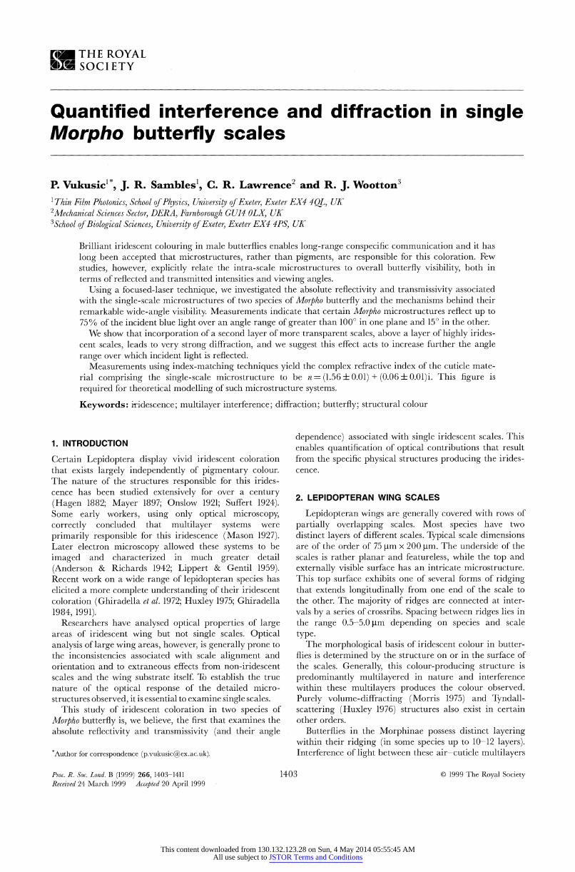
Figure 1 Optical images of (a) a magnified region of M. rhetenor wing showing the ordered arrangement of its single layer of ground scales and (b) a magnified region of M. didius wing showing the two distinct types of scales, glass scales overlying ground scales. Scale bar in (a) for (a,b), 100 Hm.
is the mechanism by which the stunning iridescent blues and violets, characteristic of this order, are produced (Anderson & Rtichards 1942). Some species within other: orders have similar structures to those in Morplho but with dimensions that support strong reflection through multi- layer interference at ultraviolet (UV) wavelengths (Ghiradella et azl. 1972).
We studied two species of iridescent blue butterflies: Morplio rlhetenor and M. didius. M. rhetenor has one layer of highly iridescent 'ground scales' (Ghiradella 1994) on its dorsal wing surface (figure 1a)) whereas M. didiues possesses both a highly iridescent ground scale, lying in a layer next to the wing substrate, and a second layer of near- transparent 'glass scales' (G:hiradella 1994) exhibiting relatively low iridescence (figure 114.
3. MATERIALS AND METHODS
A single ground scale was removed from an M. rhetenor hindwing and a ground scale and a glass scale were removed from a hindwing of M. didius. Each scale was mounted by its basal end onto a ground-down needle tip arid then examined with an optical microscope.
Scanninlg electron microscope (SEM) images were taken using a Hitachi S-3200N electron microscope. Transmission electron microscope (TEM) images were taken after fixing
samples in 3% glutaraldehyde at 210C for 2 h, followed by rinsing in sodium cacodylate buffer. Samples were then fixed in 1% osmic acid in buffer for 1 h, followed by block staining in
2% aqueous uranyl acetate for 1 h, dehydration through an acetone series ending with 100% acetone, and embedding in Spurr resin. Post-microtomed sample sections were stained with lead citrate and examined using a JEOL lOOS TEM. We then conducted detailed measurements on the optical response func- tions of an identical set of scales.
A single scale, mounted vertically on its needle, was positioned at the centre of a Euler Cradle, this centre being coincident with the path of a laser beam. In this way, optical reflection and transmission data could be taken on a single scale at any chosen angle. An achromatic lens was used to focus the incident beam at normal incidence to the scale's upper surface, giving a diffraction-limited minimum beam waist of ca. 30 Htm diameter at the scale. A micromanipulator was employed to ensure this small beam spot was consistently incident in the centre of each scale.
A detector was arranged to scan 3600 in the horizontal plane around the centrally mounted scale, to measure both its reflec- tivity and transmissivity. An Ar+ laser, three He-Ne lasers and a polarizer gave a choice of incident wavelengths and polariza- tions. Each beam was attenuated to avoid damaging the scale.
A lens with a collection angle of ? 20? was placed in front of the detector to collect the majority of the vertically spread reflected light. A vertical slit-aperture in front of this lens gave horizontal angle resolution. Signals recorded in reflection and transmission from the scale at each wavelength were integrated and compared with the integrated signal recorded without the scale present. In this way absolute reflectance and transmittance were calculated. This method was verified by replacing the butterfly scale with a small glass plate to check that the reflec- tion coefficient of the glass was equal to that calculated from its refractive index.
Attempts to fit theoretical models to experimental reflectivity data generally suffer from uncertainties in the value of the complex refractive index of cuticle material that should be used. To address this uncertainty, absolute reflection and transmission experiments with the M. rhetenor scale in air were repeated with the scale immersed in isopropyl alcohol (IPA). The scale was then removed and dried and the measurements repeated with the scale immersed in bromoform. Both liquids, of refractive indices n = 1.38 for IPA and 1.58 for bromoform, filled the spaces between the cuticle multilayering, thus changing the interfer- ence conditions that produce strong reflectance at specific wave- lengths. The additional set of optical parameters provided by these immersion experiments permitted establishment of an accurate theoretical model.
To quantify the wavelength-dependent reflectivity associated with each butterfly over a wider range than that which our lasers could accommodate, a reflectance spectrometer was employed (Perkin Elmer Lambda 900 UV/Vis/NIR with 150 mm diameter integrating sphere). Although single-scale analysis was not possible due to the spectrometer's design, large wing-area analyses covered the near-UV, a wavelength band in which the vision of some Lepidoptera and their predators shows strong sensitivity (Lutz 1924; Silberglied 1979; Bennett & Cutbill 1994).
4. RESULTS
Figure 2 shows optical images of individual scales, showing top surfaces both in reflection and transmission.
Proc. R. Soc. Lond. B (1999)
This content downloaded from 130.132.123.28 on Sun, 4 May 2014 05:55:45 AMAll use subject to JSTOR Terms and Conditions

Optical characterization ofsingle butterfly scales P. Vukusic and others 1405
., >~~~~~~~~~~~~~~~~~~~~~~~~~~~W
(a _ (b)--
Figure 2 Optical microscope images of single scales from M. rhetenor and M. didius. Top surface of the M. rhetenor iridescent ground scale in (a) reflection and (b) transmission. (c) Top surface of the MVI. didius iridescent ground scale in reflection and (d) in ti ansmission. Top surface of the M. didius glass scale in (e) reflection and (f) transmission. (Note that the camera has compensated for low light levels and the blue colour of the scale shown in (e) is artificially enhanced.) Scale bar in (a) for (a,b,c,d,ef ), 100 psm.
The characteristic blue iridescence exists only in reflection from the top surface of each scale. The blue of the M. didius glass scale (figure 2e,f), which in comparison with the blue of the ground scales is of relatively low intensity, appears equally intense due to the contrast compensation of the camera.
It is interesting to compare the transmitted colours of each scale. The ground scales of both species -are dark brown. This colouring derives both from the optical absorption associated with pigment and the strong blue reflection from the structure on the top surface. The Al rietenor ground scale appears darker than that of M. didius, due either to a higher concentration of melanin or to the presence of a more absorbing form of melanin (Nijhout 1991). The M didius glass scale appears highly transparent, indicating the absence of melanin.
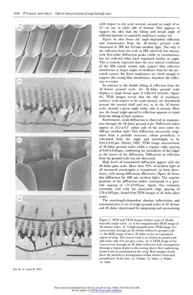
In figure 3, SEM images of the lorpho scales, and more particularly TEM images of their cross-sections, reveal the periodic upper-surface ridges cotntaining multilayers
responsible for the iridescent colour. This structure is one of the two main variants of multilayer structure found in Lepidoptera that display iridescence. The other, found for example in iridescent scales of Urania moths (Lippert & Gentil 1959), is formed from multilayers that exist within the substrate of the scale itself.
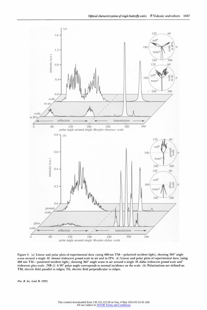
The spatial patterns of reflection and transmission from single iridescent ground scales of both butterflies and from a glass scale from M. didius are showTn in figure 4a,b. For the ground scales in air with the beam incident normal to the scale's top surface and the scale oriented so that the ridges run vertically, a broad reflection of speckle is recorded, spanning over 1000 in the horizontal plane and ca. 15' in the vertical plane.
Reflection from the M. rhetenor ground scale appears divided into two distinct lobes around the normal to the scale surface (showvn in figure 4a, for a 488nm uincident wavelength). TEM images of this scale indicate a distribu- tion of tilts of the multilayer upper and lower surfaces,
Pros. R. Soc. Lond. B (1999)
This content downloaded from 130.132.123.28 on Sun, 4 May 2014 05:55:45 AMAll use subject to JSTOR Terms and Conditions
1406 P. Vukusic and others Optical characterization ofsingle butterfly scales
" . ,S>i Sl.m.
i,2S, ' " o89 S~~~~~'' 3 MI,
4/~~~~~~~~~~~~'
(b)
(c)Of
A lmj 7/?J (d)~~~~~~~~~~~~~~~~~a
with respect to the scale normal, around an angle of ca. 150 on one or other side of normal. This appears to support the idea that the lobing and broad angle of reflected intensity is caused by multilayer surface tilt.
Figure 4a also shows the angle-dependent reflection and transmission from the M.rhetenor ground scale immersed in IPA for 455 nm incident light. Not only is the reflection from the scale in IPA relatively less intense, with first-order diffraction peaks visible in transmission, but the reflected lobes have separated further in angle. This is entirely expected since the new optical conditions of the IPA-cuticle system only support blue reflective interference at larger angles of incidence than for the air- cuticle system. Far fewer multilayers are tilted enough to support this strong blue interference, therefore the reflec- tion is weaker.
In contrast to the double lobing of reflection from the M.rhetenor ground scale, the M.didius ground scale displays a single broad span of reflected intensity (figure 4b). TEM images reveal that the tilts of multilayer surfaces, with respect to the scale normal, are distributed around the normal itself and not, as in the M. rhetenor scale, around a given angle either side of normal. Here too, the broad angle-spread in reflection appears to result from the tilting of layer surfaces.
Furthermore, weak diffraction is observed in transmis- sion through the M. didius ground scale. Diffracted orders appear at 35.5 ? O0 either side of the zero order for 488 nm incident light. This diffraction necessarily origi- nates from a periodic structure, whose periodicity is calculated from the angle and wavelength to be 0.84 + 0.01 pim (Hutley 1982). TEM image measurement of M. didius ground scales yields a regular ridge spacing of 0.83 ? 0.03 ptm, confirming the periodicity of the ridges as the source of the diffraction. Diffraction in reflection from the ground scale was not discerned.
High levels of transmitted diffraction appear with the M. didius glass scale. More than 70% of incident light of all measured wavelengths is transmitted (in both direc- tions), with strong diffraction efficiencies (figure 4b shows this diffraction for 488 nm incident light). The angular positions of the diffraction orders correspond to a peri- odic spacing of 1.71 ? 0.03 ptm. Again, this compares extremely well with the associated ridge spacing of 1.70 ? 0.05 ptm, found from TEM images of M. didius glass scales.
The wavelength-dependent absolute reflectivities and transmissivities in air of single ground scales of M. rhetenor and M. didius (determined by integrating and normalizing
Figure 3. SEM and TEM images of three types of Morpho butterfly single scales. (a) A low-magnification SEM image of M. rhetenor scales. (b) A high-magnification TEM image of a cross-section through an M. rhetenor iridescent ground scale. (c) An SEM image of three M. didius scales on a prepared region of wing. The central scale is an iridescent ground scale and either side of it are glass scales. (d) A TEM image of the cross-section through an M. didius iridescent scale arrangement showing a region of glass scales resting above their underlying ground scales as positioned on the wing. Both images clearly show the multilayer arrangement within distinct structural periodicities. Scale bars: (a) 120 ptm; (b) 3 1xm; (c) 30 [ti;
(d) 2 jsm.
Proc. R. Soc. Lond. B (1999)
This content downloaded from 130.132.123.28 on Sun, 4 May 2014 05:55:45 AMAll use subject to JSTOR Terms and Conditions
Optical characterization ofsingle butterfly scales P. Vukusic and others 1407
(a)
1.6- 120 60
refl~
180 0
1.24 -:1 0 < 1.2 ~~~~~~~~~~~~~~~~~~transn
0.8 - 120 60
refl.....0 180 0 0.4- ~ ~ ~ ~ ~ ~ ~ ~~~~~~~~~~tansn
scale/ ; s /cale
in IPA A
-: t7 ;;:0: reflection / - transmission
0 60 120 180 240 300 360 polar angle around single Morpho rhetenor scale
0.8 - (b) 120 60
ref1
0.6 - ......... 1.80... f. . 0 ... t /
transn1
0 0.4 120 60
refl
0.2 -8. I ~~~~~~~~~~~~~~~transn1
0 0 ~ ~ ~ ~ ~ ~ ~ ~ ~ ~ ~ ~ ~~4 0
ground/
polar angle around single Morpho didius scale
Figugre 4. (a) Linear and polar plots of experimental data (sing 488 nm TM-polarized incident light), showing 3600 angle scans around a single M. rhetenor iridescent ground scale in air and in IPA. (b) Linear and polar plots of experimental data (using 488 nm TM-polarized incident light), showing 360? angle scans in air around a single M. didius iridescent ground scale and iridescent glass scale. (NB (i) A 90? polar angle corresponds to normal incidence on the scale. (ii) Polarizations are defined as: TM, electric field parallel to ridges; TE, electric field perpendicular to ridges.
Proc. R. Soc. Lond. B (1999)
This content downloaded from 130.132.123.28 on Sun, 4 May 2014 05:55:45 AMAll use subject to JSTOR Terms and Conditions
1408 P. Vukusic and others Optical characterization ofsingle butterfly scales
75275 0 z3R 50 - 0
* ~~~~~~~~~25
Q 45050 0 30 4600 500 600
50 o ~~~~~~~~~~~~~~~~~~~~~~~~~~~~~~x
++ 0 ~~~~~~~~20+ +
15
10
25 s
0~~~~~~ 450 500 550 600
o hwavelength (nm)
0 450 500 550 600 650
wavelength (nm)
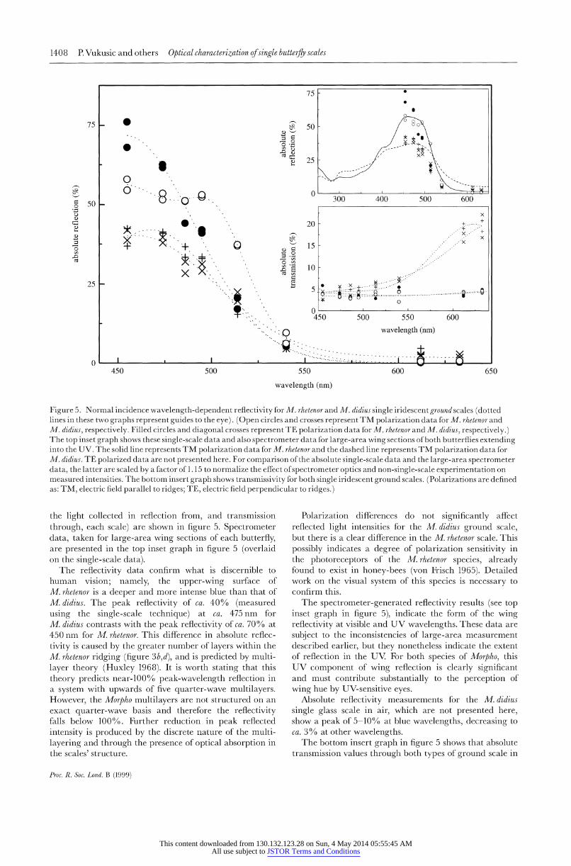
Figure 5. Normal incidence wavelength-dependent reflectivity for M. rhetenor and M. didius single iridescent ground scales (dotted lines in these two graphs represent guides to the eye). (Open circles and crosses represent TM polarization data for M. rhetenor and M. didius, respectively. Filled circles and diagonal crosses represent TE polarization data for M. rhetenor and M. didius, respectively.) The top inset graph shows these single-scale data and also spectrometer data for large-area wing sections of both butterflies extending into the UV. The solid line represents TM polarization data for M. rhetenor and the dashed line represents TM polarization data for M. didius. TE polarized data are not presented here. For comparison of the absolute single-scale data and the large-area spectrometer data, the latter are scaled by a factor of 1. 15 to normalize the effect of spectrometer optics and non-single-scale experimentation on measured intensities. The bottom insert graph shows transmissivity for both single iridescent ground scales. (Polarizations are defined as: TM, electric field parallel to ridges; TE, electric field perpendicular to ridges.)
the light collected in reflection from, and transmission through, each scale) are shown in figure 5. Spectrometer data, taken for large-area wing sections of each butterfly, are presented in the top inset graph in figure 5 (overlaid on the single-scale data).
The reflectivity data confirm what is discernible to human vision; namely, the upper-wing surface of M. rhetenor is a deeper and more intense blue than that of M. didius. The peak reflectivity of ca. 40% (measured using the single-scale technique) at ca. 475 nm for M. didius contrasts with the peak reflectivity of ca. 70% at 450 nm for M. rhetenor. This difference in absolute reflec- tivity is caused by the greater number of layers within the M. rhetenor ridging (figure 3b,d), and is predicted by multi- layer theory (Huxley 1968). It is worth stating that this theory predicts near-1000 0 peak-wavelength reflection in a system with upwards of five quarter-wave multilayers. However, the Morpho multilayers are not structured on an exact quarter-wave basis and therefore the reflectivity falls below 1000 %. Further reduction in peak reflected intensity is produced by the discrete nature of the multi- layering and through the presence of optical absorption in the scales' structure.
Polarization differences do not significantly affect reflected light intensities for the M. didius ground scale, but there is a clear difference in the M. rhetenor scale. This possibly indicates a degree of polarization sensitivity in the photoreceptors of the M. rhetenor species, already found to exist in honey-bees (von Frisch 1965). Detailed work on the visual system of this species is necessary to confirm this.
The spectrometer-generated reflectivity results (see top inset graph in figure 5), indicate the form of the wing reflectivity at visible and UV wavelengths. These data are subject to the inconsistencies of large-area measurement described earlier, but they nonetheless indicate the extent of reflection in the UV. For both species of Morpho, this UV component of wing reflection is clearly significant and must contribute substantially to the perception of wing hue by UV-sensitive eyes.
Absolute reflectivity measurements for the M.didius single glass scale in air, which are not presented here, show a peak of 5-10% at blue wavelengths, decreasing to ca. 3 % at other wavelengths.
The bottom insert graph in figure 5 shows that absolute transmission values through both types of ground scale in
Proc. R. Soc. Lond. B (1999)
This content downloaded from 130.132.123.28 on Sun, 4 May 2014 05:55:45 AMAll use subject to JSTOR Terms and Conditions
Optical characterization of single butterfly scales P. Vukusic and others 1409
0.065 80
8 .SO (;.060
0.055
'' 60 scale\ in air 500 600
wavelength (nm)
40
scale in -in -a IPA - -
0 400 500 600 '700
wavelength (nm)
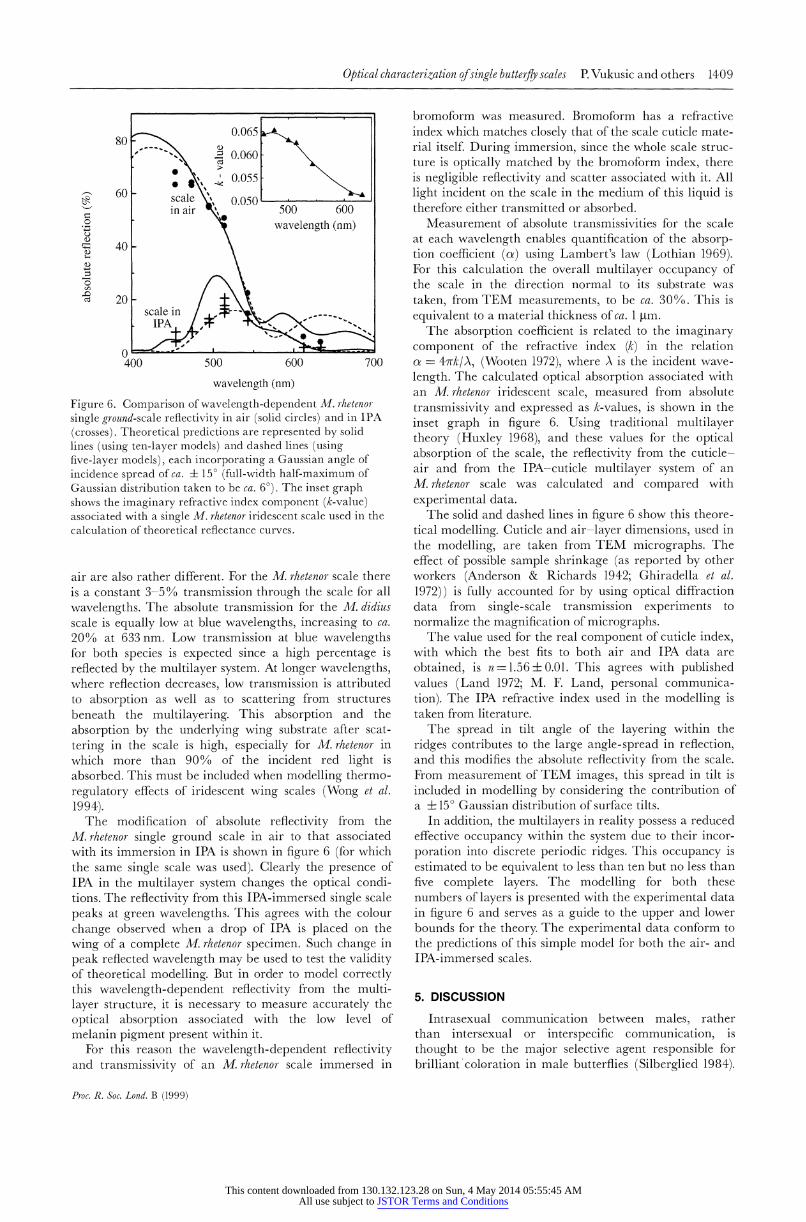
Figure 6. Comparison of wavelength-dependent M. rhetenor- single ground-scale reflectivity in air (solid circles) and in IPA (crosses). Theoretical predictions are represented by solid lines (using ten-layer models) and dashed lines (using five-layer models), each incorporating a Gaussian angle of incidence spread of ca. ? 150 (full-width half-maximum of Gaussian distribution taken to be ca. 6?). The inset graph shows the imaginary refractive index component (k-value) associated with a single M. rhetenor iridescent scale used in the calculation of theoretical reflectance curves.
air are also rather different. For the M. rhetenor scale there is a constant 3-5% transmission through the scale for all wavelengths. The absolute transmission for the M. didius scale is equally low at blue wavelengths, increasing to ca. 20% at 633 nm. Low transmission at blue wavelengths for both species is expected since a high percentage is reflected by the multilayer system. At longer wavelengths, where reflection decreases, low transmission is attributed to absorption as well as to scattering from structures beneath the multilayering. This absorption and the absorption by the underlying wing substrate after scat- tering in the scale is high, especially for M. rhetenor in which more than 90% of the incident red light is absorbed. This must be included when modelling thermo- regulatory effects of iridescent wing scales (Wong et al. 1994).
The modification of absolute reflectivity from the M. rhetenor single ground scale in air to that associated with its immersion in IPA is shown in figure 6 (for which the same single scale was used). Clearly the presence of IPA in the multilayer system changes the optical condi- tions. The reflectivity from this IPA-immersed single scale peaks at green wavelengths. This agrees with the colour change observed when a drop of IPA is placed on the wing of a complete M. rhetenor specimen. Such change in peak reflected wavelength may be used to test the validity of theoretical modelling. But in order to model correctly this wavelength-dependent reflectivity from the multi- layer structure, it is necessary to measure accurately the optical absorption associated with the low level of melanin pigment present within it.
For this reason the wavelength-dependent reflectivity and transmissivity of an M. rizetenor scale immersed in
bromoform was measured. Bromoform has a refractive index which matches closely that of the scale cuticle mate- rial itself. During immersion, since the whole scale struc- ture is optically matched by the bromoform index, there is negligible reflectivity and scatter associated with it. All light incident on the scale in the medium of this liquid is therefore either transmitted or absorbed.
Measurement of absolute transmissivities for the scale at each wavelength enables quantification of the absorp- tion coefficient (oa) using Lambert's law (Lothian 1969). For this calculation the overall multilayer occupancy of the scale in the direction normal to its substrate was taken, from TEM measurements, to be ca. 30%. This is equivalent to a material thickness of ca. 1 Htm.
The absorption coefficient is related to the imaginary component of the refractive index (k) in the relation oz = 4wrk/A, (Wooten 1972), where A is the incident wave- length. The calculated optical absorption associated with an M. rhetenor iridescent scale, measured from absolute transmissivity and expressed as k-values, is shown in the inset graph in figure 6. Using traditional multilayer theory (Huxley 1968), and these values for the optical absorption of the scale, the reflectivity from the cuticle- air and from the IPA-cuticle multilayer system of an M. rhetenor scale was calculated and compared with experimental data.
The solid and dashed lines in figure 6 show this theore- tical modelling. Cuticle and air-layer dimensions, used in the modelling, are taken from TEM micrographs. The effect of possible sample shrinkage (as reported by other workers (Anderson & Richards 1942; Ghiradella et al. 1972)) is fully accounted for by using optical diffraction data from single-scale transmission experiments to normalize the magnification of micrographs.
The value used for the real component of cuticle index, with which the best fits to both air and IPA data are obtained, is n=1.56 ? 0.01. This agrees with published values (Land 1972; M. E Land, personal communica- tion). The IPA refractive index used in the modelling is taken from literature.
The spread in tilt angle of the layering within the ridges contributes to the large angle-spread in reflection, and this modifies the absolute reflectivity from the scale. From measurement of TEM images, this spread in tilt is included in modelling by considering the contribution of a ? 15? Gaussian distribution of surface tilts.
In addition, the multilayers in reality possess a reduced effective occupancy within the system due to their incor- poration into discrete periodic ridges. This occupancy is estimated to be equivalent to less than ten but no less than five complete layers. The modelling for both these numbers of layers is presented with the experimental data in figure 6 and serves as a guide to the upper and lower bounds for the theory. The experimental data conform to the predictions of this simple model for both the air- and IPA-immersed scales.
5. DISCUSSION
Intrasexual communication between males, rather than intersexual or interspecific communication, is thought to be the major selective agent responsible for brilliant 'coloration in male butterflies (Silberglied 1984).
Pr-oc. R. Soc. Lonzd. B (1999)
This content downloaded from 130.132.123.28 on Sun, 4 May 2014 05:55:45 AMAll use subject to JSTOR Terms and Conditions
1410 P. Vukusic and others Optical characterization ofsingle butterfly scales
Experimental evidence supports this hypothesis, indi- cating that not only are males attracted to visual stimuli resembling females, but that they can be repelled from long distances by visual stimuli resembling other males (Obara 1970; Stride 1958; Rutowski 1978, 1981; Silberglied & Taylor 1978). Brilliant wing coloration appears to have the capacity to serve as an agonistic device that is used for threat (Hingston 1933) and for intimidation of rivals for prime locations (Shields 1968). It has developed in male butterflies to promote their visibility while in flight or while resting with open wings.
Although some butterfly pigmentary colours appear bright and highly visible to human eyes, their absolute visibility is substantially inferior to those wing colours that are generated through optical interference. Measure- ments in this study indicate that over 70% of incident blue light is reflected from the intra-scale structure of M. rhetenor, far above what can be achieved through pigmentary coloration alone. Selection pressures on these species have caused the evolution of large numbers of multilayers that are appropriately dimensioned for produ- cing this very significant reflectivity. Such structural coloration also enables change of hue with wing orienta- tion, narrow spectral purity, the possibility of a strong component of UV reflection, and distinct polarization effects that are also not attainable with pigments.
Sheer brightness is very likely to serve its main purpose in long-range communication. In terms of the spectral response of human eyes, Morpho can easily be seen from low-flying aircraft (Silberglied 1984), and Bates reported visibility from 'a quarter of a mile off' (Bates 1864). However, the spectral response of the eyes of Morpiho should not be inferred, simply because our eyes perceive them to be very brightly coloured (Bennett et al. 1994). In the absence of complete and detailed studies on Morpho visual perception that are not limited to the visible spec- trum, several clues should be considered. For instance, a substantial spectral sensitivity of Morpiho eyes to the UV component reflected from Morpiho wings may be assumed (Menzel 1975; Silberglied 1984). The existence, in the eyes of some species, of tapetal reflectors and interference filters whose function is to enhance colour contrast across a narrow spectral band (Miller & Bernard 1968; Bernard & Miller 1970; Bernhard et al. 1970; Ribi 1980) may also apply to Morpho eyes. The photoreceptors in the eyes of a Morpiho specimen, shown to exist at least as a trichromatic system, appear to have a maximal response to blue coloration (Swihart 1967) when tested across the human visible spectrum. It is entirely probable that not only does the blue reflection from a Morpho wing appear bright and highly visible to other male conspecifics, but that it is even more striking to them than to human eyes.
Conspicuousness also has its disadvantages. These male butterflies must also be highly visible to predatory birds, many of which have UV-sensitive vision (Bennett & Cuthill 1994). Morpho males appear to be palatable and probably suffer quite high predation. Their principal defence is thought to be their highly manoeuvrable and erratic flight (R. B. Srygley, personal communication).
This work introduces an additional element in the subject of Morpho visibility, namely the viewing angle over which they are visible. The discrete nature of the Morp7ho multilayering within scale ridging enables two distinct
effects, both of which enhance the angular spread of light reflected from the wing. First, the presence of multilayer surfaces that show a distribution of tilts with respect to the scale substrate creates a very large spread in angle over which the characteristic blue light is reflected. Similar multilayer tilting, increasing the angular spread of reflected light, has been found in the reflectors of the crab Ovalipes (Parker 1998).
In addition, the existence of a second layer of periodi- cally ridged but highly transparent glass scales above the layer of highly iridescent ground scales causes strong diffraction. This diffraction does not appear to serve the purpose of optical dispersion, but acts to spread further the angle over which incident light is reflected. A purely spectral reflection from a mirror-like surface would severely restrict the solid angle from which the wing can be observed. Long-range communication would in this respect be limited. In the case of the Morpho butterflies studied here, incorporation into the optical system of either a multilayer surface-tilt distribution, or diffraction- assisted angle broadening using glass scales, or both, enhances their overall angular visibility.
We thank Gavin Wakely at the E.M. Unit in the School of Biological Sciences of Exeter University for expert technical assistance, and David Bolton at the Royal Albert Museum, Exeter, UK, for access to specimens. This work receives funding from the BBSRC, the DERA (Farnborough) and Exeter University.
REFERENCES
Anderson, T. F. & Richards, A. G. 1942 An electron microscope study of some structural colours of insects, J. Appl. Phys. 13, 748-758.
Bates, H. W. 1864 The naturalist on the river Amazons. A record of adventures, habitats of animals, sketches of Brazilian and Indian life and aspects of nature under the equator, during elevenyears of travel, 2nd edn. London: John Murray.
Bennett, A. T. D. & Cuthill, I. C. 1994 Ultraviolet vision in birds: what is its function? Vision Res. 34, 1471-1478.
Bennett, A. T. D., Cuthill, I. C. & Norris, K. J. 1994 Sexual selec- tion and the mismeasure of colour. Am. Nat. 144, 848-860.
Bernard, G. D. & Miller, W. H. 1970 What does antenna engi- neering have to do with insect eyes? IEEE Student]. 1978, 2-8.
Bernhard, C. G., Boethius, J., Gemne, G. & Struwe, G. 1970 Eye ultrastructure, colour reception and behaviour. Nature 226, 865-866.
Ghiradella, H. 1984 Structure of iridescent lepidopteran scales: variations on several themes. Ann. Entomol. Soc. Am. 77, 637-645.
Ghiradella, H. 1991 Light and colour on the wing: structural colours in butterflies and moths. Appl. Optics 30, 3492-3500.
Ghiradella, H. 1994 Structure of butterfly scales: patterning in an insect cuticle. Microsc. Res. Tech. 27, 429-438.
Ghiradella, H., Aneshansley, D., Eisner, T., Silberglied, R. E. & Hinton, H. E. 1972 Ultra-violet reflection of a male butterfly: interference colour caused by thin layer elaboration of wing scales. Science 178, 1214-1217.
Hagen, H. A. 1882 On the colour and pattern of insects. Proc. Am. Acad. Arts Sci. 17, 234-267.
Hingston, R. W. G. 1933 The meaning of animal colour and adorn- ment. London: Edward Arnold & Co.
Hutley, M. C. 1982 Difraction gratings. London: Academic Press. Huxley, A. F. 1968 A theoretical treatment of the reflection of
light by multilayer structures. J. Exp. Biol. 48, 227-245.
Proc. R. Soc. Lond. B (1999)
This content downloaded from 130.132.123.28 on Sun, 4 May 2014 05:55:45 AMAll use subject to JSTOR Terms and Conditions
Optical characterization ofsingle butterfly scales P. Vukusic and others 1411
Huxley, J. 1975 The basis of structural colour variation in two species of Papilio. J. Entomol. A 50, 9-22.
Huxley, J. 1976 The coloration of Papilio zalmoxis and P antimachus and the discovery of Tyndall blue in butterflies. Proc. R. Soc. Lond. B 193, 441-453.
Land, M. E 1972 The physics and biology of animal reflectors. Progr. Biophys. Mol. Biol. 24, 75-106.
Lippert, W. & Gentil, K. 1959 Uber lamellare Feinstrukturen bei den Schillershuppen der Schmetterlinge vom Urania- und Morpho-typ. Z Morphol. Okol. Tiere 48, 115-122.
Lothian, G. E 1969 Absorption spectrophotometry. London: Adam Hilger Ltd.
Lutz, E E. 1924 Apparently non-selective characters and combi- nations of characters, including a study of ultra-violet in relation to the flower-visiting habits of insects. Ann. N'fAcad. Sci. 29, 181-283.
Mason, C. W. 1927 Structural colours in insects. II. Iridescent colours. _. Phys. Chem. 31, 321-354.
Mayer, A. G. 1897 On the colour and colour-patterns of moths and butterflies. Bull. Mus. Comp. Zool. 30, 169-254.
Menzel, R. 1975 Colour receptors in insects. In The compound eye and vision of insects (ed. G. A. Horridge), pp. 121-153. Oxford: Clarendon Press.
Miller, W H. & Bernard, G. D. 1968 Butterfly glow. _. Ultrastruct. Res. 24, 286-294.
Morris, R. B. 1975 Iridescence from diffraction structures in the wing scales of Callophrys rubi, the Green Hairstreak. J. Entomol. A 49, 149-154.
Nijhout, H. F. 1991 The development and evolution of butterfly wing patterns. Washington, DC: Smithsonian Institute Press.
Obara, Y. 1970 Studies on the mating behaviour of the white cabbage butterfly, Pieris rapae crucivora. III. Near-ultra-violet reflection as the signal of intra-specific communication. Z Vergl. Physiol. 69, 99-116.
Onslow, H. 1921 On a periodic structure in many insect scales and the cause of their iridescent colours. Phil. Trans. R. Soc. Lond. B 211, 1-74.
Parker, A. R. 1998 A unique form of light reflector and the evolution of signalling in Ovalipes (Crustacea: Decapoda: Portunidae). Proc. R. Soc. Lond. B 265, 861-867.
Ribi, W. A. 1980 The phenomenon of eye glow. Endeavour 5, 2-7. Rutowski, R. L. 1978 The courtship behaviour of the small
sulphur butterfly, Eurema lisa. Anim. Behav. 26, 892-903. Rutowski, R. L. 1981 Sexual discrimination using visual clues in
the checkered white butterfly (Pierisprotodice). Z Tierpsychol. 55, 325-334.
Shields, 0. 1968 Hilltopping. _. Res. Lepid. 6, 69-78. Silberglied, R. E. 1979 Communication in the ultra-violet. A.
Rev. Ecol. Syst. 10, 373-398. Silberglied, R. E. 1984 Visual communication and sexual selec-
tion among butterflies. In The biology of butterflies (ed. R. I. Vane-Wright & P. E. Ackery), pp. 207-223. Symposium of the Royal Society of London, no. 11. London: Academic Press.
Silberglied, R. E. & Taylor, 0. R. 1978 Ultraviolet reflection and its behavioural role in the courtship of the sulfur butter- flies, Colias eurytheme and C. phzilodice. Behav. Ecol. Sociobiol. 3, 203-243.
Stride, G. 0. 1958 On the courtship behaviour of a tropical mimetic butterfly, Hypolimnus misippus. Proc. Int. Congr. Entomol. 2, 419-424.
Suffert, E 1924 Morphologie und Optik der Schmetter- lingschuppen inbesondere die Schillerfarben der Schmet- terlinge. Z Morphol. Okol. Tiere 1, 171-308.
Swihart, S. L. 1967 Neural adaptations in the visual pathway of certain Heliconnine butterflies, and related forms, to variations in wing colouration. Zoologica NT 52, 1-14.
von Frisch, K. 1965 The dance language and orientation of bees. Berlin: Springer.
Wong, P. Y., Heilmann, B. D. & Miaoulis, I. N. 1994 The effect of microscale and macroscale patterns on the radiative heating of multilayer thin-film structures. Microscale Heat Transfer. ASME Heat Transfer Division 291, 27-34.
Wooten F. 1972 Optical properties of solids. London: Academic Press.
As this paper exceeds the maximum length normally permitted, the authors have agreed to contribute to production costs.
Proc. R. Soc. Lond. B (1999)
This content downloaded from 130.132.123.28 on Sun, 4 May 2014 05:55:45 AMAll use subject to JSTOR Terms and Conditions