quantitative determination of natural dipalmitoyl lecithin with dimyristoyl lecithin as internal...
TRANSCRIPT

Journal of Chromatography, 192 (1980) 185-192 © Elsevier Scientific Publishing Company, Amsterdam -- Printed in The Netherlands
CHROM. 12,561
QUANTITATIVE DETERMINATION OF NATURAL DIPALMITOYL LECI- THIN WITH DIMYRISTOYL LECITHIN AS INTERNAL STANDARD BY CAPILLARY GAS-LIQUID CHROMATOGRAPHY
ALFRED LOHNINGER Research Institute of Traumatology of the A UVA, Donaueschingenstrasse 13, A-1200 Vienna (Austria)
and ALEXEJ N1KIFOROV Institute of Organic Chemistry, University of Vienna, Vienna (Austria)
(First received May 23rd, 1979; revised manuscript received November 23rd, 1979)
SUMMARY
A quantitative determination of 1,2-dipalmitoyl-sn-glycero-3-phosphocholine from dog lungs by means of the corresponding diacyl glycerol trimethylsilyl ether derivative with 1,2-dimyristoyl-sn-glycero-3-phosphocholine as internal standard was carried out by gas-liquid chromatography with glass capillary columns. The individual diacyl glycerol species were identified by using a combination of gas-liquid chromato- graphy and mass spectrometry. In a total of 32 determinations percent deviations from the mean value between -4- 2.0 and ~ 4.4 ~ were achieved. By using glass cap- illary columns instead of conventional columns the time required for an analysis was reduced to ca. 30 ~ and separation was considerably improved. Thus the diacyl glycerol species from PC-32 were separated according to both the 1,2 and 1,3 positional isomerism and the degree of saturation of the acyl radicals.
INTRODUCTION
In several diseases such as the respiratory distress syndrome (RDS) of the newborn 1-4 or the adult RDS -- the socalled shock lung 5,6- the lung disfunction is at least in part the result of an impairment of the lung surfactant system. Dipalmitoyl lecithin (DPPC) is generally accepted as the major active component of the pulmonary surfactant 4,7-9. There is strong evidence that the reduction of DPPC is characteristic of these pulmonary infections. A treatment resulting in an increase in the phospholipid synthesis requires a quantitative determination of 1,2-dipalmitoyl-sn-glycero-3-phos- phocholine (DPPC) which has to be as exact as possible and which can be carried out in serial analyses.
The usual methods of determining disaturated phosphatidylcholine species comprise separating the corresponding diacyl glycerol acetates by argentation thin- layer chromatography (TLC) 1°-13 or by adduction with mercury acetate or osmium tetroxide combined with column chromatography or TLC, respectively 9'14'~5.

186 A. LOHNINGER, A. NIKIFOROV
Gas-liquid chromatography (GLC) with conventional columns has been used to separate the 3-sn-phosphatidylcholine species from various biological sources by means of the corresponding diacyl glycerol acetates ~6-19 or trimethylsilyl (TMS) ether derivatives 2°.
Ogino et al. 21 characterized 3-sn-phosphatidylcholine species from fetal rat and rabbit lungs as TMS ether (PC30-PCa4) by a combination of GLC and mass spectrometry (MS).
This paper describes a method for the quantitative determination of DPPC in lung tissue with 1,2-dimyristoyl-sn-glycero-3-phosphocholine as internal standard by GLC with glass capillary columns.
In accordance with the results of other authors, dimyristoyl lecithin could not be detected in natural lecithinsZ°,2k This was shown in a series of samples without the internal standard. The use of other internal standards, such as dialkyl or l-acyl-2- alkyl-sn-glycero-3-phosphocholine, has not proved possible as it is well established that plasmanylcholine also occurs in nearly all natural sources of phospholipids. As considerably better separation was achieved with glass capillary than with conven- tional columns, it was possible to separate the DPPC from the other 3-sn-phospha- tidylcholine species having the same number of carbon atoms. The individual com- ponents were identified by GLC-MS.
EXPERIMENTAL
Apparatus A Carlo Erba Fractovap 2350 gas chromatograph equipped with a hydrogen
flame detector in connection with a System 1 Integrator was used for the quantitative analyses of diacyl glycerols (Carlo Erba, Milan, Italy). The samples were analysed on a SE-30 column (12 m × 0.25 mm I.D.). The injection port and detector temperature were maintained at 350 ° . The oven temperature was programmed to rise from 24 ° to 300 ° at a rate of 4°/rain. Hydrogen was used as carrier gas at a flow-rate of 40 era/ sec.
An Ultraturax (IKA-Werk, Staufen, G.F.R.), a Kontes Micro-Ultrasonic Cell Disruptor (Kontes, Vineland, N.J., U.S.A.) and a Camag Lonomat III (Camag, Muttenz, Switzerland) were used.
A direct coupling of the glass capillary column with a Varian MAT 311A mass spectrometer in connection with a Varian 166 spectrosystem was used for the identi- fication of the individual diacyl glycerol species. The temperature of the interface was 350 °, the pressure in the ion source was 5.10 -6 Torr at a helium flow-rate of 3 ml/min. At an electron energy of 70 eV and an electron current of 3 mA every 0.7 sec, a mass spectrum in the range 20-800 mass units was recorded and evaluated by means of the spectrosystem.
Procedure The lungs of dogs were bled in situ with a physiological NaC1 solution, were
removed immediately afterwards and homogenized with an Ultraturax in 40 volumes of chloroform-methanol (2:1) and extracted overnight under nitrogen. The tissue lipid extracts were washed using the method of Folch et al. 2z.
Thin-layer chromatography. Aliquots of samples were applied as bands with

GLC OF DIPALMITOYL LECITHIN 187
1,2-dimyristoyl-sn-glycero-3-phosphocholine (100/~g) on previously cleaned TLC plates. The plates were developed in chloroform-methanol-water (60:35:5), dried and the 3-sn-phosphatidylcholine fraction was removed from the plates.
Enzymatic hydrolysis. The 1,2-diacyl-sn-glycerols of 3-sn-phosphatidylcholine were obtained by digestion with phospholipase C from Bacillus cereus. After removal from the TLC plates, the 3-sn-phosphatidylcholine samples were dried under nitrogen and 2.5 ml of the 0.1 M tris buffer, (pH 7.4, 0.03 M with respect to CaC12) was added 23,24. After sonication for ca. 15 sec, 10 #g of phospholipase C and 2.5 ml of diethyl ether were added and the mixture was stirred. After 3 h, when the reaction was complete, the ether layer was separated and the water layer was re-extracted five times with 2.5 ml of ether. The pooled ether extracts were dried under nitrogen.
Silylation. The TMS ethers of 1,2-diacyl-sn-glycerols were prepared by reacting (at room temperature for 1 h) with pyridine-hexamethyldisilazane-chlorotrimethyl- silane (12:5:2) 2°. The resulting TMS ether solutions were used directly for subsequent analyses.
Reagents. Chloroform, methanol, pyridine, hexamethylsilazane, chlorotri- methylsilane and diethyl ether were obtained from Merck (Darmstadt, G.F.R.). Phospholipase C from B. cereus was obtained from Boehringer (Mannheim, G.F.R.). 1,2-Dilauroyl-sn-glycero-3-phosphochol ine and 1,2-dimyristoyl-sn-glycero-3-phos- phocholine, 1,2-dipalmitoyl-sn-glycerol, 1,3-dipalmitoyl-sn-glycerol, 1,2-dimyristoyl- sn-glycerol were obtained from Sigma (St. Louis, Mo., U.S.A.). 1-Palmityl-sn- glycerol, 1,3-dioleyl-sn-glycerol, 1-palmityl-3-oleyl-sn-glycerol and 1-stearoyl-3- oley-sn-glycerol were obtained from Analabs, a subsidiary of New England Nuclear (Boston, Mass., U.S.A.).
RESULTS AND DISCUSSION
Fig. 1 shows a representative chromatogram of a mixture of standards of 1,2- and 1,3-diacyl glycerols. On non-polar silicone phases, GLC with glass capillary
I
IdC4.
Fig. 1. GLC of reference 1,2- and 1,3-diacyl-sn-glycerols as TMS ethers. MGL = Palmitoyl-sn-glycerol; 1 = 1,3-dimyristoyl-sn-glycerol; 2 -- 1,2-dipalmitoyl-sn-glycerol; 3 = 1,3-dipalmitoyl-sn-glycerol ; 4 1-palmitoyl-3-oleoyl-sn-glycerol; 5 = 1,3-dioleoyl-sn-glycerol; 6 = 1-stearoyl-3-oleoyl-sn-glycerol. Column, 12-m SE-30, 150°-300 °, programmed at 10°/rain, carrier gas hydrogen.

188 A. LOHNINGER, A. NIKIFOROV
columns permits the separation of the 3-sn-phosphatidylcholine species having the same number of carbon atoms according to both the 1,2 and 1,3 positional isomerism and the degree of saturation of the acyl radicals.
A good separation of the corresponding 1,2-diacyl glycerols from PC-32 was achieved on a 12-m SE-30 column, as illustrated in Fig. 2.
t ime ( ra i n ) 2b ~s ~b ~ 6
Fig. 2. GLC of the corresponding diacyl glycerols as TMS ethers from dog lung 3-sn-phosphatidyl- choline. 1 = 1,2-Dimyristoyl-sn-glycerol; 2 = 1,3-dimyristoyl-sn-glycerol; 3 = l-palmitoyl-2-palmi- toleyl-sn-glycerol; 4 = 1,2-dipalmitoyl-sn-glycerol; 5 = 1,3-dipalmitoyl-sn-glycerol. Column, 12-m SE-30, 240°-300 ° programmed at 4°/min, carrier gas hydrogen.
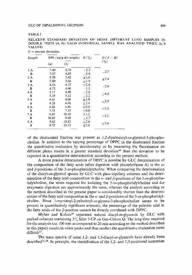
Quantitative determinations The reproducibility of the quantitative determination of dipalmitoyl lecithin
with dimyristoyl lecithin as internal standard was confirmed with eight different lung samples in duplicated tests. Each individual sample was analysed twice. The greatest deviation from the mean in these duplicated tests was 4 .4~, and the average repro- ducibility achieved was zE 3 ~o (Table I).
GLC method with glass capillary columns used as described with non-polar silicone phases (SE-30 and OV-1) can bring about a separation sufficient to carry out a quantitative determination of the 1,2-dipalmitoyl-sn-glycerol. By use of the mercury adduction method or argentation TLC, the 3-sn-phosphatidylcholine species can be separated according to the number of double bonds but not according to the number of carbon atoms. The saturated 3-sn-phosphatidylcholine species isolated by these methods contain other fatty acids in addition to palmitic acid, in particular myristic acid and stearic acid.
The following values for the percentage of palmitic acid in the fatty acids of the disaturated phosphatidylcholine of the lung have been obtained: 67.7 (ref. 14), 80-98 (ref. 10), 95 (ref. 7) and 73 (ref. 15). Henderson and Hackett zs found that 90
GLC OF DIPALMITOYL LECITHIN 189
TABLE I
RELATIVE STANDARD DEVIATION OF EIGHT DIFFERENT LUNG SAMPLES IN DOUBLE TESTS (A, B); EACH INDIVIDUAL SAMPLE WAS ANALYSED TWICE (a, b VALUES) D = percent deviation.
Sample DPL (mg/g dry weight) D (%) D (A + B) (%)
(a) (b)
1 A 7.00 6.70 ±2.2 4-2.7 B 7.07 6.85 ~1.6
2 A 5.59 5.42 4-1.6 4-3.4 B 5.80 5.66 4-1.4
3 A 4.54 4.72 ±2.0 ±2.0 B 4.72 4.60 ±1.3
4 A 5.17 4.90 4-2.6 -4-4.4 B 5.35 5.12 4-2.2
5 A 4.41 4.08 4-3.9 ±3.9 B 4.28 4.08 ±2.4
6 A 6.86 6.86 4-0.0 ±3.3 B 7.33 7.33 4-0.0
7 A 9.85 10.10 ±1.3 4-2.2 B 10.02 9.69 ±1.7
8 A 9.62 10.12 ±2.6 4-2.6 B 9.72 10.12 j2 .0
of the disaturated fraction was present as 1,2-dipalmitoyl-sn-glycerol-3-phospho- choline. In addition to the varying percentage of DPPC in the disaturated fraction the quantitative evaluation by densitometry or by measuring the fluorescence on different plates results in a greater standard deviation 26 than the deviation to be expected in a quantitative determination according to the present method.
A more precise determination of DPPC is possible by G L C determination of the composition of the fatty acids (after digestion with phospholipase A) in the c•- and/%positions of the 3-sn-phosphatidylcholine. When comparing the determination of the diacyl-sn-glycerol species by G L C with glass capillary columns and the deter- mination of the fatty acid composition in the a- and/3-positions of the 3-sn-phospha- tidylcholine, the times required for isolating the 3-sn-phosphatidylcholine and for enzymatic digestion are approximately the same, whereas the analysis according to the method described in the present paper is considerably shorter than the determi- nation of the fatty acid composition in the a- and {/-positions of the 3-sn-phosphatidyl- choline. Since 1-myristoyl-2-palmitoyl-sn-glycero-3-phosphocholine seems to be present in quantitatively significant amounts, the percentage of the palmitic acid in the fatty acids of the/3-position cannot be directly correlated with DPPC.
Myher and Kuksis 2° separated natural diacyl-sn-glycerols by GLC with packed columns containing 3 ~ Silar 5-CP on Gas-Chrom Q. The long time required for the analysis (ca. 100 rain as compared to 20 rain according to the method described in this paper) results in wider peaks and thus renders the quantitative evaluation more difficult 27.
The mass spectra of some 1,2- and 1,3-diacyl-sn-glycerols have already been describedZ~,2K In principle, the identification of the 1,2- and 1,3-positional isomerism

190 A. LOHN1NGER, A, NIKIFOROV
is possible owing to the differences in the mass spectra. The most important infor- mation disclosed by the mass spectrum are: the molecular weight (by means of the M - - 15 ion), the identification of the individual fatty acids by the RCO and RCO ÷ 74 ions, respectively, as well as the identification of the 1,3-derivatives by the preferred formation of the M - - R C O O C H 2 ion 2s.
The use of ter t . -buty l -dimethyls i ly l ethers of diacyl glycerols as reported by Myher et al. 29 enhances the ion intensities in higher mass ranges, thus making these derivatives more sensitive for characterization. However, for routine analytical use, more time consuming steps are necessary.
PC28 PC32 PC34
PC30 3 4
PC36
5 PC38
time (rain) 5 10 1'5 20
Fig. 3. A portion of the computer-reconstructed sum plot in the range from PC-28 to PC-38 recorded by GLC MS (TMS derivatives).
i16:0 16:0 14 ;0 14,0 PC3z !16:0 16:1 18 ;0 18; 1
Fig. 4. Main molecular species of PC-30-PC-34 of dog lung 3-sn-phosphatidylcholine.
1~ 16~0 • ooo 16= 1
16~0
"14=0 a ~ooo_ { 1 6 = 0
~n~ lm ln ) 5 tO 15 20
Fig. 5. M + -- 15 ion profiles of dog lung 3-sn-phosphatidylcholine of the sum plot from Fig. 3. PC- 28 (dimyristoyl-sn-glycerol) TMS derivative was used as internal standard,

GLC OF DIPALMITOYL LECITHIN 191
Fig. 3 shows the sum plot in the range from PC-28 to PC-38 reconstructed by the computer which is correlatable to the gas chromatogram. The different PC-32 species to be expected in principle are shown in Fig. 4.
The ion profiles of the M-- 15 ions in Fig. 5 show that by means of the capillary column not only the mono-unsaturated 1,2- and 1,3-PC-32 species but also the corre- sponding saturated 1,3-isomers may be separated from the saturated 1,2-species (which corresponds to DPPC). It also seems to be possible to separate 1-myristoyl-2 stearoyl species from the 1,2-dipalmitoyl species.
In addition, the formation of undesired 1,3-isomers may be suppressed to a large extent by using HMDS-TMS-pyr id ine as silylating reagent. The mass spectrum of the 1,2-dipalmitoyl-sn-glycerol shown in Fig. 6 correlates clearly with the one cited in literature which was recorded with direct inlet 2a. This confirms the correct identi- fication of the peaks. The identification of the PC-34-PC-38 species will be published elsewhere.
lOO % 129~ t 90 IRCO*74)*
8o 145 313 70
6o
5o
(RCO)* 3o 239 20 [[ L (M-RCO0)* ,o I lLrl , I I 1385 (M-15)+
o I]iI ZL ,Jh.,LL . . . . l , , , i i 62s " - " . . . . . I . . . . ' . . . . I . . . . ' . . . . I ' ' l . . . . I . . . . , . . . . i ' , i . . . . . . .
100 200 300 400 500 vvv' ' ,vv ~n • - ~ m / e
Fig. 6. Mass spectrum of ],2-dipalmitoyl-sn-glycero| TMS ether obtained by G L C - M S from dog lung 3-sn-phosphatidylcholine.
CONCLUSION
The internal standard method described proved to be reliable in determining DPPC in lung tissue. Compared with other methods, the determination by GLC with glass capillary columns permits an exact direct determination of DPPC with a mean percent deviation from the mean of ± 3 ~o, the time required for the GLC analysis being as short as c a . 20 rain.
ACKNOWLEDGEMENTS
The authors are indebted to Doz. Dr. G. Schlag, Director of the Research Institute of Traumatology of the AUVA, for his interest and criticism. We also wish to thank Mr. A. Piegler for technical assistance. This work was supported by a grant from the Lorenz-B6hler-Forschungs-Fond, and Fonds zur F6rderung der wissen- schaftlichen F6rschung, "Projects" Nos. 2696 and 3306.

192 A. LOHNINGER, A. NIKIFOROV
REFERENCES
1 F. H. Adams, T. Fujiwara and H. Latta, Biol. Neonate, 17 (1971) 198. 2 R. V. Kotas, P. M. Farrell, R. E. Ulane and R. A. Chez, J. Appl. Physiol., 43 (1977) 92. 3 P. M. Farrell and R. V. Kotas, Adv. Pediatric, 23 (1976) 213. 4 L. M. G. van Golde, Amer. Rev. Resp. Disease, 114 (1976) 977. 5 P. yon Wichert, U. Wiegers, W. Stephan, A. Huck, P. Eckert and K. Riesner, Resp. Exp. Med., 172
(1978) 223. 6 A. Lohninger, A. Nikiforov, H. Redl, G. Schlag and G. Schnells, Europ. Surgical Res., I0, Suppl.
1 (1978) 36. 7 R. Mason, G. Huber and M. Vaughan, J. Clin. Invest., 51 (1972) 68. 8 M. F. Frosolono, B. L. Charms, R. Pawlowski and S. Slivka, J. Lipid Res, 11 (1970) 439. 9 R. J. King, J. Ruch and J. A. Clements, J. Appl, Physiol., 35 (1973) 778.
10 D. F. Tierney, J. A. Clements and H. J. Trahan, Amer. J. Physiol., 213 (1967) 671. 11 G. A, E, Arvidson, Europ. J. Biochem., 4 (1968) 478. 12 L. M. G. van Golde, V. Tomasi and L. L. M. van Deenen, Chem. Phys. Lipids, 1 (1967) 282. 13 L. J. Morris, J. Lipid Res., 7 (1966) 717. 14 T. E. Morgan and L. H. Edmunds, Jr., J. Appl. Physiol., 22 (1967) 1012. 15 D. B. Gail, H. Steinkamp and D. Massard, Rcsp. Physiol., 33 (1978) 289. 16 A. Kuksis, W. C. Breckenridge, L. Marai and O. Stachnyk, J. Lipid Res., 10 (1969) 25. 17 L. Marai and A. Kuksis, J. Lipid Res., 10 (1969) 141. 18 A. Kuksis and L. Marai, Lipids, 2 (1967) 217. 19 A. Kuksis, W. C. Breckenridge, L. Marai and O. Stachnyk, J. Amer. Oil Chem. Soc., 49 (1968)
537. 20 J. J. Myher and A. Kuksis, J. Chromatogr., 37 (1975) 138. 21 H. Ogino, T. Matsumura, K. Satouchi and K. Saito, Biomed. Mass Spectrom., 4 (1977) 326. 22 J. Folch, M. Lees and G. H. S. Stanley, J. Biol. Chem., 226 (1957) 497. 23 B, Samuelsson and K. Samuelsson, J. Lipid Res., 10 (1969) 47. 24 O. Renkonen, J. Amer. Oil Chem. Soc., 42 (1965) 298. 25 R. F. Henderson and N. A Hackett, Biochem. Med., 20 (1978) 98. 26 J. Kirchner, J. Chromatogr., 82 (1973) 101. 27 A. Kuksis, Fette-Seifen-Anstrichm., 2 (1971) 130. 28 M. Barber, R. J. Chapman and A. W. Wolstenholme, J. Mass Spectrom. Ion Phys., 1 (1968) 98. 29 J. J. Myher, A. Kuksis, L. Marai and S. K. F. Yeung, Anal. Chem,, 50 (1978) 557.