radiation safety manual for the use of fluoroscopy april 2008
TRANSCRIPT

Radiation Safety Manual for the Use of Fluoroscopy April 2008

WHY THIS COURSE?.......................................................................................................3 CHAPTER 1: RADIATION PHYSICS…………………………………………………..4 Ionizing Radiation……………………………………………………………........4 X-ray Production…………………………………………………………………..4 CHAPTER 2: FLUOROSCOPY………………………………………………………….6 Automatic Brightness Control…………………………………………………….6 Imaging Modes……………………………………………………………………7 Field Size and Collimators………………………………………………………...7 Magnification Modes……………………………………………………………...8 CHAPTER 3: QUANTIFYING RADIATION:…………………………………………..9 CHAPTER 4: BIOLOGICAL EFFECTS OF RADIATION…………………………….10 Radiosensitivity…………………………………………………………………..11 Deterministic Effects…………………………………………………………….12 Stochastic Effects………………………………………………………………...13
Prenatal Effects…………………………………………………………………..14 CHAPTER 5: CASE STUDIES IN RADIATION INJURY…………………………….15 Non-symptomatic Skin Reactions………………………………………………..15 Symptomatic Skin Reactions…………………………………………………….16 Steep Fluoroscopic Angels………………………………………………………18 Multiple Procedures……………………………………………………………...18
Positions of Arms………………………………………………………………..19 Skin Sensitivity………………………………………………………………….21 Injuries to Personnel…………………………………………………………….21 CHAPTER 6: REDUCING RADIATION EXPOSURE………………………………..21 Time……………………………………………………………………………...21 Distance…………………………………………………………………………..22 Shielding…………………………………………………………………………22 Room Lighting…………………………………………………………………...22 X-ray Tube Position……………………………………………………………...23 Minimize Magnification…………………………………………………………23 Collimate Primary Beam…………………………………………………………24 Using Alternate Projections……………………………………………………...24 Optimize X-ray Tube Voltage…………………………………………………...24 CHAPTER 7: RADIATION MONITORING…………………………………………...25 CHAPTER 8: FLUOROSCOPY REQUIREMENTS…………………………………...26 New Special Procedures Equipment……………………………………………..26 Existing Special Procedures Equipment…………………………………………26 Departments Performing Special Procedures……………………………………26 CHAPTER 9: CONCLUSION…………………………………………………………..27
2

Why this Course? The safe use of fluoroscopy has become an issue as its use has increased dramatically in recent years. Advancement in medical technology through non-invasive techniques has resulted in more advanced x-ray producing devices. Personnel who have not received specialized training on the proper use of the equipment may be putting patients and staff members at risk of an overexposure to radiation. Even if the benefits of the medical procedure grossly outweigh the adverse radiation effects received from the procedure, the operators should always use means to reduce the patient dose. This manual is written as a primer for non-radiologist physicians who use fluoroscopy equipment in their practice of medicine. Contents of the manual include, but are not limited to, the basic principles of radiation physics, biology, and radiation safety procedures necessary to keep levels to the patients and staff at a minimal level. The following list of procedures utilizing extended fluoroscopy exposures:
• Percutaneous transluminal angioplasty (coronary and other vessels) • Radiofrequency cardiac catheter ablation • Vascular embolization • Stent and filter replacement • Thrombolytic and firbrinolytic procedures • Percutaneous transhepatic cholangiography • Endoscopic retrograde cholangiopancratography • Transjugular intrahepatic portosystemic shunt • Percurtaneous nephrostomy • Biliary drainage • Urinary/biliary stone removal
Physicians performing these procedures should be aware of the potential for serious radiation-induced skin injuries caused by long periods of fluoroscopy. The onset of these injuries is delayed; therefore the physician cannot detect any damage by observing the patient immediately after the procedure.
3

Radiation Physics
Ionizing Radiation X-rays fall under the category of electromagnetic radiation, just as visible light and radio waves do. They are able to penetrate human tissue, due to their high frequencies and short wavelengths. Ionization occurs when an x-ray is also considered a defined quantity of energy known as a photon. Photons interact with tissue by giving up a small portion of their energy to an electron and the remaining lower energy photon bounces (scatters) off in a new direction (Compton Scattering). Compton Scattering is the most common interaction of x-ray photons with human tissue. Scattered x-rays cause unsharp images and can cause radiation exposure to objects not in the direct beam. X-ray scatter from the patient is the major source of exposure to personnel. In radiography, lead is an effective shield against radiation. Interactions with high atomic number materials (such as lead) are primarily known as the Photoelectric Effect (absorption process). X-Ray Production X-rays are generated by causing high energy electrons to slam into a target (typically tungsten) at one end of the x-ray tube. The quantity of electron flow is known as current. The electrons are generated at the filament end of the x-ray tube by boiling them off of a heated wire. They are then given a kinetic energy by applying a high voltage between the filament and the target.
The quantity of electron flow, or current, is described in mill amperes (mA). The maximum kinectic energy of the accelerated electrons is defined in terms of kilovolts
4

peak potential (kVp). As the kVp increases, so does the intensity of the x-ray beam, i.e. more x-rays of all energies are generated. The rate of x-ray production is directly proportional to the electron flow. Higher mA values indicate more electrons are striking the tungsten target, therefore producing more x-rays. This gives a higher radiation dose to the patient. If the kVp is increased, more electrons are attracted to the filament, also increasing x-ray production. However, the relationship between kVp and mA is not directly proportional. A change in the x-ray tube current does not affect the ultimate image (actual x-ray film or video screen) contrast, while changing the tube voltage does. The total number of x-rays produced at a set kVp depends directly on the product of the mA and exposure time, also known as mA-s. The image quality is dependent on how many x-rays reach the film, while image contrast is dependent on the number of photons that get through various parts of the body. The higher the kVp, the more photons get through, but the less differentiation between tissues (contrast). The goal is to keep the mA as low as possible and the kVp as high as possible in order to negotiate between image quality (contrast) and radiation dose to the patient.
5

Fluoroscopy
The difference between fluoroscopy and conventional x-ray imaging is that fluoroscopy is viewed in real time. Instead of film, the detector is an image intensifier(II) fluorescent screen that converts the x-ray energy into light. The light output is then either distributed to a closed circuit video system (live image) or a spot film or cinematography recording system.
Automatic Brightness Control In order to maintain a constant image quality, an automatic brightness system (ABS) detects the x-ray image intensity that is reaching the detector and adjusts the mA and/or kVp. Fluoroscopy can also be operated in a manual mode, but the radiation exposure is independent of the patient size, body part imaged, and tissue type. This means that once the operator “pans” across tissues with different thickness and composition, the image quality and brightness become greatly affected. For this reason, most fluoroscopic exams are performed using automatic brightness control (ABC). Both patient and operator factors influence the number of X-rays reaching the II. The ABC compensates brightness loss by generating more x-rays (increasing radiation exposure) and/or producing more penetrating x-rays (reducing image contrast). So, for example, if the fluoroscope moves to a thinner part of the body, the x-ray intensity is reduced to avoid flooding the detector and to reduce the radiation dose to the patient.
6

Imaging Modes Normal mode is used in the majority of fluoroscopy procedures. The radiation output is adequate to provide video images for guiding procedures or observing dynamic functions. The typical exposure rate at the entrance into the patient or Entrance Exposure Rate (ESE) is 2 R/min. The Food and Drug Administartion (FDA) limits the maximum ESE to 10 R/min for routine procedures. A “boost” mode can be used if there is a situation requiring higher video image resolution. This “boost” mode generates a higher radiation rate of up to 20 R/min, which is permitted for only short periods of time. For this reason, audible alarms are activated during the “boost” mode. Cineangiography involves exposing film to the II output, thus providing a permanent record of the image. The II output required to expose cinematic film is much higher than the level needed for video imaging. As such, the x-ray production must be increased to sufficiently expose the film, therefore increasing the dose rates by 10 to 20 times (ESE of 90 R/min or greater).
Field Size and Collimators The maximum useful area of the x-ray beam (aka field size) varies with each machine. In most cases, the fluoroscopy system allows the operator to reduce the field size through lead shutters and collimators. The probability of scatter radiation is greater if a larger field size is used. A portion of this scatter will actually enter the II, creating noise and degrading the resulting image. Using collimators can also block out video “bright areas” (lung regions etc.), resulting in better resolution of other tissues.
7

Magnification Modes Magnification is achieved by electronically manipulating a smaller radiation II input area over the same II output area. The result is a lower radiation output, which also lowers image brightness. The automatic brightness control (ABC) system then compensates for the lower output brightness by increasing radiation production, subsequently exposing the patient and staff.
8

Quantifying Radiation
When x-rays interact with matter, energy is transferred to the matter in the form of kinetic energy of moving electrons. The kinetic energy then results in an excitation and change in molecular motion, also known as ionization. In tissue, the deposition of energy results in biological and chemical changes, such as breaking molecules and the generation of free radicals, etc. Radiation can be quantified three different ways, 1) exposure measured in electrons produced in a defined quantity of air, 2) absorbed dose measured as the amount of energy deposited in a defined quantity of matter, and 3) the effective dose, which takes into consideration the sensitivity of the organ irradiated and the relative importance of that organ to the well being of the human. Exposure: measured by collecting the number of electrons generated in a quantity of air. The Roentgen (R) is the classical unit and coulomb/kg (1C/kg=3876 R) is the international unit. Absorbed dose: the energy deposited in tissue. The rad (radiation absorbed dose) is the classical unit and 1 rad is 100ergs of energy deposited/gram of tissue. The gray (Gy) is the international unit. Absorbed dose can be calculated from a measurement made in roentgens by taking different absorption factors of tissue and air for a specific radiation and calculating a conversion factor. Typically, the x-ray energy used in fluoroscopy is 1. Effective dose: makes judgments about what effect radiation will have on a human. Several considerations are taken into account, including which organs were exposed, how sensitive each organ is to radiation, and the overall effect to the whole human. The classical unit of measurement is the rem (radiation equivalent man) and the international unit is the Sievert (Sv) (1 Sv = 100 rem). Background radiation comes from natural sources such as cosmic rays, radioactive materials from the earth (radium, radon, and uranium), and from our bodies. Background radiation even varies in different regions of the world. For example, the background radiation in New York City is 92 mrem/yr, while background in Denver, CO averages about 270 mrem/yr. The average background radiation for the US is 165 mrem/yr (not including radon).
9

Biological Effects of Radiation
Biological effects of radiation can be caused by the ionization of biological materials, the production of free radicals, and the direct breaking of chemical bonds. Most often, the damage can be repaired before the end of the cell’s cycle. If not, the cell may die or may survive, but with modifications. Some of these modifications result in malignancy. Repair enzymes and the immune system reduce the chances of radiation causing cancer or genetic changes, but can take time. If the radiation is received in low doses over long periods of time, it is less likely to have a biological effect compared to receiving the radiation in large doses in a short period of time.
10

Radiosensitivity Radiosensitivity is a function of the cell cycle with late S phase being the most radioresistant and G1, G2, and mitosis being more radiosensitive. According to the Law of Bergonie-Tribondeau, radiosensitivity is highest in undifferentiated and actively proliferating cells, proportional to the amount of mitotic and developmental activity that they must undergo. For example, bone marrow is much more sensitive to radiation than nerve cells, which have an extremely long cell cycle. The following list provides a relative ranking of cellular radiosensitivity:
11

Deterministic Effects A large number of ionizing radiation effects occur at high doses and seem to appear only above a threshold dose of 200 rad. The severity of these effects increases with increasing dose above the threshold. These “deterministic effects” are divided into tissue-specific local changes and whole body effects, which lead to acute radiation syndrome. Local effects include erythema, epilation, sterility, and cataracts. All but cataracts can be temporary at doses of 200 rad and permanent at doses of 600 rad. These are also seen within days or weeks after the exposure, while cataracts may appear a few years after.
The following chart summarizes what biological effects are expected at particular doses to the whole body:
12

The following table provides examples of possible radiation effects caused by fluoroscopy exposures:
The production of cataracts is of special interest to fluoroscopy operators, since the lens of the eye often receives the most significant amount of radiation. Exposures resulting in cataracts can range from 200 rad to over 750 rad. Personnel exposed to the maximum levels each year (15 rem/yr) would accumulate only 450 rem over 30 years, therefore the risk of cataracts is small. Stochastic Effects Somatic effects induced by radiation may include carcinogenesis. Experimental data suggests equal increases of dose cause a corresponding equal increase in the incidence of effects. Such effects are known as stochastic or probabilistic phenomena. Various models are used to predict the increased incidence of cancer from exposure to radiation, but the general statistics indicate that if 10,000 people receive 1 rem of radiation, there would be an increase of 8 cancers above the natural incidence. Looking at it another way, is that if an individual receives 100 rem, then that individual has an 8% increased chance of getting cancer from the radiation. But these figures must be compared to the natural risk of acquiring cancer which is about 1 out of 6.
13

Prenatal Effects Animal studies have shown that the embryo and fetus are more sensitive to radiation than adults. There are three general prenatal effects observed that are dependant upon the dose and stage of fetal development:
• Lethality • Congenital abnormalities at birth • Delayed effects, not visible at birth, but manifested later in life
250 rads or more are delivered to a human embryo before 2 to 3 weeks of
gestation will likely result in prenatal death. Those infants who do survive to term, generally do exhibit congenital abnormalities. Between 4 to 11 weeks of gestation may cause severe abnormalities to multiple organs. Irradiation during the 11th and 15th week of gestation (when the brain is developing) may result in mental retardation and microcephaly. Studies from Hiroshima and Nagasaki show that doses greater then 20 rad showed these symptoms. After the 20th week, the fetus becomes more radioresistant, but functional defects and possible leukemia may still be observed. Procedures involving radiation can be performed, but should be avoided if alternate techniques are available. If no other techniques can be used, measures should be taken to minimize patient/ fetal exposure. In order to avoid any legal complications, it is strongly suggested that physicians consult with either Board-Certified Radiologist or the Radiation Safety Officer before performing any fluoroscopy on a potentially pregnant patient. The following chart gives the maximum permissible radiation exposure allowable: Occupational Exposure 5000 mrem/yr (50 mSv/yr) General Public 100 mrem/yr (1 mSv/yr) Fetus 500 mrem/9 months Extremities 50 rem/yr Lens of eye 15 rem/yr Minor (<18 years old) 10% of adult limits
14

Case Studies of Radiation Injury
Non-Symptomatic Skin Reactions Patients may not be aware of skin changes that take place as a result of prolonged fluoroscopic procedures (Wagner 1999):
• Physical examination one year after a coronary angioplasty detected a 1 x 2.5 cm depigmented area with telangiectasia on the patient’s left shoulder. Total fluoroscopy time was 34 minutes.
• A 10 cm diameter hyperpigmented area with telangiectasia was noted on a patient’s shoulder one year after PTCA. Fluoroscopy time was 66 minutes.
Both cases involved skin changes in areas not visible to the patient and were only identified upon a physical examination conducted a year after the procedure. The threshold dose to produce skin erythema is 600 rad (6 Sv).
15

Symptomatic Skin Reactions (Saint Lukes Hospital of Kansas City, 2000) Several circumstances can lead to fluoroscopy induced injuries. The following case studies are grouped according to the possible cause of these injuries. PA Fluoroscopy:
16

Additional reported cases of radiation-induced injury (Wagner 1999):
• Following a transjugular intrahepatic portosystemic shunt (TIPS) procedure involving 90 minutes of fluoroscopy, a discharged patient developed erythema and discoloration on his back. On year after the TIPS procedure an ulcer developed, which did not heal, and two years later it was 4 cm in size. A split thickness skin graft from the right buttock was performed.
• Following a TIPS procedure lasting 6 hours and 30 minutes (no indication of total fluoroscopy time), a 16x 18 cm hyperpigmented area developed on the patient’s back and progressed over a period of several months into a central area with ulceration. After 14 months a split thickness skin graft was performed leaving a depressed scar at the surgical sight.
These case studies indicate the extensive use of fluoroscopy can induce severe skin damage, even under the most favorable geometries.
17

Steep Fluoroscopic Angles When the fluoroscope is oriented at a lateral or an oblique angle, two factors combine to increase the patient’s ESE rate. The first is that a thicker mass of body tissue must be penetrated. The second is that the skin of the patient is closer to the source because of the wider span of anatomy (Wagner 1999).
• A PA oblique angle using a C-arm involved 57 minutes of fluoroscopy. Twenty four hours later the patient reported a stabbing pain in his right thorax. Three days later an erythema developed which evolved into a superficial ulcer. At two and half months after the procedure the area was approximately 12-cm x 6.5-cm and described as a brownish pigmented area with telangiectasia, central infiltration and hyperkeratosis.
• A PA oblique angle was employed during a catheter ablation procedure involving 190 minutes of fluoroscopy. A symptomatic discoloration was noted several days after the procedure on the patient’s left upper back. In the next few weeks the area had become painful and was draining. At seven weeks the area was approximately 7- x 14-cm in size and described as a rectangular erythema with ulcers. After treatment, there was gradual lessening of tenderness with reepithelialization, leaving a mottled slightly depressed plaque.
• A steep PA oblique angle through the right shoulder was employed involving 51 minutes of fluoroscopy. Fourteen days after the procedure, and erythema appeared on the right shoulder that progressed into moist superficial ulcer with poor healing. This degenerated into a deep muscular ulcer requiring a myocutaneous skin graft approximately 14 months after the procedure.
The temporal progression of these effects is consistent with high levels of acute exposure to x-ray radiation. The temporal differences in the responses are due in part to the levels of radiation received, but are also likely due to variations in radiation sensitivity amongst the patients. Steep angled views, especially in large patients, often require penetration of large masses of tissue and dense bone, creating situations in which x-ray output rates are driven near or at the maximum (10 R/min). Multiple Procedures Although intervals between procedures should permit the skin to recover, healing might not be complete. This may lower the tolerance of the skin for further procedures (Wagner 1999):
• A patient underwent two PTCA procedures about one year apart. Skin changes appeared approximately three weeks after the second procedure. At seven weeks a cutaneous ulcer had developed over the right scapula and healed without grafting.
• A patient underwent two unsuccessful cardiac ablations involving approximately 100 minutes of fluoroscopy in a lateral oblique orientation. Approximately 12 hours after the second attempt, and erythema developed in the right axilla. At one month the area was red and blistering. At two years the area was described as a 10 x 5cm atrophic indurated plaque with lineal edges, hyper- and hypopigmentation, and telangiectasia. The patient was described as having difficulty raising her right arm.
18

• Three PTCAs were performed on the patient, the last two completed on the same day approximately 6 months after the first procedure. The total fluoroscopy time was approximately 51 minutes. Erythema was noted immediately after the last procedure. This progressed from a prolonged erythema with poor healing into a deep dermal necorsis. The patient underwent a successful split thickness skin graft two years after the last procedure.
• Past treatment of pulmonary tuberculosis often resulted in many patients undergoing extensive exposure to fluoroscopy. These patients had a demonstrated high incidence of breast cancer.
Previous procedures can lower the skin’s tolerance for future irradiation. Prior to commencing any lengthy fluoroscopy procedures, the patient’s medical history should be reviewed. The skin of the patient should be examined to ascertain if any skin damage is apparent, should the patient have a history of lengthy fluoroscopic examinations. Direct irradiation of damaged areas should be avoided when possible. Positions of arms Keeping arms out of the x-ray beam during some procedures can be a difficult objective. Careful attention must be given to providing the arms with a resting position that will not restrict circulation but will at the same time maintain the arms in an area that is outside of the radiation field (Archer 2000).
A middle-aged woman had a history of progressively worsening episodes of arrhythmia. A radiofrequency electrophysiological cardiac catheter ablation was scheduled to treat the condition. The procedure employed 20 min of beam-on time for each plane of bi-plane fluoroscope. Prior to the procedure the separator cones were removed so that the fluoroscopic c-arms could be easily rotated around the patient. The separator cone is a spacer attached to the tube housing designed to keep the patient as a reasonable distance from the x-ray source. This is done specifically to avoid the high skin-dose rates that can be encountered near the tube port.
19

The separator cone ensures that a minimal distance between the X-ray source and the patient is maintained (inverse square law effects). For some x-ray machines, the separator cone is designed to be removable in order to provide more flexibility in positioning for some special surgical procedures (e.g., portable C-arms). There is a risk of very high dose rates to the skin surface when it is removed.
20

Skin sensitivity Some patients may be hypersensitive to radiation due to pre-existing health conditions (Wagner 1999)
• Erythema developed after diagnostic angiography and liver biopsy. Skin necrosis requiring rib resection evolved in the same patient after a TIPS procedure. The wound remained open for five years before a successful cover was put in place. Investigation into the events revealed that the patient suffered from multiple problems, including Sjogren’s syndrome and mixed connective tissue disease.
Injuries to personnel The following are modern-day examples of how improper use of the fluoroscope can lead to injuries in personnel (Wagner 1999).
• Hands of physicians have incurred physiologic changes indicative of high cumulative doses of chronic low-dose-rate irradiation. Brown finger nails and epidermal degeneration are typical signs. These changes were the result of years of inserting hands into the x-ray field with the x-ray tube above the patient.
• Four cases of radiation induced cataracts have been reported in personnel from procedures utilizing the x-ray tube above the patient orientation.
Doses accumulated to the hands and eyes from frequently using the fluoroscope with the tube above the patient can be extremely high. Only routine application of proper radiation management techniques will be effective at avoiding such high doses.
Reducing Radiation Exposure
Several techniques can be used to reduce the exposure to personnel and the patient. The philosophy of As-Low-As-Reasonably-Achievable (ALARA) has been put into place to ensure all reasonable measures have been executed to reduce radiation exposure. The operator actually defines what is reasonable in each case. Even though many more techniques and factors will be discussed, the main ALARA concept stems around the concepts of time, distance, and shielding. Time Radiation exposure is directly proportional to the length of time the beam is activated. Techniques used to reduce radiation exposure include:
• Making an exposure only when viewing the TV image; • Pre-planning images. One way to do this is to ensure correct positioning of the
patient before the imaging. This avoids any unnecessary panning; • Avoid redundant views; • Maintain awareness of the 5- minute time notifications; • Use of Last-Image-Hold features allows static images to be viewed without
continuously exposing the patient and the operator’ • Short “looks” can usually accomplish the some result as a continuous exposure,
since human eye integration time (or recognition time) of a fluoroscopy image is approximately 0.2 seconds. Prolonged observation will not improve the image brightness or resolution.
21

Distance Distance associated with radiation exposure is correlated with the inverse square law. This law states the exposure rate from the point source decreases as the distance from the source squared.
Many procedures require staff to interact with the patient intermittently during exposure. The operator can reduce exposure to staff by delaying exposures until these activities are completed and/or by alerting personnel when imaging. Shielding The use of radiation shielding is a highly effective way of reducing exposure from scattered radiation. Shielding can provide exposure reductions of 90% or more if used correctly. Shields are most effective when placed as close to the radiation scatter source as possible (i.e. the patient). Many fluoroscopy tables provide side drapes or other forms of shielding. The drapes and shields function to provide the most protection as possible, while providing little difficulty or interruption with the procedure. Ceiling mounted lead acrylic face shields should be used whenever available, especially for cardiac procedures. Portable radiation shields can also be used to reduce exposure. An applicable time to use portable shields would be when nearby personnel must remain stationary during the procedure (i.e. nurse anesthetist). Lead garments reduce exposure by protecting body regions. If lead aprons, along with other lead protective equipment, were not worn, many users would exceed regulatory limits. Lead protective garments provide over 90% protection; the effectiveness is reduced when more penetrating radiation is employed. Aprons that provide “wrap-around protection” distribute the weight more evenly, while providing more effective protection for the user.
22

Thyroid shields provide similar levels of protection to the individual’s neck area. Thyroid shield use is recommended for operators who extensively use fluoroscopy. Optically clear lead glasses and face shields can reduce the operator’s eye exposure by 85-90% (Siefert 1996), but due to the high threshold for cataract development, lead glasses and face shields are only recommended for those who have high fluoroscopy workloads.
Lead gloves provide adequate protection to the user’s hands, but often sacrifice tactile feeling for the user. For this reason, several techniques can be substituted for wearing lead gloves.
• Avoid placing hands in the primary beam. • Place hands only on the top of the patient. Hands should never be placed
underneath the patient or on the table top during imaging. Consider using lead gloves if it is necessary to expose the hands in the beam for prolonged periods of time. Room Lighting Provisions should be made to eliminate any extraneous light that can interfere with the examination. Excessive light can decrease the ability of the eye to resolve detail. Room lights should be dim to enhance the visualization of the video image and the operator eyes should adapt to the dark room prior to the examination. X-ray Tube Position During fluoroscopy examinations, the operator should avoid the x-ray tube side of the table when imaging at lateral or oblique angles. In systems like portable c-arms and cardiac systems, the operator must be aware of the x-ray tube-to-patient distance. Close distances can lead to extremely high patient exposures. Minimizing the air gap between the II and the patient typically ensures that this distance is maintained. Keeping the II as close to the patient as possible significantly reduces patient and operator exposures. The II will intercept the primary beam earlier and allow less scatter to the operator and the staff. In addition, the ABC system will not need to compensate for the increased tube to II air gap. The presence of an air gap will always increase patient/operator radiation exposure. Minimize Magnification Magnification modes significantly increase radiation exposure to the patient and staff members. Magnification modes should only be used when it is necessary to see fine detail.
23

Collimate Primary Beam Collimating the beam to only the region of interest can significantly reduce the exposure. It involves adjusting lead shutters placed between the fluoroscopic tube and the patient. It also reduces noise caused by scatter. A good rule of thumb is that fluoroscopy images should not be totally “round” when collimators are available to use.
Using Alternate Projections Radiation exposure is increased when steeply angled oblique images are used because:
• X-rays must pass through more tissue before reaching the II. ABC compensates from x-ray loss caused by attenuation from the generation of more x-rays;
• Steep oblique angles are associated with increased x-ray tube to II distance. The ABC compensates for the brightness loss caused by inverse square law effects by generating more x-rays;
• Oblique views may bring the x-ray tube closer to the operator side of the table, in turn increasing the radiation exposure to the operator.
24

When similar information can be obtained, try to use alternate views when possible. They include ANT, and LAO with no tilt. Relocating operator body position when oblique views are necessary is another way of reducing exposure. Optimize X-ray Tube Voltage Selecting the adequate kVp value will allow sufficient x-ray penetration while reducing the patient’s dose rate. Most fluoroscopic procedures performed with image intensification systems use a range of 85 to 120 kVp for adult patients, but this all depends on the area being examined. Exposure factors for children could necessitate a decrease in kVp by as much as 25%. In general, the highest kVp which is consistent with the degree of contrast required. High kVp increases image contrast.
Radiation Monitoring Unlike many workplace hazards, radiation cannot be detected by the human senses, therefore personnel monitoring is provided to potentially exposed staff in the form of dosimeters or “badges”. Monitoring is very useful in improving the operator’s technique and identifying equipment problems. Monitoring also documents the level of occupational exposure.
25

Best practices and federal code require radiation monitoring when individual exposure potentially exceeds defined levels. Since conditions can change during the use of portable fluoroscopic equipment it is recommended that all operators use dosimeters. Workers recognized as exceeding 500 mrem/yr are required to have and use a whole body dosimeter. In cases where it is expected that a staff member will receive higher levels (3000 mrem) of exposure, an additional dosimeter is required for measuring neck/eye exposure as well as whole body exposure. For individuals issued a single dosimeter the badge, it should be worn on the outside of the apron on the chest or on the thyroid collar. Individuals issued a second dosimeter should wear the second badge under the apron.
FDA Fluoroscopy Requirements
New Special Procedures Equipment Fluoroscopic equipment installed after July 1, 2002 and used for special procedures, such as pacemaker implantation, and diagnostic and therapeutic cardiac procedures, must be equipped with a dose-are-product monitor that is able to record the total dose received by the patient during the procedure. Existing Special Procedures Equipment Fluoroscopic equipment installed before July 1, 2002 must be retro-fitted with a dose-are-product monitor by the patient during the procedure. Departments Performing Special Procedures Departments performing special procedures must record each patient’s fluoroscopic dose in their medical record. In addition, each department must maintain a log of fluoroscopic exposures for review by the Radiation Safety Office and the Nuclear Regulatory Commission. Adult dose exceeding 300 rads and pediatric dose exceeding 100 rads must be reported to the Radiation Safety Officer (672-6756 or 237-4347) and reviewed by the Radiation Control Committee.
26

Conclusion
Now that you are finished, what should you take away from this? Here are some key points to remember in reducing radiation exposure to yourself and the patient. X-ray equipment can only be operated by St. Vincent Healthcare privileged physicians as well as registered radiological technologists. Radiation to the patient can be reduced by considering the following factors:
Short periods of screening exposure. Digital image storage. Temporary removal of anti-scatter grid (less radiation needed for film exposure
with sacrifice in image quality). Automatic brightness control compensates for different anatomy. 90 kV and 0.5 mA should be the exposure level to deliver 1R/min (10Gy) at the
table top. X-ray tube at maximal distance from the patient. Image intensifier as close to the patient as possible. Appropriate use of the magnification mode of operation consistent with the
procedure. Collimate the beam to the smallest field. Keep beam-on time to a minimum.
Radiation to personnel can be reduced by the following:
Use local shielding around equipment. Wear protective clothing (i.e. lead aprons, thyroid shields). Exposure timing devices with audible warning. Display elapsed fluoroscopic time on the monitor screen. Personnel monitoring is there for your protection.
27

28

Examination
1. When you pan a fluoroscope from a region that transmits little radiation in the patient to one of high transmission (e.g. heart to lungs) the automatic brightness: a. Raises the dose b. Prevents flaring of the image c. Neither a or b d. Both a and b
2. The threshold for cataract production following radiation exposure over a long period of time is: a. there is no threshold b. 2 Gy (200 rad) c. 6 Gy (600 rad) d. 12 Gy (1200 rad)
3. How many extra cancers will be produced of the population of 10,000 people were irradiated to a whole body effective dose equivalent of 10 mSv (1 rem)? a. 8 b. 20 c. 40 d. no one knows
4. Using “boost” fluoroscopy: a. always decreases patient and personnel exposure b. always increases patient and personnel exposure c. may increase or decrease patient and personnel exposure dependant on the
equipment design d. increases patient exposure but decreases personnel exposure
5. The major source of radiation to the staff during fluoroscopy is: a. the patient b. the x-ray tube c. the collimator d. the image intensifier
29

6. Standing on this side of the patient during lateral fluoroscopy will reduce the scattered radiation to the staff. a. tube b. image intensifier
7. A 0.5 mm thick lead apron attenuates 90 kVp scattered x-rays by:
a. 5-15% b. 20-60% c. 80-95% d. more than 100%
8. The annual natural background (not including radon) in the U.S. is the closest to: a. 1 mSv (100 mrem) b. 2 mSv (200 mrem) c. 3 mSv (300 mrem) d. 4 mSv (400 mrem)
9. The annual whole body occupational dose limit is? a. 5 mSv (500 mrem) b. 50 mSv (5 rem) c. 150 mSv (15 rem) d. 500 mSv (50 rem)
10. What is the annual occupational dose limit to an employee’s eye? a. 5 mSv (500 mrem) b. 50 mSv (5 rem) c. 150 mSv (15 rem) d. 500 mSv (50 rem)
11. Typical entrance skin exposure rates into the patient during fluoroscopy are:
a. 0.1 R/min b. 5 R/min c. 2 R/min d. 100 R/min
12. What is the typical radiation output during high dose rate “boost” fluoroscopy?
a. 0.2 R/min b. 2 R/min c. 10 R/min d. 20 R/min
13. Where should the personnel radiation monitor be worn if only one is available?
a. under the protective apron at waist level b. outside the protective apron at waist level c. outside the protective apron on the collar
30
31
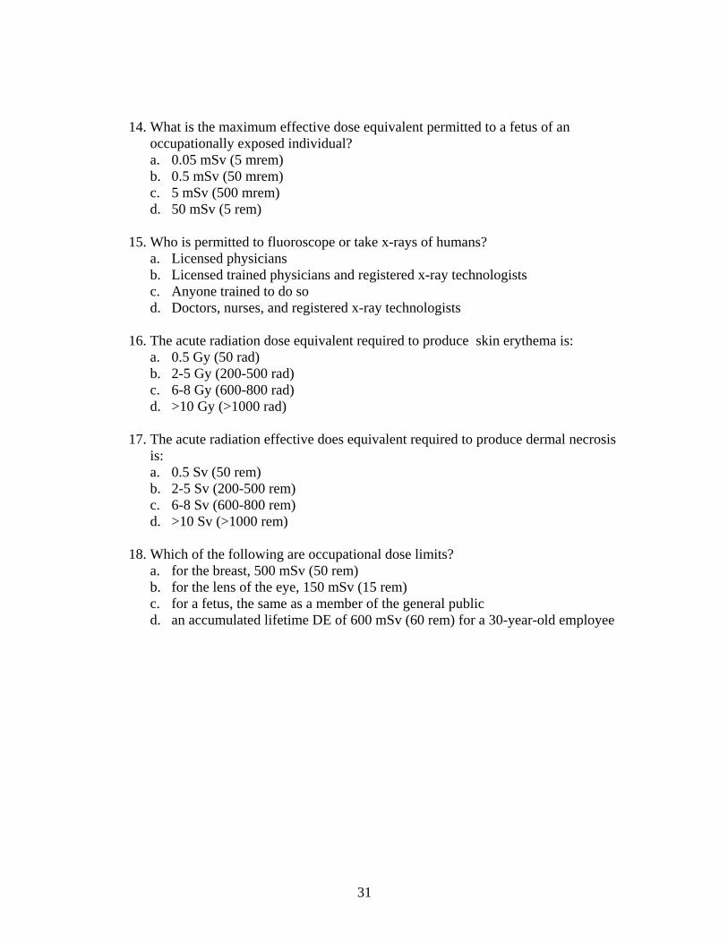
14. What is the maximum effective dose equivalent permitted to a fetus of an occupationally exposed individual? a. 0.05 mSv (5 mrem) b. 0.5 mSv (50 mrem) c. 5 mSv (500 mrem) d. 50 mSv (5 rem)
15. Who is permitted to fluoroscope or take x-rays of humans? a. Licensed physicians b. Licensed trained physicians and registered x-ray technologists c. Anyone trained to do so d. Doctors, nurses, and registered x-ray technologists
16. The acute radiation dose equivalent required to produce skin erythema is:
a. 0.5 Gy (50 rad) b. 2-5 Gy (200-500 rad) c. 6-8 Gy (600-800 rad) d. >10 Gy (>1000 rad)
17. The acute radiation effective does equivalent required to produce dermal necrosis
is: a. 0.5 Sv (50 rem) b. 2-5 Sv (200-500 rem) c. 6-8 Sv (600-800 rem) d. >10 Sv (>1000 rem)
18. Which of the following are occupational dose limits?
a. for the breast, 500 mSv (50 rem) b. for the lens of the eye, 150 mSv (15 rem) c. for a fetus, the same as a member of the general public d. an accumulated lifetime DE of 600 mSv (60 rem) for a 30-year-old employee