radiologic findings of complications of ercp -...
TRANSCRIPT
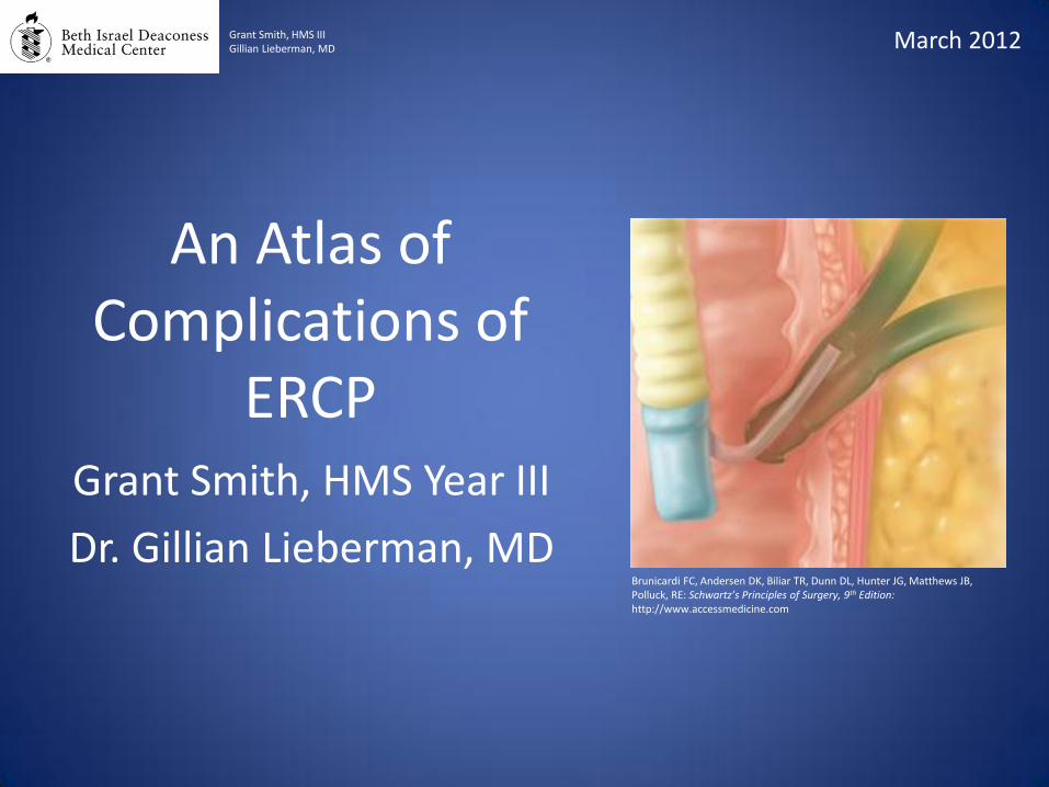
An Atlas of Complications of
ERCP Grant Smith, HMS Year III
Dr. Gillian Lieberman, MD
March 2012 Grant Smith, HMS III Gillian Lieberman, MD
Brunicardi FC, Andersen DK, Biliar TR, Dunn DL, Hunter JG, Matthews JB, Polluck, RE: Schwartz’s Principles of Surgery, 9th Edition: http://www.accessmedicine.com
Teaching Goals
1) Understand how ERCP is performed.
2) Become familiar with the indications for ERCP.
3) Know the common complications of ERCP.
4) Identify the menu of radiologic tests used to diagnose complications of ERCP.
5) Recognize radiologic findings of ERCP complications.
Grant Smith, HMS III Gillian Lieberman, MD
2
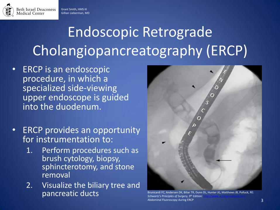
Endoscopic Retrograde Cholangiopancreatography (ERCP)
• ERCP is an endoscopic procedure, in which a specialized side-viewing upper endoscope is guided into the duodenum.
• ERCP provides an opportunity
for instrumentation to: 1. Perform procedures such as
brush cytology, biopsy, sphincterotomy, and stone removal
2. Visualize the biliary tree and pancreatic ducts
Grant Smith, HMS III Gillian Lieberman, MD
Brunicardi FC, Andersen DK, Biliar TR, Dunn DL, Hunter JG, Matthews JB, Polluck, RE: Schwartz’s Principles of Surgery, 9th Edition: http://www.accessmedicine.com Abdominal Fluoroscopy during ERCP 3
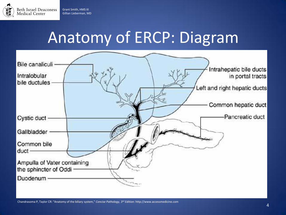
Anatomy of ERCP: Diagram
Grant Smith, HMS III Gillian Lieberman, MD
Chandrasoma P, Taylor CR: “Anatomy of the biliary system,” Concise Pathology, 3rd Edition: http://www.accessmedicine.com 4
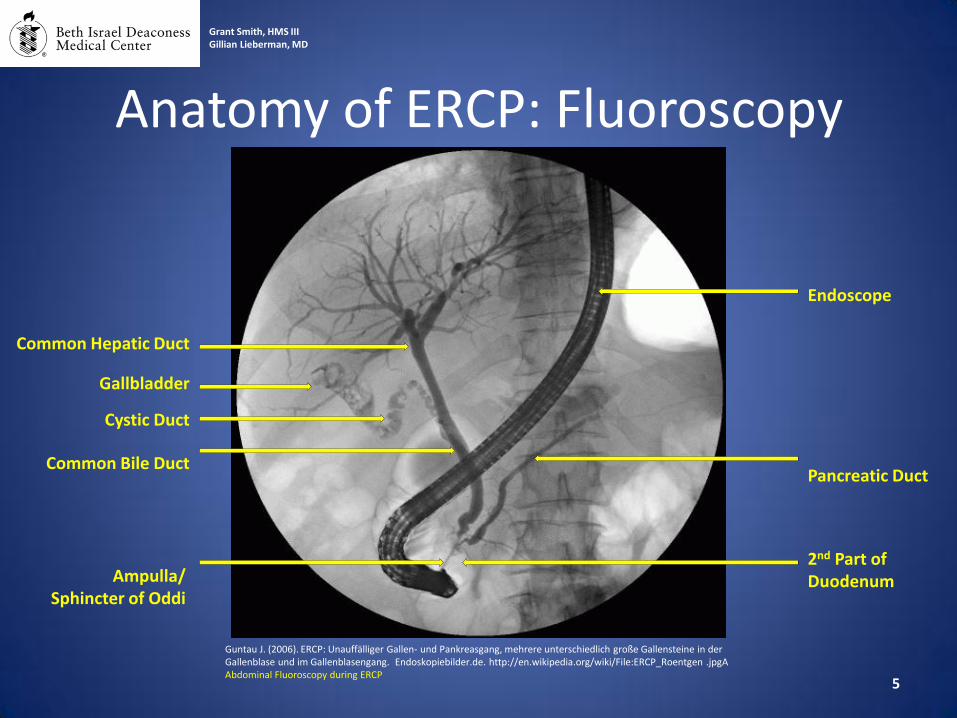
Anatomy of ERCP: Fluoroscopy
Grant Smith, HMS III Gillian Lieberman, MD
Guntau J. (2006). ERCP: Unauffälliger Gallen- und Pankreasgang, mehrere unterschiedlich große Gallensteine in der Gallenblase und im Gallenblasengang. Endoskopiebilder.de. http://en.wikipedia.org/wiki/File:ERCP_Roentgen .jpgA Abdominal Fluoroscopy during ERCP
Endoscope
Pancreatic Duct
2nd Part of Duodenum
Cystic Duct
Common Bile Duct
Gallbladder
Ampulla/ Sphincter of Oddi
Common Hepatic Duct
5
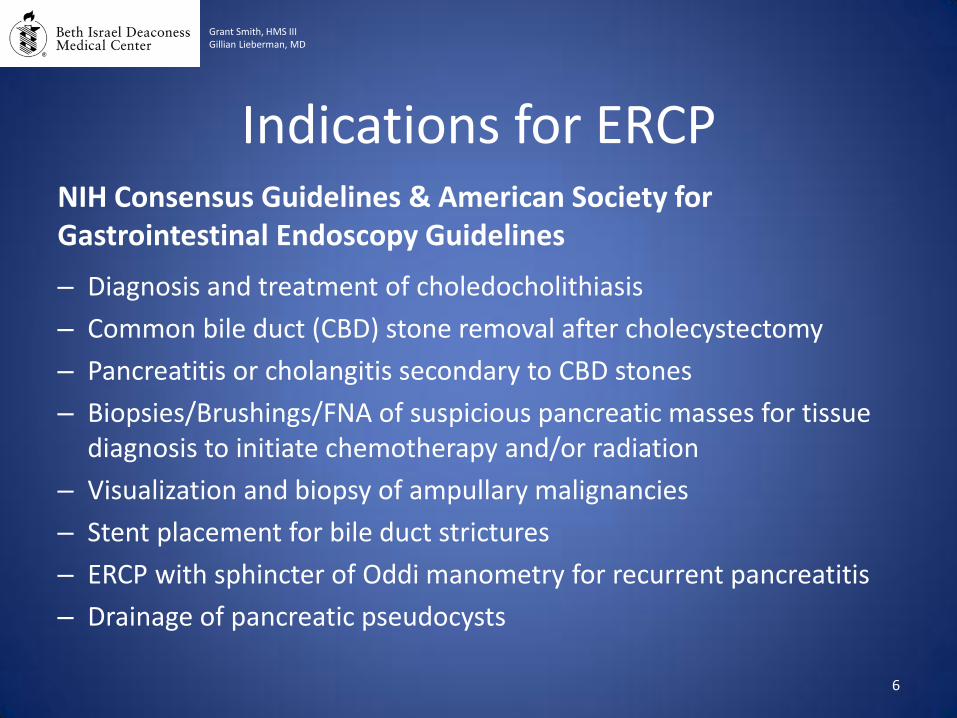
Indications for ERCP NIH Consensus Guidelines & American Society for Gastrointestinal Endoscopy Guidelines
– Diagnosis and treatment of choledocholithiasis
– Common bile duct (CBD) stone removal after cholecystectomy
– Pancreatitis or cholangitis secondary to CBD stones
– Biopsies/Brushings/FNA of suspicious pancreatic masses for tissue diagnosis to initiate chemotherapy and/or radiation
– Visualization and biopsy of ampullary malignancies
– Stent placement for bile duct strictures
– ERCP with sphincter of Oddi manometry for recurrent pancreatitis
– Drainage of pancreatic pseudocysts
Grant Smith, HMS III Gillian Lieberman, MD
6
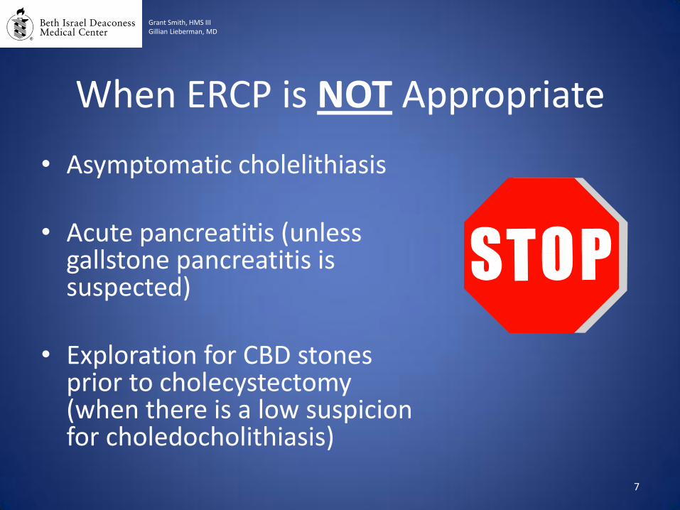
When ERCP is NOT Appropriate
• Asymptomatic cholelithiasis
• Acute pancreatitis (unless gallstone pancreatitis is suspected)
• Exploration for CBD stones prior to cholecystectomy (when there is a low suspicion for choledocholithiasis)
7
Grant Smith, HMS III Gillian Lieberman, MD
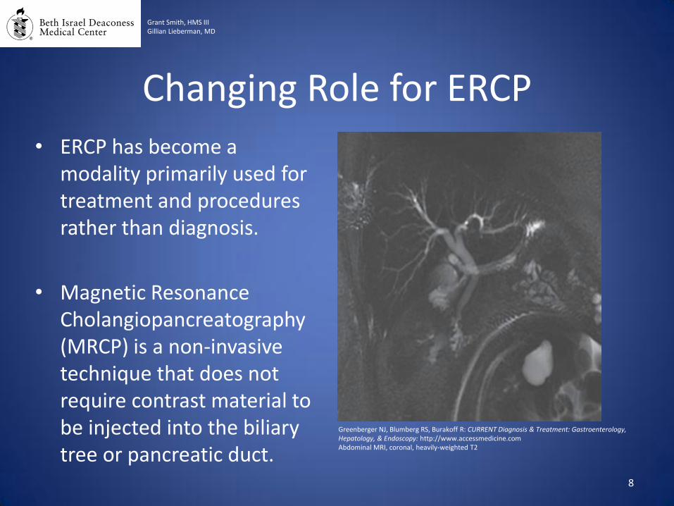
Changing Role for ERCP
• ERCP has become a modality primarily used for treatment and procedures rather than diagnosis.
• Magnetic Resonance Cholangiopancreatography (MRCP) is a non-invasive technique that does not require contrast material to be injected into the biliary tree or pancreatic duct.
8
Grant Smith, HMS III Gillian Lieberman, MD
Greenberger NJ, Blumberg RS, Burakoff R: CURRENT Diagnosis & Treatment: Gastroenterology, Hepatology, & Endoscopy: http://www.accessmedicine.com Abdominal MRI, coronal, heavily-weighted T2
COMPLICATIONS OF ERCP
9
Grant Smith, HMS III Gillian Lieberman, MD
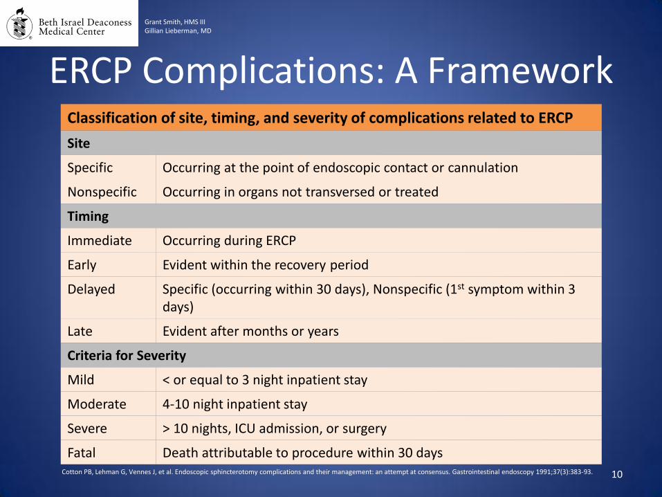
ERCP Complications: A Framework
10
Grant Smith, HMS III Gillian Lieberman, MD
Classification of site, timing, and severity of complications related to ERCP
Site
Specific Occurring at the point of endoscopic contact or cannulation
Nonspecific Occurring in organs not transversed or treated
Timing
Immediate Occurring during ERCP
Early Evident within the recovery period
Delayed Specific (occurring within 30 days), Nonspecific (1st symptom within 3 days)
Late Evident after months or years
Criteria for Severity
Mild < or equal to 3 night inpatient stay
Moderate 4-10 night inpatient stay
Severe > 10 nights, ICU admission, or surgery
Fatal Death attributable to procedure within 30 days Cotton PB, Lehman G, Vennes J, et al. Endoscopic sphincterotomy complications and their management: an attempt at consensus. Gastrointestinal endoscopy 1991;37(3):383-93.
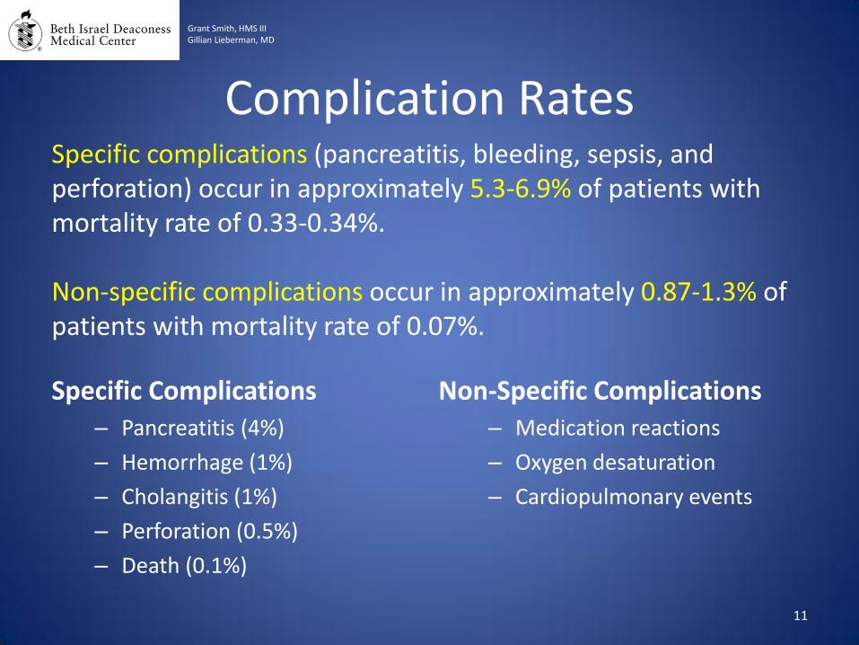
Complication Rates
Specific Complications – Pancreatitis (4%)
– Hemorrhage (1%)
– Cholangitis (1%)
– Perforation (0.5%)
– Death (0.1%)
Non-Specific Complications – Medication reactions
– Oxygen desaturation
– Cardiopulmonary events
11
Grant Smith, HMS III Gillian Lieberman, MD
Specific complications (pancreatitis, bleeding, sepsis, and perforation) occur in approximately 5.3-6.9% of patients with mortality rate of 0.33-0.34%. Non-specific complications occur in approximately 0.87-1.3% of patients with mortality rate of 0.07%.
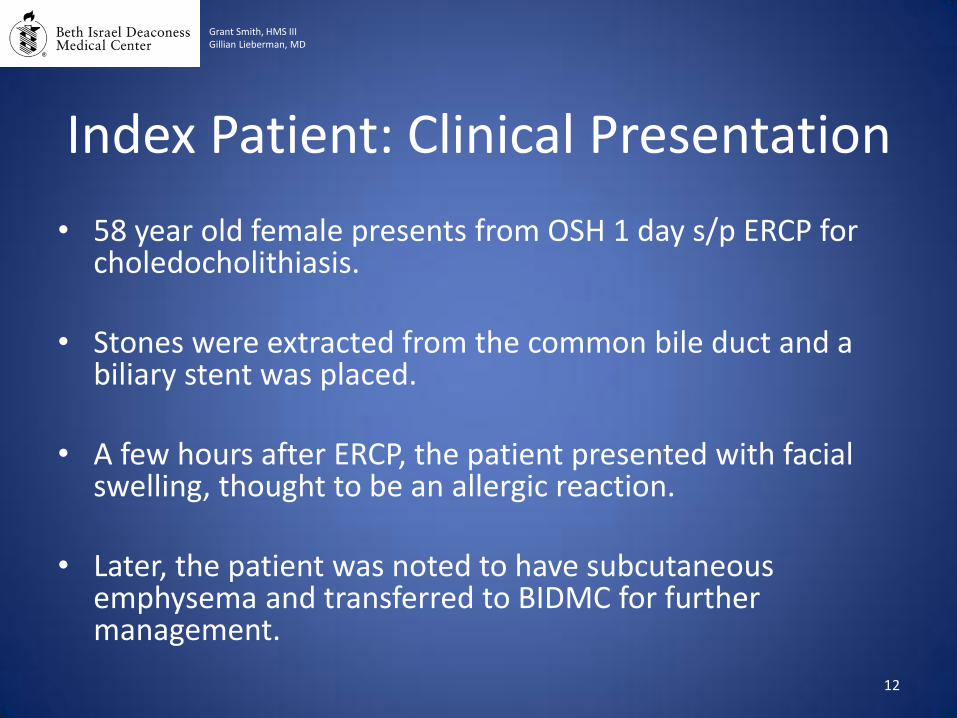
Index Patient: Clinical Presentation
• 58 year old female presents from OSH 1 day s/p ERCP for choledocholithiasis.
• Stones were extracted from the common bile duct and a biliary stent was placed.
• A few hours after ERCP, the patient presented with facial swelling, thought to be an allergic reaction.
• Later, the patient was noted to have subcutaneous emphysema and transferred to BIDMC for further management.
12
Grant Smith, HMS III Gillian Lieberman, MD

Subcutaneous air in cervical
area
Pneumo-mediastinum
“Gingko Sign” Air between muscle fibers of pectoralis major indicating subcutaneous emphysema
13
Grant Smith, HMS III Gillian Lieberman, MD
Index Patient: Initial CXR
BIDMC, PACS Chest X-ray, Portable
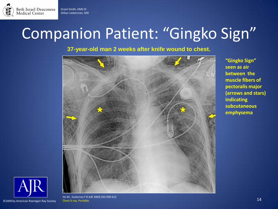
37-year-old man 2 weeks after knife wound to chest.
Ho M , Gutierrez F R AJR 2009;192:599-612 Chest X-ray, Portable ©2009 by American Roentgen Ray Society 14
Companion Patient: “Gingko Sign”
Grant Smith, HMS III Gillian Lieberman, MD
“Gingko Sign” seen as air between the muscle fibers of pectoralis major (arrows and stars) indicating subcutaneous emphysema
15
Subcutaneous emphysema
Grant Smith, HMS III Gillian Lieberman, MD
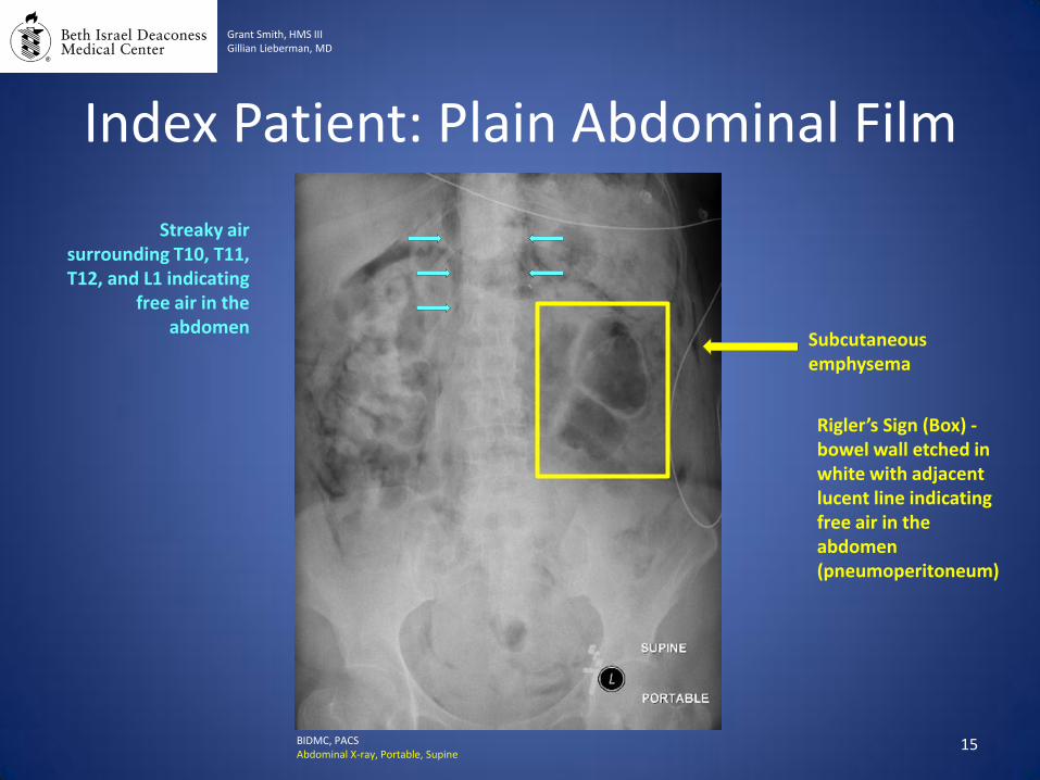
Index Patient: Plain Abdominal Film
BIDMC, PACS Abdominal X-ray, Portable, Supine
Rigler’s Sign (Box) - bowel wall etched in white with adjacent lucent line indicating free air in the abdomen (pneumoperitoneum)
Streaky air surrounding T10, T11, T12, and L1 indicating
free air in the abdomen
16
Grant Smith, HMS III Gillian Lieberman, MD
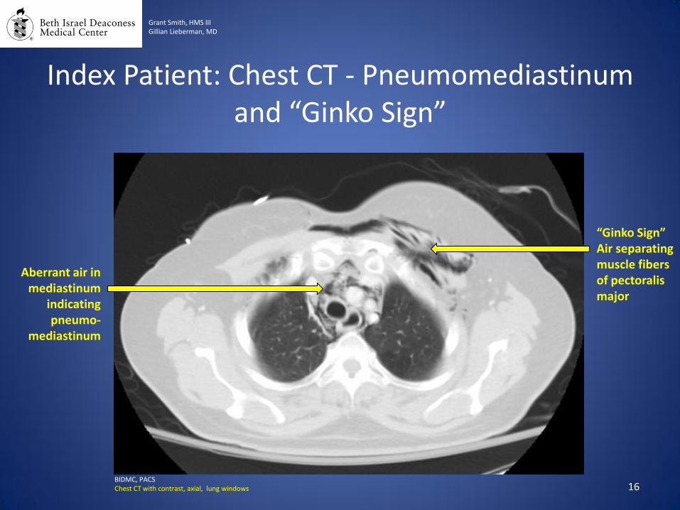
Index Patient: Chest CT - Pneumomediastinum and “Ginko Sign”
Aberrant air in mediastinum
indicating pneumo-
mediastinum
“Ginko Sign” Air separating muscle fibers of pectoralis major
BIDMC, PACS Chest CT with contrast, axial, lung windows

17
Grant Smith, HMS III Gillian Lieberman, MD
Index Patient: Chest CT - Pneumothorax
Small pneumothorax
“Ginko Sign” showing air separating muscle fibers of pectoralis major
Pneumo-mediastinum
BIDMC, PACS Chest CT with contrast, axial, lung windows
18
Grant Smith, HMS III Gillian Lieberman, MD
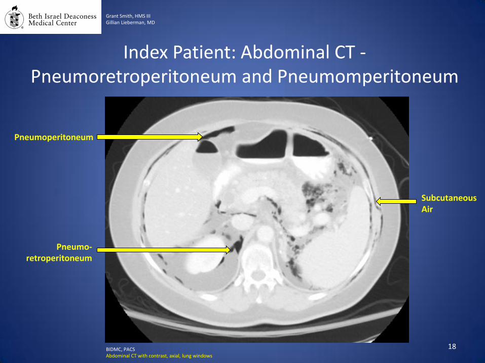
Index Patient: Abdominal CT - Pneumoretroperitoneum and Pneumomperitoneum
Subcutaneous Air
Pneumo-retroperitoneum
Pneumoperitoneum
BIDMC, PACS Abdominal CT with contrast, axial, lung windows
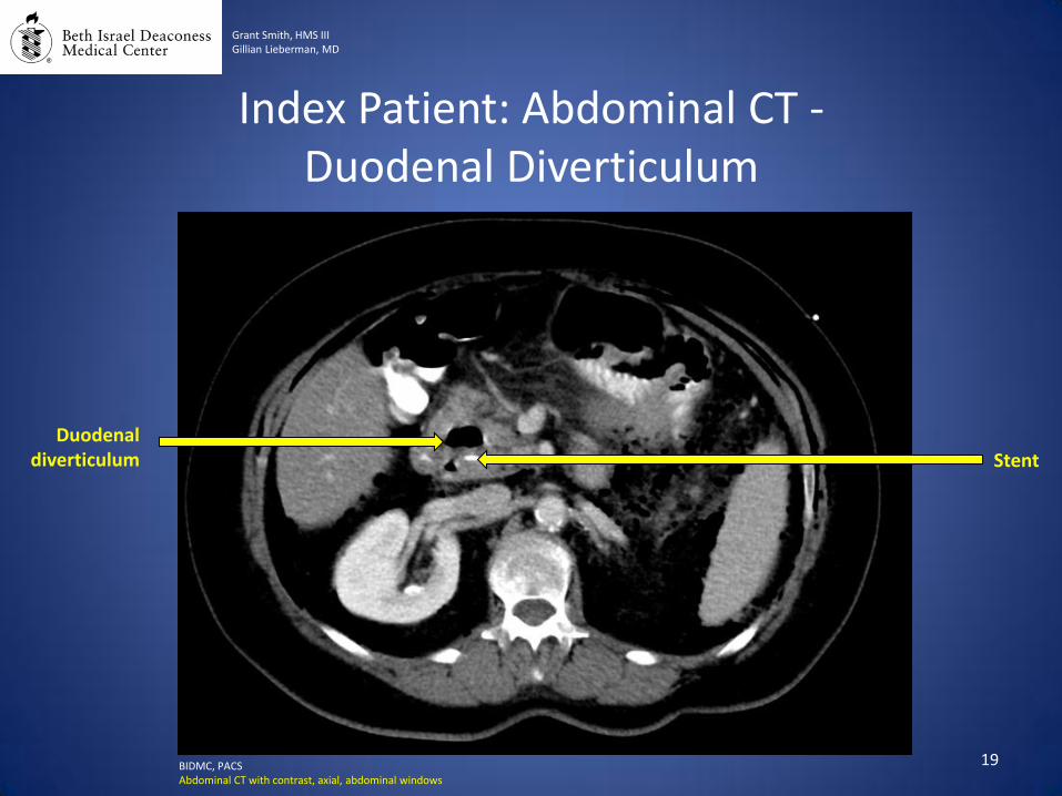
Duodenal diverticulum
19
Grant Smith, HMS III Gillian Lieberman, MD
Index Patient: Abdominal CT - Duodenal Diverticulum
Stent
BIDMC, PACS Abdominal CT with contrast, axial, abdominal windows
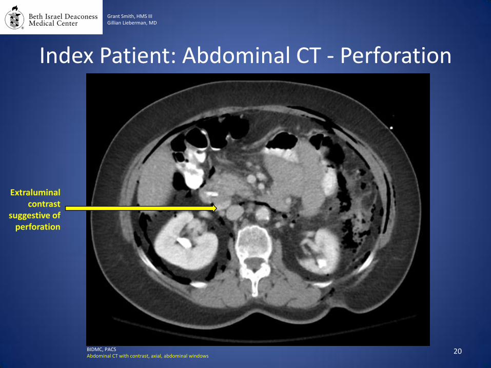
Extraluminal contrast
suggestive of perforation
20
Grant Smith, HMS III Gillian Lieberman, MD
Index Patient: Abdominal CT - Perforation
BIDMC, PACS Abdominal CT with contrast, axial, abdominal windows
Perforation: Overview • Incidence – Approx. 1.3% of cases
• Clinical Manifestations
– Abdominal pain – Elevated serum amylase
• Risk Factors – Sphincterotomy – Sphincter of Oddi dysfunction – Dilated CBD – Long procedure – Biliary stricture dilatation – Duodenal diverticula – Aberrant biliary anatomy – Post-surgical anatomy (Roux-en-Y gastric bypass) 21
Grant Smith, HMS III Gillian Lieberman, MD
22
• Menu of Radiologic Tests – Plain Abdominal Film
• Free air seen as “Rigler’s Sign” (bowel wall outlined by air) or the “Football Sign” (central lucency with visualization of falciform and medial umbilical ligaments). – Free air is best seen on upright films or left lateral decubitus (if
unable to stand) or cross-table lateral view.
– Computed Tomography
• Ability to see tiny foci of free air not seen on plain films. • Recommended if patient has increase WBC count or pain
and fever. • Bile infection and bile leakage through a perforation seen on
CT correlates with increased mortality.
Perforation: Radiologic Findings
Grant Smith, HMS III Gillian Lieberman, MD
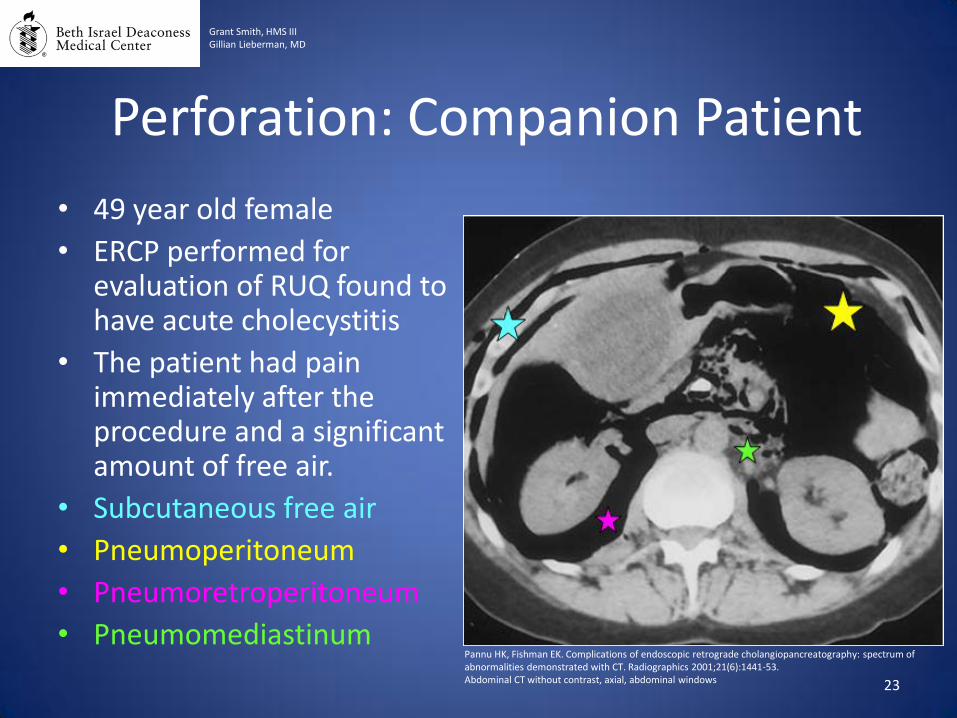
Perforation: Companion Patient
• 49 year old female
• ERCP performed for evaluation of RUQ found to have acute cholecystitis
• The patient had pain immediately after the procedure and a significant amount of free air.
• Subcutaneous free air
• Pneumoperitoneum
• Pneumoretroperitoneum
• Pneumomediastinum
23
Grant Smith, HMS III Gillian Lieberman, MD
Pannu HK, Fishman EK. Complications of endoscopic retrograde cholangiopancreatography: spectrum of abnormalities demonstrated with CT. Radiographics 2001;21(6):1441-53. Abdominal CT without contrast, axial, abdominal windows

Pancreatitis: Overview • Incidence – Approx. 5% of diagnostic cases and 10% of
therapeutic cases • Clinical Manifestations
– Abdominal pain for >24hrs s/p ERCP • Often epigastric or back pain with nausea
– Elevated serum amylase and lipase (3x normal)
• Risk Factors – Operator-Related: inadequate training, lack of experience, low case
volume – Patient-Related: younger age, females, recurrent pancreatitis, history
of post-ERCP pancreatitis, Sphincter of Oddi dysfunction – Procedure-Related factors: difficulty with cannulation, pancreatic duct
infection, precut/pancreatic/minor papilla sphincterotomy, or biliary balloon sphincteroplasty.
24
Grant Smith, HMS III Gillian Lieberman, MD
25
• Menu of Radiologic Tests
– Computed Tomography
• Heterogeneous enhancement and gland enlargement.
• Peripancreatic fat has increased attenuation due to extravasation of pancreatic secretions.
• Glandular necrosis appears as hypoattenuation.
• Infected necrosis appears as bubbles of gas in devitalized parenchyma.
Pancreatitis: Radiologic Findings
Grant Smith, HMS III Gillian Lieberman, MD
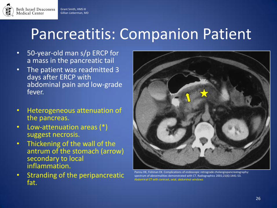
Pancreatitis: Companion Patient
26
Grant Smith, HMS III Gillian Lieberman, MD
Pannu HK, Fishman EK. Complications of endoscopic retrograde cholangiopancreatography: spectrum of abnormalities demonstrated with CT. Radiographics 2001;21(6):1441-53. Abdominal CT with contrast, axial, abdominal windows
• 50-year-old man s/p ERCP for a mass in the pancreatic tail
• The patient was readmitted 3 days after ERCP with abdominal pain and low-grade fever.
• Heterogeneous attenuation of the pancreas.
• Low-attenuation areas (*) suggest necrosis.
• Thickening of the wall of the antrum of the stomach (arrow) secondary to local inflammation.
• Stranding of the peripancreatic fat.
27
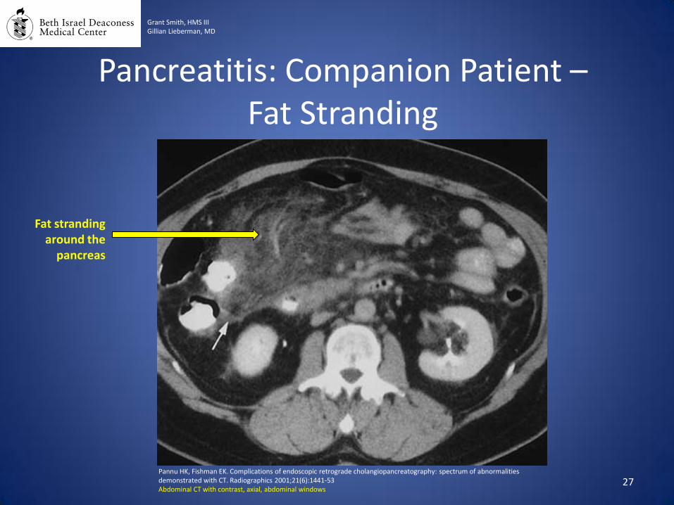
Pancreatitis: Companion Patient – Fat Stranding
Grant Smith, HMS III Gillian Lieberman, MD
Fat stranding around the
pancreas
Pannu HK, Fishman EK. Complications of endoscopic retrograde cholangiopancreatography: spectrum of abnormalities demonstrated with CT. Radiographics 2001;21(6):1441-53 Abdominal CT with contrast, axial, abdominal windows

Pancreatitis: Index Patient
Grant Smith, HMS III Gillian Lieberman, MD
Infected pancreatic necrosis with gas
28 BIDMC, PACS Abdominal CT with contrast, axial, abdominal windows
Hemorrhage: Overview • Incidence – Approx. 1.3% (with about 29% of bleeds requiring >5
units of transfusions or intervention)
• Clinical Manifestations – Drop in hemoglobin/hematocrit – Melena or hematemesis
• Risk Factors
– Sphincterotomy – Evidence of bleeding at time of sphincterotomy – Prior sphincterotomy – Prolonged PTT (at least 2x above normal) – Periampullary diverticulum – Cholangitis
29
Grant Smith, HMS III Gillian Lieberman, MD
Hemorrhage: Radiologic Findings
• Menu of Radiologic Tests and Findings
– Computed Tomography (CT) • Typically not performed to diagnose hemorrhage; but bleeding
may be detected if CT is performed.
• Acute hemorrhage is hyperattenuating on noncontrast CT, which can become iso/hypoattenuating in later stages.
• Non-contrast CT is used to assess for hematoma, while contrast-enhanced CT angiography is used to assess for site of active extravasation.
30
Grant Smith, HMS III Gillian Lieberman, MD

• 67-year-old woman s/p ERCP with unsuccessful cannulation of the common bile duct
• The patient experienced pain after the procedure.
• High attenuation area between the duodenum and pancreas (arrow) representing bleeding
31
Hemorrhage: Companion Patient #1
Grant Smith, HMS III Gillian Lieberman, MD
Pannu HK, Fishman EK. Complications of endoscopic retrograde cholangiopancreatography: spectrum of abnormalities demonstrated with CT. Radiographics 2001;21(6):1441-53 Abdominal CT with contrast, axial, abdominal windows
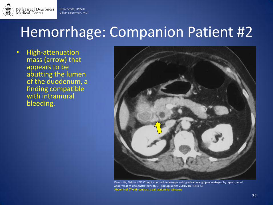
• High-attenuation mass (arrow) that appears to be abutting the lumen of the duodenum, a finding compatible with intramural bleeding.
32
Hemorrhage: Companion Patient #2
Grant Smith, HMS III Gillian Lieberman, MD
Pannu HK, Fishman EK. Complications of endoscopic retrograde cholangiopancreatography: spectrum of abnormalities demonstrated with CT. Radiographics 2001;21(6):1441-53 Abdominal CT with contrast, axial, abdominal windows
Infection/Cholangitis: Overview
33
Grant Smith, HMS III Gillian Lieberman, MD
• Infection can include many complications • Ascending cholangitis is the most frequent infectious complication of
ERCP
• Incidence – Approx 1.4% (with range of 0.4-10% depending on the study)
• Clinical Manifestations – Typically occur 24-72 hours after ERCP – Fever – Jaundice – Abdominal pain (RUQ) – May develop confusion and hypotension – Elevated WBC count
• Risk Factors – Biliary stents – Combined percutaneous and endoscopic procedures – Unsuccessful drainage of the biliary system (retained stones)
Charcot’s Triad Reynold’s Pentad
Infection/Cholangitis: Radiologic Findings
34
Grant Smith, HMS III Gillian Lieberman, MD
• Menu of Radiologic Tests
– Computed Tomography (CT) • Bile ducts may appear dilated and bile itself may appear
hyperattenuated due to increased debris.
• Thickening of wall of ducts and pneumobilia.
• Peri-biliary hyperattenuation.
• Abscesses may also appear with enhancing capsules.

• 67 year old man with common bile duct stones
• In this case, has not undergone ERCP but demonstrates findings of biliary obstruction
• Diffuse, mottled enhancement of the liver parenchyma
• Dilatation of the intrahepatic bile ducts (arrows)
35
Infection/Cholangitis: Companion Patient
Grant Smith, HMS III Gillian Lieberman, MD
Kim SW, Shin HC, Kim HC, Hong MJ, Kim IY. Diagnostic performance of multidetector CT for acute cholangitis: evaluation of a CT scoring method. The British journal of radiology. Abdominal CT with contrast, axial, abdominal windows
Summary
• ERCP is an endoscopic procedure primary used for therapeutic intervention.
• ERCP is appropriately used to remove stones from the CBD, assist in diagnosis of pancreatic/ampullary masses, and stent placement for biliary obstruction.
• The main complications of ERCP include perforation, pancreatitis, hemorrhage, and infection.
• Computed tomography is a good first choice for investigating for complications of ERCP when patients become acutely ill within 24-48 hours after ERCP.
36
Grant Smith, HMS III Gillian Lieberman, MD
References 1. Cohen S, Bacon BR, Berlin JA, et al. National Institutes of Health State-of-the-Science Conference Statement: ERCP for
diagnosis and therapy, January 14-16, 2002. Gastrointestinal endoscopy 2002;56(6):803-9. 2. Adler DG, Baron TH, Davila RE, et al. ASGE guideline: the role of ERCP in diseases of the biliary tract and the pancreas.
Gastrointestinal endoscopy 2005;62(1):1-8. 3. Cotton PB, Lehman G, Vennes J, et al. Endoscopic sphincterotomy complications and their management: an attempt at
consensus. Gastrointestinal endoscopy 1991;37(3):383-93. 4. Andriulli A, Loperfido S, Napolitano G, et al. Incidence rates of post-ERCP complications: a systematic survey of
prospective studies. The American journal of gastroenterology 2007;102(8):1781-8. 5. Williams EJ, Taylor S, Fairclough P, et al. Risk factors for complication following ERCP; results of a large-scale, prospective
multicenter study. Endoscopy 2007;39(9):793-801. 6. Wang P, Li ZS, Liu F, et al. Risk factors for ERCP-related complications: a prospective multicenter study. The American
journal of gastroenterology 2009;104(1):31-40. 7. Zissin R, Shapiro-Feinberg M, Oscadchy A, Pomeranz I, Leichtmann G, Novis B. Retroperitoneal perforation during
endoscopic sphincterotomy: imaging findings. Abdominal imaging 2000;25(3):279-82. 8. Cohen SA, Siegel JH, Kasmin FE. Complications of diagnostic and therapeutic ERCP. Abdominal imaging 1996;21(5):385-
94. 9. Scarlett PY, Falk GL. The management of perforation of the duodenum following endoscopic sphincterotomy: a proposal
for selective therapy. The Australian and New Zealand journal of surgery 1994;64(12):843-6. 10. Pannu HK, Fishman EK. Complications of endoscopic retrograde cholangiopancreatography: spectrum of abnormalities
demonstrated with CT. Radiographics 2001;21(6):1441-53 11. Balthazar EJ. CT diagnosis and staging of acute pancreatitis. Radiologic clinics of North America 1989;27(1):19-37. 12. Balthazar EJ, Freeny PC, vanSonnenberg E. Imaging and intervention in acute pancreatitis. Radiology 1994;193(2):297-
306. 13. Testoni PA, Mariani A, Giussani A, et al. Risk factors for post-ERCP pancreatitis in high- and low-volume centers and
among expert and non-expert operators: a prospective multicenter study. The American journal of gastroenterology;105(8):1753-61
14. Kim SW, Shin HC, Kim HC, Hong MJ, Kim IY. Diagnostic performance of multidetector CT for acute cholangitis: evaluation of a CT scoring method. The British journal of radiology.
15. Carr-Locke DL. Therapeutic role of ERCP in the management of suspected common bile duct stones. Gastrointestinal endoscopy 2002;56(6 Suppl):S170-4.
16. Ho ML, Gutierrez FR. Chest radiography in thoracic polytrauma. Ajr 2009;192(3):599-612.
37
Grant Smith, HMS III Gillian Lieberman, MD
Special Thanks To:
• Dr. Gillian Lieberman
• Dr. Mai-Lan Ho
• Dr. Mark Ashkan
38