radiology packet 4 cardiac enlargement. 9 year old shetland sheepdog “alt” hx: presented for...
Post on 21-Dec-2015
217 views
TRANSCRIPT

Radiology Packet 4
Cardiac Enlargement

9 year old shetland sheepdog “Alt”
• Hx: Presented for coughing, exercise intolerance and has a systolic murmur


9 year old shetland sheepdog “Alt”
• RF– Lateral
• Elevation of the trachea.• Heart is taller than it should be.• Marked loss of the caudal cardiac waist with soft tissue structure
overlying the caudal heart base region.• Upward deviation toward the heart of the caudal vena cava.• Increased cardiophrenic contact.• Prominent pulmonary vessels.
– DV• Large soft tissue opacity of the hilar region, between the mainstem
bronchi, which can be followed over to the 3 o’clock position (left atrium and auricle).
• R/O– Mitral insufficiency and secondary heart enlargement
• Next: Cardiac ultrasound
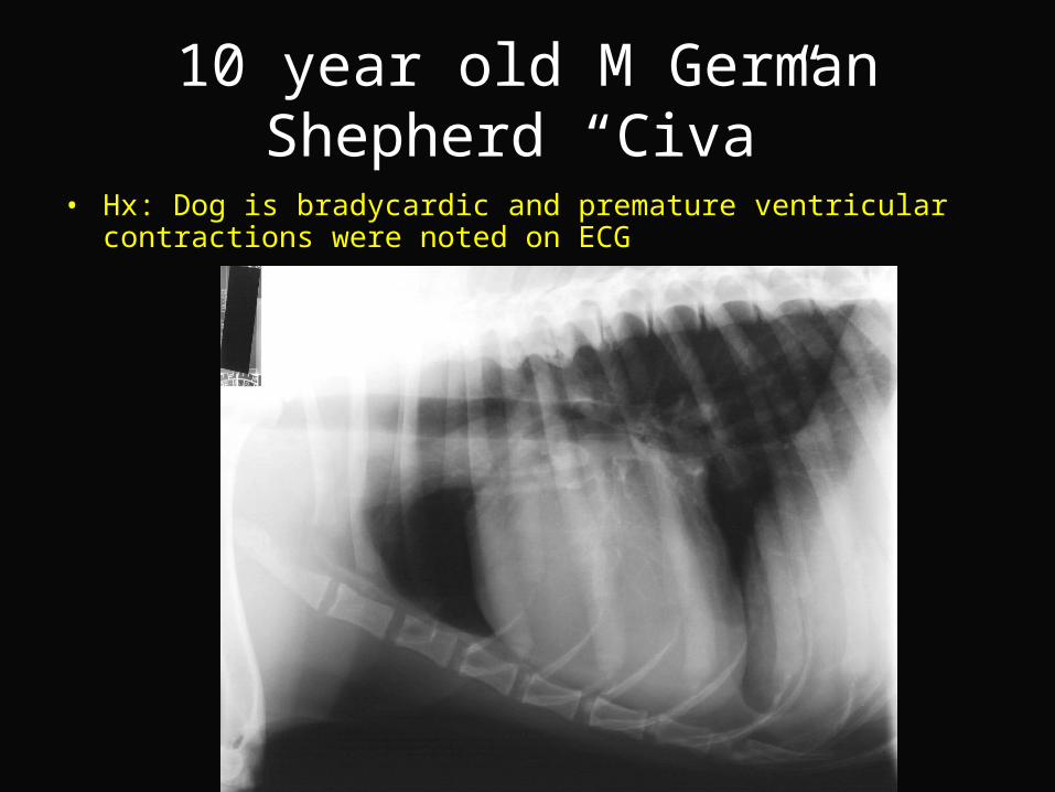
10 year old M German Shepherd “Civa”
• Hx: Dog is bradycardic and premature ventricular contractions were noted on ECG


10 year old M German Shepherd “Civa”
• RF – Mild generalized cardiac enlargement.– Incidental finding of spondylosis at several sites in the
thoracic spine.
• R/O– Dilatative cardiomyopathy – Pericardial effusion
• Next: ECG

10 year old cocker spaniel MN “Sprocket”
• Hx: Presented for evaluation of a chronic cough. No signs of systemic illness and no heart murmur is ausculted. The patient has been treated with Furosemide (Lasix), Enalapril (cardiac drug) and Hycodan (cough suppressent)

.

10 year old cocker spaniel MN “Sprocket”
• RF– Right ventricular enlargement.
– Trachea is mildly elevated so right atrial enlargement may also be present.
– Diffuse mild interstitial lung pattern consistent with normal age change.
– Liver is mildly enlarged.
• R/O– Right sided cardiac enlargement
• Cor pulmonale• Aquired right-sided cardiac enlargement (relatively uncommon)
– Bronchitis

6 year old FS Bernese Mountain Dog
• Hx: Presented for surgical repair of a cranial cruciate ligament rupture. While under general anesthesia occasional ventricular pre-mature contractions were noted.


6 year old FS Bernese Mountain Dog
• RF- Microcardia and small size of the pulmonary vessels.
- Lung fields are over-exposed making evaluation more difficult
• RD– hypovolemia
• R/O– Dehydration
– Shock
– Addison’s disease (hypoadrenocorticism)
• Next: ACTH stimulation test

4 year old German Shorthair Pointer “Autumn”
• Hx: Has had 2 episodes of mild weakness/collapse associated with intense exercise. A persistent cough is also reported. A systolic murmur with the point of maximal intensity at the aortic valve was noted ~1 year ago.


4 year old German Shorthair Pointer “Autumn”
• RD– Mild right-sided cardiac enlargement– Moderate diffuse interstitial lung pattern with bronchial markings (not prominent
enough to call a broncho-interstitial lung pattern)
• R/O– Tricuspid valve insufficiency – Cor pulmonale (increased size of the right heart due to pulmonary hypertension)– Presence of interstitial pattern is non-specific and R/O include lungworm
infestation, interstitial pneumonia and interstitial changes due to prior pulmonary disease
• Next:– Echocardiography– Baermann fecal exam

12 year old DSH “Tuffy”
• Hx: Presented for mild ataxia. The owners report the cat seems to be breathing rapidly. Auscultation of the thorax was unrewarding.


12 year old DSH “Tuffy”
• RF– Increased sternal contact of the heart and the aorta has a prominent
appearance.– Cardiac silhouette is wider than normal.– Cranial border is slightly square on the lateral view.– Heart has a “Valentine” shape.– The cranial pulmonary vessels are seen and at the upper limits of
normal size.
• RD– Hypertrophic cardiomyopathy
• Next– Echocardiogram

6-year old MN DSH“Fatty Lumpkin”
• Hx: Presented for evaluation of lethargy and increased respiratory rate


6-year old MN DSH“Fatty Lumpkin”
• RF– Cardiac silhouette is partially obscured by increased opacity within the
thoracic cavity.
– The atrial region of the heart appears wide.
– The trachea is elevated.
– Mild pulmonary vascular congestion is present as well as free pleural fluid.
• RD– Hypertrophic cardiomyopathy
– Congestive heart failure

9-year old MN Schipperke“Robbie”
• Hx: Presented for evaluation of a cardiac murmur. The murmur was not present the previous year. It is described as a grade 2-3 of 6 systolic murmur.


9-year old MN Schipperke“Robbie”
• RF– Caudal mainstem bronchi are elevated and there is loss of the caudal
cardiac waste.– In VD the left atrium is visible as a large round structure caudal to the
mainstem bronchi bifurcation.– The entire trachea is elevated and the caudal cardiac margin is
elongated.– In the VD there is mild bulging in the left ventricular region.
• RD– Left atrial and ventricular enlargement
• R/O– Mitral regurgitation secondary to mitral valve endocardiosis

12 year old M Saluki“Linca”
• Hx: Presented with a 1 week history of lethargy and poor appetite. On PE the abdomen appears distended and a fluid wave can be balloted. On auscultation the heart sounds are muffled and of variable intensity.


12 year old M Saluki“Linca”
• RF– Cardiac silhouette is enlarged and very round.– In the lateral view a single pleural fissure line is visible.
• RD– Globoid cardiac silhouette– Pericardial effusion
• R/O– Hemangiosarcoma of the right atrium– Heart base mass (lymphosarcoma) within the pericardial sac– Idiopathic pericardial effusion– Pericardial effusion leading to right sided heart failure– Underlying cardiac disease such as dilated cardiomyopathy and tricuspid
dysplasia– Peritoneopericardial diaphragmatic hernia