real-time pcr assay and rapid diagnostic tests for the diagnosis
TRANSCRIPT
RESEARCH Open Access
Real-time PCR assay and rapid diagnostic testsfor the diagnosis of clinically suspected malariapatients in BangladeshMohammad Shafiul Alam1*, Abu Naser Mohon1, Shariar Mustafa1, Wasif Ali Khan1, Nazrul Islam2,Mohammad Jahirul Karim2, Hamida Khanum3, David J Sullivan Jr4 and Rashidul Haque1
Abstract
Background: More than 95% of total malaria cases in Bangladesh are reported from the 13 high endemic districts.Plasmodium falciparum and Plasmodium vivax are the two most abundant malaria parasites in the country. Toimprove the detection and management of malaria patients, the National Malaria Control Programme (NMCP) hasbeen using rapid diagnostic test (RDT) in the endemic areas. A study was conducted to establish a SYBR Green-based modified real-time PCR assay as a gold standard to evaluate the performance of four commercially-availablemalaria RDTs, along with the classical gold standard- microscopy.
Methods: Blood samples were collected from 338 febrile patients referred for the diagnosis of malaria by theattending physician at MatirangaUpazila Health Complex (UHC) from May 2009 to August 2010. Paracheck RDT and microscopy were performed atthe UHC. The blood samples were preserved in EDTA tubes. A SYBR Green-based real-time PCR assay wasperformed and evaluated. The performances of the remaining three RDTs (Falcivax, Onsite Pf and Onsite Pf/Pv)were also evaluated against microscopy and real-time PCR using the stored blood samples.
Result: In total, 338 febrile patients were enrolled in the study. Malaria parasites were detected in 189 (55.9%) and188 (55.6%) patients by microscopy and real-time PCR respectively. Among the RDTs, the highest sensitivity for thedetection of P. falciparum (including mixed infection) was obtained by Paracheck [98.8%, 95% confidence interval(CI) 95.8-99.9] and Falcivax (97.6%, 95% CI 94.1-99.4) compared to microscopy and real-time PCR respectively.Paracheck and Onsite Pf/Pv gave the highest specificity (98.8%, 95% CI 95.7-99.9) compared to microscopy andOnsite Pf/Pv (98.8, 95% CI 95.8-99.9) compared to real-time PCR respectively for the detection of P. falciparum. Onthe other hand Falcivax and Onsite Pf/Pv had equal sensitivity (90.5%, 95% CI 69.6-98.8) and almost 100%specificity compared to microscopy for the detection of P. vivax. However, compared to real-time PCR assay RDTsand microscopy gave low sensitivity (76.9%, 95% CI 56.4-91) in detecting of P. vivax although a very high specificitywas obtained (99- 100%).
Conclusion: The results of this study suggest that the SYBR Green-based real-time PCR assay could be used as analternative gold standard method in a reference setting. Commercially-available RDTs used in the study are quitesensitive and specific in detecting P. falciparum, although their sensitivity in detecting P. vivax was not satisfactorycompared to the real-time PCR assay.
* Correspondence: [email protected] Laboratory, ICDDR,B, GPO Box 128, Dhaka-1000, BangladeshFull list of author information is available at the end of the article
Alam et al. Malaria Journal 2011, 10:175http://www.malariajournal.com/content/10/1/175
© 2011 Alam et al; licensee BioMed Central Ltd. This is an Open Access article distributed under the terms of the Creative CommonsAttribution License (http://creativecommons.org/licenses/by/2.0), which permits unrestricted use, distribution, and reproduction inany medium, provided the original work is properly cited.
BackgroundMalaria is still considered a major public-health problem inthe eastern districts of Bangladesh, bordering India andMyanmar. These districts experience a perennial transmis-sion of malaria with two peaks in pre-monsoon (March-May) and post-monsoon (September-November) periods[1]. In the changing climatic situation and in absence ofmajor malaria vectors, such as Anopheles minimus andAnopheles baimaii a number of Anopheles species havebeen incriminated and playing a role in the transmission ofmalaria in the country [2]. Plasmodium falciparum andPlasmodium vivax are two main malaria parasites in thecountry as reported by a nationwide prevalence survey in2007. The survey showed that contribution of P. falciparumwas 90.18%, followed by P. vivax (5.29%), and the remain-ing (4.53%) was mixed infection of these two species [3].The Giemsa-stained blood slide using thin and thick
smears for malaria parasites has been the gold standardmethod for nearly a century [4]. No alternative methodstill could be established to replace this universally-accepted gold standard method. Such a laboratory tech-nique to confirm the clinical suspicion of malaria islabour-intensive [5] and sometimes unreliable due tolack of skilled microscopists, limited supplies, inade-quate maintenance of microscopes and reagents, andinadequate or absence of quality-control systems [6].In recent time, lateral flow immunochromatographic-
based rapid diagnostic test (RDT) has been developedfor the diagnosis of suspected malaria patients and arewidely used in remote areas across the world [7]. MostRDTs are intended to react with antigens commonlyreleased from or enzymes present in parasitized redblood cells. In the case of P. falciparum, the water solu-ble histidine-rich protein-2 (HRP-2) antigen is com-monly used as it is specific to P. falciparum associatedinfection. Non-falciparum malaria or mixed infectionswith P. falciparum are commonly detected by Plasmo-dium lactate dehydrogenase (pLDH) [8,9]. In the GlobalFund sponsored malaria control programme RDT isrecommended and being widely used for detectingmalaria cases in the endemic areas of Bangladesh [1].The molecular detection method, such as polymerase
chain reaction (PCR) has been developed to diagnose Plas-modium spp. and has been performed in several places forroutine diagnosis or for evaluating the performance ofmicroscopy or RDT [10-14]. In recent time, real-time PCRmethod has been established for the quantitative detectionof malaria parasites [15-19]. Real-time PCR is reliable andyield high sensitivity and specificity when compared withmicrocopy or nested PCR [15,19,20].This study demonstrated a SYBR Green-based modi-
fied real-time method to use it as a gold standard, alongwith conventional microscopy to evaluate four RDTs for
diagnosis of malaria from suspected febrile patients.Such a study has never been done before in Bangladesh.The study would provide additional support to theNMCP for monitoring and evaluation of the perfor-mances of the diagnostic methods used in their ongoingmalaria control programme.
MethodsStudy area and populationThe study was conducted at Matiranga Upazila (sub-dis-trict) of Khagrachari district situated at the south-east-ern part of Bangladesh. Febrile patients referred tomicroscopy for malaria diagnosis at Matiranga UpazilaHealth Complex (UHC) from May 2009 to August 2010were enrolled. The recent malaria prevalence survey,Matiranga showed high prevalence of asymptomaticmalaria cases (21.6%) [21].
Sample collectionFive ml of blood was taken from an adult subject and incase of children or minor subjects three ml of bloodwas obtained through venipuncture by an experiencedmedical technologist. Two drops of sample were usedfor preparing thick and thin smear slides, one drop wasused for Paracheck RDT, and the remaining sampleswere preserved in an EDTA tube and stored at -20°C.
MicroscopyThe blood film was stained with Giemsa in phosphatebuffer saline and examined under the compound micro-scope at a magnification of ×1,000 for malaria parasites.Blood films were defined as negative if no parasite wasobserved in 100× oil immersion fields (magnification,×1,000) on thin film by an experienced microscopist[22]. Declaring a slide positive or negative and initialspeciation was routinely based on the examination of200 fields in the Giemsa-stained thick film. A slide wasconsidered positive when at least one parasite wasfound. After finding the first parasite, another 200 fieldswere completed for any mixed infection. If no parasitewas found in 200 oil fields, the slide was considerednegative. Density of the parasite was measured fromthick blood smears by counting the number of parasitesper 200 leukocytes and expressed as parasites/μl. In thecase of 10 or less parasites, 500 leukocytes werecounted. Each slide was assessed by two independentmicroscopists; one of them was employed by the studyand the other person was posted at Matiranga UHC. Aslide was considered positive only when these twomicroscopists were in agreement. There was a provisionfor third microscopist posted at the Khagrachari CivilSurgeon’s office situated 20 km away from MatirangaUHC for any disagreement between them.
Alam et al. Malaria Journal 2011, 10:175http://www.malariajournal.com/content/10/1/175
Page 2 of 9
Rapid diagnostic testsIn the present investigation four RDTs (device) wereused. These were Paracheck (Orchid Biomedical System,India), FalciVax Pf (Zephyr Biomedicals, India), OnsitePf (CTK Biotech Inc, USA) and Onsite Pf/Pv (CTK Bio-tech Inc, USA). Paracheck and Onsite Pf used P. falci-parum-specific HRP-2 antigen. FalciVax and Onsite Pf/Pv used P. vivax- specific pLDH together with P. falci-parum-specific HRP-2.All the RDTs were used following the instructions of
the manufacturers. ‘Paracheck’ is being used by theNational Malaria Control Programme (NMCP) in theendemic areas and was available at the MUHC. Para-check test was performed at Matiranga UHC concur-rently with the microscopy. The remaining three RDTswere performed using the stored samples as per theinstructions of the manufacturers.
DNA extractionDNA was extracted from 200 μl EDTA preserved bloodsamples using the QiaAmp blood mini kit (QIAGEN,Inc., Germany) following the manufacturer’s instructionsat the Parasitology Laboratory of ICDDR,B. DNA samplewas stored at 4°C until PCR could be completed.
Real-time PCRReal-time PCR was done by the primer sets described byPerandin et al [15] with some modification to a single-plex reaction. Instead of TaqMan probe, SYBR Green Idye was used for visualizing the amplification. PCR con-dition was also modified slightly to fit with Platinum®
SYBR® Green qPCR SuperMix-UDG (Invitrogen Cor-poration, USA) following the instructions of the manu-facturer. Purified DNA templates were amplified in aBioRad CFX-96 real time system (BioRad, USA) with aspecies-specific primer set. Briefly, a 25-μl PCR mixturewas prepared using 1 μl of template DNA, 12.5 μl Plati-num SYBR Green qPCR supermix (PlatinumR TaqDNA polymerase, SYBR Green I dye, Tris-HCl, KCl, 6mM MgCl2, 400 μM dGTP, 400 μM dATP, 400 μMdCTP, 800 μM dUTP, uracil DNA glycosylase, and sta-bilizers), 320 nM concentration of each of parasite spe-cies-specific primer set. Amplification and detectionwere performed as follows: 50°C for 2 min and 95°C for2 min. After that 95°C for 1 min, 58°C for 1 min and72°C for 1 min 30 sec for a single cycle were performed.40 cycles were considered for P. falciparum and 35cycles for P. vivax. The plate read was taken after theextension at 72°C. The melt curve was prepared from50°C to 95°C with an increment of 0.5°C each after fiveseconds.To establish the minimum number of parasites detect-
able by the Plasmodium SYBR Green assay (detectionlimit), blood samples from two patients infected,
respectively, with P. falciparum (one patient) and P.vivax (one patient) were collected, and parasitaemia wascalculated using 200 WBC count as reference. Theinfected blood samples were diluted with uninfected ery-throcytes from healthy individuals with known baselineerythrocyte counts. Ten-fold serial dilution was made toobtain a final parasitaemia of 1% (1 parasite/μl of blood)for each sample. All DNA aliquots purified from thedilutions were treated in duplicate for real-time PCRassay. To estimate the analytical specificity of the Plas-modium real-time PCR assay, DNA from in vitro culturesamples of other protozoan parasites, such as Enta-moeba histolytica and Leishmania donovani were used.The clinical sensitivity and specificity of the modifiedPlasmodium real-time PCR assay for detecting and iden-tifying malaria parasites were calculated on 338 whole-blood samples, microscopy as the gold standard andvice- versa.
Analysis of dataThe performance of each method was calculated bymeans of sensitivity, specificity, positive predictive value(PPV), and negative predictive value (NPV) using micro-scopy and modified real-time PCR as gold standard.SPSS software version 11.5 (SPSS Inc., USA) was usedfor calculating the kappa coefficient (k) of the tests foreach association using the X2 test. Sensitivity, specificity,positive predictive value and negative predictive valuewere calculated using the ‘diagt’ command of theSTATA software version 10 (Stata Corp, USA)[23].
Ethical approvalThe study was approved by the Research Review Com-mittee and Ethical Review Committee of ICDDR,B.Approval was also obtained from the NMCP for thestudy. Informed consent was obtained from all adultsubjects, and assent was obtained from the legal guar-dians in the case of minor subjects before the collectionof blood sample. Good clinical and laboratory practiceswere followed in all the procedures.
ResultsEnrollmentIn total, 338 febrile patients were recruited for the studyfrom May 2009 to August 2010. Of them, 50.3% werefemale. The age of the patients ranged from 18 monthsto 82 years, with a median age of 14 years.
MicroscopyMalaria parasites were detected in 189 (55.9%) patientsby microscopy. Of them 168 (88.9%) were infected by P.falciparum, 18 (9.5%) patients by P. vivax and remainingthree (1.6%) patients had a mixed infection of P. falci-parum and P. vivax (Table 1). Overall, high parasite
Alam et al. Malaria Journal 2011, 10:175http://www.malariajournal.com/content/10/1/175
Page 3 of 9

count was observed in microscopy. Parasite count ran-ged from 16 to 261,480 parasites/μl of blood. A mediannumber of parasite count of 19,960 [interquartile range(IQR) 6,280-48,320] parasites/μl of blood was found in171 P. falciparum positive patients. Only six (3.5%) of thesamples were below 100 parasites/ μl whereas 118 (69%)had a count of more than 10,000 parasites/ μl of blood.Of 21 P. vivax positive slides, the parasite count rangedfrom 32 to 25,120 parasites/μl of blood, with a median of5,040 (IQR 520-17,160) parasites/μl of blood. One (4.8%)sample had a parasite count below 100 parasites/ μlwhereas 15 (71.4%) of the sample had a count of morethan 1,000 parasites/μl of blood (see Additional file 1).
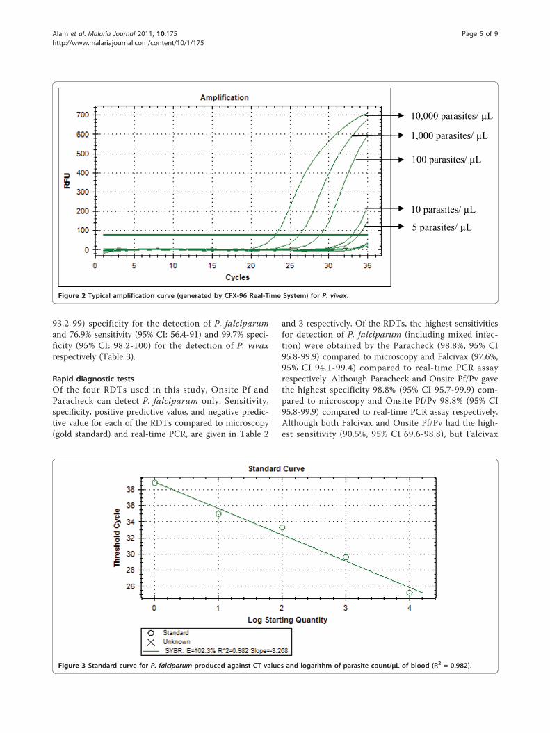
Real-time PCRTypical displays (amplification plots) for P. falciparumand P. vivax by the SYBR Green I PCR assay providedby Bio Rad CFX-96 are shown in Figures 1 and 2. Posi-tive signals by means of cycle threshold [CT] value wereobtained for all dilutions, with a detection limit of 5-10
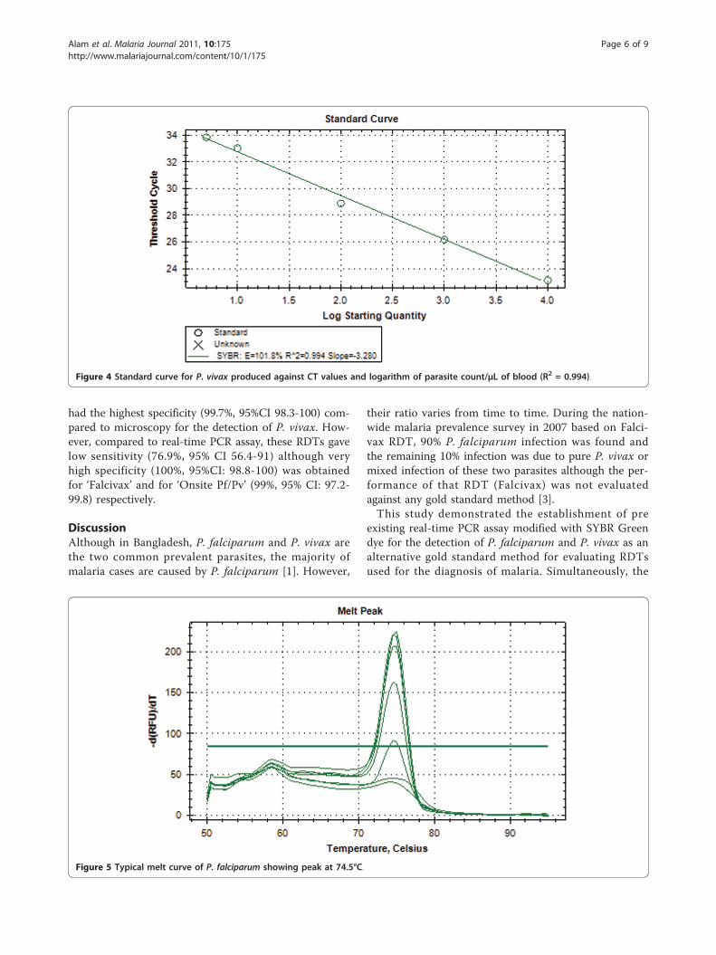
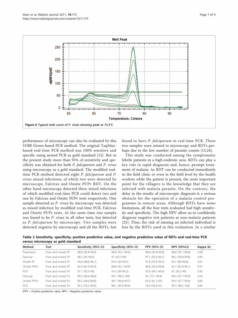
parasites/μl for P. falciparum and P. vivax in differentexperiments. Reproducible linearity of over a 10,000-foldrange was shown by CT values. A significant correlationcoefficient was found for the mean CT values and para-sitaemia (P. falciparum, R2 = 0.982; P. vivax, R2 = 0.994)(Figures 3 and 4). For non-Plasmodium protozoan DNA(E. histolytica and L. donovani) and blood DNA samplesof healthy human subjects no signal was obtained by theSYBR Green real-time PCR. The melt peak for P. falci-parum and P. vivax was found at 74.5°C and 75.5°Cfrom the corresponding positive controls respectively(Figures 5 and 6). Any amplification other than thesetwo melting temperatures was excluded as falseamplification.Using the real-time PCR assay results 188 (55.6%)
samples were found positive for any malarial infection(Table 1). Of the 188 PCR positive samples 162 (86.2%)were infected by P. falciparum, 18 (9.5%) were infectedby P. vivax and the remaining 8 (4.3%) samples weremixed infection with P. falciparum and P. vivax (Table1). Sensitivity, specificity, positive predictive value, nega-tive predictive value, and kappa (k) of PCR assay com-pared to microscopy are given in table 2. For thedetection of P. falciparum (including mixed infection),modified real-time PCR assay had 97.1% (95% CI: 93.3-99) sensitivity and 97.6% (95% CI: 94-99.3) specificityrespectively. While for the detection of P. vivax (includ-ing mixed infection) modified real-time PCR showed95.2% (95% CI: 76.2-99.9) sensitivity and 98.1% (95% CI:95.9-99.6) specificity respectively (Table 2).Compared to real-time PCR assay, microscopy had
97.6% sensitivity (95% CI: 94.1-99.4) and 97% (95%CI
Table 1 Results for different tests used in the study
Test Negative Positive
N (%) Pf (%) Pv (%) Pf + Pvmixed (%)
Total (%)
Microscopy 149 (44.1) 168 (49.7) 18 (5.3) 3 (0.9) 189(55.9)
Paracheck 167 (49.4) 171 (50.6) N/A N/A 171 (50.6)
Onsite Pf 174 (51.5) 164(48.5) N/A N/A 164(48.5)
Falcivax 147 (43.5) 171 (50.6) 18 (5.3) 2 (0.6) 191 (56.5)
Onsite Pf/Pv 160 (47.3) 155 (45.9) 18 (5.3) 5 (1.5) 178 (52.7)
Real-time PCR 150 (44.4) 162 (47.9) 18 (5.3) 8 (2.4) 188(55.6)
100 parasites/ μL
10 parasites/ μL
1000 parasites/ μL
1 parasite/μL
Figure 1 Typical amplification curve (generated by CFX-96 Real-Time System) for P. falciparum.
Alam et al. Malaria Journal 2011, 10:175http://www.malariajournal.com/content/10/1/175
Page 4 of 9
93.2-99) specificity for the detection of P. falciparumand 76.9% sensitivity (95% CI: 56.4-91) and 99.7% speci-ficity (95% CI: 98.2-100) for the detection of P. vivaxrespectively (Table 3).
Rapid diagnostic testsOf the four RDTs used in this study, Onsite Pf andParacheck can detect P. falciparum only. Sensitivity,specificity, positive predictive value, and negative predic-tive value for each of the RDTs compared to microscopy(gold standard) and real-time PCR, are given in Table 2
and 3 respectively. Of the RDTs, the highest sensitivitiesfor detection of P. falciparum (including mixed infec-tion) were obtained by the Paracheck (98.8%, 95% CI95.8-99.9) compared to microscopy and Falcivax (97.6%,95% CI 94.1-99.4) compared to real-time PCR assayrespectively. Although Paracheck and Onsite Pf/Pv gavethe highest specificity 98.8% (95% CI 95.7-99.9) com-pared to microscopy and Onsite Pf/Pv 98.8% (95% CI95.8-99.9) compared to real-time PCR assay respectively.Although both Falcivax and Onsite Pf/Pv had the high-est sensitivity (90.5%, 95% CI 69.6-98.8), but Falcivax
10,000 parasites/ μL
1,000 parasites/ μL
100 parasites/ μL
10 parasites/ μL
5 parasites/ μL
Figure 2 Typical amplification curve (generated by CFX-96 Real-Time System) for P. vivax.
Figure 3 Standard curve for P. falciparum produced against CT values and logarithm of parasite count/μL of blood (R2 = 0.982).
Alam et al. Malaria Journal 2011, 10:175http://www.malariajournal.com/content/10/1/175
Page 5 of 9
had the highest specificity (99.7%, 95%CI 98.3-100) com-pared to microscopy for the detection of P. vivax. How-ever, compared to real-time PCR assay, these RDTs gavelow sensitivity (76.9%, 95% CI 56.4-91) although veryhigh specificity (100%, 95%CI: 98.8-100) was obtainedfor ‘Falcivax’ and for ‘Onsite Pf/Pv’ (99%, 95% CI: 97.2-99.8) respectively.
DiscussionAlthough in Bangladesh, P. falciparum and P. vivax arethe two common prevalent parasites, the majority ofmalaria cases are caused by P. falciparum [1]. However,
their ratio varies from time to time. During the nation-wide malaria prevalence survey in 2007 based on Falci-vax RDT, 90% P. falciparum infection was found andthe remaining 10% infection was due to pure P. vivax ormixed infection of these two parasites although the per-formance of that RDT (Falcivax) was not evaluatedagainst any gold standard method [3].This study demonstrated the establishment of pre
existing real-time PCR assay modified with SYBR Greendye for the detection of P. falciparum and P. vivax as analternative gold standard method for evaluating RDTsused for the diagnosis of malaria. Simultaneously, the
Figure 4 Standard curve for P. vivax produced against CT values and logarithm of parasite count/μL of blood (R2 = 0.994).
Figure 5 Typical melt curve of P. falciparum showing peak at 74.5°C.
Alam et al. Malaria Journal 2011, 10:175http://www.malariajournal.com/content/10/1/175
Page 6 of 9
performance of microscopy can also be evaluated by thisSYBR Green-based PCR method. The original TaqMan-based real-time PCR method was 100% sensitive andspecific using nested PCR as gold standard [15]. But inthe present study more than 95% of sensitivity and spe-cificity was obtained for both P. falciparum and P. vivaxusing microscopy as a gold standard. The modified real-time PCR method detected eight P. falciparum and P.vivax mixed infections, of which two were detected bymicroscopy, Falcivax and Onsite Pf/Pv RDT. On theother hand microscopy detected three mixed infectionsof which modified real-time PCR could detect two andone by Falcivax and Onsite Pf/Pv tests respectively. Onesample detected as P. vivax by microscopy was detectedas mixed infection by modified real-time PCR, Falcivaxand Onsite Pf/Pv tests. At the same time one samplewas found to be P. vivax in all other tests, but detectedas P. falciparum by microscopy. Two samples weredetected negative by microscopy and all the RDTs, but
found to have P. falciparum in real-time PCR. Thesetwo samples were missed in microscopy and RDTs per-haps due to the low number of parasite counts [15,24].This study was conducted among the symptomatic
febrile patients in a high-endemic area. RDTs can play akey role in rapid diagnosis and, hence, prompt treat-ment of malaria. As RDT can be conducted immediatelyin the field clinic or even in the field level by the healthworkers while the patient is present, the most importantpoint for the villagers is the knowledge that they areinfected with malaria parasite. On the contrary, thedelay in the results of microscopic diagnosis is a seriousobstacle for the operation of a malaria control pro-gramme in remote areas. Although RDTs have somelimitations, all the four tests evaluated had high sensitiv-ity and specificity. The high NPV allow us to confidentlydiagnose negative test patients as non-malaria patients[25]. Thus, the risk of missing an infected individual isless by the RDTs used in this evaluation. In a similar
Table 2 Sensitivity, specificity, positive predictive value, and negative predictive value of RDTs and real-time PCRversus microscopy as gold standard
Method Test Sensitivity (95% CI) Specificity (95% CI) PPV (95% CI) NPV (95%CI) Kappa (k)
Paracheck Pure and mixed Pf 98.8 (95.8-99.9) 98.8 (95.7-99.9) 98.8 (95.8-99.9) 98.8 (95.7-99.8) 0.98
Falcivax Pure and mixed Pf 98.2 (95-99.6) 97 (93.2-99) 97.1 (93.4-99.1) 98.2 (94.8-99.6) 0.95
Onsite Pf Pure and mixed Pf 93.6 (88.8-96.7) 97.6 (94-99.3) 97.6 (93.9-99.3) 93.7 (89-96.8) 0.91
Onsite Pf/Pv Pure and mixed Pf 92.4 (87.4-95.9) 98.8 (95.7-99.9) 98.8 (95.6-99.8) 92.7 (87.8-96.1) 0.91
PCR Pure and mixed Pf 97.1 (93.3-99) 97.6 (94-99.3) 97.6 (94.1-99.4) 97 (93.2-99) 0.95
Falcivax Pure and mixed Pv 90.5 (69.6-98.8) 99.7 (98.3-100) 95 (75.1-99.9) 99.4 (97.7-99.9) 0.92
Onsite Pf/Pv Pure and mixed Pv 90.5 (69.6-98.8) 98.7 (96.8-99.7) 82.6 (61.2-95) 99.4 (97.7-99.9) 0.85
PCR Pure and mixed Pv 95.2 (76.2-99.9) 98.1 (95.9-99.6) 76.9 (56.4-91) 99.7 (98.2-100) 0.84
(PPV = Positive predictive value, NPV = Negative predictive value).
Figure 6 Typical melt curve of P. vivax showing peak at 75.5°C.
Alam et al. Malaria Journal 2011, 10:175http://www.malariajournal.com/content/10/1/175
Page 7 of 9

study in India, high NPV was also recorded for FalcivaxRDT [13].Overall, 55.9% of the febrile patients with suspected
malaria in the present study had a positive blood slide,indicating that over half of the suspected cases referredto this hospital (Matiranga UHC) had malaria. A highpercentage of malaria cases among the febrile cases ofthis area could be due to a high prevalence of asympto-matic malaria cases at the community [21].Pf-HRP 2 based Paracheck is currently being used in
the country’s NMCP, although there is a necessity of aRDT for detecting multiple malaria infections in thecountry [1]. In the present study Parachek showed highsensitivity and specificity compared to a study in Malawiwhere low specificity was reported [26]. However, sincethe control programme is now targeting for RDTs thatcan detect multiple infections, this study could providea valuable guideline to them.Onsite duo as newly developed test had never been
evaluated earlier in any part of the world gave satisfac-tory results in the present study. Although Onsite Pf/Pvfailed to diagnose some P. falciparum positive samplewhich undoubtedly affects its sensitivity compared to itscounterpart Falcivax. However, Onsite Pf/Pv gave almosta similar result as like as Falcivax for detecting P. vivax.Low sensitivity of the two RDTs (Falcivax and Onsite
Pf/Pv) for the detection of P. vivax compared to real-time PCR assay in this present study is similar to studiespublished earlier [20,26]. This could be due to theinherent limitations of pLDH assay to detect low parasi-taemia in the clinical specimens [27]. Miscroscopistssimilar to RDTs also missed P. vivax cases compared toreal-time PCR assay perhaps due to the same reason(low parasitaemia)[28].RDTs do not depend on the operator like microscopy.
It was evolved to overcome or reduce the limitations ofmicroscopy. They have brought a revolution in the fieldof malaria diagnosis. However, these must achieve >95% sensitivity to prove their usefulness [29]. It has
been estimated that over 70 million RDTs are soldacross the world. There are a number of companies pro-ducing RDTs for the diagnosis of malaria which wasinitiated by a single company in 1993 [30]. The worldhealth organization listed approximately 50 RDTs, ofwhich only a few had PvLDH antigen-based tests thatdistinguish between P. falciparum and P. vivax are com-mercially available [26].
ConclusionsFindings of the study suggest that the SYBR Green-based real-time PCR and RDTs used in the study aresensitive and specific for the detection of P. falciparum.However, RDTs and microscopy were not sensitiveenough compared to real-time PCR assay for the detec-tion of P. vivax. The SYBR Green-based real-time PCRcould be a useful tool for monitoring the performanceof different malaria diagnostic tests in a reference settingby the NMCP. Efforts should be given to increase theaccuracy of RDTs as well as microscopy for diagnosis ofP. vivax in the field level. As more than one malariaparasites are present in the endemic areas of Bangla-desh, it is imperative to deploy a RDT that can detectmultiple malaria infections by the NMCP.
AcknowledgementsThis research study was funded by ICDDR,B and its donors which provideunrestricted support to ICDDR,B for its operations and research. Currentdonors providing unrestricted support include: Australian Agency forInternational Development (AusAID), Government of the People’s Republicof Bangladesh; Canadian International Development Agency (CIDA), SwedishInternational Development Cooperation Agency (Sida), and the Departmentfor International Development, UK (DFID). We gratefully acknowledge thesedonors for their support and commitment to ICDDR,B’s research efforts.The authors are grateful to NMCP for their permission to conduct the studyin their facilities and also for providing Paracheck RDT. The authors are alsograteful to CTK Biotech Inc, USA, for providing ‘Onsite Pf’ and ‘Onsite Pf/Pv’RDT as a donation. The authors are also indebted to the people ofMatiranga who had consented to participate in the study and the doctorsand staff of Matiranga UHC for their extended supports. The authors alsoappreciate the contribution of Mamun Kabir, Khaja Mohiuddin, A. E. M.Rubayet Elahi, Milka Patracia Podder, Shihab U. Sobuz, and Md. GulamMusawwir Khan for their valuable contributions to the study.
Table 3 Sensitivity, specificity, positive predictive value and negative predictive value of RDTs and microscopy versusreal-time PCR as gold standard
Method Test Sensitivity (95% CI) Specificity (95% CI) PPV (95% CI) NPV (95%CI) Kappa (k)
Paracheck Pure and mixed Pf 97.1 (93.3-99) 96.4 (92.4-98.7) 96.5 (92.5-98.7) 97 (93.2-99) 0.94
Falcivax Pure and mixed Pf 97.6 (94.1-99.4) 95.8 (91.6-98.3) 96 (91.8-98.4) 97.6 (93.9-99.3) 0.94
Onsite Pf Pure and mixed Pf 94.1 (89.4-97.1) 97.6 (94-99.3) 97.6 (93.9-99.3) 94.3 (89.7-97.2) 0.92
Onsite Pf/Pv Pure and mixed Pf 92.9 (88-96.3) 98.8 (95.8-99.9) 98.8 (95.6-99.8) 93.3 (88.5-96.5) 0.92
Microscopy Pure and mixed Pf 97.6 (94.1-99.4) 97 (93.2-99) 97.1 (93.3-99) 97.6 (94-99.3) 0.95
Falcivax Pure and mixed Pv 76.9 (56.4-91) 100 (98.8-100) 100 (83.2-100) 98.1 (95.9-99.3) 0.86
Onsite Pf/Pv Pure and mixed Pv 76.9 (56.4-91) 99 (97.2-99.8) 87 (66.4-97.2) 98.1 (95.9-99.3) 0.80
Microscopy Pure and mixed Pv 76.9 (56.4-91) 99.7 (98.2-100) 95.2 (76.2-99.9) 98.1 (95.8-99.3) 0.84
(PPV = Positive predictive value, NPV = Negative predictive value).
Alam et al. Malaria Journal 2011, 10:175http://www.malariajournal.com/content/10/1/175
Page 8 of 9

Author details1Parasitology Laboratory, ICDDR,B, GPO Box 128, Dhaka-1000, Bangladesh.2Malaria and Parasitic Disease Control Unit, Directorate General of HealthServices, Mohakhali, Dhaka 1212, Bangladesh. 3Department of Zoology,University of Dhaka, Dhaka 1000, Bangladesh. 4Johns Hopkins MalariaResearch Institute, Bloomberg School of Public Health, Baltimore, MD, USA.
Authors’ contributionsMSA conceptualized and designed the study, collected and identifiedsamples, analysed data, drafted the manuscript and made final revisions.MSA, ANM, WAK, NI, MJK, HK, DS, RH did sample analysis and made criticalrevision of the manuscript. SM organized the field activities, analysed dataand helped revise the manuscript. MSA and RH drafted the manuscript. Allthe authors read the final version of the manuscript and approved.
Competing interestsThe authors declare that they have no competing interests.
Received: 17 April 2011 Accepted: 26 June 2011Published: 26 June 2011
References1. M&PDC: Strategic Plan for Malaria Control Programme Bangladesh 2008-
2015. 2008, 28, Ministry of Health and Family Welfare: Govt. of Bangladesh.2. Alam MS, Khan MG, Chaudhury N, Deloer S, Nazib F, Bangali AM, Haque R:
Prevalence of anopheline species and their Plasmodium infection statusin epidemic-prone border areas of Bangladesh. Malar J 2010, 9:15.
3. Haque U, Ahmed SM, Hossain S, Huda M, Hossain A, Alam MS, Mondal D,Khan WA, Khalequzzaman M, Haque R: Malaria prevalence in endemicdistricts of Bangladesh. PLoS One 2009, 4:e6737.
4. Murray CK, Bell D, Gasser RA, Wongsrichanalai C: Rapid diagnostic testingfor malaria. Trop Med Int Health 2003, 8:876-883.
5. Bojang KA, Obaro S, Morison LA, Greenwood BM: A prospective evaluationof a clinical algorithm for the diagnosis of malaria in Gambian children.Trop Med Int Health 2000, 5:231-236.
6. Coleman RE, Maneechai N, Rachaphaew N, Kumpitak C, Miller RS,Soyseng V, Thimasarn K, Sattabongkot J: Comparison of field and expertlaboratory microscopy for active surveillance for asymptomaticPlasmodium falciparum and Plasmodium vivax in western Thailand. Am JTrop Med Hyg 2002, 67:141-144.
7. Fogg C, Twesigye R, Batwala V, Piola P, Nabasumba C, Kiguli J, Mutebi F,Hook C, Guillerm M, Moody A, Guthmann JP: Assessment of three newparasite lactate dehydrogenase (pan-pLDH) tests for diagnosis ofuncomplicated malaria. Trans R Soc Trop Med Hyg 2008, 102:25-31.
8. Huong NM, Davis TM, Hewitt S, Huong NV, Uyen TT, Nhan DH, Cong le D:Comparison of three antigen detection methods for diagnosis andtherapeutic monitoring of malaria: a field study from southern Vietnam.Trop Med Int Health 2002, 7:304-308.
9. van den Broek I, Hill O, Gordillo F, Angarita B, Hamade P, Counihan H,Guthmann JP: Evaluation of three rapid tests for diagnosis of P.falciparum and P. vivax malaria in Colombia. Am J Trop Med Hyg 2006,75:1209-1215.
10. Berry A, Fabre R, Benoit-Vical F, Cassaing S, Magnaval JF: Contribution ofPCR-based methods to diagnosis and management of imported malaria.Med Trop (Mars) 2005, 65:176-183.
11. Gatti S, Gramegna M, Bisoffi Z, Raglio A, Gulletta M, Klersy C, Bruno A,Maserati R, Madama S, Scaglia M: A comparison of three diagnostictechniques for malaria: a rapid diagnostic test (NOW Malaria), PCR andmicroscopy. Ann Trop Med Parasitol 2007, 101:195-204.
12. Mehlotra RK, Lorry K, Kastens W, Miller SM, Alpers MP, Bockarie M,Kazura JW, Zimmerman PA: Random distribution of mixed species malariainfections in Papua New Guinea. Am J Trop Med Hyg 2000, 62:225-231.
13. Singh N, Shukla MM, Shukla MK, Mehra RK, Sharma S, Bharti PK, Singh MP,Singh A, Gunasekar A: Field and laboratory comparative evaluation ofrapid malaria diagnostic tests versus traditional and moleculartechniques in India. Malar J 2010, 9:191.
14. Snounou G, Viriyakosol S, Jarra W, Thaithong S, Brown KN: Identification ofthe four human malaria parasite species in field samples by thepolymerase chain reaction and detection of a high prevalence of mixedinfections. Mol Biochem Parasitol 1993, 58:283-292.
15. Perandin F, Manca N, Calderaro A, Piccolo G, Galati L, Ricci L, Medici MC,Arcangeletti MC, Snounou G, Dettori G, Chezzi C: Development of a real-time PCR assay for detection of Plasmodium falciparum, Plasmodiumvivax, and Plasmodium ovale for routine clinical diagnosis. J Clin Microbiol2004, 42:1214-1219.
16. Rougemont M, Van Saanen M, Sahli R, Hinrikson HP, Bille J, Jaton K:Detection of four Plasmodium species in blood from humans by 18SrRNA gene subunit-based and species-specific real-time PCR assays. JClin Microbiol 2004, 42:5636-5643.
17. Swan H, Sloan L, Muyombwe A, Chavalitshewinkoon-Petmitr P, Krudsood S,Leowattana W, Wilairatana P, Looareesuwan S, Rosenblatt J: Evaluation of areal-time polymerase chain reaction assay for the diagnosis of malaria inpatients from Thailand. Am J Trop Med Hyg 2005, 73:850-854.
18. Mangold KA, Manson RU, Koay ES, Stephens L, Regner M, Thomson RB Jr,Peterson LR, Kaul KL: Real-time PCR for detection and identification ofPlasmodium spp. J Clin Microbiol 2005, 43:2435-2440.
19. Shokoples SE, Ndao M, Kowalewska-Grochowska K, Yanow SK: Multiplexedreal-time PCR assay for discrimination of Plasmodium species withimproved sensitivity for mixed infections. J Clin Microbiol 2009,47:975-980.
20. Khairnar K, Martin D, Lau R, Ralevski F, Pillai DR: Multiplex real-timequantitative PCR, microscopy and rapid diagnostic immuno-chromatographic tests for the detection of Plasmodium spp:performance, limit of detection analysis and quality assurance. Malar J2009, 8:284.
21. Haque U, Huda M, Hossain A, Ahmed SM, Moniruzzaman M, Haque R:Spatial malaria epidemiology in Bangladeshi highlands. Malar J 2009,8:185.
22. Warhurst DC, Williams JE: ACP Broadsheet no 148. July 1996. Laboratorydiagnosis of malaria. J Clin Pathol 1996, 49:533-538.
23. Seed P: DIAGT: Stata module to report summary statistics for diagnostictests compared to true disease status. Statistical Software Components.2001.
24. Cnops L, Jacobs J, Van Esbroeck M: Validation of a four-primer real-timePCR as a diagnostic tool for single and mixed Plasmodium infections.Clin Microbiol Infect 2010, 17 doi: 10.1111/j.1469-0691.2010.03344..
25. Kilian AH, Kabagambe G, Byamukama W, Langi P, Weis P, vonSonnenburg F: Application of the ParaSight-F dipstick test for malariadiagnosis in a district control program. Acta Trop 1999, 72:281-293.
26. Meena M, Joshi D, Joshi R, Sridhar S, Waghdhare S, Gangane N, Kalantri SP:Accuracy of a multispecies rapid diagnostic test kit for detection ofmalarial parasite at the point of care in a low endemicity region. Trans RSoc Trop Med Hyg 2009, 103:1237-1244.
27. Hopkins H, Kambale W, Kamya MR, Staedke SG, Dorsey G, Rosenthal PJ:Comparison of HRP2- and pLDH-based rapid diagnostic tests for malariawith longitudinal follow-up in Kampala, Uganda. Am J Trop Med Hyg2007, 76:1092-1097.
28. Postigo M, Mendoza-Leon A, Perez HA: Malaria diagnosis by thepolymerase chain reaction: a field study in south-eastern Venezuela.Trans R Soc Trop Med Hyg 1998, 92:509-511.
29. Ochola LB, Vounatsou P, Smith T, Mabaso ML, Newton CR: The reliability ofdiagnostic techniques in the diagnosis and management of malaria inthe absence of a gold standard. Lancet Infect Dis 2006, 6:582-588.
30. Perkins MD, Bell DR: Working without a blindfold: the critical role ofdiagnostics in malaria control. Malar J 2008, 7(Suppl 1):S5.
doi:10.1186/1475-2875-10-175Cite this article as: Alam et al.: Real-time PCR assay and rapid diagnostictests for the diagnosis of clinically suspected malaria patients inBangladesh. Malaria Journal 2011 10:175.
Alam et al. Malaria Journal 2011, 10:175http://www.malariajournal.com/content/10/1/175
Page 9 of 9