red scaly rash the papulosquamous eruption
TRANSCRIPT

1
Red scaly rash: the papulosquamous eruption
Basic Dermatology Curriculum
Last updated January 2015

2
Module Instructions
The following module contains a number of blue, underlined terms which are hyperlinked to the dermatology glossary, an illustrated interactive guide to clinical dermatology and dermatopathology.
We encourage the learner to read all the hyperlinked information.

3
Goals and Objectives
The purpose of this module is to help medical students develop a clinical approach to the evaluation and initial management of patients presenting with red scaling patches or plaques on the body.
After completing this module, the learner will be able to: • Recognize the importance of KOH exam for red, round scaling
rashes on the body • Identify differentiating features of common papulosquamous
eruptions • Determine when to refer to a patient with a red scaly rash to a
dermatologist

Papulosquamous eruption: differential diagnosis
Common causes: • Tinea corporis • Pityriasis rosea • Secondary syphilis • Psoriasis • Nummular eczema • and others
Patients that are systemically ill and/or eruptions that are rapidly progressing should be referred to dermatology
4
The morphology and distribution are important clues
to determine the diagnosis (see table on next slide, try printing it and
using it on the following cases)

Red Scaling Rashes Differential Diagnosis Table
Morphology Distribution Diagnostics (KOH, RPR, Biopsy)
Treatment
Psoriasis Pink-red Dry, silvery scale Macules-patches Papules-plaques
Scalp, Elbows, Knees, Nails
Anywhere
consider KOH, biopsy
Topical steroid Topical calcineurin
inhibitor
Eczema (including nummular eczema)
Varies depending on chronicity, acute = bullous, chronic = pink lichenified
Variable depending on type, areas of allergen exposure vs flexures
in atopic
consider KOH, biopsy
Topical steroids Topical calcineurin
inhibitor
Pityriasis rosea Pink, annular Fine trailing scale
Papules, patches, plaques
Herald patch, “Christmas tree”
KOH RPR
Observe Topical steroid
Antibiotics
Lupus Pink-Red-Brown, Annular Variable scale
Variable Scarring
DLE: Sunny SCLE: Chest, back
Biopsy ANA, etc
SPF Topical steroids
Antimalarials
Tinea Pink annular, patches and plaques with
advancing scale
Anywhere with stratified squamous
epithelium
KOH, consider biopsy if KOH is
negative and you’re suspicious
Topical or oral antifungal
5

A simple approach Is it tinea corporis? First, do KOH to rule out fungus If KOH is negative, is it psoriasis?
• Examine classic areas and ask about strep • A biopsy at this point could confirm this diagnosis or another skin
disease Could it be pityriasis rosea?
• Teens/20s, + herald patch, follows skin lines • Rule out secondary syphilis
Most other causes are steroid-responsive, either representing eczema, a drug eruption, or cutaneous lymphoma. Treat with topical steroids if symptomatic and refer to a dermatologist.
6
7
Case One Celia Miner
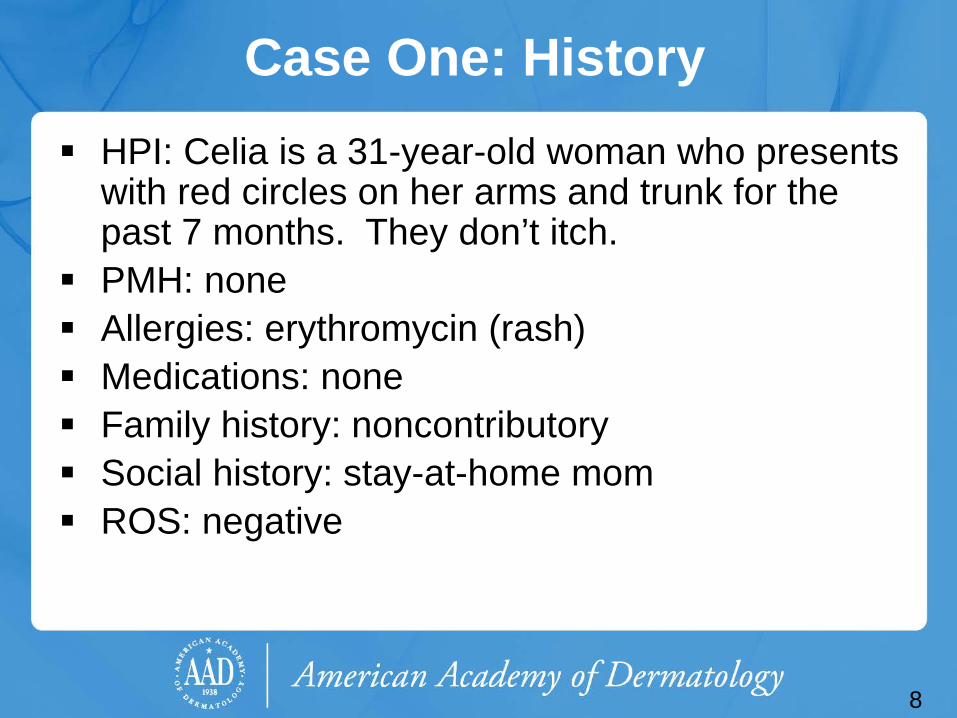
Case One: History HPI: Celia is a 31-year-old woman who presents
with red circles on her arms and trunk for the past 7 months. They don’t itch.
PMH: none Allergies: erythromycin (rash) Medications: none Family history: noncontributory Social history: stay-at-home mom ROS: negative
8
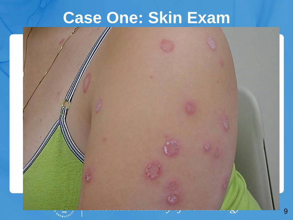
Case One: Skin Exam
9
10
Case One, Question 1
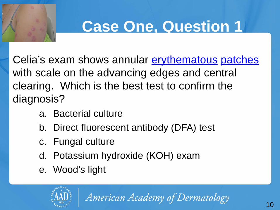
Celia’s exam shows annular erythematous patches with scale on the advancing edges and central clearing. Which is the best test to confirm the diagnosis?
a. Bacterial culture b. Direct fluorescent antibody (DFA) test c. Fungal culture d. Potassium hydroxide (KOH) exam e. Wood’s light

Case One, Question 1
Answer: d Mrs. Majocchi’s exam shows annular erythematous patches with scale on the advancing edges and central clearing. Which is the best test to confirm the diagnosis?
a. Bacterial culture (would only get normal skin flora) b. Direct fluorescent antibody (DFA) test (for herpes virus) c. Fungal culture (more expensive, takes longer) d. Potassium hydroxide (KOH) exam (can make the
diagnosis in the office; inexpensive test) e. Wood’s light (this does not fluoresce)
11

KOH exam
Excellent choice. You decide to do a KOH exam in the office.
Scrape the leading edge for fine scale Click here to learn how to properly perform
a KOH exam
12
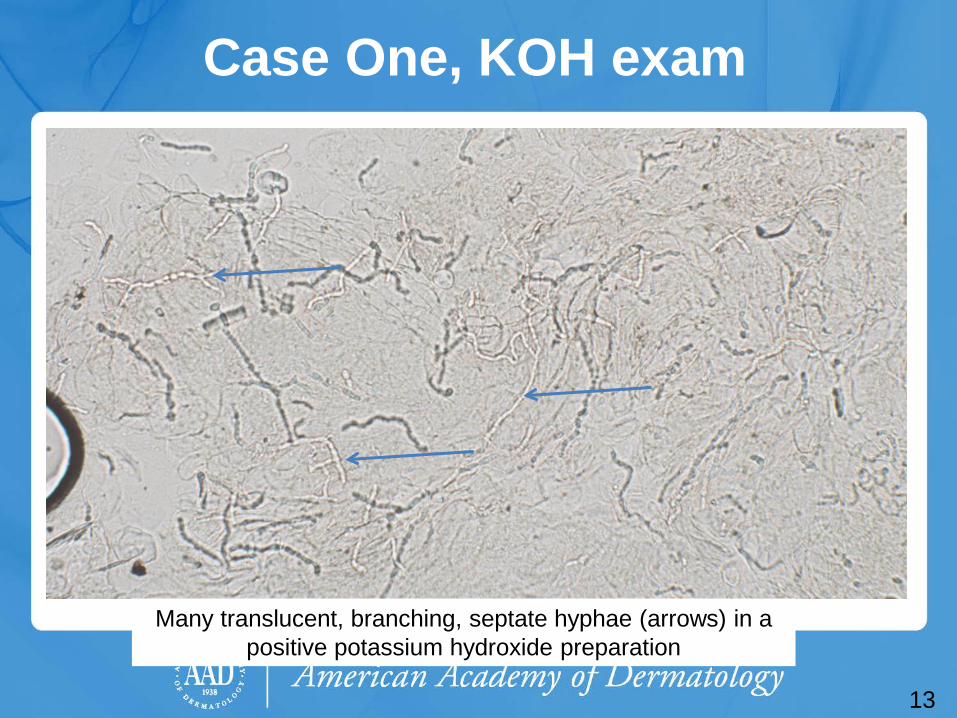
Case One, KOH exam
13
Many translucent, branching, septate hyphae (arrows) in a positive potassium hydroxide preparation

Potassium hydroxide exam
The first step in diagnosing a scaling annular rash on the body is to perform a KOH exam to rule out fungus
“All that scales must be scraped” is a common mantra in dermatology
Rule out dermatophyte infections before moving forward on scaling rashes
14

Case One, Question 2
Mrs. Majocchi’s KOH exam is positive. Which of the following questions are important to ask?
a. Do you have a rash in your groin? b. Do you have a rash on your feet? c. Do you have any pets? d. Do you take care of young children? e. All of the above
15

Case One, Question 2
Answer: e Mrs. Majocchi’s KOH exam is positive. Which of the following questions are important to ask?
a. Do you have a rash in your groin? (associated tinea cruris) b. Do you have a rash on your feet? (associated tinea pedis) c. Do you have any pets? (cats can transmit a type of
ringworm) d. Do you take care of young children? (kids may have fungal
infections on the scalp) e. All of the above
16

17
Tinea corporis
Tinea corporis (“ringworm”) classically presents as annular patches with peripheral scaling at the advancing edge and central clearing • Complete skin exam often reveals tinea cruris (“jock itch”) and
tinea pedis (“athlete’s foot”). Check these areas on full skin exam.
Tinea corporis is usually caused by Trichophyton and Microsporum species. • These dermatophytes appear
as branching, septate hyphae. They do not have yeast forms as seen in tinea versicolor, caused by Malassezia spp.

18
Tinea cruris

Tinea pedis
19
Vesicles and bullae on the feet can be caused by tinea, eczema and other types of dermatoses.

20
Tinea capitis

21
Case One, Question 3
Mrs. Majocchi has athlete’s foot and extensive tinea corporis on her abdomen, chest, back, and arms. What is the best therapy for her?
a. Nystatin cream b. Oral terbinafine c. Terbinafine cream d. Triamcinolone cream a. Ultraviolet B (UVB) phototherapy
22
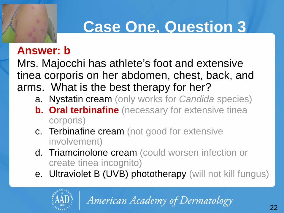
Case One, Question 3 Answer: b Mrs. Majocchi has athlete’s foot and extensive tinea corporis on her abdomen, chest, back, and arms. What is the best therapy for her?
a. Nystatin cream (only works for Candida species) b. Oral terbinafine (necessary for extensive tinea
corporis) c. Terbinafine cream (not good for extensive
involvement) d. Triamcinolone cream (could worsen infection or
create tinea incognito) e. Ultraviolet B (UVB) phototherapy (will not kill fungus)
23
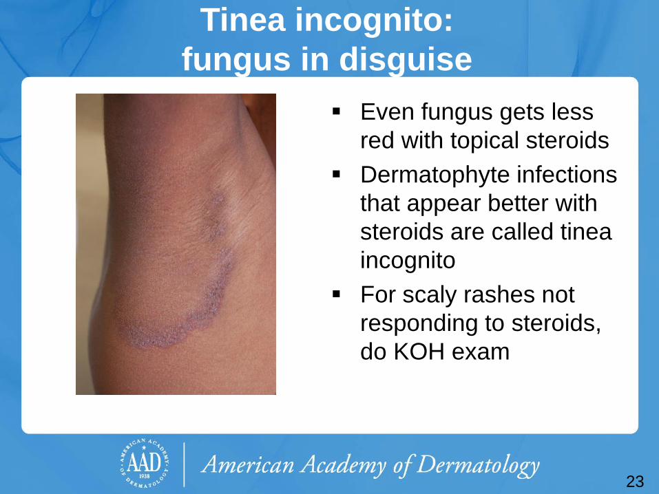
Tinea incognito: fungus in disguise
Even fungus gets less red with topical steroids
Dermatophyte infections that appear better with steroids are called tinea incognito
For scaly rashes not responding to steroids, do KOH exam

Tinea corporis treatment Counsel on foot care to prevent recurrence Localized involvement
• Azoles (miconazole, clotrimazole) are fungistatic and must be used BID
• Allylamines (terbinafine, naftifine) and benzylamines (butenafine) are fungicidal • These have higher cure rates and lower
recurrence rates than azoles Extensive involvement may require oral
antifungals (14-28 days of terbinafine)
24

25
Case Two Jacques Gibert
Case Two: History HPI: Jacques Gibert is a 21-year-old French foreign
exchange student visiting the United States. He presents for a routine college physical but mentions a scaly patch on his abdomen for the past few days. It itches a little bit.
PMH: tonsillectomy as a child Allergies: penicillin (rash) Medications: none Family history: noncontributory Social history: college student; plays lacrosse; sexually
active; moderate alcohol intake ROS: recent cold symptoms after moving into dorms
26
27
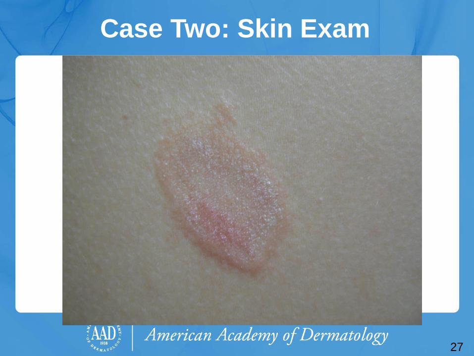
Case Two: Skin Exam

28
Case Two, Question 1
Jacques’s exam shows a single erythematous oval plaque with scaling. What is the first test should you get during his office visit?
a. Bacterial culture b. Gonorrhea culture c. Potassium hydroxide (KOH) exam d. Rapid plasma reagin e. Shave biopsy
29
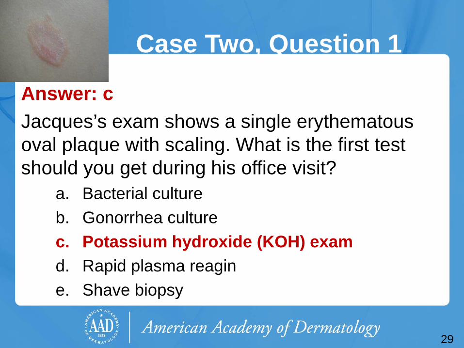
Case Two, Question 1
Answer: c Jacques’s exam shows a single erythematous oval plaque with scaling. What is the first test should you get during his office visit?
a. Bacterial culture b. Gonorrhea culture c. Potassium hydroxide (KOH) exam d. Rapid plasma reagin e. Shave biopsy
Potassium hydroxide preparation
Great job! You remembered that “All that scales must be scraped”
Rule out dermatophyte infections before moving forward on scaling rashes
30

31
Potassium hydroxide preparation
• Negative KOH
Case Two
In this case, the KOH exam is negative This does not bother him much, so you
give him a mid-potency topical steroid cream to use BID as needed for itching
32

33
Case Two, continued Jacques returns in 3 days for a
rash on his chest, abdomen, and back (larger picture next slide)
It only itches a little bit Exam shows oval salmon-
colored patches with minor scale; the oval patches follow skin tension lines on the back
Palms and soles are normal Repeat KOH exam is negative

34
Case Two
35
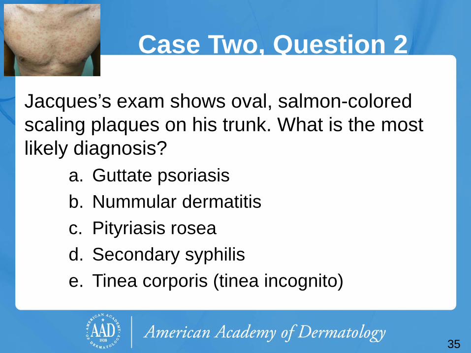
Case Two, Question 2
Jacques’s exam shows oval, salmon-colored scaling plaques on his trunk. What is the most likely diagnosis?
a. Guttate psoriasis b. Nummular dermatitis c. Pityriasis rosea d. Secondary syphilis e. Tinea corporis (tinea incognito)
36
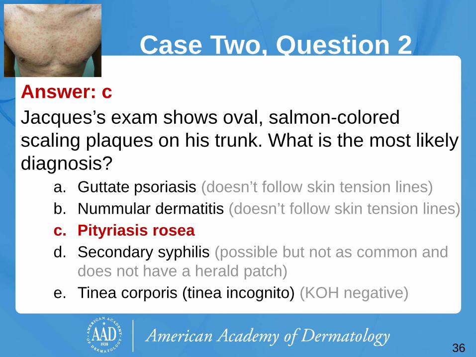
Case Two, Question 2 Answer: c Jacques’s exam shows oval, salmon-colored scaling plaques on his trunk. What is the most likely diagnosis?
a. Guttate psoriasis (doesn’t follow skin tension lines) b. Nummular dermatitis (doesn’t follow skin tension lines) c. Pityriasis rosea d. Secondary syphilis (possible but not as common and
does not have a herald patch) e. Tinea corporis (tinea incognito) (KOH negative)

Pityriasis rosea
Pityriasis rosea (PR) is an acute exanthematous eruption that mainly occurs in young people • Most patients are between the ages of 10 and 35 • The peak incidence is in late teens and early 20s • Some studies suggest a possible viral etiology, but this has not
been definitively proven Usually asymptomatic, but patients may have associated
flu-like symptoms • Malaise, nausea, loss of appetite, gastrointestinal upset, upper
respiratory symptoms • Less commonly fever, swollen lymph nodes, pain, or sore throat
are noted
37
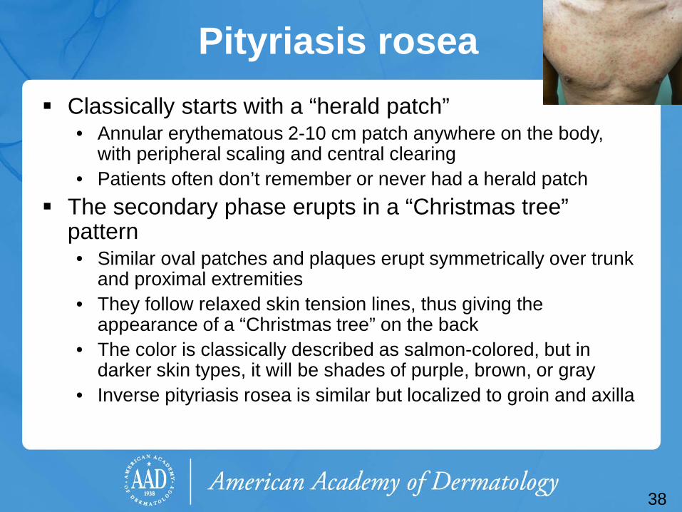
Pityriasis rosea Classically starts with a “herald patch”
• Annular erythematous 2-10 cm patch anywhere on the body, with peripheral scaling and central clearing
• Patients often don’t remember or never had a herald patch The secondary phase erupts in a “Christmas tree”
pattern • Similar oval patches and plaques erupt symmetrically over trunk
and proximal extremities • They follow relaxed skin tension lines, thus giving the
appearance of a “Christmas tree” on the back • The color is classically described as salmon-colored, but in
darker skin types, it will be shades of purple, brown, or gray • Inverse pityriasis rosea is similar but localized to groin and axilla
38

39
Now let’s look at a few examples of pityriasis rosea

40
The elusive “herald patch”
40

41 41
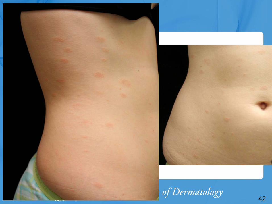
42

43

44
Pityriasis rosea
Note that in darker skin types the color varies in shades of brown, gray, or purple
Some call it lilac or violaceous
Pityriasis rosea treatment Pityriasis rosea is self-limiting
• The mean duration is about 5 weeks • More than 80% resolve by 8 weeks without treatment • Most patients only need to be reassured
About 25% request treatment for mild to severe pruritus • Soothing anti-itch lotions available over-the-counter, topical
steroids, and oral antihistamines may help • Quality evidence for these treatments is lacking
Erythromycin given for 2 weeks helped improve the rash in one study, but subsequent studies failed to validate this
45

46
Case Three Melvin Biette

Case Three: History HPI: Melvin Biette is a 46-year-old man who presents
with a scaly red rash on his chest, abdomen, and back, for the past week. It does not itch.
PMH: back pain Allergies: none Medications: ibuprofen as needed Family history: noncontributory Social history: works in office administration ROS: vague symptoms of headache, lack of appetite
47

48
Case Three: Skin Exam
48
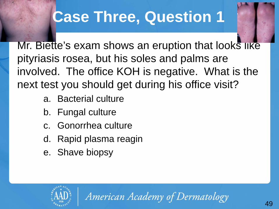
Case Three, Question 1
Mr. Biette’s exam shows an eruption that looks like pityriasis rosea, but his soles and palms are involved. The office KOH is negative. What is the next test you should get during his office visit?
a. Bacterial culture b. Fungal culture c. Gonorrhea culture d. Rapid plasma reagin e. Shave biopsy
49

Case Three, Question 1 Answer: d Mr. Biette’s exam shows an eruption that looks like pityriasis rosea, but his soles and palms are involved. The office KOH is negative. What is the next test you should get during his office visit?
a. Bacterial culture (no crusts to suggest impetigo) b. Fungal culture (not likely positive if KOH negative) c. Gonorrhea culture (joint pain, petechiae suggest this) d. Rapid plasma reagin e. Shave biopsy (not the best screening test for this)
50

Secondary syphilis
Primary syphilis begins with a painless chancre • Patients often do not notice or recall the chancre
The secondary phase comes weeks later • Prodrome may include malaise, fever, headache,
stiff neck, myalgias, arthralgias, runny nose and eyes, and mental changes
• The clinical rashes created by secondary syphilis are highly variable. Consider secondary syphilis in a patient with a new onset red scaly eruption.
51
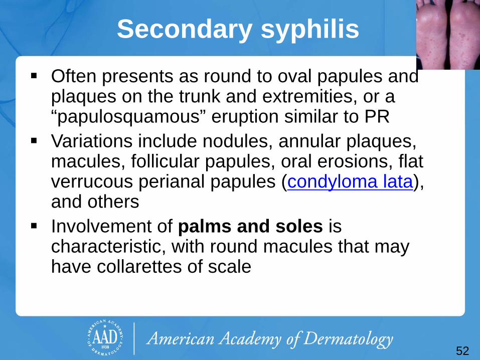
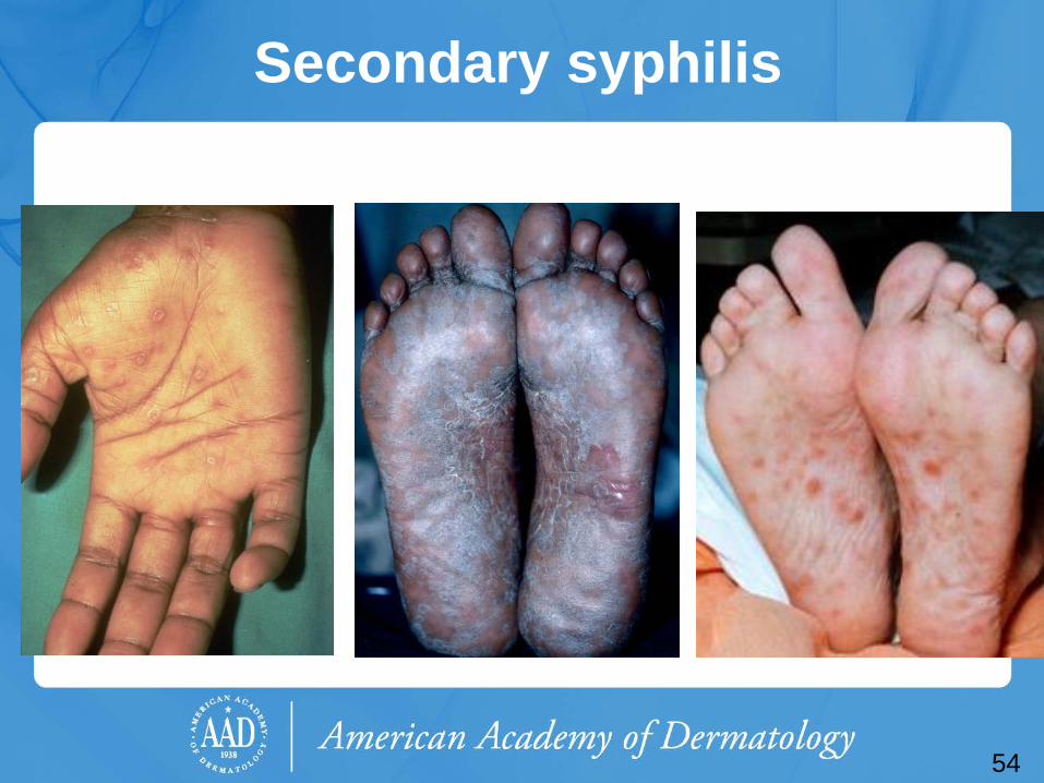
Secondary syphilis Often presents as round to oval papules and
plaques on the trunk and extremities, or a “papulosquamous” eruption similar to PR
Variations include nodules, annular plaques, macules, follicular papules, oral erosions, flat verrucous perianal papules (condyloma lata), and others
Involvement of palms and soles is characteristic, with round macules that may have collarettes of scale
52
53
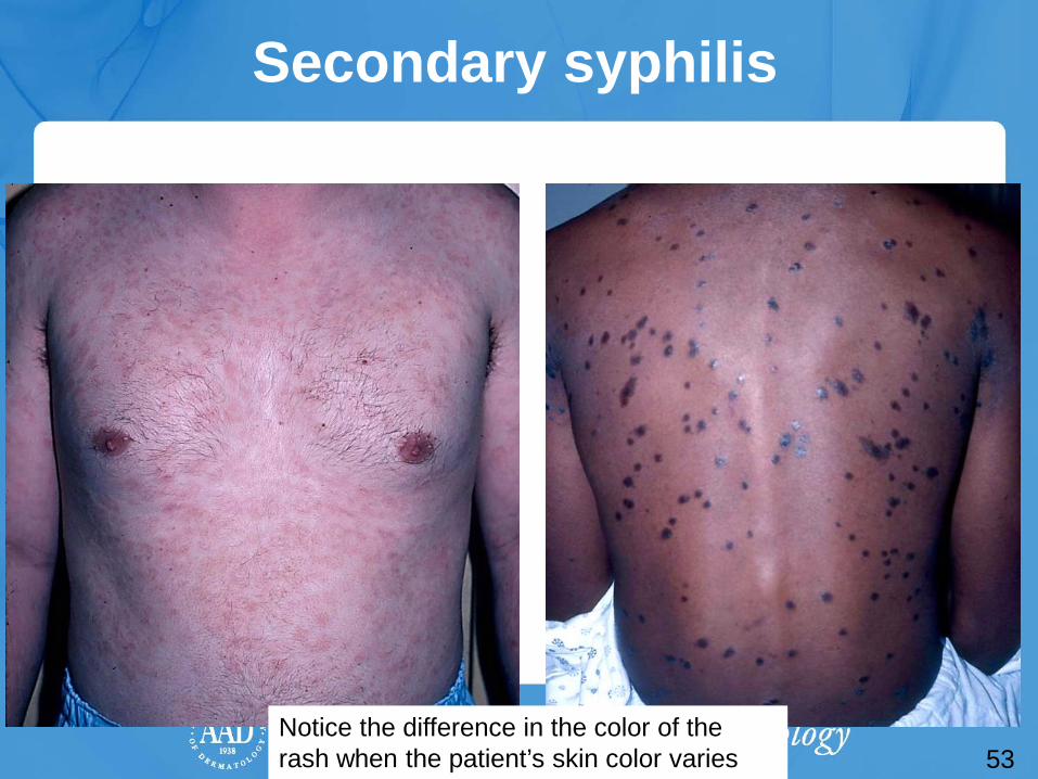
Secondary syphilis
53
Notice the difference in the color of the rash when the patient’s skin color varies
54
Secondary syphilis
Secondary syphilis: testing and treatment
Consider syphilis with papulosquamous eruptions that look like pityriasis rosea • Especially if palms, soles, or mouth
involved Confirm with serologic tests for syphilis Treatment for early syphilis remains
intramuscular benzathine penicillin G
55
56
Case Four Chris Koebner
Case Four: History HPI: Captain Chris Koebner is a 35-year-old Air Force
pilot who presents with one week of small pink scaly round spots on his chest, abdomen, back, upper arms, thighs, and forehead. They itch somewhat.
PMH: none Allergies: none Medications: none Family history: daughter had strep throat a month ago Social history: lives with wife and three children (ages 3,
7, and 9) on military base. Recent travel to Japan, Singapore, and Thailand, on military mission.
ROS: mild sore throat for past two weeks
57
58
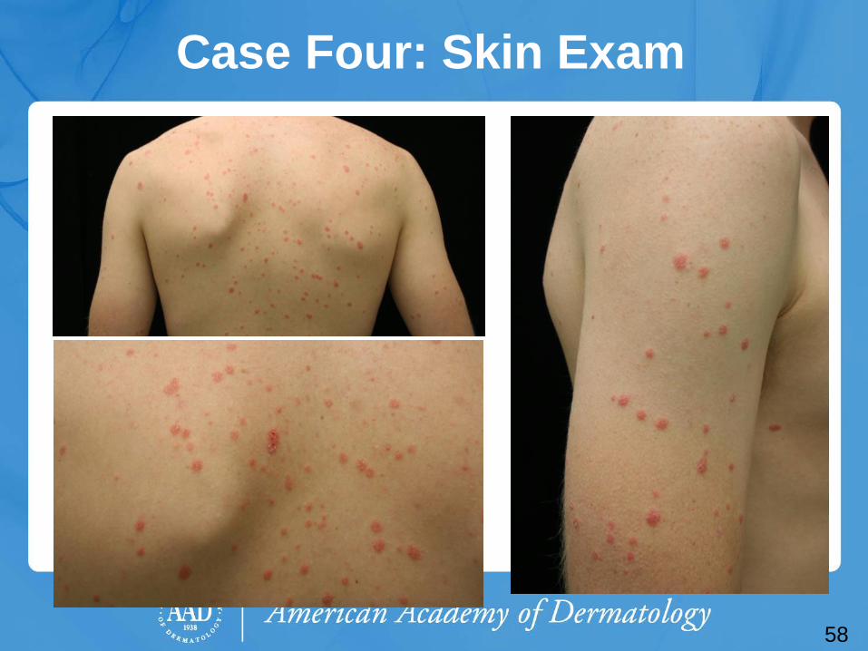
Case Four: Skin Exam

Case Four, Question 1
Captain Koebner’s exam shows many guttate (raindrop-like) scaly pink to bright red papules. They do not follow skin tension lines, and there is no oral, palm, or sole involvement. What is the first test you should perform?
a. Fungal culture b. Potassium hydroxide (KOH) exam c. Rapid plasma reagin d. Shave biopsy
59
Case Four, Question 1 Answer: b Captain Koebner’s exam shows many guttate (raindrop-like) scaly pink to bright red circinate papules. They do not follow skin tension lines, and there is no oral, palm, or sole involvement. What is the first test you should perform?
a. Fungal culture b. Potassium hydroxide (KOH) exam c. Rapid plasma reagin d. Shave biopsy
60
Case Four, Question 2
You perform a KOH exam to rule out tinea corporis, and it is negative. What is the most likely diagnosis for Captain Koebner?
a. Guttate psoriasis b. Nummular dermatitis c. Pityriasis rosea d. Secondary syphilis e. Tinea corporis
61
Case Four, Question 2 Answer: a You perform a KOH exam to rule out tinea corporis, and it is negative. What is the most likely diagnosis for Captain Koebner?
a. Guttate psoriasis b. Nummular dermatitis (not associated with strep throat) c. Pityriasis rosea (follows skin tension lines) d. Secondary syphilis (can present this way and it is never
wrong to order a screening test; no strep throat) e. Tinea corporis (should have positive KOH)
62

Psoriasis
Psoriasis is a common, chronic, inflammatory multi-system disease that mostly involves skin and joints
Classic plaque psoriasis presents as pink to bright red well-demarcated plaques with silvery scale • Usually located on extensor knees and elbows • Commonly involves scalp, umbilicus, gluteal cleft, and nails
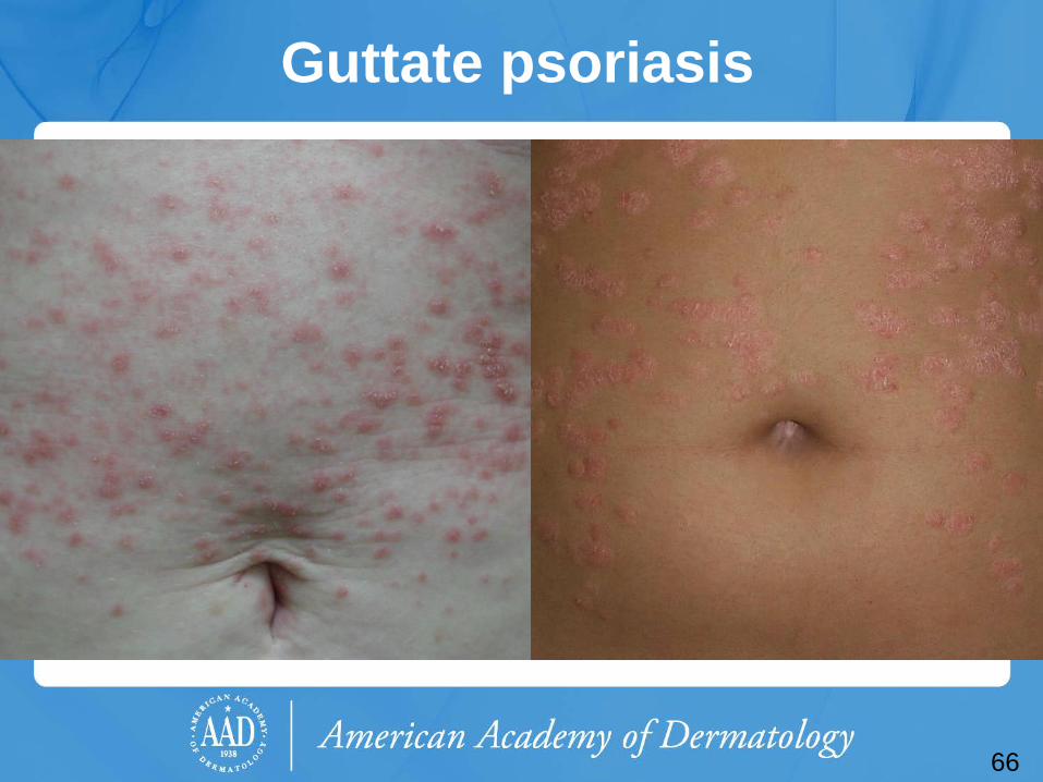
Guttate psoriasis presents as small “drop-like” scaly papules and plaques mostly on the trunk and extremities • Often follows group A beta hemolytic streptococcal
infections
63

64
Now let’s look at a few examples of different psoriasis
presentations

65
Classic psoriasis
Psoriasis vulgaris favors the extremities, nails, and interestingly, the umbilicus and the gluteal cleft
66
Guttate psoriasis
67
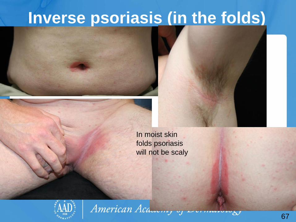
Inverse psoriasis (in the folds)
67
In moist skin folds psoriasis will not be scaly
Case Four, Question 3
You correctly suspect guttate psoriasis, and obtain a throat culture. What treatment would you recommend for Captain Koebner’s psoriasis?
a. Desonide cream b. Oral prednisone c. Oral terbinafine d. Ultraviolet B (UVB) phototherapy
68
Case Four, Question 3
Answer: d You correctly suspect guttate psoriasis, and obtain a throat culture. What treatment would you recommend for Captain Koebner’s psoriasis?
a. Desonide cream (not strong enough) b. Oral prednisone (makes worse upon
withdrawal) c. Oral terbinafine (not fungal) d. Ultraviolet B (UVB) phototherapy
69
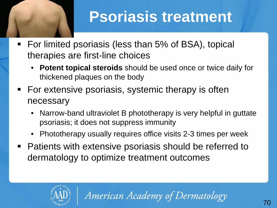
Psoriasis treatment For limited psoriasis (less than 5% of BSA), topical
therapies are first-line choices • Potent topical steroids should be used once or twice daily for
thickened plaques on the body
For extensive psoriasis, systemic therapy is often necessary • Narrow-band ultraviolet B phototherapy is very helpful in guttate
psoriasis; it does not suppress immunity • Phototherapy usually requires office visits 2-3 times per week
Patients with extensive psoriasis should be referred to dermatology to optimize treatment outcomes
70
71
Case Five Jay Sulzberger

Case Five: History HPI: Jay Sulzberger is a 35-year-old man who
presents with three months of an extremely itchy red rash on his arms and legs.
PMH: seasonal allergic rhinitis, childhood eczema Allergies: peanuts Medications: loratadine Family history: brother with asthma Social history: lives with wife and two children ROS: negative
72

73
Case Five: Skin Exam
73
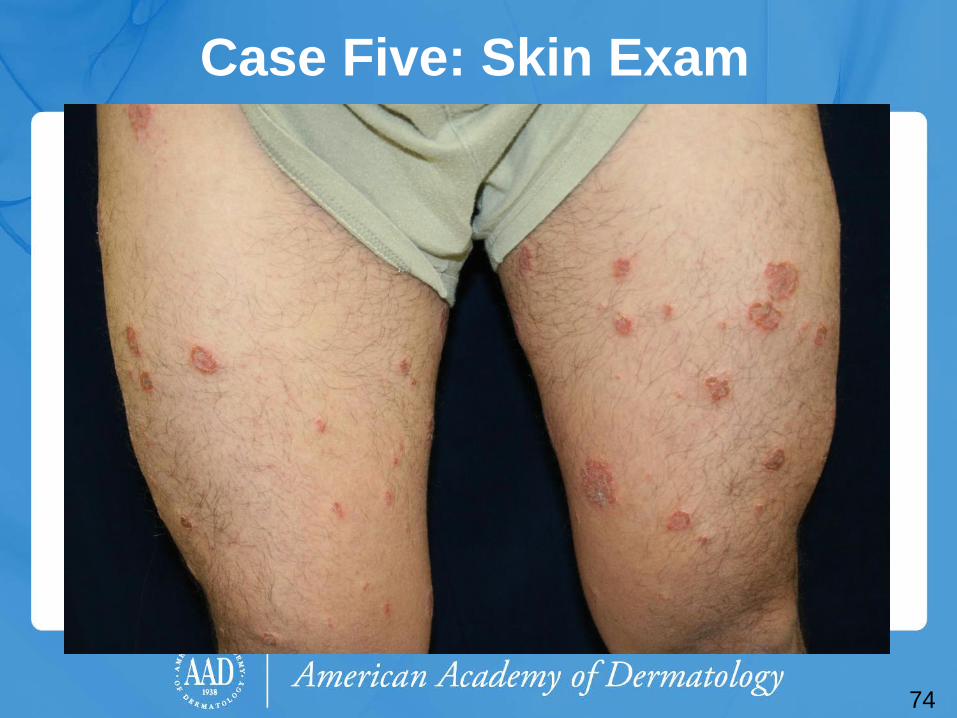
Case Five: Skin Exam
74

75
Case Five
Since this rash is scaly, you correctly start with a KOH exam, which is negative.
The round eczematous plaques are on his arms, legs, and back.
His scalp, umbilicus, nails, palms, and soles are unaffected.
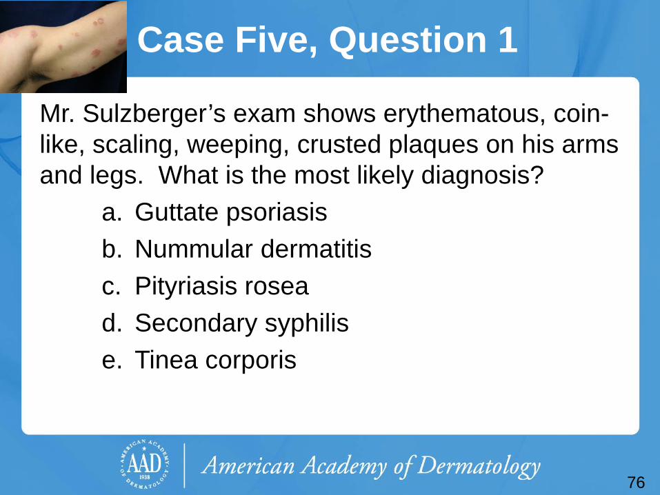
Case Five, Question 1
Mr. Sulzberger’s exam shows erythematous, coin-like, scaling, weeping, crusted plaques on his arms and legs. What is the most likely diagnosis?
a. Guttate psoriasis b. Nummular dermatitis c. Pityriasis rosea d. Secondary syphilis e. Tinea corporis
76

Case Five, Question 1 Answer: b Mr. Sulzberger’s exam shows erythematous, coin-like, scaling, weeping, crusted plaques on his arms and legs. What is the most likely diagnosis?
a. Guttate psoriasis (usually not weeping, crusted) b. Nummular dermatitis c. Pityriasis rosea (does not last this long, or weep) d. Secondary syphilis (does not last this long, or weep) e. Tinea corporis (KOH is negative, no central clearing)
77

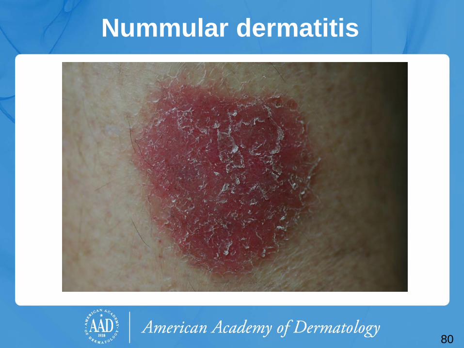
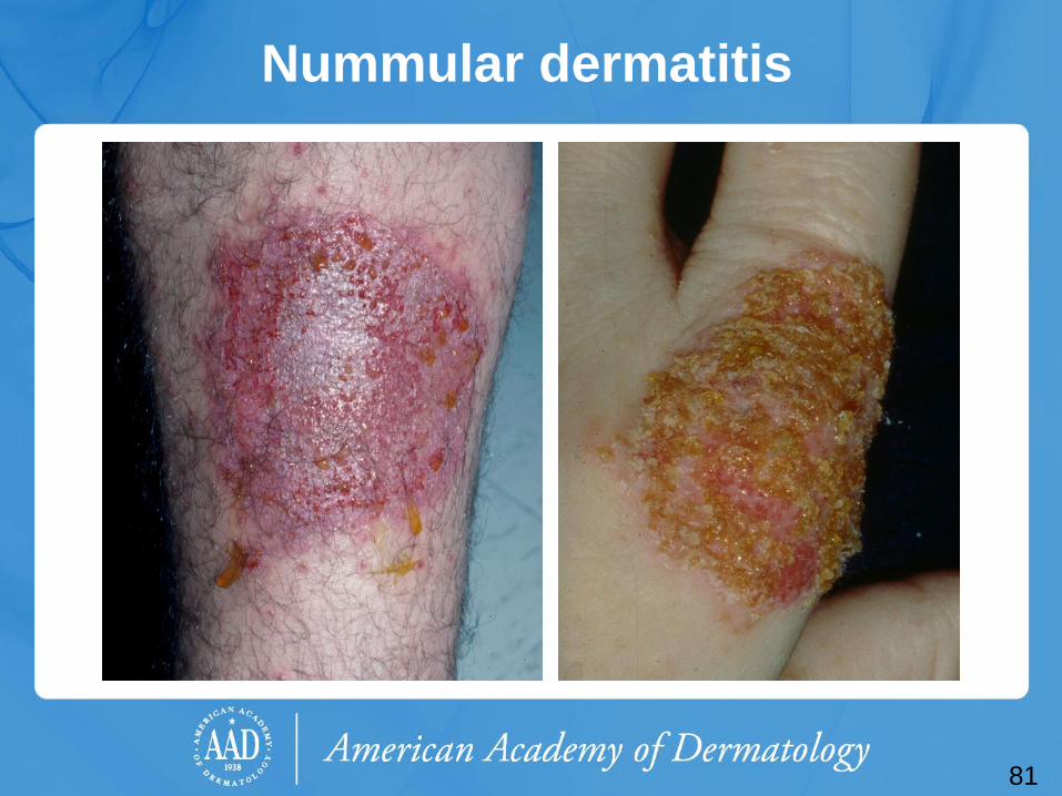
Nummular dermatitis
Nummular dermatitis presents as multiple coin-shaped eczematous plaques on the extremities and trunk
• May be scaly but lacks the central clearing seen in tinea corporis and is KOH negative
• Very pruritic • May exhibit weeping, cracking, vesicles, or
crusts • Pathology shows spongiotic dermatitis
78

79
Now let’s look at a few examples of nummular dermatitis
Nummular dermatitis
80
81
Nummular dermatitis
82
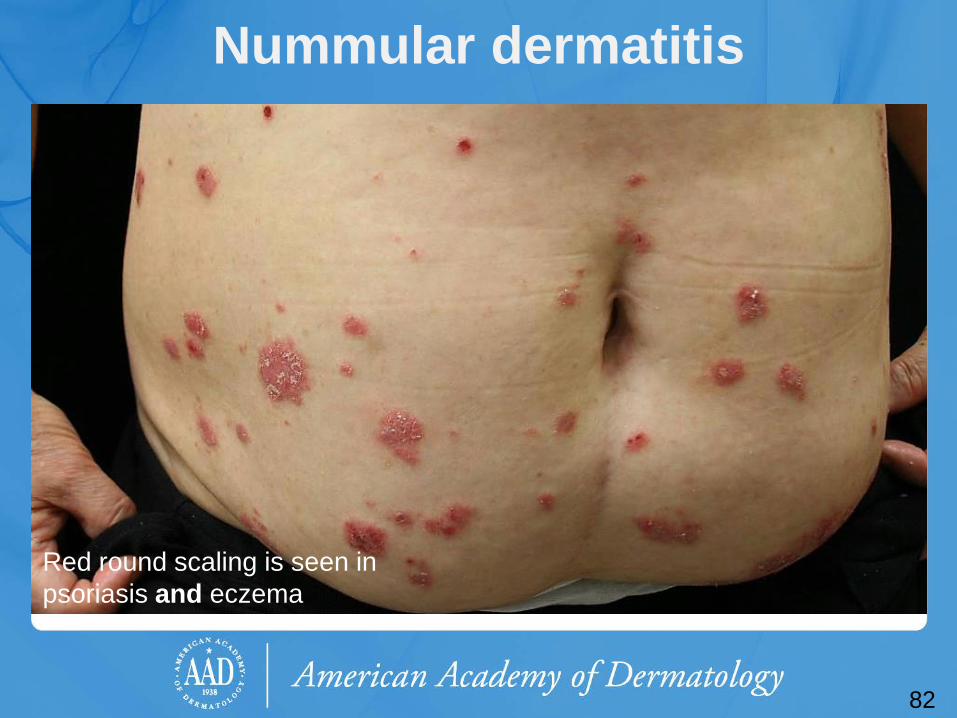
Nummular dermatitis
Red round scaling is seen in psoriasis and eczema

Case Five, Question 2
You diagnose Mr. Sulzberger with nummular dermatitis. What treatment would you recommend?
a. Desonide cream b. Fluocinonide ointment (potent steroid) c. Oral erythromycin d. Oral terbinafine e. Ultraviolet B (UVB) phototherapy
83

Case Five, Question 2
Answer: b You diagnose Mr. Sulzberger with nummular dermatitis. What treatment would you recommend?
a. Desonide cream (not strong enough) b. Fluocinonide ointment (potent steroid) c. Oral erythromycin (for pityriasis rosea) d. Oral terbinafine (not fungal) e. Ultraviolet B (UVB) phototherapy (for guttate
psoriasis)
84
Nummular dermatitis
Treat like atopic dermatitis or any other eczema
Potent topical steroids are necessary to control this type of eczematous eruption
Apply emollients twice a day
85

Wrap-Up: After managing the previous five patients, can you fill-in the missing information?
Morphology Distribution Pathophysiology Diagnostics (KOH, RPR, Biopsy)
Treatment
Psoriasis Pink-red Dry, silvery scale Macules-patches Papules-plaques
consider KOH, biopsy Topical steroid Topical calcineurin
inhibitor
Eczema (including nummular eczema)
Variable depending on type, areas of
allergen exposure vs flexures in atopic
consider KOH, biopsy
Pityriasis rosea
Pink, annular Fine trailing scale Papules, patches,
plaques
KOH RPR
Observe Topical steroid
Antibiotics
Lupus Pink-Red-Brown, Annular
Variable scale Variable Scarring
DLE: Sunny SCLE: Chest, back
Biopsy ANA, etc
Tinea Anywhere with stratified squamous
epithelium
KOH, consider biopsy if KOH negative and
you’re suspicious
Topical or oral antifungal
86

Wrap-Up: Red Scaling Rashes Morphology Distribution Pathophysiology Diagnostics
(KOH, RPR, Biopsy) Treatment
Psoriasis Pink-red Dry, silvery scale Macules-patches Papules-plaques
Scalp, Elbows, Knees, Nails Anywhere
Multifactorial inflammatory immune condition, with
genetic and environmental influence.
Favors Th17, 22-mediated immunity
consider KOH, biopsy
Topical steroid Topical calcineurin
inhibitor
Eczema (including nummular eczema)
Varies depending on chronicity,
acute = bullous, chronic = pink
lichenified
Variable depending on type, areas of allergen exposure
vs flexures in atopic
Multifactorial inflammatory immune condition, with
genetic and environmental influence.
Favors Th2-mediated immunity
consider KOH, biopsy
Topical steroids Topical calcineurin
inhibitor
Pityriasis rosea
Pink, annular Fine trailing scale Papules, patches,
plaques
Herald patch, “Christmas tree”
Possibly a reaction to a viral infection (HHV-6 or 7)
KOH RPR
Observe Topical steroid
Antibiotics
Lupus Pink-Red-Brown, Annular
Variable scale Variable Scarring
DLE: Sunny SCLE: Chest, back
Classical autoimmune disease (B-cell mediated)
Biopsy ANA, etc
SPF Topical steroids
Antimalarials
Tinea Pink annular, patches and plaques with
advancing scale
Anywhere with stratified squamous
epithelium
Fungal infection (dermatophyte) of the
epidermis
KOH, consider biopsy if KOH
negative and you’re suspicious
Topical or oral antifungal
87

Guidelines for consulting dermatology on the red scaly rash
1. The morphology and distribution are clues to the diagnosis, so it helps to critically inspect the individual lesions and perform a full skin exam.
2. “If it scales, scrape it” • For scaly patches, especially annular patches, perform a
KOH • KOH can be difficult to interpret at first – you will get
better with time & experience (and enough scale on the slide)
3. Try to avoid combination steroid-antifungal creams. Steroids can exacerbate cutaneous fungal infections.
88

Acknowledgements
This module was developed by the American Academy of Dermatology Medical Student Core Curriculum Workgroup from 2008-2012.
Primary author: Patrick McCleskey, MD, FAAD. Peer reviewers: Timothy G. Berger, MD, FAAD;
Peter A. Lio, MD, FAAD; Daniel S. Loo, MD, FAAD; Sarah D. Cipriano, MD, MPH.
Revisions: Joslyn S. Kirby, MD. Last revised October 2014.
89

References Berger T, Hong J, Saeed S, Colaco S, Tsang M, Kasper R. The Web-Based
Illustrated Clinical Dermatology Glossary. MedEdPORTAL; 2007. Available from: www.mededportal.org/publication/462
Usatine, RP, Marcellin, L. Psoriasis. First Consult. Philadelphia: Reed Elsevier, 2013.
Marks Jr JG, Miller JJ. Lookingbill and Marks’ Principles of Dermatology, 4th ed. Elsevier; 2006:187-197.
Stulberg, DL, Wolfrey J. Pityriasis Rosea. Am Fam Physician 2004;69:87-92,94.
Drago F, Broccolo F, Rebora A. Pityriasis rosea: an update with a critical appraisal of its possible herpesviral etiology. J Am Acad Dermatol 2009;61:303-18.
Domantay-Apostol GP, Handog EB, Gabriel MTG. Syphilis: the international challenge of the great imitator. Dermatol Clin 2008;191-202.
Menter A, Korman NJ, Emets CA, et al. Guidelines of care for the management of psoriasis and psoriatic arthritis (Section 3). J Am Acad Dermatol 2009:60:643-59.
Feldman SR, Koo JYM, Menter A, Bagel J. Decision points for the initiation of systemic treatment for psoriasis. J Am Acad Dermatol 2005;53:101-7.
90

To take the quiz, click on the following link: https://www.aad.org/quiz/red-scaly-rash-learners-
91