redox-responsive polyanhydride micelles for cancer … · · 2014-07-10redox-responsive...
TRANSCRIPT

lable at ScienceDirect
Biomaterials 35 (2014) 3080e3090
Contents lists avai
Biomaterials
journal homepage: www.elsevier .com/locate/biomateria ls
Redox-responsive polyanhydride micelles for cancer therapy
Jie Wang a,1, Guang Yang a,1, Xing Guo a, Zhaomin Tang a, Zhendong Zhong b,Shaobing Zhou a,*
aKey Laboratory of Advanced Technologies of Material, Ministry of Education, School of Materials Science and Engineering, Southwest Jiaotong University,Chengdu 610031, Chinab Sichuan Academy of Medical Sciences & Sichuan Provincial People’s Hospital, Chengdu 610212, China
na r t i c l e i n f o
Article history:Received 10 October 2013Accepted 13 December 2013Available online 3 January 2014
Keywords:MicelleRedox-responsiveNanocarrierDrug deliveryCancer therapy
* Corresponding author. Tel.: þ86 28 87634068; faE-mail addresses: [email protected],
(S. Zhou).1 These authors contributed equally to this work.
0142-9612/$ e see front matter � 2013 Elsevier Ltd.http://dx.doi.org/10.1016/j.biomaterials.2013.12.025
pm.co
m.ca b s t r a c t
Biodegradable polyanhydrides possess unique features like those that they can predominantly undergosurface erosion, and the payloads can be released by a steady speed. However, there is little work that hasbeen published to describe the polyanhydride micelles with redox-responsiveness as a nanocarrier fordrug delivery. In this study, we develop one type of new amphiphilic polyanhydride copolymer con-taining disulfide bonds between the hydrophilic and hydrophobic segments. The copolymer can self-assemble into stable micelles with well-defined coreeshell structure and a uniform size distributionwith an average diameter of 69 nm. The disassembly behaviors of the micelles triggered by glutathioneare evaluated from the changes of the micellar size, morphology and molecular weight. An approximatezero-order in vitro drug release mode with a fast speed can be achieved in a reducing and acid envi-ronment similar with that of tumor cells. In vitro cytotoxicity analysis demonstrate that the Cur-loadedmicelles are of great efficiency in inhibiting the growth of cancer cells due to the rapidly intracellulardelivery of therapeutic agent. Both the qualitative and quantitative results of the antitumor activity in4T1 tumor-bearing BALB/c mice reveal that the redox-responsive micelles have a more significanttherapeutic effect to artificial solid tumor compared to the redox-insensitive micelles. This study pro-vides a new insight into the biomedical application of polyanhydrides in drug delivery.
� 2013 Elsevier Ltd. All rights reserved.
s . www1. IntroductionNanocarriers have shown tremendous promise in drug and genedelivery [1,2], since they have an ability of delivering their cargospreferentially into cancer tissues or targeted location, and in turnsignificantly enhance the therapeutic efficacy or transfection effi-cacy while substantially reducing side effects [3]. Among thesenanoscaled drug delivery systems, polymeric micelles with distinctcore/shell architecture self-assembled from amphiphilic co-polymers have been extensively explored [4e8]. They presentexceptional advantages as nanocarriers, including relatively highstability due to low critical micelle concentration (CMC), animproved solubility of poorly water soluble drugs, and prolongedblood circulation owing to their high water solubility [9e12].
In the micelle systems, the hydrophobic segments of amphi-philic copolymers mostly studied are aliphatic polyesters, such as
x: þ86 28 [email protected]
All rights reserved.
poly ( 3-caprolactone) (PCL), poly(D,L-lactide) (PLA), and the hydro-philic segments are generally polyether, such as polyethylene glycol(PEG) [13,14]. Hydrophobic polyanhydrides are the most suitablecandidate as a vesicle in drug delivery system due to their uniquefeatures like those that they can predominantly undergo surfaceerosion, and the drug can be released by a steady speed from thepolymer matrix [15,16]. However, nowadays they are seldomemployed to act as the hydrophobic segments of amphiphilic co-polymers due to the worry of the fast degradation of poly-anhydrides and subsequent instability of their nanoparticles.Nevertheless, our previous work demonstrated that the polyether-polyanhydride copolymer composed of methoxypolyethylene gly-cols (mPEG), 1,3-bis(carboxyphenoxy) propane (CPP) and sebacicacid (SA) could self-assemble into micelles with good stability[17,18]. Furthermore, in most cases of cancer therapy using nano-carriers, the payloads of therapeutic agents need to be releasedquickly into cancer cells so as to efficiently induce death of variouscancer cells [19,20], and this process generally takes a short periodof time from several hours to several days. It is still a challengingtask for the nanoparticle-based drug delivery system that can befast degraded for nanocarriers or be rapidly disassembled for this

m
J. Wang et al. / Biomaterials 35 (2014) 3080e3090 3081
www.sp
system in response to environmental stimuli after these nano-particles are endocytosed into cancer cells.
Recently, various stimuli-responsive (e.g. temperature [21], pH[14], redox [13,22e24].) polymeric micelles have been highly con-cerned. Among all applied stimuli, redox potential is a potentstimuli due to the difference in glutathione (GSH) concentrationexisting between the reducing intracellular space (approximately2e10 mM) and mildly oxidizing in extracellular space (approxi-mately 2e20 mM) [25,26]. Redox-sensitive polymers usually connectwith characteristic disulfide linkages or diselenide groups whichcan maintain adequate stability in the circulation and extracellularmilieu and tend to rapid cleavage under the reducing intracellularspace [27e30].
In this study, we developed a new redox-responsive drug de-livery system based on amphiphilic PEG-based ether-anhydridecopolymer (mPEG-ss-CPP-SA) containing disulfide bonds betweenmPEG and CPP/SA segments. It can self-assemble into micelles inaqueous solution and the reduction-triggered disassembly behav-iors were systematically investigated with GSH. Curcumin (Cur), alipophilic polyphenol drug, were loaded into micelles by physicalmixing method. In comparison to the redox-insensitive micelles,the intracellular drug release, cell apoptosis and cell cycle inducedby the Cur-loadedmicelles, antitumor effect both in vitro and in vivoand histological assessment of the redox-responsive mPEG-ss-CPP-SAmicelles were systematically evaluated after being applied to theBALB/c mice bearing 4T1 tumor model.
2. Materials and methods
2.1. Materials
Monomethyl poly (ethylene glycol) (mPEG, MW ¼ 2.0 kDa) was purchased fromSigmaeAldrich (USA). Curcumin (Cur, 95%) was purchased from Chengdu laboooCompany (Chengdu, China). Glutathione (GSH), 3, 30-dithiodipropionic acid (DTDP)were purchased from Adamas-beta. Pyrene, 1, 30-dibromopropane, p-hydrox-ybenzoic acid and sebacic acid (SA) were obtained from Chengdu KeLong ChemicalReagent Company (Chengdu, China). Cell Cycle and Annexin V-FITC ApoptosisDetection Kits were purchased from KeyGEN Biotech (Nanjing, China) and used asreceived. All other chemicals were of analytical grade andwere usedwithout furtherpurification.
2.2. Cell lines and culture conditions
The human cervical cancer cell line HeLa, osteoblasts (OB) cells and murinebreast cancer cells 4T1 were purchased from Sichuan University (Chengdu, China).Cells were cultured in RPMI 1640 supplemented with 10% newborn calf serum (NCS,Gibco, USA) at 37 �C and 5% CO2 under fully humidified conditions. All experimentswere performed on cells in the exponential growth phase.
2.3. Animals
Male BALB/c mice (20 � 2 g) used for in vivo antitumor tests were purchasedfrom Experimental Animal Center of Sichuan University. Animals were providedwith standard laboratory chow and tap water ad libitum. All animal procedures wereperformed according to the protocol approved by the Institutional Animal Care andTreatment Committee of Sichuan University.
2.4. Synthesis procedures
2.4.1. Synthesis of prepolymers1, 3-Bis-(carboxyphenoxy) propane (CPP) was prepared according to our earlier
studies with a yield of 40e50% [17,18]. As shown in Scheme S1in Support Infor-mation (SI), the dicarboxylic acid monomers are converted to the mixed anhydrideof acetic acid by refluxing in excess acetic anhydride. Briefly, preweighed dicar-boxylic acid monomers (SA, CPP, DTDP) were respectively refluxed in excess aceticanhydride (w/v ¼ 1:10) under the protection of argon, followed by filtration andconcentrated under vacuum at 60�C to remove the unreacted diacid and aceticanhydride residues. After dried under vacuum, the prepolymers were obtained withyields ranging from 56% to 63%.
2.4.2. Synthesis of copolymersThe final mPEG-ss-CPP-SA copolymer was synthesized via classical melt-
condensation polymerization (Scheme S1). In brief, mPEG, DTDP prepolymer, SAprepolymer and CPP prepolymer were mixed in a weight feed ratio of 40:5:35:20 ina three-neck round-bottom flask. The flask was immersed in an oil bath at 180 �C,after the prepolymers were melted, high vacuum was applied. At the end of the
.com
.cn
reaction, the mixtures were allowed to cool completely and dissolved in dichloro-methane. Finally the copolymer was precipitated using anhydrous ether and driedunder vacuum with yields ranging from 62% to 70%. At the same time, the redox-insensitive copolymer, mPEG-CPP-SA, was also synthesized with the same proce-dure as described above. All products obtained were stored at�20�C for further use.
2.5. Micelle formation
The blank micelles of mPEG-ss-CPP-SA and mPEG-CPP-SA were prepared bydropwise addition of 5.0 mL block copolymer solution in THF to 10.0 mL deionizedwater under stirring, followed by the removal of THF at room temperature.
The hydrophobic anticancer drug Cur was used as a model for fabricating Cur-loaded mPEG-ss-CPP-SA (Cur-ssM) and mPEG-CPP-SA micelles (Cur-M). The pro-cedure is almost similar with that of blank micelles. The differences are that Cur wasdissolved in THF together with copolymer, and the final micelle solution wastransferred into dialysis bag (MWCO 1000) against deionized water for 36 h toremove the free Cur. Finally, the samples were lyophilized and stored at �20�C forfuture use.
2.6. Characterization
Fourier transform infrared spectrometer (FT-IR) spectra were recorded on aNicolet 5700 spectrometer by KBr sample holder method. Nuclear magnetic reso-nance (1H NMR) spectra were obtained on a Bruker AM 300 apparatus with CDCl3 orD2O as solvents and TMS as an internal reference. The number-average molecularweight (Mn), weight-average molecular weight (Mw), andMw/Mn were measured bygel permeation chromatography (GPC). For GPC (Waters 1515, USA) measurement,dimethylformamide (DMF)/LiBr was used as the eluent at a flow rate of 1 mL/min at40 �C, and polymethyl methacrylate (PMMA) was used as reference. All sampleswith concentration of 1.00 mg/mL were filtered with a 0.22 mm syringe filter beforemeasurement. Dynamic light scattering (DLS) measurements were performed inaqueous solution using a Malvern Zetasizer Nano-ZS90 apparatus. Transmissionelectron microscopy (TEM) studies were performed with a JEOL 2100F instrument(JEOL Ltd., Japan) operated at 200 kV. Atomic force microscopy (AFM) (CSPM5000,Beijing, China) was employed with tapping-mode to further observe themorphology. Critical micelle concentration (CMC)was determinedwith a Fluoromaxspectrometer (F-7000, Hitachi, Japan) at room temperature.
Drug loading content (LC) and encapsulation efficiency (EE) were measuredwith UVevis spectrophotometer (UV-2550, Shimadzu, Japan). The amount ofreleased drug in the incubation mediumwas quantified by high performance liquidchromatography (HPLC, Agilent 1260 Infinity) with UV detector.
2.7. Reduction-triggered disassembly behaviors of mPEG-ss-CPP-SA micelles
To evaluate the reduction-triggered disassembly behaviors of mPEG-ss-CPP-SAmicelles, the size change of micelles (3 mL, 1 mg/mL) in response to different GSHconcentration (20 mM, 0.5 mM, 10 mM) in PBS buffer (pH ¼ 7.4, 0.01 M) was investi-gated by DLS measurement. The excitation spectra of the solutions with 10 mM GSHwere recorded with a Fluoromax spectrometer. The morphology change of micellebefore and after adding 10 mM GSH for 4 h was measured by TEM.
2.8. Reduction-triggered drug release in vitro
The in vitro release kinetics of Cur from micelles were investigated in fourdifferent media all containing Tween-80 (0.5% w/w), i.e. phosphate buffer saline(PBS, 0.01 M, pH 7.4) with or without 10mMGSH and acetate buffer saline (ABS, 0.2 M,pH 5.0) with orwithout 10mMGSH. The freeze-dried samplewas diluted to 1mg/mLand 1.0 mL of this solution was transferred into a dialysis bag (MWCO 1000). It wasimmersed into a tube containing 30 mL of incubation media in a shaking bed at100 rpm and 37 �C. The amount of released drug in the incubation medium wasquantified by HPLC.
2.9. In vitro biocompatibility evaluation
2.9.1. Cytotoxicity assayThe cytotoxicity studies were carried out to investigate whether the copolymers
affect cell proliferation of the HeLa cells and OB cells based on Alamar blue assay.Cells were seeded into 48-well plate at a density of 1 �104 cells/well, and the blankmicelles at the concentrations ranging from 40 mg/mL to 800 mg/mL were added toincubate for future 24 h. A sample of 200 mL of Alamar blue solution was pipettedinto a 96-well plate after 4 h incubation and the absorbances were read in anautomated microplate spectrophotometer (ELX800 Biotek, USA) at 570 nm (exci-tation)/600 nm (emission). The cell morphology was observed with an invertedphase contrast microscope (Nikon Eclipse TS100, Japan).
2.9.2. Hemocompatibility testThe hemolysis properties of both the blank micelle and drug-loaded micelle
were examined by spectrophotometry with the similar procedure described by Guoet al. [14]. The absorbance of the samples was measured in an automated microplatespectrophotometer at 540 nm and the hemolysis ratio (HR) was calculated usingFormula (1):

J. Wang et al. / Biomaterials 35 (2014) 3080e30903082
HRð%Þ ¼ Asample � Anegative control
Apositive control � Anegative control� 100 (1)
m
www.sp
wherein A means the absorbance.
2.10. In vitro antitumor activity assay
The antitumor activity of Cur-loadedmicelles (Cur-ssM, Cur-M) and free Cur wasstudied with the HeLa cells with Alamar blue assay. Briefly, HeLa cells were plated ina 48-well plate at a density of 1 � 104 cells/well. After an overnight attachmentperiod, the Cur-loaded micelles with Cur dosages varying from 0.1 mg/mL to 50 mg/mL were added to incubate for future 48 h. The cell viabilities were determined asthe cytotoxicity studies described above.
2.11. Intracellular drug release
The cellular uptake and intracellular release behaviors of the Cur-loaded mi-celles were investigated by fluorescence microscopy (FM), confocal laser scanningmicroscopy (CLSM) and flow cytometry (FCM) against HeLa cells. After differentincubation time intervals, the cells were then fixed with 2.5% glutaraldehyde for50min and cell nuclei were stainedwith 4, 6-diamidino-2-phenylindole (DAPI, blue)for 7 min. The fluorescence images were taken using an inverted fluorescence mi-croscope (Olympus, CKX41) and CLSM (FV1000, Olympus, Japan). For flow cyto-metric analysis, after presetting incubation time, the cells were washed three timeswith PBS, followed by trypsin and centrifugation at 2000 rpm for 3 min to harvestthe cells, then ten thousands cells were gated and analyzed for fluorescent intensityper cells with the flow cytometer (BD AcCuri C6, USA).
2.12. Cell apoptosis and cell cycle assay
To confirm the therapeutic effect of the three Cur formulations, we performedthe cell apoptosis and cell cycle assays by FCM. First, HeLa cells were seeded in 6-well plates at a density of 1 � 105 cells/well and incubated at 37 �C for 24 h. Thenthe cells were treated with free Cur, Cur-M and Cur-ssM containing 20 mg/mLequivalent Cur to incubate for future 24 h. The rate of apoptosis was measured byusing Annexin V-FITC Apoptosis Detection Kit in accordance with the manufac-turer’s protocol.
2.13. In vivo tumor growth and biodistribution studies
To establish tumor models, 1 � 106 4T1cells in 100 mL medium were inoculatedsubcutaneously at the lower right flank of the BALB/c mice. About 10 days postinoculation, tumors in the mice were detachable and reached a volume around50 mm3 (designated as the day 0). Mice were randomly divided into five groups(n ¼ 7) and received intravenous administration of saline, blank micelle, free Cur,Cur-M and Cur-ssM at 10 mg/kg equivalent Cur, respectively. The treatments wereperformed every two days and lasted for 4 times. Mice weight and tumor size werealso monitored every two days after the first treatment. The tumor size wasmeasured using a vernier caliper across its longest (a) and shortest (b) diametersand calculated with the equation volume ¼ ab2/2. For biodistribution evaluation,tumor-bearing mice were randomized into three groups (n ¼ 3) and injectedintravenously with 100 mL saline at a dosage of 10 mg Cur/kg body weight of freeCur, Cur-M and Cur-ssM (10 mg/kg), respectively. At indicated times, the mice weresacrificed by cervical dislocation. Tissue samples (i.e. heart, liver, spleen, lung,kidney and tumor) were collected and washed with saline, blotted with filter pa-per, weighed, then placed in 1 mL saline and homogenized, respectively. Then thehomogenized samples were extracted with ethyl acetate and vortexed for 5 minand centrifugated for 5 min at 3500 r/min, respectively. The supernatants werecollected and evaporated to dryness. The obtained dry residues were then dis-solved in methanol and filtered through a 0.22 mm syringe filter. 20 mL of filtratewas collected for HPLC analysis. Tissue distribution was expressed as the amount ofCur per gram of the tissues.
2.14. Histological assessment
At day 21, portions of mice were sacrificed and their tumor tissues werecollected in 4% formaldehyde and embedded in paraffin blocks after dehydratingwith gradient ethanol. Then tissue sections of 5 mmwere dewaxed and stained withhematoxylin and eosin (H&E) or detected with the In Situ Cell Death Detection Kit,POD (Roche Diagnostics GmbH, Mannheim, Germany) for microscopic observation.The mean optical density (OD) values were measured from three pictures of eachsample with Image-Pro Plus 6.0 software and the results were the average data withstandard deviations.
2.15. Statistical analysis
Data were expressed as the mean value � standard deviation. Single factorialanalysis of variance (ANOVA) was performed to determine statistical significance ofthe data.
.com
.cn
3. Results and discussion
3.1. Characterization of mPEG-ss-CPP-SA copolymers
The composition and structure of redox-sensitive mPEG-ss-CPP-SA copolymer were confirmed by FT-IR, 1H NMR, and GPC. InFig. S1A in SI, the FT-IR spectra display some clear signals attrib-utable to the characteristic absorption bands of anhydride carbonyl(1810 cm�1, 1739 cm�1), ether bond (1110 cm�1, 1084 cm�1) andmethylene (2921 cm�1), respectively. With further characterizationof 1H NMR (Fig. S1B), we can find that inmPEG-ss-CPP-SA spectrumtwo new peaks at 2.89 and 2.86 ppm occurred in contrast to thespectrum of mPEG-CPP-SA, suggesting that the successful intro-duction of disulfide bond into this copolymer. The other peaks ofboth copolymers are consistent with previous reports [17,18]. Theactual ratio of mPEG, disulfide bonds, CPP and SA componentscalculated from 1H NMR is 30.3:3.9:22.2:43.6, which is almost inagreement with feed ratio. The molecular weight and its distribu-tion of the copolymers were further verified by GPC (Fig. S1C), andthe analysis results are summarized in Table S1 in SI. We can findthat both the copolymers have a narrow distribution with poly-dispersity of 1.3e1.07, also indicating that both polyanhydride co-polymers possess a defined chemical structure. The number-averagemolecular weight (Mn) of mPEG-CPP-SA andmPEG-ss-CPP-SA is 11,200 and 13,100 g/mol, respectively.
3.2. Characterization of micelles
A series of measurements were employed to verify the forma-tion of the stable micelles self-assembled from amphiphilic mPEG-CPP-SA andmPEG-ss-CPP-SA copolymers. Firstly, the CMC values ofmicelles derived from the plot of I339/I333 ratio vs copolymer con-centration using pyrene as a probe were shown in Fig. S2 in SI. Thelow CMC values (2.04 mg/L and 3.01 mg/L) can guarantee themicelle to retain a good stabilization under very dilute aqueousmilieu such as blood and body fluids before reaching the targetingsite. Secondly, another evidence for the micelle formation wasperformed by the 1H NMR in D2O as shown in Fig. 1A. The signals ofthe hydrogen atoms of methylene group in mPEG units and thesolvent peak of D2O were detected at 3.55 ppm and 4.64 ppm andthe signals of the hydrogen atoms in the polyanhydride segmentalmost disappeared in D2O as compared with that of the copolymerin CDCl3 shown in Fig. S1B, which demonstrated that the copolymerformed a well-defined micellar structure with hydrophobic innercore (polyanhydride segments) and a hydrophilic outer shell(mPEG segments). Thirdly, DLS measurement further revealed thatthe mPEG-ss-CPP-SA micelles had a uniform size distribution withan average diameter of 69 nm (Fig. 1B). The small size of particles ismost suitable for nanocarriers passively accumulating in tumortissues via the enhanced penetration and retention (EPR) effect[1,31]. Finally, both the TEM and AFM images in Fig. 1C, D showedthat the mPEG-ss-CPP-SA micelles are relatively uniform sphericalin shape and have a smaller diameter in size compared with thatdetermined by DLS, which is attributed to the fact that the dehy-dration of the micelles during sample preparation can cause theshrinkage of the mPEG shell.
3.3. Disassembly behaviors of micelles triggered by glutathione(GSH)
To demonstrate the disassembly behaviors of mPEG-ss-CPP-SAmicelles triggered by GSH, a comprehensive characterization wasconducted with a variety of ways. First, we examined the micellarsize changes triggered by GSH with various concentrations by DLS.As shown in Fig. 2A, there was no significant change in size within

mco
m.cn
Fig. 1. 1H NMR spectrum of mPEG-ss-CPP-SA copolymer in D2O (A), Particle size distribution of mPEG-ss-CPP-SA micelles determined by DLS (B), and The typical morphology ofmPEG-ss-CPP-SA micelles detected by TEM (C) and AFM (D). The inserted scale bar is 50 nm.
J. Wang et al. / Biomaterials 35 (2014) 3080e3090 3083
www.sp
4 h under the concentration of 20 mM GSH which is the same withthe concentration of GSH in normal tissues in the body such asblood, almost an astomotic with that in the absence of GSH. Incontrast, the micellar size increased from 69 nm to 175 nm, and thesize distribution shifted from a unimodal peak tomultimodal peaksafter adding 10 mM GSH during 4 h (Fig. 2B), indicating the rapiddisassembly of these micelles triggered by the mimicking reducingenvironment of tumor cells [32].
To further demonstrate the destruction of these micelles,fluorescence excitation spectra of the mPEG-ss-CPP-SA micellarsolution were recorded by fluorescence spectrometer with a pyr-ene concentration of 2.0 � 10�6
M and the I339/I333 ratio atdifferent time intervals after addition of 10 mM GSH (Fig. 2C, D). Itcould be clearly observed that I339/I333 abruptly decreased at acertain concentration after 10 mM GSH added. This indicated thatthe surrounding environment of pyrene was changed from thehydrophobic micelle core to the polar aqueous solution, mostprobably due to the redox-sensitive cleavage of the disulfidebonds of the copolymer and subsequently the disassembly of themicelles. Fig. 2E clearly illustrated the micellar morphology withirregular-shape and larger-size fragments upon exposure to 10 mM
GSH for 4 h. Furthermore, from GPC analysis (Fig. 2F), we can alsofind that with increasing time after GSH added, the main peaks onthe GPC curve moved to the small molecular weight region, alsoattributing to the cleavage of the disulfide bonds. In a word, themPEG-ss-CPP-SA micelles have an ability of the redox-sensitivityand are suitable as a nanocarrier for intracellular delivery ofdrug in cancer therapy.
3.4. In vitro drug release from Cur-loaded micelles
The aqueous insolubility and instability and thereby poorbioavailability prevent Cur from effectively clinical applications[33,34]. The limitations can be significantly improved by
.encapsulating Cur into polymeric micelles, and in the mPEG-ss-CPP-SA (Cur-ssM) and mPEG-CPP-SA (Cur-M) micelles, the Curloading content and encapsulation efficiency were approximately8% and 80%, respectively. In vitro Cur release behaviors of the drug-loaded micelles were performed in PBS (pH ¼ 7.4, 0.01 M) and ABS(pH ¼ 5.0, 0.2 M) with or without 10 mM GSH, which were used tosimulate the tumor acidic and normal physiological environment,respectively. Tween-80 was selected as an emulsifier on the basis ofmaximum in vitro bioaccessibility of Cur [35]. As shown in Fig. 3A,Cur-ssM and Cur-M were considerably stable in PBS at pH 7.4 andless than 10% of Cur was released in 72 h in the absence of GSH,while with the addition of 10 mM GSH, the release percentage wassignificantly increased to nearly 25%. Moreover, the drug releaserate was promoted significantly in ABS at pH 5.0. It is noteworthythat these redox-responsive polyanhydride micelles possess anapproximately steady and sustained release, and no obvious burstrelease can be found in contrast to the copolymer micelles derivedfrom PEG and aliphatic polyesters most frequently studied[14,25,32]. As it has been proposed that polyanhydrides can pre-dominantly undergo surface erosion and erode heterogeneously atrates suitable for controlled release. Moreover, with the activationof GSH, the cleavage of the disulfide bonds occurred, and in turn thebare hydrophobic cores loaded with drug were generated (Fig. 3B).So, the drug release at a steady speed was acquired due to the factthat polyanhydride is surface eroding with a predictable degrada-tion rate [16,36,37]. Additionally, the stability of Cur is pH-dependent and the degradation occurs faster at neutral-basicconditions [38]. Under acidic conditions, its structure can bemaintained, which endows it the efficiency for the cancer treat-ment. Herein, we also find that the release of the Cur was faster atpH 5.0 than that at pH 7.4. This release profile is just expected in thecancer treatment since the pH value of early endosome in tumorcells is around 5e6 while the late lysosome is much more acidiccompartment (pH 4e5) [39,40].

ww.spm
.com
.cn
Fig. 2. The disassembly behaviors of mPEG-ss-CPP-SA micelles in response to GSH: Time-dependent changes in micellar diameter in the absence or presence of 20 mM, 0.5 mM and20 mM GSH in PBS at pH 7.4 at 37 �C (A), Change of size distribution profiles of these micelles triggered with 10 mM GSH (B), Fluorescence excitation spectra of the micellar solutionat different time intervals after addition of 10 mM GSH (C), The I339/I333 ratio vs time upon exposure to 10 mM GSH (D), The morphological change detected by TEM after incubationwith 10 mM GSH for 4 h (E) and GPC curves of mPEG-ss-CPP-SA recorded at different time intervals after addition of 10 mM GSH (F). (n ¼ 3).
J. Wang et al. / Biomaterials 35 (2014) 3080e30903084
w3.5. Biocompatibility analysis
Fig. S3AeD in SI showed the viability and representative opticalmicroscope images of HeLa and OB cells treated with blank mi-celles. We can find that more than 85% of the cells remained viable,and their growth is healthy, suggesting that the copolymer isnoncytotoxic. Moreover, the blank and drug-loaded micellesshowed negligible hemolysis rates, even at a very high concentra-tion of 2.0 g/L, suggesting that the micelles had excellent bloodcompatibility and could be suitable for in vivo drug delivery (Fig. S4in SI).
3.6. In vitro antitumor effect
A dose-dependent manner in the tumor cell viability wasobserved in all cases (Fig. 3C). The IC50 values for the Cur-M, Cur-ssM and free Cur were found to be 15.4, 8.9 and 7.7 mg/mL,respectively. The significant difference of IC50 between Cur-M andCur-ssM may be contributed to the unequal cumulative Cur releaseas shown in Fig. 3A. Nevertheless, the Cur-ssM exerted a
comparable cytotoxic effect on HeLa cells after incubation for 48 hin contrast to free Cur at the same dose. Besides, from the in vitroCur release profile of the redox-responsive micelles (Fig. 3A), wecan also speculate that Cur-ssM can exert continuous suppressioneffect on cancer cells with sustained Cur release compared withfree drug. It is noted that in this study free Cur was dissolved withthe aid of DMSO and the final concentration of DMSO in the me-dium was ensured at less than 0.1%, which had no effect on cellproliferation [33,41].
3.7. Intracellular release of Cur
FM was firstly employed to qualitatively observe the internali-zation process as a function of time in HeLa cells cultured with freeCur, Cur-M and Cur-ssM (Fig. S4eS6 in SI). We can find that thecellular uptake of the three cases showed a time-dependent in-crease trend during the observation time. CLSM images were usedto further follow the cellular localization of Cur. After 0.5 h of in-cubation with free Cur, Cur-M and Cur-ssM, green fluorescence ofCur was visibly observed in cell cytoplasm and mainly in nuclei

mco
m.cn
Fig. 3. In vitro Cur release from Cur-ssM and Cur-M micelles triggered with 10 mM GSH or without in PBS (pH 7.4) and ABS (pH 5.0) at 37 �C (A), Schematic illustration of the releasemechanism of the redox-responsive micelles (B), and Cytotoxicity of HeLa cells treated by free Cur and Cur-loaded micelles as a function of Cur concentration (C). (n ¼ 3).
J. Wang et al. / Biomaterials 35 (2014) 3080e3090 3085
www.sp
(Fig. 4A). We can deduce that the green fluorescence located innucleus is ascribed to the released Cur molecules rather than thedrug-loaded micelles, since the nuclear pore size is much smallerthan the drug-loaded micelles. When the incubation period waselongated to 3 h, the Cur fluorescence in the nuclei slightlyincreased for the three cases (Fig. 4B). It was noted that the cellsexhibited obvious morphological changes with cell rounding,chromatin condensation and nuclear shrinkage compared withthese at 0.5 h incubation, in particular for Cur-ssM group. Thecellular accumulation of Cur in HeLa cells was similar between thetreatments with Cur-ssM and Cur-M. However, the fluorescenceintensity of cells treated with Cur-ssM was higher than that in cellstreated with Cur-M according to the quantitative analysis by FCM(Fig. 4C and D), which could be attributed to the rapid release of Curtriggered by the high concentration of GSH in tumor cells. Thecellular uptake of free Cur was more than that of Cur-loaded mi-celles at initial 0.5 h but less than those at 3 h. Thismay be due to thedifferent endocytosis mechanisms of the two cases. Free Cur mol-ecules can quickly diffuse into the cell through cell membrane,while the drug-loaded nanoparticles are mainly internalized byendocytosis [42]. The intracellular release result indicates thatredox-responsive mPEG-ss-CPP-SA micelle has a capability ofdelivering and rapidly releasing the antitumor agents in cancer cells.
3.8. Cell apoptosis and cell cycle assay
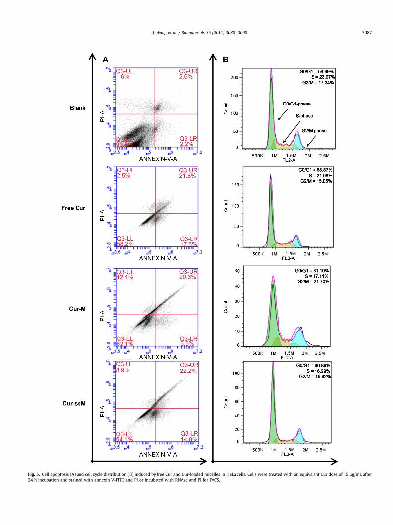
Cur can inhibit the proliferation and survival of almost all typesof tumor cells and the corresponding mechanisms about how Curkills tumor cells are controversial [43]. To date, cell cycle arrest isone of the universally accepted mechanisms. To evaluate theimpact of Cur-loaded micellar formulation on pharmacological ef-fect, cell apoptosis and cell cycle distribution analyses induced byboth the free Cur and Cur-loaded micelles were conducted, asshown in Fig. 5. From Fig. 5A, we can find that after 24 h incubation,
.the total apoptotic HeLa cell populations induced by free Cur andCur-ssM were 39.3% and 37.0%, respectively, which were slightlyhigher than that (25.8%) of Cur-M. Analysis of cell cycle indicatedthat the three Cur formulations could effectively arrest HeLa cells inthe G0/G1 phase after 24 h incubation. Compared to free Cur, Cur-ssM had a relatively higher arrest proportion of cells in the G0/G1phase but without significant differences between these groups(Fig. 5B). This may be due to the fact that free Cur diffused andaccumulated directly at action sites of tumor cells while Cur-ssMshowed a rapid release of Cur inside cancer cell and thereby acti-vated apoptotic signals to induce tumor cell death. These results areconsistent with the documented report that Cur is able to arrestcells in the G0/G1 phase and inhibit the proliferation of rhabdo-myosarcoma cells in a p53-independent manner [44].
3.9. Biodistribution and antitumor efficiency in vivo
The experimental process of the in vivo tumor inhibition wasshown in Fig. 6A. To measure the accumulated Cur in major organsand tumors at 1, 6, and 24 h after intravenous administration of freeCur, Cur-M and Cur-ssM at 10 mg/kg Cur equivalent, the tissuesamples were analyzed according to the report by Song et al. [45]and the final results were gathered in Fig. 6B. After administra-tion for 1 h, 6 h and 24 h, the accumulations of Cur-ssM and Cur-Min tumors were all significantly enhanced in comparison to free Cur.This can be attributed to two factors, one is the protection ofmicellar carrier with bulky hydrophilic outer shell to evade specificrecognition by the reticuloendothelial system (RES) [46], the otheris the well-known enhanced penetration and retention (EPR) effectin tumor tissues with tortuous and leaky vasculatures [47]. Besidesthe tumor tissues, the main accumulation of Cur in normal tissueswas observed in lung, liver and spleen, which are the major dis-tribution sites and target organs that are involved in the nonspecificuptake by the RES for nanomaterials [48,49].

www.spm
.com
.cn
Fig. 4. Representative confocal microscopy images and flow cytometry analyses contrasting the level of cellular uptake between HeLa cells incubated with free Cur, Cur-M and Cur-ssM formulations for 0.5 h (A, C) and 3 h (B, D). (Scale bars: 50 mm).
J. Wang et al. / Biomaterials 35 (2014) 3080e30903086
3.10. Antitumor efficiency in vivo and histological analysis
Although both the Cur-M and Cur-ssM showed no significantdifference in the in vivo distribution, the Cur-ssM group producedthe most noticeable efficiency in regressing tumor growth with 2.3and 3.5 times higher inhibition efficiency than Cur-M and free Cur
groups (Fig. 6C). From the discussion in Figs. 3A and 4, we knowthat the Cur-ssM could disassemble immediately in the reducingand acid microenvironment and subsequently release the payloadsrapidly, leading the cancer cells death. Free Cur could not achieveobvious antitumor effect due to its poor hydrophilicity and shorthalf-life in blood [45,50]. The weak antitumor efficiency in Cur-M
www.spm
.com
.cn
Fig. 5. Cell apoptosis (A) and cell cycle distribution (B) induced by free Cur and Cur-loaded micelles in HeLa cells. Cells were treated with an equivalent Cur dose of 15 mg/mL after24 h incubation and stained with annexin V-FITC and PI or incubated with RNAse and PI for FACS.
J. Wang et al. / Biomaterials 35 (2014) 3080e3090 3087

www.spm
.com
.cnFig. 6. In vivo biodistribution and therapeutic efficacy of these nanoformulations: A experimental schedule for antitumor study in vivo (A), Biodistribution in tissue in mice after i.v.administration of free Cur, Cur-ssM and Cur-M for different time (B), Changes of tumor volume (C), body weight (D) and survival rate (E) of tumor-bearing BABL/c mice receivingintravenous injection of saline, blank micelle and free Cur, Cur-ssM, Cur-M at a Cur dose of 10 mg/kg (*p < 0.05 and **p < 0.01).
Fig. 7. H&E staining and TUNEL analysis of tumor sections at the 21st days after the first treatment (A) and the mean optical density measured with Image-Pro Plus 6.0 softwarebased on TUNEL images (B). **p < 0.01 vs. saline group. (Scale bars: 200 mm).
J. Wang et al. / Biomaterials 35 (2014) 3080e30903088

J. Wang et al. / Biomaterials 35 (2014) 3080e3090 3089
group could be owing to the low Cur intracellular release from theredox-insensitive micelles as discussed in Fig. 3A. In addition, thebody weight of mice in the Cur-ssM group increased obviously, inthe Cur-M group almost kept no change while the other threegroups decreased slightly as shown in Fig. 6D, also suggesting thatthe redox-responsive micelle can reduce the toxicity of therapeuticagent to normal tissues or organs. The survival rate of themice aftertreatments shown in Fig. 6E also demonstrated that the therapeuticeffect of Cur-ssMwas remarkably better than that of Cur-M and freeCur.
To further confirm the therapeutic efficacy, histological analysisof tumor sections was performed at the 21st day after the first in-jection and the results were shown in Fig. 7. From the H&E stainingin Fig. 7A, we can find that in comparison with saline and blankmicelle groups, Cur-induced groups including free Cur, Cur-M andCur-ssM exhibited distinct apoptosis degrees, along with cellsbecoming smaller, nuclei lysis, vacuoles appearing and membraneintegrity destruction, especially for the Cur-ssM treated group.Apoptotic cells in dark brown were also identified by TUNEL assay(Fig. 7A). For Cur-ssM group, we can clearly observe that nuclearmembrane cracked and chromatin condensed, marginalized anddivided into blocks or apoptotic bodies, which is an obvious signalfor the apoptosis of tumor cells as previous reports [51,52]. Inbiology, OD is commonly used for immunohistochemical exami-nation. Usually the deeper the stained lesion, the larger the opticaldensity. To quantify the apoptosis of each group, the lesions withdark brown represents the apoptotic cells in TUNEL assay imagesand the mean OD of these lesions was calculated with Image-ProPlus 6.0 software and shown in Fig. 7B. The OD value (0.17924) ofthe Cur-ssM group is the highest and has significant differencewiththe other groups, especially compared to the saline group(0.00629), also indicating the excellent superiority in inducingapoptosis of cancer cells in vivo.
mwww.sp
4. Conclusion
In summary, we have successfully developed one type ofredox-responsive polyanhydride copolymer, and acquired stablemicelles with well-defined coreeshell structure by self-assemblyof this copolymer. The desirable redox-sensitivity of micelles isverified by changing the concentration of the reducing agent GSH.In vitro release profiles demonstrate that the therapeutic agent Curcan be released from the polymer matrix at a steady and rapidspeed in a reducing and acid environment. In vitro cytotoxicityanalysis reveals that Cur loaded redox-responsive micelles aremore effective in inhibiting the growth of tumor cells due to therapid release of therapeutic agent in tumor microenvironment incontrast to redox-insensitive micelles. In vivo antitumor activitydemonstrates that the redox-responsive micelles possess highersafety to the body and a better therapeutic effect in the inductionof tumor necrosis compared with redox-insensitive micelles.Therefore, the study provides a facile strategy towards the designand engineering of the next-generation nanomedicines for cancertherapy.
Acknowledgments
This work was partially supported by National Basic ResearchProgram of China (973 Program, 2012CB933600), National NaturalScience Foundation of China (51173150, 51373138), National KeyProject of Scientific and Technical Supporting Programs Funded byMSTC (2012BAI17B06), Research Fund for the Doctoral Program ofHigher Education of China (20120184110029) and FundamentalResearch Funds for The Central Universities (SWJTU11ZT10).
.com
.cn
Appendix A. Supplementary data
Supplementary data related to this article can be found at http://dx.doi.org/10.1016/j.biomaterials.2013.12.025.
References
[1] Peer D, Karp JM, Hong S, Farokhzad OC, Margalit R, Langer R. Nanocarriers asan emerging platform for cancer therapy. Nat Nanotechnol 2007;2:751e60.
[2] Newland B, Zheng Y, Jin Y, Abu-Rub M, Cao H, Wang W, et al. Single cyclizedmolecule versus single branched molecule: a simple and efficient 3D “knot”polymer structure for nonviral gene delivery. J Am Chem Soc 2012;134:4782e9.
[3] Farokhzad OC, Langer R. Impact of nanotechnology on drug delivery. ACSNano 2009;3:16e20.
[4] Duncan R. The dawning era of polymer therapeutics. Nat Rev Drug Discov2003;2:347e60.
[5] Fox ME, Szoka FC, Frechet JM. Soluble polymer carriers for the treatment ofcancer: the importance of molecular architecture. Acc Chem Res 2009;42:1141e51.
[6] Deng C, Jiang Y, Cheng R, Meng F, Zhong Z. Biodegradable polymeric micellesfor targeted and controlled anticancer drug delivery: promises, progress andprospects. Nano Today 2012;7:467e80.
[7] Wang C, Wang Z, Zhang X. Amphiphilic building blocks for self-assembly:from amphiphiles to supra-amphiphiles. Acc Chem Res 2012;45:608e18.
[8] Nicolas J, Mura S, Brambilla D, Mackiewicz N, Couvreur P. Design, function-alization strategies and biomedical applications of targeted biodegradable/biocompatible polymer-based nanocarriers for drug delivery. Chem Soc Rev2013;42:1147e235.
[9] Kazunori K, Glenn SK, Masayuki Y, Teruo O, Yasuhisa S. Block copolymermicelles as vehicles for drug delivery. J Control Release 1993;24:119e32.
[10] Allen TM, Cullis PR. Drug delivery systems: entering the mainstream. Science2004;303:1818e22.
[11] Hu X, Liu S, Chen X, Mo G, Xie Z, Jing X. Biodegradable amphiphilic blockcopolymers bearing protected hydroxyl groups: synthesis and characteriza-tion. Biomacromolecules 2008;9:553e60.
[12] Basarkar A, Singh J. Poly (lactide-co-glycolide)-polymethacrylate nano-particles for intramuscular delivery of plasmid encoding interleukin-10 toprevent autoimmune diabetes in mice. Pharm Res 2009;26:72e81.
[13] Sun Y, Yan X, Yuan T, Liang J, Fan Y, Gu Z, et al. Disassemblable micelles basedon reduction-degradable amphiphilic graft copolymers for intracellular de-livery of doxorubicin. Biomaterials 2010;31:7124e31.
[14] Guo X, Shi C, Wang J, Di S, Zhou S. pH-triggered intracellular release fromactively targeting polymer micelles. Biomaterials 2013;34:4544e54.
[15] Rosen HB, Chang J, Wnek GE, Linhardt RJ, Langer R. Bioerodible poly-anhydrides for controlled drug delivery. Biomaterials 1983;4:131e3.
[16] Kumar N, Langer RS, Domb AJ. Polyanhydrides: an overview. Adv Drug DelivRev 2002;54:889e910.
[17] Zhao A, Zhou S, Zhou Q, Chen T. Thermosensitive micelles from PEG-basedether-anhydride triblock copolymers. Pharm Res 2010;27:1627e43.
[18] Chen T, Guo X, Liu X, Shi S, Wang J, Shi C, et al. A strategy in the design ofmicellar shape for cancer therapy. Adv Healthcare Mater 2012;1:214e24.
[19] Griset AP, Walpole J, Liu R, Gaffey A, Colson YL, Grinstaff MW. Expansilenanoparticles: synthesis, characterization, and in vivo efficacy of an acid-responsive polymeric drug delivery system. J Am Chem Soc 2009;131:2469e71.
[20] Wang J, Sun X, Mao W, Sun W, Tang J, Sui M, et al. Tumor redoxheterogeneity-responsive prodrug nanocapsules for cancer chemotherapy.Adv Mater 2013;25:3670e6.
[21] Yang Y, Wang J, Zhang X, Lu W, Zhang Q. A novel mixed micelle gel withthermo-sensitive property for the local delivery of docetaxel. J Control Release2009;135:175e82.
[22] Dai J, Lin S, Cheng D, Zou S, Shuai X. Interlayer-crosslinked micelle withpartially hydrated core showing reduction and pH dual sensitivity for pin-pointed intracellular drug release. Angew Chem Int Ed Engl 2011;50:9404e8.
[23] Yoon S, Kim WJ, Yoo HS. Dual-responsive breakdown of nanostructures withhigh doxorubicin payload for apoptotic anticancer therapy. Small 2013;9:284e93.
[24] Liu J, Pang Y, Zhu Z, Wang D, Li C, Huang W, et al. Therapeutic nanocarrierswith hydrogen peroxide-triggered drug release for cancer treatment. Bio-macromolecules 2013;14:1627e36.
[25] Cheng R, Feng F, Meng F, Deng C, Feijen J, Zhong Z. Glutathione-responsivenano-vehicles as a promising platform for targeted intracellular drug andgene delivery. J Control Release 2011;152:2e12.
[26] Wei H, Zhuo R-X, Zhang X-Z. Design and development of polymeric micelleswith cleavable links for intracellular drug delivery. Prog Polym Sci 2013;38:503e35.
[27] Gilbert HF. In: Lester P, editor. Thiol/disulfide exchange equilibria and disul-fidebond stability. Methods Enzymol Academic Press; 1995. pp. 8e28.
[28] Shim MS, Kwon YJ. Stimuli-responsive polymers and nanomaterials for genedelivery and imaging applications. Adv Drug Deliv Rev 2012;64:1046e59.
[29] Liu J, Pang Y, Chen J, Huang P, Huang W, Zhu X, et al. Hyperbranched poly-diselenide as a self assembling broad spectrum anticancer agent. Biomaterials2012;33:7765e74.

.
J. Wang et al. / Biomaterials 35 (2014) 3080e30903090
[30] Liu J, Huang W, Pang Y, Huang P, Zhu X, Zhou Y, et al. Molecular self-assemblyof a homopolymer: an alternative to fabricate drug-delivery platforms forcancer therapy. Angew Chem Int Ed Engl 2011;50:9162e6.
[31] Cheng Z, Al Zaki A, Hui JZ, Muzykantov VR, Tsourkas A. Multifunctionalnanoparticles: cost versus benefit of adding targeting and imaging capabil-ities. Science 2012;338:903e10.
[32] Meng F, Hennink WE, Zhong Z. Reduction-sensitive polymers and bio-conjugates for biomedical applications. Biomaterials 2009;30:2180e98.
[33] Tang H, Murphy CJ, Zhang B, Shen Y, Van Kirk EA, Murdoch WJ, et al. Cur-cumin polymers as anticancer conjugates. Biomaterials 2010;31:7139e49.
[34] Murphy CJ, Tang H, Van Kirk EA, Shen Y, Murdoch WJ. Reproductive effects ofa pegylated curcumin. Reprod Toxicol 2012;34:120e4.
[35] Yu H, Huang Q. Improving the oral bioavailability of curcumin using novelorganogel-based nanoemulsions. J Agric Food Chem 2012;60:5373e9.
[36] Akbari H, D’Emanuele A, Attwood D. Effect of geometry on the erosioncharacteristics of polyanhydride matrices. Int J Pharm 1998;160:83e9.
[37] Hanes J, Chiba M, Langer R. Degradation of porous poly(anhydride-co-imide)microspheres and implications for controlled macromolecule delivery. Bio-materials 1998;19:163e72.
[38] Wang YJ, Pan MH, Cheng AL, Lin LI, Ho YS, Hsieh CY, et al. Stability of curcuminin buffer solutions and characterization of its degradation products. J PharmBiomed Anal 1997;15:1867e76.
[39] Fleige E, Quadir MA, Haag R. Stimuli-responsive polymeric nanocarriers forthe controlled transport of active compounds: concepts and applications. AdvDrug Deliv Rev 2012;64:866e84.
[40] Hubbell JA. Enhancing drug function. Science 2003;300:595e6.[41] Sahoo SK, Ma W, Labhasetwar V. Efficacy of transferrin-conjugated paclitaxel-
loaded nanoparticles in a murine model of prostate cancer. Int J Cancer2004;112:335e40.
[42] Yan Y, Johnston AP, Dodds SJ, Kamphuis MM, Ferguson C, Parton RG, et al.Uptake and intracellular fate of disulfide-bonded polymer hydrogel
www.spm
cn
capsules for Doxorubicin delivery to colorectal cancer cells. ACS Nano2010;4:2928e36.
[43] Ravindran J, Prasad S, Aggarwal BB. Curcumin and cancer cells: how manyways can curry kill tumor cells selectively? AAPS J 2009;11:495e510.
[44] Beevers CS, Li F, Liu L, Huang S. Curcumin inhibits the mammalian target ofrapamycin-mediated signaling pathways in cancer cells. Int J Cancer2006;119:757e64.
[45] Song Z, Feng R, Sun M, Guo C, Gao Y, Li L, et al. Curcumin-loaded PLGA-PEG-PLGA triblock copolymeric micelles: preparation, pharmacokinetics and dis-tribution in vivo. J Colloid Interface Sci 2011;354:116e23.
[46] Smola M, Vandamme T, Sokolowski A. Nanocarriers as pulmonary drug de-livery systems to treat and to diagnose respiratory and non respiratory dis-eases. Int J Nanomed 2008;3:1.
[47] Maeda H, Wu J, Sawa T, Matsumura Y, Hori K. Tumor vascular permeabilityand the EPR effect in macromolecular therapeutics: a review. J Control Release2000;65:271e84.
[48] Lu J, Huang Y, Zhao W, Marquez RT, Meng X, Li J, et al. PEG-derivatizedembelin as a nanomicellar carrier for delivery of paclitaxel to breast andprostate cancers. Biomaterials 2013;34:1591e600.
[49] Wang B, He X, Zhang Z, Zhao Y, Feng W. Metabolism of nanomaterialsin vivo:blood circulation and organ clearance. Acc Chem Res 2013;46:761e9.
[50] Li L, Braiteh FS, Kurzrock R. Liposome-encapsulated curcumin: in vitro andin vivo effects on proliferation, apoptosis, signaling, and angiogenesis. Cancer2005;104:1322e31.
[51] Wang W, Cheng D, Gong F, Miao X, Shuai X. Design of multifunctional micellefor tumor-targeted intracellular drug release and fluorescent imaging. AdvMater 2012;24:115e20.
[52] Fan H, Hu QD, Xu FJ, LiangWQ, Tang GP, YangWT. In vivo treatment of tumorsusing host-guest conjugated nanoparticles functionalized with doxorubicinand therapeutic gene pTRAIL. Biomaterials 2012;33:1428e36.
.com