intracellular drug delivery of - royal society of chemistry · mmp-2 responsive polymeric micelles...
TRANSCRIPT

S1
Electronic Supplementary Information
of
MMP-2 Responsive Polymeric Micelles for Cancer-Targeted
Intracellular Drug Delivery
Wei-Hai Chen, Guo-Feng Luo, Qi Lei, Hui-Zhen Jia, Sheng Hong, Qing-Rong Wang*,
Ren-Xi Zhuo and Xian-Zheng Zhang*
Key Laboratory of Biomedical Polymers of Ministry of Education & Department of
Chemistry, Wuhan University, Wuhan 430072, P. R. China
*Corresponding author. Tel./fax: +86-27-68754509.
E-mail addresses: [email protected] (W.Q.R.), [email protected] (Z.X.Z.).
Electronic Supplementary Material (ESI) for ChemComm.This journal is © The Royal Society of Chemistry 2014

S2
1. Materials
Benzotriazol-1-yl-oxytripyrrolidinophosphonium hexafluorophosphate (PyBOP), 1-
hydroxybenzotriazole (HOBt), trifluoroacetic acid (TFA) and diisopropylethylamine
(DIEA) were purchased from GL Biochem. Ltd. (Shanghai, China) and used as
received. N,N-Dimethylformamide (DMF), methanol, dimethyl sulfoxide (DMSO),
tetrahydrofuran (THF) and dichloromethane (DCM) were provided by Shanghai
Chemical Co. (China) and distilled prior to use.
Doxorubicin hydrochloride (DOX·HCl) was purchased from Zhejiang Hisun
Pharmaceutical Co. (China). The MMP-2 protease sensitive peptide substrate Ac-Cys-
Pro-Leu-Gly-Leu-Ala-Gly-Gly-DOX (Ac-CPLGLAGG-DOX: peptide-DOX) was
prepared according to our previous report.[S1] The Purity (95.8%) of the peptide-DOX
(Fig. S1) was confirmed by high-pressure liquid chromatography (HPLC). 6-
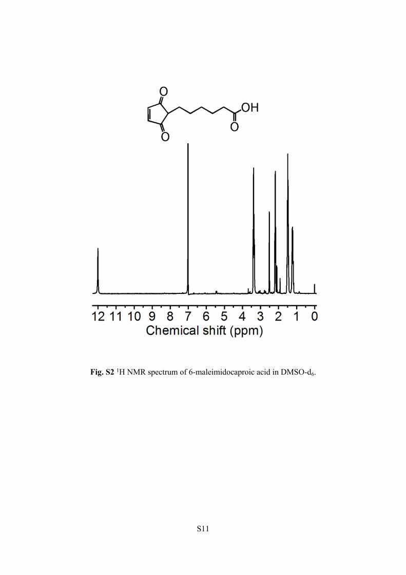
Maleimidocaproic acid was also synthesized according to the previous report.[S2] The
1H NMR spectrum (Fig. S2) demonstrated that 6-maleimidocaproic acid was
synthesized successfully. Biotin-poly(ethylene glycol)-NH2-3400 (Biotin-PEG-NH2)
was purchased from Laysan Bio Inc. 1,10-Phenanthroline monohydrate and Brij35
were purchased from Aladdin Reagent Co. Ltd. (Shanghai, China). Matrix
metalloproteinase (MMP-2) was purchased from RD-SYSTEMS. N(ε)-
benzyloxycarebonyl-L-lysine (H-Lys(Z)-OH) and TIMP-2 were purchased from
Sigma-Aldrich. Dulbecco’s Modified Eagle’s Medium (DMEM), fetal bovine serum
(FBS), 3-[4,5-dimethylthiazol-2-yl]-2,5-diphenyltetra-zoliumbromide (MTT), trypsin,
penicillin streptomycin, molecular probe (Hoechst 33258) and Dulbecco’s phosphate
buffered saline (PBS) were purchased from Invitrogen. All other reagents and
solvents were of analytical grade and used directly.
2. Synthesis of Biotin-poly(ethylene glycol)-blocked-poly(L-lysine) (Biotin-PEG-
S3
b-PLL)
Brifly, H-Lys(Z)-OH (4.0 g) was suspended in the distilled THF (40 mL) and the
mixture was stirred at 50 oC under N2 atmosphere. Then a solution of triphosgene (3.0
g) in THF (15 mL) was added dropwise to the suspension. And when the suspension
became a clear solution, the reaction was stopped and then poured into excess dried n-
hexane to obtain crude Z-Lys-NCA. The crude product was further recrystallized
from dried THF/n-hexane twice and dried under vacuum. The polymerization of Z-
Lys-NCA (4.5 g) by Biotin-PEG-NH2 (0.1 g) was carried out in 20 mL anhydrous
N,N-dimethylformamide (DMF) at 50 oC under N2 atmosphere for 72 h. Then the
solution was precipitated in an excess of diethyl ether three times to obtain Biotin-
PEG-b-PLL(Z). Subsequently, 15 mL TFA was added to dissolve Biotin-PEG-b-
PLL(Z) (600 mg) in an ice bath. After stirring for 15 min, HBr (5 mL of a 33 wt%
solution in acetic acid) was added dropwise and stirred for another 1 h. Then the
solution was precipitated in diethy ether. After collecting and drying, the product was
resuspended in 10 mL DMF and the solution was dialyzed (MWCO: 3500 Da) against
distilled water and finally lyophilized to obtain Biotin-PEG-b-PLL.
3. Synthesis of the 6-maleimidocaproic acid modified Biotin-PEG-b-PLL (Biotin-
PEG-b-PLL(Mal))
Brifly, the 6-maleimidocaproic acid (48 mg, 1 eq. to amino groups of Biotin-PEG-
b-PLL), DIEA (0.45 mL), PyBOP (177.4 mg) and HOBt (45.7 mg) were dissolved in
distilled DMSO (5 mL) and stirred at room temperature. After 30 min, a solution of
Biotin-PEG-b-PLL (50 mg) in DMSO (10 mL) was added and stirred for another 48 h.
Then the mixture solution was dialyzing against distilled water (MWCO: 3500 Da).
Biotin-PEG-b-PLL(Mal) was obtained after lyophilized.
4. Preparation of the MMP-2 protease sensitive Biotin-PEG-b-PLL(Mal)-
S4
peptide-DOX
The Biotin-PEG-b-PLL(Mal)-peptide-DOX was synthesized via Michael addition
according to a previous reference.[S3] Briefly, a solution of the previous prepared
Biotin-PEG-b-PLL(Mal) (30 mg) containing the substrate Ac-CPLGLAGG-DOX
(18.8 mg) in 15 mL DMSO was stirred at room temperature for 24 h. Then the
mixture solution was dialyzing against DMSO/water (1:1, V/V) (MWCO: 3500 Da)
for 24 h. Finally, the Biotin-PEG-b-PLL(Mal)-peptide-DOX was obtained after
lyophilized as red solid.
5. Fabrication of the multifunctional polymeric micelles
The multifunctional polymeric micelles were prepared by directly dissolving the
Biotin-PEG-b-PLL(Mal)-peptide-DOX (0.5 mg/mL) in the PBS (0.01 M, pH 7.4) and
subsequently standing in the dark. The inherent amphiphilicity property of the Biotin-
PEG-b-PLL(Mal)-peptide-DOX provided the opportunity to self-assemble into
micelles. The hydrophobic peptide-DOX composed the core and the hydrophilic PEG
formed the shell.
6. Characterizations
1H NMR spectra were recorded on a Varian Unity 300 MHz spectrometer by using
dimethyl sulfoxide-d6 (DMSO-d6) or D2O as the solvent. The molecular weight and
polydispersity index (PDI) of Biotin-PEG-b-PLL(Z) were evaluated by gel
permeation chromatographic (GPC) system consisting of Waters 2690D separations
module and Waters 2410 refractive index detector. Tetrahydrofuran (THF) was used
as the eluent at a flow rate of 0.3 mL/min. Fluorescence analysis was performed on a
RF-530/PC spectrofluorophotometer (Shimadzu). The particle size and zeta potential
were measured using Malvern Zetasizer Nano-ZS ZEN3600. The morphologies of
polymer micelles were viewed on transmission electron microscopy (TEM, JEM-

S5
2100).
7. Determination of critical micelle concentration (CMC)
The CMC of Biotin-PEG-b-PLL(Mal)-peptide-DOX micelles was estimated by
fluorescence spectra, which was recorded on an RF-530/PC spectrofluorophotometer
(Shimadzu) and using pyrene as a hydrophobic fluorescent probe. 50 µL of pyrene
solutions (6 × 10⁻⁷ M in acetone) were added to containers, after the acetone
evaporated, 1 mL aqueous solution of Biotin-PEG-b-PLL(Mal)-peptide-DOX
polymeric micelles at particular concentration varying from 1 × 10⁻⁴ to 1 mg/mL was
added to the container. The sample solutions containing pyrene residues were kept at
room temperature for 24 h to reach the equilibrium of pyrene partition between water
and micelles. For the pyrene excitation spectra, the emission wavelength was carried
out at 390 nm, and the excitation spectra of samples were recorded ranging from 300
nm to 360 nm. The fluorescence intensity ratio of the third and first vibronic bands
(I3/I1) was plotted against the logarithm of Biotin-PEG-b-PLL(Mal)-peptide-DOX
concentrations, and the CMC value was estimated from the intersection of the tangent
to the curve at the inflection with the horizontal tangent through the points at low
concentration.
8. Cell culture
SCC-7 (squamous cell carcinoma) cancer cells and COS7 normal cells (African
green monkey kidney fibroblast cells) were incubated in DMEM medium with 10%
FBS and 1% antibiotics (penicillin-streptomycin, 10000 U/mL) at 37 °C in a
humidified atmosphere containing 5% CO2.
9. The MMP-2 protease responsive behaviors of the multifunctional micelles
To investigate the MMP-2 protease responsive property of Biotin-PEG-b-
PLL(Mal)-peptide-DOX micelles, the morphology and the size changes of the

S6
micelles were detected after the micelles incubated with the MMP-2 protease for 6 h.
10. In vitro drug release study
In vitro drug release experiments were carried out in three different media: 1) 3 mL
TCNB buffer solution (composed of 100 mM Tris, 5 mM calcium chloride, 200 mM
NaCl, 0.1% Brij35) and 100 μL MMP-2 (2 μg/mL); 2) 3 mL TCNB buffer solution,
100 μL MMP-2 (2 μg/mL) and with the MMP-2 protease inhibitor; and 3) only 3 mL
TCNB buffer solution, respectively. For each release study, 1.5 mg of the Biotin-
PEG-b-PLL(Mal)-peptide-DOX micelles were dispersed in the above solutions. Then
the solutions were put into the dialysis tube (MWCO: 3500 Da) and subsequently
immersed into 10 mL incubation medium and maintained at 37 oC. After particular
time intervals, fluorescence intensity of the incubation medium was analyzed by RF-
5301PC spectrofluorophotometer. The emission and excitation slit widths were set at
5 nm with λex=470 nm.
11. Co-incubation of Biotin-PEG-b-PLL(Mal)-peptide-DOX micelles with cells
SCC-7 cancer cells and COS7 normal cells were seeded respectively in a glass
bottom dish at a density of 1 × 10⁵ cells/well for 24 h. As the negative control, SCC-7
cells and COS7 cells were incubated with the excess biotin (1 mM) for 4 h in advance.
Thereafter, the Biotin-PEG-b-PLL(Mal)-peptide-DOX micelles (containing 3 µg/mL
of Dox) dispersed in DMEM containing 10% FBS and 1% antibiotics were added and
the cells were incubated at 37 °C for another 3 h or 6 h. After removing the medium
and washing with PBS, the nucleus were stained with Hoechst 33258 at 37 oC for 15
min. Then the cells were observed by confocal laser scanning microscopy.
12. Quantitative analysis of cellular uptake DOX by flow cytometry
SCC-7 cancer cells and COS7 normal cells were seeded in 24-well plates at a
density of 5 × 10⁴ cells/well and cultured with 1 mL of DMEM containing 10% FBS
S7
for 1 day. As the negative control, SCC-7 cells and COS7 cells were incubated with
excess biotin (1 mM) for 4 h in advance. Then all cells were cultured with Biotin-
PEG-b-PLL(Mal)-peptide-DOX micelles (containing 3 µg/mL of Dox) for another 3 h
or 6 h. After that, the medium was removed and the cells were washed 3 times with
PBS. Then all the cells were digested by trypsin and collected in centrifuge tubes by
centrifugating at 1000 rpm for 3 mins. The supernatant was discarded and the bottom
cells were washed with PBS 3 times to remove excess micelles. Then the suspended
cells were filtrated and detected for red fluorescence (PE-A) by flow cytometry (BD
FACSAria TM III, USA). Cells untreated with micelles were used as the control. The
fluorescence scan was performed with 1 × 10⁴ cells.
13. In vitro cytotoxicity studies
In vitro cytotoxicity was performed with SCC-7 cancer cells and COS7 normal
cells by MTT assay. Briefly, SCC-7 and COS7 cells were seeded in 96-well plates at
a density of 6000 cells/well, and then cells were incubated in 100 μL DMEM
containing 10% FBS and 1% antibiotics for 1 day prior to adding Biotin-PEG-b-
PLL(Mal)-peptide-DOX micelles or Biotin-PEG-b-PLL. After co-incubation for 2
days, the medium was replaced with 200 μL of fresh medium. Then 20 μL MTT
solutions (5 mg/mL) was added to each well and further incubated for 4 h. After that,
the medium was removed and 200 μL DMSO was added. The absorbance was
measured at 570 nm using a microplate reader (Bio-Rad, Model 550, USA). The
relative cell viability was calculated as: cell viability = (OD570 (samples)/OD570 (control)) ×
100%, where OD570 (control) was obtained in the absence of the micelles or Biotin-PEG-
b-PLL, and OD570 (samples) was obtained in the presence of the micelles or Biotin-PEG-
b-PLL. Each value was averaged from four independent experiments.

S8
Supplementary References
[S1] W. H. Chen, X. D. Xu, H. Z. Jia, Q. Lei, G. F. Luo, S. X. Cheng, R. X. Zhuo and
X. Z. Zhang, Biomaterials, 2013, 34, 8798.
[S2] D. Willner, P. A. Trail, S. J. Hofstead, H. D. King, S. J. Lasch, G. R, Braslawsky,
R. S. Greenfield, T. Kaneko and R. A. Firestone, Bioconjug. Chem., 1993, 4,
521.
[S3] Q. Lin, C. Bao, S. Cheng, Y. Yang, W. Ji and L. Zhu, J. Am. Chem. Soc., 2012,
134, 5052.

S9
Supplementary Figures and Schemes
Scheme S1. Synthesis of (a) Z-Lys-NCA and (b) Biotin-PEG-PLL(Mal)-peptide-
DOX.

S10
Fig. S1 HPLC profile of peptide-DOX (Ac-Cys-Pro-Leu-Gly-Leu-Ala-Gly-Gly-DOX)
and the free DOX as the control. The purity of the peptide-DOX was 95.8%.
S11
Fig. S2 1H NMR spectrum of 6-maleimidocaproic acid in DMSO-d6.

S12
Fig. S3 1H NMR spectrum of Biotin-PEG-b-PLL(Z) in DMSO-d6.

S13
Fig. S4 1H NMR spectrum of Biotin-PEG-b-PLL in D2O.
S14
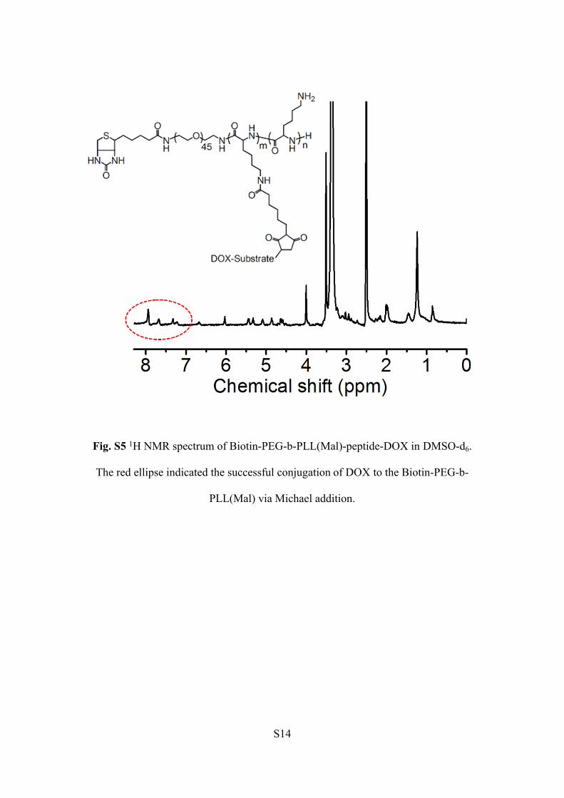
Fig. S5 1H NMR spectrum of Biotin-PEG-b-PLL(Mal)-peptide-DOX in DMSO-d6.
The red ellipse indicated the successful conjugation of DOX to the Biotin-PEG-b-
PLL(Mal) via Michael addition.

S15
Fig. S6 The CMC of Biotin-PEG-b-PLL(Mal)-peptide-DOX polymeric micelles
determined by fluorescence spectra and using pyrene as a hydrophobic fluorescent
probe. Plot of the intensity ratio I3/I1 vs log C, the tested CMC value was 3.47 × 10⁻²
mg/mL.

S16
Fig. S7 Monitoring the diameter (a) and PDI (b) changes of Biotin-PEG-b-PLL(Mal)-
peptide-DOX polymeric micelles by DLS.

S17
Fig. S8 Confocal laser scanning microscopy (CLSM) images of COS7 normal cells
incubated with Biotin-PEG-b-PLL(Mal)-peptide-DOX micelles for 3 h (A-A2) and 6 h
( B-B2), and 6 h for cells pre-treated with the excess of biotin in advance ( C-C2). (A-
C) blue fluorescence images of nuclei; (A1-C1) red fluorescence images of DOX; (A2-
C2) the merge images of blue and red fluorescence. The scale bar is 14 μm.

S18
Fig. S9 Quantitative flow cytometry analysis of the cellular DOX red fluorescence
mean fluorescence intensity (MFI) values of SCC-7 cancer cells (a) and COS7 normal
cells (b), respectively.

S19
Fig. S10 The cell viability of SCC-7 cancer cells and COS7 normal cells after
incubated with Biotin-PEG-b-PLL 48 h by MTT assay.