regulation of constitutive and alternative splicing by...
TRANSCRIPT

Regulation of constitutive and alternativesplicing by PRMT5 reveals a rolefor Mdm4 pre-mRNA in sensing defectsin the spliceosomal machinery
Marco Bezzi,1,2 Shun Xie Teo,1 Julius Muller,1 Wei Chuen Mok,1 Sanjeeb Kumar Sahu,1
Leah A. Vardy,3,4 Zahid Q. Bonday,5 and Ernesto Guccione1,2,6
1Division of Cancer Genetics and Therapeutics, Laboratory of Chromatin, Epigenetics, and Differentiation, Institute ofMolecular and Cell Biology (IMCB), A*STAR (Agency for Science, Technology, and Research), Singapore 138673, Singapore;2Department of Biochemistry, Yong Loo Lin School of Medicine, National University of Singapore, Singapore 119074, Singapore;3Institute of Medical Biology (IMB), A*STAR, Singapore 138673, Singapore; 4School of Biological Sciences, NanyangTechnological University, Singapore 637551, Singapore; 5Lilly Research Laboratories, Eli Lilly and Company, Indianapolis, Indiana46285, USA
The tight control of gene expression at the level of both transcription and post-transcriptional RNA processingis essential for mammalian development. We here investigate the role of protein arginine methyltransferase5 (PRMT5), a putative splicing regulator and transcriptional cofactor, in mammalian development. Wedemonstrate that selective deletion of PRMT5 in neural stem/progenitor cells (NPCs) leads to postnatal deathin mice. At the molecular level, the absence of PRMT5 results in reduced methylation of Sm proteins, aberrantconstitutive splicing, and the alternative splicing of specific mRNAs with weak 59 donor sites. Intriguingly,the products of these mRNAs are, among others, several proteins regulating cell cycle progression. We identifyMdm4 as one of these key mRNAs that senses the defects in the spliceosomal machinery and transduces thesignal to activate the p53 response, providing a mechanistic explanation of the phenotype observed in vivo. Ourdata demonstrate that PRMT5 is a master regulator of splicing in mammals and uncover a new role for theMdm4 pre-mRNA, which could be exploited for anti-cancer therapy.
[Keywords: PRMT5; arginine methylation; development; splicing; p53; MDM4]
Supplemental material is available for this article.
Received May 8, 2013; revised version accepted July 26, 2013.
Arginine methylation is a post-translational modificationknown to play a key role in both transcription and post-transcriptional RNA processing by mediating epigeneticcontrol of chromatin and functionally regulating RNA-binding proteins and components of the splicing machin-ery (Cheng et al. 2007; Kouzarides 2007; Bedford andClarke 2009; Migliori et al. 2010).
Initial attempts to identify arginine-methylated pro-teins have generated lists of putative protein argininemethyltransferase (PRMT) targets (Boisvert et al. 2003;Ong et al. 2004). These studies failed to identify residuesmethylated by specific PRMT family members and todistinguish between symmetric and asymmetric dimeth-ylation. However, they did shed light on the relevance of
arginine methylation in splicing regulation by identifyingas targets key components of the constitutive splicingmachinery (e.g., Sm proteins) as well as several additionalregulators of alternative splicing (e.g., FUS/TLS, SF2, andmembers of the heterogeneous nuclear ribonucleoprotein[hnRNP] family).
The splicing complexity occurring in the mammalianbrain is a remarkable product of evolution and distinc-tively distinguishes the human species from others(Barbosa-Morais et al. 2012; Dillman et al. 2013). Properfunctioning of all splicing-associated proteins allows asignificant increase in the complexity of the cell pro-teome. On the other hand, mutations in proteins involvedin RNA processing have been causally linked to neuro-degenerative disorders such as spinal muscular atrophy(SMA) and amyotrophic lateral sclerosis (ALS), to men-tion a few (Zhang et al. 2008; Vance et al. 2009; Da Cruzand Cleveland 2011). These disease-causing mutationsemphasize the pivotal role of RNA processing in highervertebrate brain development, urging researchers to
6Corresponding authorE-mail [email protected] is online at http://www.genesdev.org/cgi/doi/10.1101/gad.219899.113.Freely available online through the Genes & Development Open Accessoption.
GENES & DEVELOPMENT 27:1903–1916 � 2013, Published by Cold Spring Harbor Laboratory Press; ISSN 0890-9369/13; www.genesdev.org 1903
Cold Spring Harbor Laboratory Press on June 11, 2020 - Published by genesdev.cshlp.orgDownloaded from
investigate the role of novel splicing regulators in theCNS.
PRMT5 is a type II arginine methyltransferase able tosymmetrically dimethylate several nuclear and cytoplas-mic proteins (Bedford and Clarke 2009; Karkhanis et al.2011). In the cytoplasm, PRMT5 acts together with pIClnand WDR77 as part of the methylosome, which mainlymethylates Sm proteins (SmB/B9, SmD1, and SmD3),increasing their affinity for the Tudor domain of spinalmotor neuron 1 (SMN1) (Friesen et al. 2001a,b, 2002;Meister et al. 2001). SMN1 deficiency results in earlylethality (embryonic day 3.5 [E3.5]) in the mouse embryo,similarly to Prmt5 constitutive deletion (Hsieh-Li et al.2000; Tee et al. 2010), while lack of SMN1 in SMA mousemodels leads to splicing defects in not only spinal motorneurons, but several organs (Zhang et al. 2008). Given thedirect connection between PRMT5 and SMN1 and therelevance of arginine methylation in regulating splicingproteins, it is of extreme relevance to assess the role, ifany, of PRMT5 in the CNS.
Here we demonstrate that selective deletion of PRMT5in the CNS leads to the death of the animal 14 d afterbirth. We first show that genetic deletion of p53 in aPrmt5-null background partially rescues the develop-mental defects. Second, we show that the absence ofPRMT5 in neural stem/progenitor cells (NPCs) leads todefects in the core splicing machinery. This results inreduced constitutive splicing and the alternative splicingof specific mRNAs, which have weak 59 donor sites. Weidentify Mdm4 as one of these mRNAs that acts as asensor of the splicing defects. Specifically, the Mdm4alternative splicing event results in the generation of theunstable Mdm4s product, the reduction of the full-lengthprotein, and the transduction of the p53 signaling cas-cade. We finally expand our findings to other cell typesand tissues, demonstrating that Mdm4/Mdm4s alterna-tive splicing senses the absence of PRMT5 also in mouseembryonic fibroblasts (MEFs) in several organs duringembryo development and in human cancer cell lines.
We believe our data provide an underlying mechanismfor many observations on PRMT5 biology (Jansson et al.2008; Scoumanne et al. 2009) and, more in general, onperturbation of the splicing machinery (Allende-Vegaet al. 2013) and their link to the p53 pathway that werepreviously ignored.
Results
PRMT5 deficiency in the CNS results in early postnatallethality
To address the effect of PRMT5 depletion in mammals,we made use of a conditional knockout mouse (Whiteet al. 2013) harboring LoxP (F/F) sequences flanking exon 7in the Prmt5 gene and studied the effect of its conditionaldeletion in the CNS. We used a Nestin-Cre (Nes) trans-genic mouse strain that expresses Cre recombinase undera neural-specific enhancer of the Nestin promoter, lead-ing to an efficient recombination event in precursorsof neurons and glia starting at E10.5 (Graus-Porta et al.2001). All of the Prmt5F/FNes mice were obtained from
Prmt5F/F 3 Prmt5+/FNes crosses, and, as expected, thePrmt5+/FNes mice were viable and fertile, and we couldnot observe any evident defects. Single-site insertion wasverified by Southern blotting, and CNS-specific deletionof PRMT5 was confirmed by genomic PCR and Westernblotting (Supplemental Fig. S1).
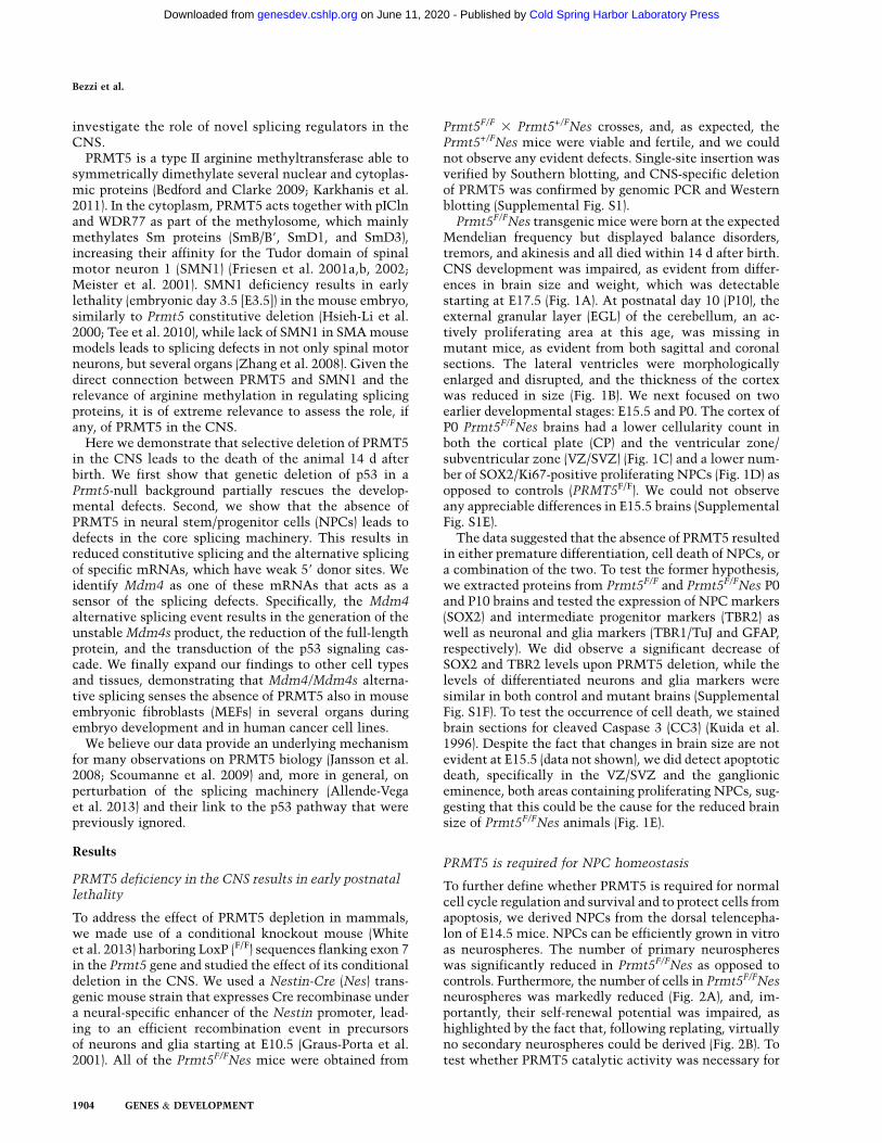
Prmt5F/FNes transgenic mice were born at the expectedMendelian frequency but displayed balance disorders,tremors, and akinesis and all died within 14 d after birth.CNS development was impaired, as evident from differ-ences in brain size and weight, which was detectablestarting at E17.5 (Fig. 1A). At postnatal day 10 (P10), theexternal granular layer (EGL) of the cerebellum, an ac-tively proliferating area at this age, was missing inmutant mice, as evident from both sagittal and coronalsections. The lateral ventricles were morphologicallyenlarged and disrupted, and the thickness of the cortexwas reduced in size (Fig. 1B). We next focused on twoearlier developmental stages: E15.5 and P0. The cortex ofP0 Prmt5F/FNes brains had a lower cellularity count inboth the cortical plate (CP) and the ventricular zone/subventricular zone (VZ/SVZ) (Fig. 1C) and a lower num-ber of SOX2/Ki67-positive proliferating NPCs (Fig. 1D) asopposed to controls (PRMT5F/F). We could not observeany appreciable differences in E15.5 brains (SupplementalFig. S1E).
The data suggested that the absence of PRMT5 resultedin either premature differentiation, cell death of NPCs, ora combination of the two. To test the former hypothesis,we extracted proteins from Prmt5F/F and Prmt5F/FNes P0and P10 brains and tested the expression of NPC markers(SOX2) and intermediate progenitor markers (TBR2) aswell as neuronal and glia markers (TBR1/TuJ and GFAP,respectively). We did observe a significant decrease ofSOX2 and TBR2 levels upon PRMT5 deletion, while thelevels of differentiated neurons and glia markers weresimilar in both control and mutant brains (SupplementalFig. S1F). To test the occurrence of cell death, we stainedbrain sections for cleaved Caspase 3 (CC3) (Kuida et al.1996). Despite the fact that changes in brain size are notevident at E15.5 (data not shown), we did detect apoptoticdeath, specifically in the VZ/SVZ and the ganglioniceminence, both areas containing proliferating NPCs, sug-gesting that this could be the cause for the reduced brainsize of Prmt5F/FNes animals (Fig. 1E).
PRMT5 is required for NPC homeostasis
To further define whether PRMT5 is required for normalcell cycle regulation and survival and to protect cells fromapoptosis, we derived NPCs from the dorsal telencepha-lon of E14.5 mice. NPCs can be efficiently grown in vitroas neurospheres. The number of primary neurosphereswas significantly reduced in Prmt5F/FNes as opposed tocontrols. Furthermore, the number of cells in Prmt5F/FNesneurospheres was markedly reduced (Fig. 2A), and, im-portantly, their self-renewal potential was impaired, ashighlighted by the fact that, following replating, virtuallyno secondary neurospheres could be derived (Fig. 2B). Totest whether PRMT5 catalytic activity was necessary for
Bezzi et al.
1904 GENES & DEVELOPMENT
Cold Spring Harbor Laboratory Press on June 11, 2020 - Published by genesdev.cshlp.orgDownloaded from

the observed phenotype, we infected primary NPCsderived from Prmt5F/FNes mice with wild-type humanPRMT5 (hPRMT5) or a catalytically inactive mutant(hPRMT5AAA). Only cells infected with hPRMT5 wereable to grow and could be propagated into secondary neuro-spheres, and when expanded into tertiary neurospheres,cells expressing hPRMT5 grew as efficiently as NPCs de-rived from Prmt5F/F control litters (Fig. 2C). To confirm theresults obtained in vivo, we first counted the percentage ofpyknotic nuclei and then stained Prmt5F/FNes-derivedNPCs for CC3 to verify that they were undergoing apopto-sis, confirming the requirement for PRMT5 to suppress celldeath (Fig. 2D).
To understand the molecular mechanism underpinningthe observed apoptotic phenotype, we next performeda gene expression analysis of Prmt5F/FNes NPCs. Approx-imately 2500 genes were differentially expressed whencompared with control, showing up-regulation of thep53 pathway and down-regulation of genes involved
in cell cycle progression and replication (SupplementalFig. S2).
We then generated a second conditional knockout strainby crossing the Prmt5F/F mice to the ROSA26:CreERt2(ER) mice, which allowed the triggering of a recombinationevent in both live animals and, ex vivo, primary cells byusing 4-hydroxytamoxifen (OHT). We switched to thePrmt5F/FER system for three main reasons: First, it allowedus to look at cell-autonomous defects. Second, we couldderive a much larger number of cells amenable for furthermechanistic studies. Third, it allowed us to focus on earlytime points after PRMT5 depletion in order to detectcausal defects. In all experiments described hereafter inwhich Prmt5F/FER-derived cells, tissues, or embryos wereanalyzed, we always used the ER counterparts as negativecontrols, making sure that the addition of OHT or tamox-ifen (TAM) was not toxic (data not shown).
p53 is a transcription factor that drives the expressionof several downstream targets in response to a variety of
Figure 1. PRMT5 deficiency in the CNS results in early postnatal lethality. Nestin-Cre-induced deletion of the PRMT5 gene in theCNS. (A) Weight in milligrams of wild-type (Prmt5F/F) and Prmt5-deleted (Prmt5F/FNes) brains at three different time points (E17.5, P0,and P10). Brain sizes of P10 Prmt5F/F and Prmt5F/FNes mice are shown as an example in the right panel. (B) Hematoxylin and eosin(H&E)-stained sagittal and coronal sections of P10 cerebrum (Cr) and the cerebellum (Cb) from Prmt5F/F (right) and Prmt5F/FNes (left).(C) Coronal sections of P0 brains. Cellularity of the cortical plate (CP) and VZ/SVZ are indicated in wild-type (wt; black) and mutant(red) brains. (MZ) Marginal zone; (SP) subplate; (IZ) intermediate zone. (D) SOX2 and Ki67 immunohistochemistry (IHC) staining in P0brains. (E) Cleaved Caspase 3 (CC3) staining is shown in both the cortex and the gangliomic eminence.
PRMT5 role in mammalian development
GENES & DEVELOPMENT 1905
Cold Spring Harbor Laboratory Press on June 11, 2020 - Published by genesdev.cshlp.orgDownloaded from

stimuli, including activation of the DNA damage re-sponse (DDR) (Lane 1992). Much is known about theregulation of p53 by post-translational modifications, andmany of them, including phosphorylation and acetyla-tion, are known to regulate its protein stability, leadingto transcriptional activation. We first checked whether,upon PRMT5 deletion, we could detect DDR activationand whether p53 would be stabilized. We did observea modest p53 protein stabilization and p53 phosphoryla-tion (P-p53) as well as basal levels of H2AX phosphoryla-tion (gH2AX). As a positive control, we used a DNA-damaging agent (etoposide), which, as expected, greatlystabilized p53 and increased the levels of gH2AX. Nota-bly, despite a minor activation of the DDR, the absence ofPRMT5 caused an even greater induction of the p53target gene p21 as compared with etoposide (Fig. 2E, cf.lanes 2 and 3).
p53 deletion partially rescues Prmt5F/FNesdevelopmental defects
The data indicated that PRMT5 deficiency triggered ap53 response and that the phenotypic outcome in NPCsled to cell death. To formally prove this conclusion, wecrossed Prmt5F/FNes mice into a p53-null background.Prmt5F/FNes;p53�/�mice survived, on average, 1 wk longerthan Prmt5F/FNes;p53wt, while mice heterozygous forp53 (Prmt5F/FNes;p53+/�) displayed an intermediate phe-
notype (Fig. 3A). When stained for activated Caspase 3,E15.5 Prmt5F/FNes;p53�/� embryos showed a complete res-cue of the apoptotic response, with levels of staining similarto wild type (Fig. 3B). Importantly, the number of SOX2-positive cells in the VZ/SVZ of Prmt5F/FNes;p53�/� embryoswas increased when compared with Prmt5F/FNes;p53wt
brains (Fig. 3C). However, we did not observe a significantrescue of proliferating KI67-positive cells, suggesting ap53-independent impairment in cell cycle progression,which most likely accounts for the lethality of theanimals 20–22 d after birth (Fig. 3C, right panel). Indeed,when we derived NPCs from mice with different p53backgrounds and cultured them as neurospheres, p53deficiency led to a significant but not complete rescuein the number of proliferating cells (Fig. 3D). The samewas true in P10 brains, where defects in EGL morpho-genesis in the cerebellum and, in general, in brain devel-opment were only partially rescued in the absence ofp53 (Fig. 3E). We can conclude that p53 plays an importantrole in regulating the apoptotic response in Prmt5F/FNescells. The fact that we still observed death of the animals,although significantly delayed, however, pointed at addi-tional proliferative defects in targeted cells. To gainfurther insight, we first checked by RT-qPCR the levelof transcriptional up-regulation of p53 targets in bothPrmt5F/FNes and Prmt5F/FER in a p53�/� background.Activation of cell cycle inhibitor p21, proapoptotic Noxa,Puma (Akhtar et al. 2006), and all other target genes was
Figure 2. PRMT5 is required for NPC homeostasis. (A) Number of primary neurospheres and total number of cells from cultures ofE14.5 dorsal telencephalon NPCs derived from Prmt5F/F and Prmt5F/FNes embryos. Each bar represents an average of at least threeexperiments. (B) Number of secondary neurospheres, as in A. (C) Primary neurospheres (left panel) from Prmt5F/FNes mice infectedwith empty vector (EV), wild-type PRMT5 (hPRMT5), or a catalytically inactive PRMT5 mutant (hPRMT5AAA) and passaged to derivesecondary and tertiary neurospheres (right panel). (D) Neurospheres derived from Prmt5F/F or Prmt5F/FNes NPCs were stained withDAPI and CC3, and the percentage of pyknotic nuclei was counted. (E) Protein levels upon treatment with OHT and subsequentPRMT5 depletion for 4 d. The antibodies used are indicated on the right of each panel. As a positive control, p53 and the DDR wereinduced by treating cells with 10 mM etoposide for 2 h.
Bezzi et al.
1906 GENES & DEVELOPMENT
Cold Spring Harbor Laboratory Press on June 11, 2020 - Published by genesdev.cshlp.orgDownloaded from

completely muted in the absence of p53, excluding com-pensation by other transcription factors such as p53family members p63 and p73 (Fig. 3F; Supplemental Fig.S3A). In the absence of p53, PRMT5 depletion led to thereduction of the number of BrdU-positive cells and theirexit from the cell cycle, and, consistently, we observed areduction in the levels of apoptotic cells (Fig. 3G; Supple-mental Fig. S3B). These data confirm that, despite in-activation of the p53 response, a second checkpointmechanism prevents these cells from proliferating.
PRMT5 loss leads to malfunction of the constitutivesplicing machinery and to alternative splicing events
We looked for defects that could mechanistically un-derpin both the activation of the p53 pathway and theadditional proliferation defects. In Drosophila and HeLacells, PRMT5 is known to symmetrically dimethylate Smproteins (Gonsalvez et al. 2006, 2007; Deng et al. 2010;Sanchez et al. 2010). We first tested whether this was alsorelevant during mammalian development. We treatedPrmt5F/FER NPCs with either ethanol (EtOH = wild-typePRMT5 levels) or OHT (OHT = PRMT5-depleted) andobserved that despite constant levels of SmD1 and SmD3proteins (Fig. 4A, top panels), there was a reduction in the
levels of symmetric arginine dimethylation by day 4, asdetectable by two independent antibodies (SYM10 andY12) (Fig. 4A, bottom panels). We analyzed cells at thisearly time point for further experiments. Consistent withthe fact that the SMN1 Tudor domain binds arginine-methylated SmB/B9, SmD1, and SmD3, we observed areduced binding of SMN1 to SmD1 and SmD3 (Fig. 4B),suggesting that PRMT5-depleted NPCs would have sub-optimal small nuclear ribonucleoprotein (snRNP) matu-ration. This is indeed the case, since we observed a clearreduction of assembled Sm proteins by 35S pulse-chaseassay (Supplemental Fig. S4A). In order to mechanisti-cally understand what could link the splicing defects toapoptosis, we then generated libraries for pair-end RNAsequencing (RNA-seq) from samples treated with EtOHor OHT in order to delete PRMT5. We identified 2416genes being differentially expressed between the twoconditions (Supplemental Table S1). Consistently, thefunctional annotation of the up-regulated and down-regulated genes looked similar to the one from Prmt5F/FNescells and showed the activation of the p53 pathway as thetop up-regulated category (Supplemental Fig. S2B).
In contrast to what was reported in plants and Dro-sophila, where PRMT5 regulates splice site selection
Figure 3. p53 deletion partially rescues Prmt5F/FNes developmental defects. (A) Kaplan-Meier survival analysis of Prmt5F/FNes micein a p53wt (n = 14), p53+/� (n = 24), or p53�/� (n = 14) background. (B) Nestin-Cre-induced deletion of the PRMT5 gene in the CNS ofp53�/� embryos. Coronal sections of E15.5 brains stained for CC3 (B) and P0 brains stained for SOX2 and Ki67 (C) to identify stem cellsand assess their proliferation status. The antibodies used are indicated for each panel. (D) Total number of NPC cells grown as primaryneurospheres derived from Prmt5F/FNes;p53wt, Prmt5F/FNes;p53+/�, and Prmt5F/FNes;p53�/� as indicated. (E) H&E-stained coronal brainsections of PRMT5F/FNes mice with different p53 backgrounds. The cerebellum is shown at a higher magnification in the inset. (F)Expression of p53 up-regulated target genes in NPCs from different genotypes as indicated. The activation of the genes is expressed asthe average fold change of three embryos/NPCs, normalized against Prmt5F/F;p53wt and HK. (G) NPCs treated with OHT to deletePrmt5 were stained with propidium iodide and subjected to FACS. Bars indicate the increase in sub-G1/apoptotic cell populations,normalized to EtOH-treated cells. P53 genotypes are indicated.
PRMT5 role in mammalian development
GENES & DEVELOPMENT 1907
Cold Spring Harbor Laboratory Press on June 11, 2020 - Published by genesdev.cshlp.orgDownloaded from

without greatly affecting constitutive pre-mRNA splicing(Sanchez et al. 2010), we observed that the compilednumber of reads in introns was elevated in the absence ofPRMT5, with 1682 introns being significantly affected(Fig. 4C). We then proceeded to characterize the splicingdefects in more detail using Multivariate Analysis of
Transcript Splicing (MATS) (Fig. 4D–F; SupplementalFig. S4B; Shen et al. 2012). Prmt5F/FER mice were noton a pure C57BL/6 genetic background; hence, we se-quenced three independent NPC populations and firstchecked the variability in splicing among embryos. Inthe absence of PRMT5, we observed an overlap of 320
Figure 4. PRMT5 loss leads to malfunction of the constitutive splicing machinery and to alternative splicing events. (A) PRMT5,SmD1, SmD3, and SMN1 levels were assessed in Prmt5F/FER NPC cells depleted of PRMT5 after 2, 3, and 4 d after OHT treatment.Levels of symmetric arginine dimethylation were assessed by staining SmB/B9, SmD1, and SmD3 with SYM10 and Y12 antibodies. (B)Coimmunoprecipitation between SMN, SmD3, and SmD1, as indicated, in the presence ([E] EtOH) or absence ([O] OHT) of PRMT5. (C)Total number of reads in introns (red) or genes (blue) expressed as fold change of the events in NPCs lacking PRMT5 over control (wild-type PRMT5). A smooth density estimate is drawn as calculated by a Gaussian kernel. (D) Number of genes affected by alternativesplicing events in each NPC population (derived from independent embryos). (Right panel) (Snapshot; full figure is in Supplemental Fig.S4B.) Network representation of the differentially spliced genes upon Prmt5 deletion in NPCs. The gene ontology (GO) terms arerepresented as nodes based on their k scores. The edges represent the relationships between the GO terms and the shared genes.(E) Shapiro (CV) score of 59 donor sites of the RI events in NPCs identified by MATS. A smooth density estimate is drawn ascalculated by a Gaussian kernel. The top panels depict the sequence logo of the 59 donor of all RI events (left) and the 59 donor of theRI events detected upon PRMT5 deletion (right, indicated by the red arrow). The CV score of the downstream donor site is displayedfor direct comparison. (F) Same as in E. Shapiro (CV) score of 59 donor sites of the SE events (in red). The CV scores of the exclusionsite (left, indicated by the blue arrow) and the downstream donor site (right, indicated by the green arrow) are displayed for directcomparison.
Bezzi et al.
1908 GENES & DEVELOPMENT
Cold Spring Harbor Laboratory Press on June 11, 2020 - Published by genesdev.cshlp.orgDownloaded from

genes affected by alternative splicing in two out ofthree embryos. These alternatively spliced genes arenot random but belonged to specific biological pathways.Importantly, network analysis revealed that these genesare involved in post-transcriptional RNA processing,membrane organization, and negative regulation of cellcycle processes (Fig. 4D; Supplemental Fig. S4C). Thelatter included transduction of the p53 signaling path-way, suggesting that early problems with the coresplicing machinery can be sensed by key alternativelyspliced mRNAs to instruct cell cycle arrest or apoptosis(Fig. 4D).
In the absence of PRMT5, we observed a majority ofretained intron (RI) and skipped exon (SE) events in allthree embryos (Supplemental Fig. S4B), and we couldvalidate 18 of 20 SE events (Supplemental Fig. S4D) and21 of 21 RI events (Supplemental Fig. S4E), confirmingthat despite the observed embryo-to-embryo variabil-ity, we identified a robust set of conserved alterna-tively spliced events. Both the RI (Fig. 4E) and SE (Fig. 4F)events detected in the absence of PRMT5 are charac-terized by a weak 59 donor site, as quantified by theirlow CV score (Shapiro score) (Shapiro and Senapathy1987), their low MaxEntScan (Yeo and Burge 2004) andH-Bond (Freund et al. 2003) scores (Supplemental Fig.S4F), and an overall randomization of the key bases atpositions �1, �2, +4, and +5. What distinguishes SEfrom RI events is the length of the affected intron, whichis significantly shorter in the case of RI events (Supple-mental Fig. S4G). Hence, absence of PRMT5 leads toselective retention of introns and skipping of exons withweak donor sites.
The Mdm4 alternative splicing event is a sensorof PRMT5 depletion and defects in the constitutivesplicing machinery
MDM4 (also known as MDMX) has been reported to bedown-regulated upon direct depletion of spliceosomecomponents (Allende-Vega et al. 2013), and perturbationof its levels stood out as potentially recapitulating theactivation of the p53 response that we observed in vivo.Importantly, the phenotype of the Mdm4�/� conditionalCNS deletion is remarkably similar to what was observedfor Prmt5F/FNes, and the most up-regulated gene in theabsence of PRMT5 is Ptprv (Supplemental Fig. S2), whichwas originally identified as deregulated in Mdm4�/� em-bryos (Doumont et al. 2005).
We thus decided to focus our attention on the alterna-tive splicing of Mdm4 (Supplemental Fig. S5) for the restof the study. MDM4 is a direct regulator of p53 activity; itbinds to p53 and inhibits its function by blocking itstransactivation capabilities (Francoz et al. 2006; Xionget al. 2006). Mdm4 undergoes alternative splicing at exon7 in Prmt5F/FER OHT-treated cells, resulting in the pro-duction of a shorter MDM4 isoform that has been pre-viously described as MDM4S (Supplemental Fig. S5A;Rallapalli et al. 2003; Lenos and Jochemsen 2011). Mdm4exon 7 is located within a 1-kb genomic region that ishighly conserved in vertebrates (as assessed by PhyloP)
(Supplemental Fig. S5B), suggesting a common mechanismto regulate the abundance of the differentially splicedisoform. To verify that the alternative splicing event ofMdm4/Mdm4s was not a consequence, rather than a cause,of p53 activation and apoptosis, we derived Prmt5F/FNesNPCs with different p53 backgrounds. Reassuringly, thedegree of alternative splicing was even greater in p53�/�
cells (Fig. 5A). This suggests that the cells in which Mdm4alternative splicing takes place are rapidly eliminated dueto p53 activation. Importantly, all alternative splicing eventsthat we validated were not a consequence of p53 activationor apoptosis (Supplemental Fig. S4D,E).
The literature on the MDM4S protein isoform is quitecontroversial (Rallapalli et al. 2003; Lenos and Jochemsen2011), with some reports suggesting its possible role as apotent p53 inhibitor, and others stating that the MDM4Sproduct is unstable. Notably, all of the data are based onforced overexpression experiments and negative results(failure to detect the endogenous protein product). Wethus decided to address this issue by looking at theMdm4s mRNA stability. We performed polysome pro-filing of cells upon PRMT5 deletion and noted that thefull-length Mdm4 product was present in the polysomefractions (F4–F5), while the Mdm4s mRNA was associ-ated with fractions containing significantly fewer poly-somes (F3–F4) (Supplemental Fig. S5C). This result sug-gested two possibilities: either a low level of translationof the Mdm4s RNA or the fact that this RNA would betargeted for nonsense-mediated decay (NMD) (Chiu et al.2004). To test the latter possibility, we treated cells withcyclohexamide (CHX), an inhibitor of protein synthesisknown to block NMD-mediated mRNA degradation, andlater with Actinomycin D, which blocks RNA polymer-ase II (Pol II) transcription. The data in Figure 5B demon-strate that the Mdm4s isoform is less stable than the full-length product and is targeted for NMD.
Our data suggest that upon PRMT5 depletion, the Mdm4mRNA undergoes alternative splicing, giving rise to theunstable Mdm4s product. Indeed, this leads to the re-duction of the full-length MDM4 protein (Fig. 5C). Toextend our findings beyond perturbation of the splicingmachinery through PRMT5, we treated NPCs with well-characterized splicing inhibitors (TG003 and SpliceostatinA) (Muraki et al. 2004; Kaida et al. 2007) and consistentlyobserved Mdm4/Mdm4s alternatively splicing. As con-trols, neither p53 activation by Nutlin nor the induction ofDNA damage by etoposide generated similar results (Fig.5D). These results are in contrast with previous reports(Allende-Vega et al. 2013) and provide a direct mechanisticlink between perturbation of the splicing machinery anddownstream activation of p53.
To confirm our hypothesis, we demonstrated that thep53 transcriptional response (Fig. 5E) and the induction ofapoptosis (Fig. 5F) caused by PRMT5 deletion could berescued by reintroducing full-length MDM4 into NPCs.Not surprisingly, the rescue was only partial due to otheralternative splicing events induced by the absence ofPRMT5 (Supplemental Fig. S4).
To further prove that loss of PRMT5 leads to reducedlevels of spliceosomal snRNPs and that this leads to
PRMT5 role in mammalian development
GENES & DEVELOPMENT 1909
Cold Spring Harbor Laboratory Press on June 11, 2020 - Published by genesdev.cshlp.orgDownloaded from

changes in splicing patterns, we decided to deplete snRNPsalternatively by directly knocking down SmB/B9. Weobtained efficient knockdown in NPCs with four indepen-dent shRNA-expressing vectors (Supplemental Fig. S5D).Importantly, these phenocopy the loss of PRMT5 andinduce reduction of full-length Mdm4 (Supplemental Fig.S5E), activation of p53 target genes (Supplemental Fig.S5F), and increased apoptosis (Supplemental Fig. S5G).
PRMT5 depletion triggers Mdm4 alternative splicingand p53 activation in multiple tissues
So far, we dissected the role of PRMT5 in the developingCNS and showed that it plays a key role in ensuring theproper splicing of Mdm4 in proliferating NPCs. We nextasked what the effect would be of deleting PRMT5 inother cell types or in different organs in the mouse em-bryo. First, we could confirm in MEFs most of what wasobserved in NPCs (Supplemental Fig. S6). The most notabledifferences were that MEFs displayed less splicing defects
when compared with NPCs and phenotypically did notundergo cell death but rather exited the cell cycle. De-spite these differences, the overlap of genes with in-creased intronic reads was remarkable (57%), and thep53-independent exon skipping event on Mdm4 was fullyconserved between the two cell types (Supplemental Fig.S6; Supplemental Table S1).
To assess the effect of PRMT5 depletion during organo-genesis, Prmt5 was selectively deleted in Prmt5F/FER em-bryos from pregnant Prmt5F/F females crossed to Prmt5+/FERmales following TAM injection. CRE-ER was activatedefficiently in different organs (Fig. 6A), and mutant em-bryos were readily recognizable by their smaller size, palecolor, and growth retardation (Fig. 6B). Upon Prmt5 de-letion, we did observe a switch in the ratio of the full-length over the Mdm4s isoform in most samples (Fig. 6C,bottom panel). Importantly, the degree of Mdm4 alterna-tive splicing upon PRMT5 deletion correlates with the up-regulation of p53 targets (Fig. 6C, top panel). The effect wasmore pronounced in actively proliferative organs such as
Figure 5. Mdm4 alternative splicing event is a sensor of PRMT5 depletion and defects in the constitutive splicing machinery. (A) PCRvalidation and relative quantification of the alternative splicing event taking place on the Mdm4 mRNA upon PRMT5 deletion indifferent p53 genetic backgrounds. (B) Semiquantitative PCR of the indicated transcripts upon CHX (100 mg/mL) treatment to blockNMD. Cells were pretreated for 3 h and then for the indicated time with 5 mg/mL Actinomycin D to block transcription. (C) MDM4full-length protein levels are reduced upon PRMT5 deletion. (O) OHT. Tubulin was used as a loading control. (D) PCR detecting bothMdm4 and Mdm4s in wild-type (wt) and mutant NPCs upon inhibition of the core splicing machinery, 100 mM TG003, and 30 ng/mLSpliceostatin A (SSA), or p53 stabilization (Nutlin and 5 mM etoposide). (D) DMSO; (M) MetOH. (E) Full-length Mdm4, re-expressed inPRMT5-depleted NPCs, is able to partially rescue the activation of the p53 response. PCR quantification of p53 target genes uponPRMT5 deletion in cells re-expressing full-length Mdm4 (gray and blue bars) or negative control, empty vector plasmid (black and redbars). A representative experiment of three is shown as an example. (F) NPCs infected with a retroviral vector stably expressing MDM4or empty vector (EV) control. Prmt5 was deleted (OHT), and cells were stained with propidium iodide and subjected to FACS. Barsindicate the increase in sub-G1/apoptotic cell populations, normalized to EtOH-treated cells.
Bezzi et al.
1910 GENES & DEVELOPMENT
Cold Spring Harbor Laboratory Press on June 11, 2020 - Published by genesdev.cshlp.orgDownloaded from

the lung and liver. The latter, at this stage of develop-ment, is populated by hematopoietic progenitor cells,recognizable by their dark-purple color in the hematox-ylin and eosin (H&E) staining (Fig. 6D), and the impair-ment of their homeostasis is consistent with the palecolor of the embryos (Fig. 6B). Phenotypically, we observedboth activation of the apoptotic response (CC3 stain-ing) and exit from the cell cycle (reduced Ki67 staining)(Fig. 6D).
Finally, we cloned the region surrounding mouse exon7 into a minigene construct (Supplemental Fig. S7). Exon7 is skipped upon PRMT5 depletion (OHT) in MEFs, reca-pitulating what was observed at the endogenous level.
Mdm4 pre-mRNA senses defects in the spliceosomalmachinery in cancer lines
Activation of the p53 pathway is important in cell homeo-stasis as well as in development but is certainly bestknown for its aberrant deregulation in human cancer.PRMT5 has been described as a potential oncogene inhuman malignancies (Karkhanis et al. 2011). Given thehigh degree of conservation of the region around thealternative splicing exon 7 on mouse Mdm4 (Supplemen-tal Fig. S5B), we tested whether the orthologous humanexon 6 conserved a similar sensing mechanism. UponPrmt5 knockdown and treatment with the splicing in-
Figure 6. PRMT5 depletion triggers Mdm4 alternative splicing and p53 activation in multiple organs. (A) Experimental strategy usedto delete PRMT5 at mid-gestation (E10.5). Embryos were analyzed at E15.5 and E17.5. Upon TAM injection, no pups were born alive.(Bottom panel) Efficiency of CRE recombination taking place in different organs. (B) Weight of PRMT5 wild-type (EtOH) or PRMT5-deleted (TAM) whole embryos at E15.5 and E17.5. (Right panel) Representative example of E15.5 embryos with wild-type (left) ordeleted PRMT5 (right). (C) Quantitative PCR (qPCR) quantification of p53 targets in the indicated organs upon PRMT5 deletion.(Bottom panel) PCR validation and relative quantification of the alternative splicing event taking place on the Mdm4 mRNA uponPRMT5 deletion in the same organs. (D) H&E staining of wild-type and knockout E15.5 lung and liver sections. In the liver, light-purple-stained hepatocytes and dark-purple-stained hematopoietic precursor cells are easily detectable. Note the dramatic loss of thelatter and the corresponding loss of ki67 staining. Below each H&E staining are the IHC stainings of lung and liver sections froma representative embryo. CC3 was used to detect apoptotic cells, and Ki67 was used to detect proliferating cells. Mdm4 pre-mRNAsenses defects in the spliceosomal machinery in cancer lines. (E) PCR quantification of the alternative splicing event taking place onthe Mdm4 mRNA upon PRMT5 knockdown (KD). Scramble shRNA was used as a control (Scr). Treatment with the splicing inhibitorTG003 (or with DMSO vehicle control) was used as an alternative way of perturbing the splicing machinery. The experiments wereperformed in the indicated human cancer cells (shown at the top). GAPDH was used as a loading control. (F) Quantification of Mdm4fl/Mdm4s splicing levels 4 d after infection and 2-d selection in 1 mg/mL puromycin upon knockdown with three different shRNAlentiviral constructs (Sh1–Sh3) in U2OS cells. (Scr) Scrambled control shRNA.
PRMT5 role in mammalian development
GENES & DEVELOPMENT 1911
Cold Spring Harbor Laboratory Press on June 11, 2020 - Published by genesdev.cshlp.orgDownloaded from
hibitor TG003, we observed a similar alternative splicingevent occurring on the human Mdm4 transcript in cancercell lines derived from different tissues (osteosarcoma,gastric, breast, and glioma) (Fig. 6E). Notably, in accor-dance with what was observed in NPCs (SupplementalFig. S5D–G), knockdown of SmB/B9 in U2OS cells leads toMdm4 alternative splicing (Fig. 6F).
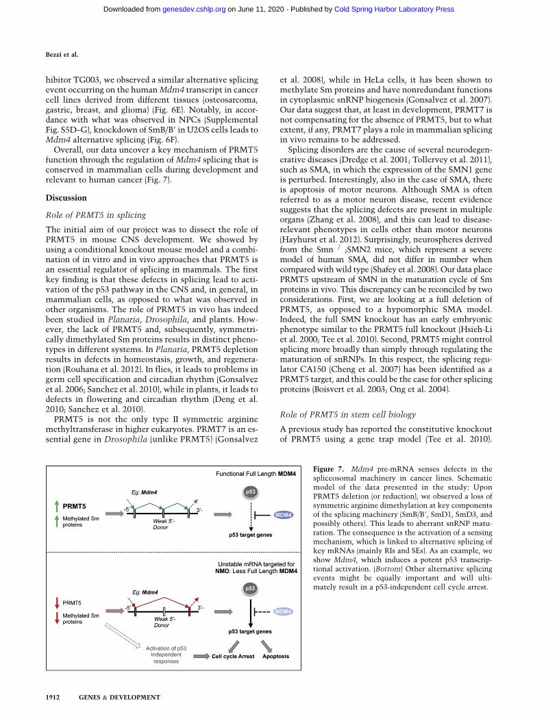
Overall, our data uncover a key mechanism of PRMT5function through the regulation of Mdm4 splicing that isconserved in mammalian cells during development andrelevant to human cancer (Fig. 7).
Discussion
Role of PRMT5 in splicing
The initial aim of our project was to dissect the role ofPRMT5 in mouse CNS development. We showed byusing a conditional knockout mouse model and a combi-nation of in vitro and in vivo approaches that PRMT5 isan essential regulator of splicing in mammals. The firstkey finding is that these defects in splicing lead to acti-vation of the p53 pathway in the CNS and, in general, inmammalian cells, as opposed to what was observed inother organisms. The role of PRMT5 in vivo has indeedbeen studied in Planaria, Drosophila, and plants. How-ever, the lack of PRMT5 and, subsequently, symmetri-cally dimethylated Sm proteins results in distinct pheno-types in different systems. In Planaria, PRMT5 depletionresults in defects in homeostasis, growth, and regenera-tion (Rouhana et al. 2012). In flies, it leads to problems ingerm cell specification and circadian rhythm (Gonsalvezet al. 2006; Sanchez et al. 2010), while in plants, it leads todefects in flowering and circadian rhythm (Deng et al.2010; Sanchez et al. 2010).
PRMT5 is not the only type II symmetric argininemethyltransferase in higher eukaryotes. PRMT7 is an es-sential gene in Drosophila (unlike PRMT5) (Gonsalvez
et al. 2008), while in HeLa cells, it has been shown tomethylate Sm proteins and have nonredundant functionsin cytoplasmic snRNP biogenesis (Gonsalvez et al. 2007).Our data suggest that, at least in development, PRMT7 isnot compensating for the absence of PRMT5, but to whatextent, if any, PRMT7 plays a role in mammalian splicingin vivo remains to be addressed.
Splicing disorders are the cause of several neurodegen-erative diseases (Dredge et al. 2001; Tollervey et al. 2011),such as SMA, in which the expression of the SMN1 geneis perturbed. Interestingly, also in the case of SMA, thereis apoptosis of motor neurons. Although SMA is oftenreferred to as a motor neuron disease, recent evidencesuggests that the splicing defects are present in multipleorgans (Zhang et al. 2008), and this can lead to disease-relevant phenotypes in cells other than motor neurons(Hayhurst et al. 2012). Surprisingly, neurospheres derivedfrom the Smn�/�;SMN2 mice, which represent a severemodel of human SMA, did not differ in number whencompared with wild type (Shafey et al. 2008). Our data placePRMT5 upstream of SMN in the maturation cycle of Smproteins in vivo. This discrepancy can be reconciled by twoconsiderations. First, we are looking at a full deletion ofPRMT5, as opposed to a hypomorphic SMA model.Indeed, the full SMN knockout has an early embryonicphenotype similar to the PRMT5 full knockout (Hsieh-Liet al. 2000; Tee et al. 2010). Second, PRMT5 might controlsplicing more broadly than simply through regulating thematuration of snRNPs. In this respect, the splicing regu-lator CA150 (Cheng et al. 2007) has been identified as aPRMT5 target, and this could be the case for other splicingproteins (Boisvert et al. 2003; Ong et al. 2004).
Role of PRMT5 in stem cell biology
A previous study has reported the constitutive knockoutof PRMT5 using a gene trap model (Tee et al. 2010).
Figure 7. Mdm4 pre-mRNA senses defects in thespliceosomal machinery in cancer lines. Schematicmodel of the data presented in the study: UponPRMT5 deletion (or reduction), we observed a loss ofsymmetric arginine dimethylation at key componentsof the splicing machinery (SmB/B9, SmD1, SmD3, andpossibly others). This leads to aberrant snRNP matu-ration. The consequence is the activation of a sensingmechanism, which is linked to alternative splicing ofkey mRNAs (mainly RIs and SEs). As an example, weshow Mdm4, which induces a potent p53 transcrip-tional activation. (Bottom) Other alternative splicingevents might be equally important and will ulti-mately result in a p53-independent cell cycle arrest.
Bezzi et al.
1912 GENES & DEVELOPMENT
Cold Spring Harbor Laboratory Press on June 11, 2020 - Published by genesdev.cshlp.orgDownloaded from
Phenotypically, lack of PRMT5 leads to derepression ofdifferentiation genes in mouse embryonic stem cells(mESCs) due, at least in part, to the lack of symmetricArg3 methylation on histone H2A (H2AR3me2s). How-ever, because of the early embryonic lethality, the in-vestigators were not able to perform large-scale molec-ular experiments, and thus whether PRMT5 plays anyrole in controlling splicing in mESCs remains to beexplored. It is of note that PRMT5 has been used to im-prove induced pluripotent stem cell (iPSC) derivation incombination with klf4 and Oct3/4 (Nagamatsu et al. 2011).Reducing p53 activity is known to be very important toenhance iPSC derivation (Hong et al. 2009; Kawamura et al.2009). We thus believe that our data, which link PRMT5methyltransferase activity to the regulation of MDM4abundance by controlling its alternative splicing, willprovide new insights into this expanding field of research.
Role of PRMT5 in regulating cell cycle progressionand cell death
We described here how cells can sense general defects inthe core splicing machinery (such as the one caused byPRMT5 depletion) by regulating the alternative splicingof a key p53 activator such as Mdm4. This alternativesplicing event reduces the full-length MDM4 protein andgives rise to the unstable MDM4S product (Lenos andJochemsen 2011), thus activating the p53 transcriptionalprogram. Our findings describe for the first time the linkbetween the methylosome (Friesen et al. 2001b), the coresplicing machinery, alternative splicing, and activation ofa p53 response in mammalian development.
What we uncovered here is indeed a much broaderpicture of how cells can activate the alternative splicingof sensor mRNAs (e.g., Mdm4) at key exons, character-ized by weak 59 donor sites. This occurs upon perturba-tion of the general splicing machinery—whether becauseof PRMT5 deletion or chemical inhibition (Fig. 5D)—toarrest growth and/or induce apoptosis. Besides Mdm4,there are other mRNAs that can potentially play a similarrole. To mention a few, the SE event observed in themRNA of 59 cap-binding protein eIF4E, which is a rate-limiting component in the translation process, couldaffect genes involved in apoptosis and cell cycle arrest(Supplemental Fig. S4D; Mamane et al. 2004), while theRI events observed in Dvl1 might lead to the inactivationof the Wnt/Dvl1/b-catenin signaling pathway, which isknown to support NPCs’ growth and self-renewal poten-tial (Supplemental Fig. S4E; Faigle and Song 2013).
We described here how the complete depletion of PRMT5in mouse cells leads to a minor induction of gH2AX buta strong activation of p53 target genes such as p21 evenwhen compared with etoposide. This is in contrast towhat has been observed previously using siRNA/shRNAstrategies to reduce the levels of PRMT5 in human cancerlines (Jansson et al. 2008; Scoumanne et al. 2009). Thisdiscrepancy could be due to differences between mouseand human cells and between primary cells and cancercells and the fact that PRMT5 levels have to fall belowa certain threshold in order to exhibit an activation of the
p53 pathway. The latter concept has already been ob-served for other splicing regulators such as SMN (Zhanget al. 2008).
Both the concept that perturbation of the core splicingmachinery can lead to regulation of alternative splicing(Saltzman et al. 2011) and the concept that apoptosis isregulated by alternative splicing (Schwerk and Schulze-Osthoff 2005; Moore et al. 2010) have been describedpreviously. The fundamental advance that we describehere is that the two pathways are directly linked becauseMdm4, the target of alternative splicing, unlike Bcl2-likefactors, caspases, death receptors, and proapoptotic li-gands, is as a direct upstream regulator of p53.
Splicing disorders have been estimated to occur in 50%of tumors (Ritchie et al. 2008; David and Manley 2010;Ward and Cooper 2010). These can contribute to tumorprogression by giving rise to alternative isoforms of on-cogenes or tumor suppressors. At the same time, whilethe perturbation of the splicing machinery was known toactivate the p53 pathway, the underlying mechanisms bywhich this occurred were unknown (Allende-Vega et al.2013). Our data uncover the mechanism of p53 activationby identifying Mdm4 as a key sensor mRNA. We believethese data to be extremely relevant for the entire p53field. Mdm4 is indeed up-regulated in several p53wt tu-mors (Gembarska et al. 2012), and our findings providenew therapeutic avenues to alter its protein levels byaffecting its splicing pattern.
In conclusion, this study expands our understanding ofthe complex network regulating correct splicing and cellfate decisions in mammalian development as well as inhuman cancer lines, providing new possibilities to targetthe arginine methyltransferase family to treat neurode-generative diseases (Dredge et al. 2001; Tollervey et al.2011) and cancer (Ritchie et al. 2008; David and Manley2010; Ward and Cooper 2010).
Materials and methods
Mouse strains and genotyping
The PRMT5 knockout first mice were obtained from theEuropean Conditional Mouse Mutagenesis Program (EUCOMM;http://www.knockoutmouse.org). To generate the PRMT5 FLOXallele, the bgal-neomycin cassette was removed by crossing PRMT5knockout first mice with b-actin-Flpe transgenic mice (Rodriguezet al. 2000) [strain name: B6.Cg-Tg(ACTFLPe) 9205Dym/J; stockno. 005703, The Jackson Laboratory]. These were then crossed toNestin-CRE [B6.Cg-Tg(Nes-cre)1Kln/J; JAX Laboratory]. 4-OHT-inducible conditional knockouts were created by crossing PRMT5F/F
mice with ER transgenic mice (Hameyer et al. 2007) in a mixedC57BL/6 3 129S1/SvlmJ background. Southern blotting wascarried out according to a standard protocol (Southern 2006).
Immunohistochemistry (IHC) staining
Automated IHC staining and counterstaining was performed onthe Leica Bond-Max autostainer. Detailed protocols are availablein the Supplemental Material.
Cell culture
NPCs Neurosphere cultures were established as previouslydescribed (Lim and Kaldis 2012). Briefly, E14.5 embryos were
PRMT5 role in mammalian development
GENES & DEVELOPMENT 1913
Cold Spring Harbor Laboratory Press on June 11, 2020 - Published by genesdev.cshlp.orgDownloaded from

harvested, and cortices were carefully dissected in ice-cold PBSand incubated in trypsin (Invitrogen, 25300120) for 10 min at37°C. The tissue was then mechanically dissociated, and singlecells were cultured into complete NSC medium (DMEM, LifeTechnologies, 11965118 + 2% B-27, Life Technologies, 17504-044), 1% penicillin/streptomycin (Life Technologies, 15140122),20 ng/mL recombinant human epidermal growth factor (EGF)(Peprotech, 100-15), and 20 ng/mL recombinant human fibro-blast growth factor-basic (FGF-2) (Peprotech, 100-18B). PRMT5F/F
ER day 4 neurospheres were treated with either 50 nM 4-OHT(Sigma, H7904) or the equivalent volume of EtOH for 24 h beforesplitting to induce PRMT5 knockout.
MEFs Primary MEFs were prepared from E14.5 embryos aspreviously described (Xu 2005) and maintained in a humidified5% CO2 atmosphere at 37°C in DMEM (Life Technologies,11965118) supplemented with 10% fetal bovine serum (FBS)(Hyclone, SH30070.03) and 1% penicillin/streptomycin (LifeTechnologies, 15140122). To induce PRMT5 knockout, MEFs(passage 1) were grown to confluence in 15-cm dishes, and either50 nM 4-OHT (H7904; Sigma) or the equivalent volume of EtOHwas added 24 h before splitting.
Human cell lines HEK293T, Phoenix-Eco, A549, U87, U2OS,and HCT116 were obtained from American Type Culture Collec-tion (ATCC) and propagated according to ATCC data sheets.
Microarray analysis
The expression data from quadruplicate Illumina MouseRef-8 V2microarrays were quantile-normalized, and only probes with anabsolute fold change >1.5-fold and a Q-value <0.01 were labeledas significantly differentially expressed.
RNA-seq library preparation and splicing analysis
For RNA-seq library preparation, we followed the IlluminaTruSeq RNA Sample Preparation kit version 2 manual. At least70 million, 51-base-pair (bp)-long paired-end reads were mappedto the NCBI37/mm9 version of the mouse genome per replicate.Genes were labeled as significantly differentially expressed if theP-value as called by cuffdiff (Trapnell et al. 2012) was <0.05.
To determine differential splicing events, MATS 3.0.6 beta(Shen et al. 2012) was used for counting junction reads and readsfalling into the tested region within ENSEMBL version 65 genedefinitions. Matching embryos were analyzed individually, andonly significant events occurring in at least two replicates wereconsidered. Splicing events were labeled significant if the sumof the reads supporting a specific event exceeded 10 reads, theP-value was <0.05, and the minimum inclusion level differenceas determined by MATS was >0.2. All sequencing and micro-array data have been submitted to the Gene Expression Omnibusrepository and are available under accession number GSE45285.
Functional annotation and network analysis
The functional annotation of the significant microarray andRNA-seq genes was performed with the Database for Annota-tion, Visualization, and Integrated Discovery (DAVID) (Huanget al. 2009) using Kyoto Encyclopedia of Genes and Genomics(KEGG) pathway (Kanehisa et al. 2012) representations. Networkrepresentations were generated using Cytoscape 3.0.1 (Shannonet al. 2003) with the plug-in ClueGO (Bindea et al. 2009). Onlygene ontology terms with at least five members and a k score>0.3 were used.
Polysome purification
Polysomes were isolated and separated as previously described(Zhang et al. 2012).
TAM injections
A single pulse of 2 mg of TAM (Sigma, T5648) plus 1 mg ofprogesterone (Sigma, P8783) in mineral oil (Sigma, M5904) wasgiven to pregnant females at E10.5. Embryos were harvested andanalyzed at E15.5 and E17.5.
Competing interest statement
The research of Z.Q.B. was funded by Lilly Research Laborato-ries, and he is an employee of Eli Lilly and Company.
Acknowledgments
We thank P.R. Kaldis, S.H. Lim, K. Diril, and U. Surana forsharing protocols and for helpful discussions. We thank L. Toraand P.R. Kaldis for critically reading the manuscript; K. Rogers,S. Rogers, and E.W. Sim for help with histopathology work; andM. Al-Haddawi for help with the pathology description of thePRMT5F/FNes brains. We thank P. Kaldis for sharing the Nestin-CRE and ER transgenic animals, V. Tergaonkar for sharing thep53-null mice, D. Lane and C.F. Cheok for the kind gift of Nutlin,Y. Minoru for the kind gift of Spliceostatin A, E. Makeyev forsharing the RFP-minigene backbone construct, and the BRCShared facilities for technical support. We are grateful to X. Ruanand the GIS Genome Sequencing Team for help with the Solexahigh-throughput sequencing, and the entire E.G. laboratory forcritical discussion. This work was supported by an AGA-SINGA(Singapore Graduate Award) fellowship to M.B. and by IMCB,A-STAR. E.G. acknowledges support from JCO-ASTAR grantsnumber 1134c001 and number 11/03/FG/07/04.
References
Akhtar RS, Geng Y, Klocke BJ, Latham CB, Villunger A,Michalak EM, Strasser A, Carroll SL, Roth KA. 2006. BH3-only proapoptotic Bcl-2 family members Noxa and Pumamediate neural precursor cell death. J Neurosci 26: 7257–7264.
Allende-Vega N, Dayal S, Agarwala U, Sparks A, Bourdon JC,Saville MK. 2013. p53 is activated in response to disruptionof the pre-mRNA splicing machinery. Oncogene 32: 1–14.
Barbosa-Morais NL, Irimia M, Pan Q, Xiong HY, Gueroussov S,Lee LJ, Slobodeniuc V, Kutter C, Watt S, Colak R, et al. 2012.The evolutionary landscape of alternative splicing in verte-brate species. Science 338: 1587–1593.
Bedford MT, Clarke SG. 2009. Protein arginine methylation inmammals: Who, what, and why. Mol Cell 33: 1–13.
Bindea G, Mlecnik B, Hackl H, Charoentong P, Tosolini M,Kirilovsky A, Fridman WH, Pages F, Trajanoski Z, GalonJ. 2009. ClueGO: A Cytoscape plug-in to decipher function-ally grouped gene ontology and pathway annotation net-works. Bioinformatics 25: 1091–1093.
Boisvert FM, Cote J, Boulanger MC, Richard S. 2003. A proteo-mic analysis of arginine-methylated protein complexes. MolCell Proteomics 2: 1319–1330.
Cheng D, Cote J, Shaaban S, Bedford MT. 2007. The argininemethyltransferase CARM1 regulates the coupling of tran-scription and mRNA processing. Mol Cell 25: 71–83.
Chiu SY, Lejeune F, Ranganathan AC, Maquat LE. 2004. Thepioneer translation initiation complex is functionally distinct
Bezzi et al.
1914 GENES & DEVELOPMENT
Cold Spring Harbor Laboratory Press on June 11, 2020 - Published by genesdev.cshlp.orgDownloaded from

from but structurally overlaps with the steady-state trans-lation initiation complex. Genes Dev 18: 745–754.
Da Cruz S, Cleveland DW. 2011. Understanding the role of TDP-43 and FUS/TLS in ALS and beyond. Curr Opin Neurobiol
21: 904–919.David CJ, Manley JL. 2010. Alternative pre-mRNA splicing
regulation in cancer: Pathways and programs unhinged.Genes Dev 24: 2343–2364.
Deng X, Gu L, Liu C, Lu T, Lu F, Lu Z, Cui P, Pei Y, Wang B,Hu S, et al. 2010. Arginine methylation mediated by theArabidopsis homolog of PRMT5 is essential for proper pre-mRNA splicing. Proc Natl Acad Sci 107: 19114–19119.
Dillman AA, Hauser DN, Gibbs JR, Nalls MA, McCoy MK,Rudenko IN, Galter D, Cookson MR. 2013. mRNA expres-sion, splicing and editing in the embryonic and adult mousecerebral cortex. Nat Neurosci 16: 499–506.
Doumont G, Martoriati A, Beekman C, Bogaerts S, Mee PJ,Bureau F, Colombo E, Alcalay M, Bellefroid E, Marchesi F,et al. 2005. G1 checkpoint failure and increased tumor sus-ceptibility in mice lacking the novel p53 target Ptprv. EMBOJ 24: 3093–3103.
Dredge BK, Polydorides AD, Darnell RB. 2001. The splice oflife: Alternative splicing and neurological disease. Nat Rev
Neurosci 2: 43–50.Faigle R, Song H. 2013. Signaling mechanisms regulating adult
neural stem cells and neurogenesis. Biochim Biophys Acta
1830: 2435–2448.Francoz S, Froment P, Bogaerts S, De Clercq S, Maetens M,
Doumont G, Bellefroid E, Marine JC. 2006. Mdm4 andMdm2 cooperate to inhibit p53 activity in proliferating andquiescent cells in vivo. Proc Natl Acad Sci 103: 3232–3237.
Freund M, Asang C, Kammler S, Konermann C, Krummheuer J,Hipp M, Meyer I, Gierling W, Theiss S, Preuss T, et al. 2003.A novel approach to describe a U1 snRNA binding site.Nucleic Acids Res 31: 6963–6975.
Friesen WJ, Massenet S, Paushkin S, Wyce A, Dreyfuss G. 2001a.SMN, the product of the spinal muscular atrophy gene, bindspreferentially to dimethylarginine-containing protein tar-gets. Mol Cell 7: 1111–1117.
Friesen WJ, Paushkin S, Wyce A, Massenet S, Pesiridis GS, VanDuyne G, Rappsilber J, Mann M, Dreyfuss G. 2001b. Themethylosome, a 20S complex containing JBP1 and pICln,produces dimethylarginine-modified Sm proteins. Mol Cell
Biol 21: 8289–8300.Friesen WJ, Wyce A, Paushkin S, Abel L, Rappsilber J, Mann M,
Dreyfuss G. 2002. A novel WD repeat protein component ofthe methylosome binds Sm proteins. J Biol Chem 277: 8243–8247.
Gembarska A, Luciani F, Fedele C, Russell EA, Dewaele M,Villar S, Zwolinska A, Haupt S, de Lange J, Yip D, et al. 2012.MDM4 is a key therapeutic target in cutaneous melanoma.Nat Med 18: 1239–1247.
Gonsalvez GB, Rajendra TK, Tian L, Matera AG. 2006. TheSm-protein methyltransferase, dart5, is essential for germ-cell specification and maintenance. Curr Biol 16: 1077–1089.
Gonsalvez GB, Tian L, Ospina JK, Boisvert FM, Lamond AI,Matera AG. 2007. Two distinct arginine methyltransferasesare required for biogenesis of Sm-class ribonucleoproteins.J Cell Biol 178: 733–740.
Gonsalvez GB, Praveen K, Hicks AJ, Tian L, Matera AG. 2008.Sm protein methylation is dispensable for snRNP assemblyin Drosophila melanogaster. RNA 14: 878–887.
Graus-Porta D, Blaess S, Senften M, Littlewood-Evans A, DamskyC, Huang Z, Orban P, Klein R, Schittny JC, Muller U. 2001.b1-class integrins regulate the development of laminae and
folia in the cerebral and cerebellar cortex. Neuron 31: 367–379.
Hameyer D, Loonstra A, Eshkind L, Schmitt S, Antunes C,Groen A, Bindels E, Jonkers J, Krimpenfort P, Meuwissen R,et al. 2007. Toxicity of ligand-dependent Cre recombinasesand generation of a conditional Cre deleter mouse allowingmosaic recombination in peripheral tissues. Physiol Geno-
mics 31: 32–41.Hayhurst M, Wagner AK, Cerletti M, Wagers AJ, Rubin LL.
2012. A cell-autonomous defect in skeletal muscle satellitecells expressing low levels of survival of motor neuronprotein. Dev Biol 368: 323–334.
Hong H, Takahashi K, Ichisaka T, Aoi T, Kanagawa O, NakagawaM, Okita K, Yamanaka S. 2009. Suppression of induced plu-ripotent stem cell generation by the p53–p21 pathway. Nature
460: 1132–1135.Hsieh-Li HM, Chang JG, Jong YJ, Wu MH, Wang NM, Tsai CH,
Li H. 2000. A mouse model for spinal muscular atrophy. Nat
Genet 24: 66–70.Huang DW, Sherman BT, Lempicki RA. 2009. Systematic and
integrative analysis of large gene lists using DAVID bioin-formatics resources. Nat Protoc 4: 44–57.
Jansson M, Durant ST, Cho EC, Sheahan S, Edelmann M,Kessler B, La Thangue NB. 2008. Arginine methylationregulates the p53 response. Nat Cell Biol 10: 1431–1439.
Kaida D, Motoyoshi H, Tashiro E, Nojima T, Hagiwara M,Ishigami K, Watanabe H, Kitahara T, Yoshida T, NakajimaH, et al. 2007. Spliceostatin A targets SF3b and inhibits bothsplicing and nuclear retention of pre-mRNA. Nat Chem Biol
3: 576–583.Kanehisa M, Goto S, Sato Y, Furumichi M, Tanabe M. 2012.
KEGG for integration and interpretation of large-scale mo-lecular data sets. Nucleic Acids Res 40: D109–D114.
Karkhanis V, Hu YJ, Baiocchi RA, Imbalzano AN, Sif S. 2011.Versatility of PRMT5-induced methylation in growthcontrol and development. Trends Biochem Sci 36: 633–641.
Kawamura T, Suzuki J, Wang YV, Menendez S, Morera LB, RayaA, Wahl GM, Izpisua Belmonte JC. 2009. Linking the p53tumour suppressor pathway to somatic cell reprogramming.Nature 460: 1140–1144.
Kouzarides T. 2007. Chromatin modifications and their func-tion. Cell 128: 693–705.
Kuida K, Zheng TS, Na S, Kuan C, Yang D, Karasuyama H,Rakic P, Flavell RA. 1996. Decreased apoptosis in the brainand premature lethality in CPP32-deficient mice. Nature384: 368–372.
Lane DP. 1992. Cancer. p53, guardian of the genome. Nature
358: 15–16.Lenos K, Jochemsen AG. 2011. Functions of MDMX in the
modulation of the p53-response. J Biomed Biotechnol 2011:876173.
Lim S, Kaldis P. 2012. Loss of Cdk2 and Cdk4 induces a switchfrom proliferation to differentiation in neural stem cells.Stem Cells 30: 1509–1520.
Mamane Y, Petroulakis E, Rong L, Yoshida K, Ler LW, SonenbergN. 2004. eIF4E—from translation to transformation. Onco-gene 23: 3172–3179.
Meister G, Eggert C, Buhler D, Brahms H, Kambach C, FischerU. 2001. Methylation of Sm proteins by a complex contain-ing PRMT5 and the putative U snRNP assembly factorpICln. Curr Biol 11: 1990–1994.
Migliori V, Phalke S, Bezzi M, Guccione E. 2010. Arginine/lysine-methyl/methyl switches: Biochemical role of histonearginine methylation in transcriptional regulation. Epige-nomics 2: 119–137.
PRMT5 role in mammalian development
GENES & DEVELOPMENT 1915
Cold Spring Harbor Laboratory Press on June 11, 2020 - Published by genesdev.cshlp.orgDownloaded from
Moore MJ, Wang Q, Kennedy CJ, Silver PA. 2010. An alternativesplicing network links cell-cycle control to apoptosis. Cell142: 625–636.
Muraki M, Ohkawara B, Hosoya T, Onogi H, Koizumi J,Koizumi T, Sumi K, Yomoda J, Murray MV, Kimura H,et al. 2004. Manipulation of alternative splicing by a newlydeveloped inhibitor of Clks. J Biol Chem 279: 24246–24254.
Nagamatsu G, Kosaka T, Kawasumi M, Kinoshita T, Takubo K,Akiyama H, Sudo T, Kobayashi T, Oya M, Suda T. 2011. Agerm cell-specific gene, Prmt5, works in somatic cell repro-gramming. J Biol Chem 286: 10641–10648.
Ong SE, Mittler G, Mann M. 2004. Identifying and quantifyingin vivo methylation sites by heavy methyl SILAC. NatMethods 1: 119–126.
Rallapalli R, Strachan G, Tuan RS, Hall DJ. 2003. Identificationof a domain within MDMX-S that is responsible for its highaffinity interaction with p53 and high-level expression inmammalian cells. J Cell Biochem 89: 563–575.
Ritchie W, Granjeaud S, Puthier D, Gautheret D. 2008. Entropymeasures quantify global splicing disorders in cancer. PLoSComput Biol 4: e1000011.
Rodriguez CI, Buchholz F, Galloway J, Sequerra R, Kasper J,Ayala R, Stewart AF, Dymecki SM. 2000. High-efficiencydeleter mice show that FLPe is an alternative to Cre-loxP.Nat Genet 25: 139–140.
Rouhana L, Vieira AP, Roberts-Galbraith RH, Newmark PA.2012. PRMT5 and the role of symmetrical dimethylargininein chromatoid bodies of planarian stem cells. Development139: 1083–1094.
Saltzman AL, Pan Q, Blencowe BJ. 2011. Regulation of alterna-tive splicing by the core spliceosomal machinery. Genes Dev25: 373–384.
Sanchez SE, Petrillo E, Beckwith EJ, Zhang X, Rugnone ML,Hernando CE, Cuevas JC, Godoy Herz MA, Depetris-ChauvinA, Simpson CG, et al. 2010. A methyl transferase links thecircadian clock to the regulation of alternative splicing.Nature 468: 112–116.
Schwerk C, Schulze-Osthoff K. 2005. Regulation of apoptosis byalternative pre-mRNA splicing. Mol Cell 19: 1–13.
Scoumanne A, Zhang J, Chen X. 2009. PRMT5 is required forcell-cycle progression and p53 tumor suppressor function.Nucleic Acids Res 37: 4965–4976.
Shafey D, MacKenzie AE, Kothary R. 2008. Neurodevelop-mental abnormalities in neurosphere-derived neural stemcells from SMN-depleted mice. J Neurosci Res 86: 2839–2847.
Shannon P, Markiel A, Ozier O, Baliga NS, Wang JT, Ramage D,Amin N, Schwikowski B, Ideker T. 2003. Cytoscape: Asoftware environment for integrated models of biomolecularinteraction networks. Genome Res 13: 2498–2504.
Shapiro MB, Senapathy P. 1987. RNA splice junctions of differentclasses of eukaryotes: Sequence statistics and functional im-plications in gene expression. Nucleic Acids Res 15: 7155–7174.
Shen S, Park JW, Huang J, Dittmar KA, Lu ZX, Zhou Q, CarstensRP, Xing Y. 2012. MATS: A Bayesian framework for flexibledetection of differential alternative splicing from RNA-seqdata. Nucleic Acids Res 40: e61.
Southern E. 2006. Southern blotting. Nat Protoc 1: 518–525.Tee WW, Pardo M, Theunissen TW, Yu L, Choudhary JS,
Hajkova P, Surani MA. 2010. Prmt5 is essential for earlymouse development and acts in the cytoplasm to maintainES cell pluripotency. Genes Dev 24: 2772–2777.
Tollervey JR, Wang Z, Hortobagyi T, Witten JT, Zarnack K,Kayikci M, Clark TA, Schweitzer AC, Rot G, Curk T, et al.2011. Analysis of alternative splicing associated with aging
and neurodegeneration in the human brain. Genome Res 21:1572–1582.
Trapnell C, Hendrickson DG, Sauvageau M, Goff L, Rinn JL,Pachter L. 2012. Differential analysis of gene regulation attranscript resolution with RNA-seq. Nat Biotechnol 31:46–53.
Vance C, Rogelj B, Hortobagyi T, De Vos KJ, Nishimura AL,Sreedharan J, Hu X, Smith B, Ruddy D, Wright P, et al. 2009.Mutations in FUS, an RNA processing protein, cause familialamyotrophic lateral sclerosis type 6. Science 323: 1208–1211.
Ward AJ, Cooper TA. 2010. The pathobiology of splicing.J Pathol 220: 152–163.
White JK, Gerdin AK, Karp NA, Ryder E, Buljan M, Bussell JN,Salisbury J, Clare S, Ingham NJ, Podrini C, et al. 2013.Genome-wide generation and systematic phenotyping ofknockout mice reveals new roles for many genes. Cell 154:452–464.
Xiong S, Van Pelt CS, Elizondo-Fraire AC, Liu G, Lozano G.2006. Synergistic roles of Mdm2 and Mdm4 for p53 in-hibition in central nervous system development. Proc NatlAcad Sci 103: 3226–3231.
Xu J. 2005. Preparation, culture, and immortalization of mouseembryonic fibroblasts. Curr Protoc Mol Biol 70: 28.1.1–28.1.8.
Yeo G, Burge CB. 2004. Maximum entropy modeling of shortsequence motifs with applications to RNA splicing signals.J Comput Biol 11: 377–394.
Zhang Z, Lotti F, Dittmar K, Younis I, Wan L, Kasim M, DreyfussG. 2008. SMN deficiency causes tissue-specific perturbationsin the repertoire of snRNAs and widespread defects in splicing.Cell 133: 585–600.
Zhang D, Zhao T, Ang HS, Chong P, Saiki R, Igarashi K, Yang H,Vardy LA. 2012. AMD1 is essential for ESC self-renewal andis translationally down-regulated on differentiation to neuralprecursor cells. Genes Dev 26: 461–473.
Bezzi et al.
1916 GENES & DEVELOPMENT
Cold Spring Harbor Laboratory Press on June 11, 2020 - Published by genesdev.cshlp.orgDownloaded from

10.1101/gad.219899.113Access the most recent version at doi: 27:2013, Genes Dev.
Marco Bezzi, Shun Xie Teo, Julius Muller, et al. machinery
pre-mRNA in sensing defects in the spliceosomalMdm4role for Regulation of constitutive and alternative splicing by PRMT5 reveals a
Material
Supplemental
http://genesdev.cshlp.org/content/suppl/2013/09/06/27.17.1903.DC1
References
http://genesdev.cshlp.org/content/27/17/1903.full.html#ref-list-1
This article cites 71 articles, 21 of which can be accessed free at:
License
Commons Creative
.http://creativecommons.org/licenses/by-nc/3.0/License (Attribution-NonCommercial 3.0 Unported), as described at
, is available under a Creative CommonsGenes & DevelopmentThis article, published in
ServiceEmail Alerting
click here.right corner of the article or
Receive free email alerts when new articles cite this article - sign up in the box at the top
© 2013, Published by Cold Spring Harbor Laboratory Press
Cold Spring Harbor Laboratory Press on June 11, 2020 - Published by genesdev.cshlp.orgDownloaded from