regulation of the cell cycle and centrosome biology by
TRANSCRIPT
Regulation of the cell cycle and centrosome biology by deubiquitylases.
Sarah Darling, Andrew B Fielding, Dorota Sabat-Pospiech, Ian A Prior, Judy M
Coulson*
Cellular and Molecular Physiology, Institute of Translational Medicine, University of
Liverpool, Liverpool, L69 3BX, UK
*Corresponding author: Cellular & Molecular Physiology, Institute of Translational
Medicine, University of Liverpool, Crown St, Liverpool, L69 3BX, UK. Phone: 44-151-
794-5850, Fax: 44-151-794-4434, E-mail: [email protected].
Running title: DUBs in the cell and centrosome cycles
Key words: ubiquitin, post-translational modification, centrosomes, cell cycle, cancer
Subject area: Cell Cycle, Growth & Proliferation, Cancer, Molecular Bases of Health
& Disease, Organelles & Localization, Post-Translational Modifications
brought to you by COREView metadata, citation and similar papers at core.ac.uk
provided by University of Liverpool Repository

Abstract
Post-translational modification of proteins by ubiquitylation is increasingly recognised
as a highly complex code that contributes to the regulation of diverse cellular
processes. In humans, a family of almost one hundred deubiquitylase enzymes
(DUBs) are assigned to six sub-families and many of these DUBs can remove
ubiquitin from proteins to reverse signals. Roles for individual DUBs have been
delineated within specific cellular processes, including many that are dysregulated in
diseases, particularly cancer. As potentially druggable enzymes, disease-associated
DUBs are of increasing interest as pharmaceutical targets. The biology, structure and
regulation of DUBs have been extensively reviewed elsewhere, so here we focus
specifically on roles of DUBs in regulating cell cycle processes in mammalian cells.
Over a quarter of all DUBs, representing four different families, have been shown to
play roles either in the unidirectional progression of the cell cycle through specific
checkpoints, or in the DNA damage response and repair pathways. We catalogue
these roles and discuss specific examples. Centrosomes are the major microtubule
nucleating centres within a cell, and play a key role in forming the bipolar mitotic
spindle required to accurately divide genetic material between daughter cells during
cell division. To enable this mitotic role, centrosomes undergo a complex replication
cycle that is intimately linked to the cell division cycle. Here we also catalogue and
discuss DUBs that have been linked to centrosome replication or function, including
centrosome clustering, a mitotic survival strategy unique to cancer cells with
supernumerary centrosomes.

Reversible ubiquitylation
The post-translational attachment of ubiquitin moieties to substrate proteins, termed
ubiquitylation, involves the covalent conjugation of ubiquitin, most commonly to lysine
(K) residues. In the simplest form, ubiquitylation is the addition of an ubiquitin
monomer, termed monoubiquitylation. However, the ubiquitin signal can be highly
complex and is linked to a plethora of cellular processes (1, 2). Polyubiquitin chains
linked through K48 target proteins for proteasomal degradation. However, ubiquitin
possesses seven lysine residues (K6, K11, K27, K29, K33, K48 and K63) enabling
the formation of diverse polyubiquitin chains that may be homotypic or heterotypic in
nature, and can have alternative functions, as comprehensively reviewed in (3). The
world of ubiquitylation is multifaceted and each layer relies upon families of proteins
to write, read or erase this ubiquitin code. The steps to write the ubiquitin code are
highly conserved, relying on an E1 ubiquitin activating enzyme, an E2 ubiquitin
conjugating enzyme and an E3 ubiquitin protein ligase with substrate specificity. As
reviewed in (1), the human genome encodes two ubiquitin E1 enzymes,
approximately forty E2 enzymes and more than six hundred E3 ligases, a clear
depiction of the complexity involved in functional ubiquitylation.
Ubiquitylation is a reversible post-translational modification, with removal of the
ubiquitin signal catalysed by deubiquitylase enzymes (DUBs). The human genome
encodes approximately one hundred DUBs, that we refer to here as the DUBome.
These DUBs belong to six families: the ubiquitin specific protease (USP), ubiquitin C-
terminal hydrolase (UCH), ovarian tumour protease (OTU), Josephin (JOS),
JAB1/MPN/MOV34 (JAMM) families (4), or the newly discovered motif-interacting
with Ub (MIU)-containing novel DUB (MINDY) family (5). As reviewed in (4), whilst
most DUB families are thiol proteases harbouring a catalytic triad, the JAMM
metalloproteases require a zinc ion to facilitate ubiquitin chain removal. As editors of
ubiquitin signalling, DUBs are regulators of varied essential cellular processes,
notably many have been assigned roles in DNA damage repair and cell cycle
progression. As these processes are often dysregulated in cancer, DUBs, as
potentially druggable enzymes, have quickly become the focus of several
pharmaceutical companies vying to develop new cancer therapies.
The cell division cycle
The cell cycle coordinates cellular events to duplicate the genetic material and divide
the cellular contents to create two identical daughter cells. The cycle comprises four
stages. After division, cells undergo an initial growth phase (G1), followed by the

replication of the genome (S-phase). A second growth phase (G2) prepares the cell
for division and assembles cytoskeletal structures, before the genetic material
divides between the daughter cells during mitosis (M). The unidirectional progression
through these cell cycle phases is dependent upon the periodic activation and
inactivation of substrate proteins by kinases (including CDKs and PLKs) and
ubiquitin-mediated degradation of key effectors by E3 ligases (including the APC/C
and SCF complexes). Accordingly, cell cycle effectors are regulated through protein-
protein interactions, phosphorylation-dependent activation and ubiquitylation-
dependent degradation, all working in concert to achieve an exquisite level of control
throughout the cycle (6). Once initiated, the cell cycle can be viewed as a series of
autonomic cellular events that cascade until the eventual division into two daughter
cells. However, checkpoints are inherent in the system, to temporarily halt the cell
cycle if conditions are unfavourable. Many DUBs have direct or indirect roles during
the cell cycle (7-9). We discuss here selected examples of those regulating cell
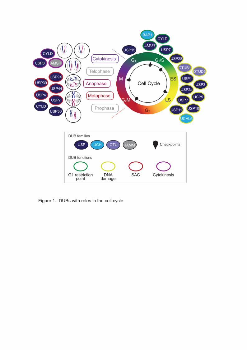
cycle progression and checkpoint maintenance, as summarised in Figure 1.
DUBs and the G1 restriction point
Upon entering G1, cells are not committed to a subsequent round of cell division, as
entry into the cell cycle requires sufficient mitogenic signalling to overcome a
restriction point in late G1. Rb, the mediator of this restriction point, inhibits the E2F
transcription factor during G1 (10). Upon CDK4/6 activation by the G1-cyclin CCND
and then by CCNE, Rb is increasingly phosphorylated. This results in progression
through the restriction point as hyperphosphorylated Rb dissociates from E2F,
causing the transcription of S-phase genes (importantly CCNE and CCNA) (11). In
addition to phosphorylation, Rb is regulated by ubiquitylation, being a target of the E3
ligase MDM2 (12, 13) which promotes its proteasomal degradation (14). The DUB
USP7 directly antagonises MDM2-mediated polyubiquitylation of Rb, stalling the cell
cycle in G1 (15). USP7 is not the only DUB that may govern the restriction point, the
tumour suppressor BAP1 also indirectly regulates the activity of E2F, via the
deubiquitylation of HCF-1, an important transcriptional co-regulator at E2F promoter
sites (16, 17). CYLD, another well-established tumour suppressor, plays a protective
role during G1 via the transcription factor BCL-3. CYLD deubiquitylates BCL-3
inhibiting its nuclear translocation and so decreases the transcription of BCL-3 target
genes including CCND (18). CYLD therefore indirectly decreases CCND levels
preventing cells from passing through the restriction point. During G1, the APC/C
polyubiquitylates the S-phase cyclin CCNA, targeting it for degradation in order to
prevent the cell from entering S-phase. The DUB USP37 directly regulates S-phase
entry through antagonising activity of the APC/CCDH1 in G1 by removing polyubiquitin
chains to stabilise CCNA (19).
DUBs and DNA damage checkpoints
Key to successful cell division is maintaining the integrity of the genome during DNA
replication in S-phase, and this is monitored by a number of quality control
mechanisms. If DNA becomes damaged, checkpoints stall the cell cycle and activate
DNA damage repair (DDR) pathways. This response revolves around p53, which is
stabilised and activated by DNA damage checkpoint signalling following a range of
genotoxic insults. Under normal conditions, p53 is continuously synthesized but
maintained at a low level by MDM2 polyubiquitylation targeting p53 for proteasomal
degradation (20). Under genotoxic stress, these regulatory mechanisms are
reversed, to allow p53 to stall the cell cycle to enable repair or trigger apoptosis. At
sites of DNA damage, sensors (e.g. 53BP1) facilitate the activation of DNA damage
kinases (notably ATM and CHK2) resulting in p53 phosphorylation. This abolishes
the interaction between p53 and MDM2, increasing p53 levels and inducing
transcription of p53 target genes (21), as well as activating transcription-independent
roles of p53 in many of the major DDR pathways (22).
Given the integral role of p53 in cell cycle fate, it is perhaps unsurprising that many
DUBs have been highlighted as direct or indirect p53 regulators, including USP2a,
USP5, USP7 (HAUSP), USP10, USP11, USP28, OTUB1 and OTUD5. USP7, a
predominantly nuclear DUB, was the first to be associated with the p53-dependent
DDR via directly antagonising MDM2 polyubiquitylation of p53 (23). However, USP7
also directly deubiquitylates the auto-polyubiquitylated MDM2, stabilising the E3
ligase as well as its substrate (24). Although this may seem counterintuitive, USP7
exhibits a preference for MDM2 over p53 in unstressed cells ensuring p53 levels are
maintained at a low level. Upon DNA damage, USP7 is dephosphorylated by PPM1G
reducing activity towards MDM2, leading to increased auto-polyubiquitylation and
degradation of MDM2, and the subsequent accumulation of p53 (25).
Other DUBs, including USP10, USP11 and OTUD5, also directly interact with,
deubiquitylate and stabilise p53. Interestingly USP10, a predominantly cytoplasmic
DUB, is involved in homeostasis of cytoplasmic p53 in unstressed cells, but following
DNA damage a fraction of USP10 can translocate into the nucleus where it
contributes to p53 activation (26). As described for USP7, other DUBs indirectly
control p53 levels via MDM2. For example, USP2a negatively regulates p53 levels

through the stabilisation of MDM2, whilst exhibiting no deubiquitylating activity
towards p53 directly (27). OTUB1, another cytoplasmic DUB, can directly interact
with p53, but predominantly stabilises p53 indirectly in the cytoplasm, through a non-
catalytic mechanism. OTUB1 does this by binding and supressing polyubiquitylation
through the MDM2 associated E2 enzyme UbcH5 (28). In contrast, USP28 was
shown to interact with and stabilise both the damage sensor 53BP1 and the
checkpoint kinase CHK2, that activate p53 under genotoxic conditions (29). USP5
uses perhaps the most indirect mechanism to stabilise p53 without physically
interacting with components of the p53-MDM2 axis. It primarily disassembles
unanchored polyubiquitin chains, and loss of USP5 results in accumulation of these
chains that compete with ubiquitylated p53, but not MDM2, for proteasome
recognition and degradation so that p53 is selectively stabilised (30).
In addition, many DUBs have also been associated with executing specific DDR
pathways (8). For example, USP1 can support repair through both the Fanconi
anaemia and translesion repair pathways (31). An RNAi-based study has linked
USP3 with double-strand DNA break repair; USP3 directly interacts with and
removes monoubiquitylation from histones H2A and H2B, and possibly other DDR
effectors, to coordinate DNA repair (32). Some DUBs exhibit a more global effect on
DDR pathways, for example one screen revealed that UCHL5 was recruited to sites
of DNA damage in addition to being involved in double-strand break resection (33).
DUBs with roles in mitotic progression and cytokinesis
Following replication of the genome, and assuming checkpoints are satisfied in G2,
the cell enters mitosis, where the newly replicated sister chromatids must be divided
into each daughter cell. To achieve this, the cell passes through a sequence of
distinct mitotic phases: prophase, metaphase, anaphase, telophase and cytokinesis
(Figure 1). Prior to mitosis, the mitotic kinase CDK1 is held in an inactivate state by
WEE1 phosphorylation, until SCFβTrCP-mediated ubiquitylation and degradation of
WEE1 triggers mitotic entry; USP50 can repress mitotic entry through stabilising
WEE1 (34). Subsequently, USP7 can indirectly regulate the levels of Aurora A, a
kinase required for correct maturation of the bipolar mitotic spindle, by stabilising
CHFR, an E3 ligase that targets Aurora A for degradation (35).
USP44 was one of the first DUBs to be linked to mitotic progression, with a role in
metaphase-anaphase transition (36). Anaphase entry is stimulated by the APC/C
and results in the separation of sister chromatids. To ensure the correct chromosome

complement is distributed to each daughter cell, the spindle assembly checkpoint
(SAC) monitors attachment of each chromosome pair to opposite poles of the mitotic
spindle. Anaphase is arrested until the SAC is satisfied, preventing premature and
inaccurate division of genomic content. Three key proteins, MAD2, BUBR1 and
BUB3, comprise the mitotic checkpoint complex (MCC) (37). The MCC sequesters
the APC/C activator CDC20 at unattached chromosomes, thus inhibiting the APC/C
until chromosomes are correctly attached (37). Once the SAC is satisfied, CDC20 is
ubiquitylated and subsequently dissociates from the MCC to activate the APC/C (38).
USP44 plays a protective role at the SAC, directly antagonising CDC20
ubiquitylation, and so promoting MCC inhibition of the APC/C (36). Once the SAC is
satisfied, USP44 dephosphorylation decreases its activity towards CDC20, initiating
mitotic exit through APC/C activation (39).
USP44 is not the only DUB that contributes to regulation of the SAC, for example
USP39 and USP9X are also essential for the correct alignment of chromosomes at
the mitotic spindle and their accurate division during anaphase. The mitotic kinase
Aurora B is a key regulator of the attachment of sister chromatids to microtubules in
the mitotic spindle. It exists in a complex with Survivin, the ubiquitylation status of
which mediates interaction of the complex with chromosomes (40). Depletion of
USP39 results in decreased transcription and consequently lower levels of Aurora B
kinase in cycling cells (41), whilst USP9X-mediated deubiquitylation of Survivin is
required for dissociation from the chromosomes once correctly aligned (42). Another
DUB, USP4, plays an indirect role in the SAC through regulating correct splicing of
mRNA transcripts, including for the mitotic checkpoint kinase BUB1 (43).
The DUB CYLD plays roles during both metaphase and cytokinesis. CYLD directly
interacts with the catalytic domain of HDAC6, inhibiting alpha-tubulin deacetylation
and therefore indirectly increasing the stability of microtubules. This governance of
microtubule stability by CYLD plays a role in spindle orientation during metaphase
(44) and regulates the rate of cytokinesis (45). Lastly, USP8 and AMSH, two DUBs
that are usually recruited to endosomes, have an important role in cytokinesis. The
scission of the two daughter cells requires components of the ESCRT machinery
including VAMP8, which co-localises with, and is deubiquitylated by, both USP8 and
AMSH during cytokinesis (46).

The centrosome cycle
Centrosomes are cytoplasmic organelles which act as the dominant microtubule-
organizing centres (MTOCs) in animal cells. During the cell cycle, centrosomes
determine spatial arrangement of the microtubule arrays to influence cell shape,
polarity, motility and organization of the mitotic spindle (47, 48). The core
components are two centrioles, small barrel-shaped organelles that are embedded in
pericentriolar material (PCM). Each centriole consists of nine microtubule triplets
arranged in a highly conserved rotational symmetry, imparted by SAS-6 during
centriole assembly (49, 50). The PCM is a dense protein matrix composed of various
proteins and exhibiting a high level of spatial organization, its major function is
recruitment of gamma-tubulin complexes which are essential for microtubule
nucleation (51, 52).
Centrosome replication is strictly coordinated with cell cycle progression (Figure 2).
Duplication of the single G1 centrosome begins at the G1/S transition and is
completed during S-phase so that two centrosomes are present in G2. These
facilitate bipolar spindle formation at metaphase and are then segregated, one into
each daughter cell, during cytokinesis (53, 54). Key to centrosome replication is
centriole duplication, as the pre-existing mother centriole duplicates itself to form a
daughter centriole. The kinase PLK4 and two SCF ubiquitin E3 ligases ensure that
only a single replication event normally occurs. SCFFBXW5 ubiquitylates SAS-6 to
target it for proteasomal degradation, preventing centriole over-duplication. SCFFBXW5
activity is limited by PLK4 to prevent premature SAS-6 degradation. Following G1/S
transition, PLK4 homodimerises and trans-autophosphorylates, signalling recruitment
of SCFβTrCP which ubiquitylates and degrades PLK4. Decreased PLK4 levels restore
SCFFBXW5 activity and block re-duplication (49, 55-58). Once duplicated, the daughter
centriole elongates during S-phase and G2. This process is controlled by several
genes including the multifunctional centriolar protein CP110, which becomes
ubiquitylated by SCFcyclinF during G2 and mitosis. Centrosomes then undergo a
maturation process which requires recruitment of PCM. Finally, the centrosomes
separate during G2, through KIF11 kinesin activity, which also facilitates bipolar
spindle formation during mitosis (49, 59, 60).
Many human cells also display cilia in a cell cycle-dependent manner. During G1 (or
G0 in terminally-differentiated cells) centrosomes migrate to the cell cortex, where the
mother centriole matures into a basal body which acts as a template for cilia
elongation. During S-phase, both mother and daughter centrioles undergo
duplication as normal. Then, prior to mitosis, cilia disassemble and the centrioles
migrate back to the cell interior, ready to act as spindle poles during mitosis (61).
Various cell division errors, such as centrosome over-duplication, cytokinesis failure,
or cell fusion can cause centrosome amplification, which is observed in many human
cancers. The notion that, in addition to acting as MTOCs, centrosomes may function
as signalling hubs (62) suggests one way in which amplification of centrosomes may
benefit cancer cells. However, supernumerary centrosomes may cause multipolar
spindle formation, impaired cell division, aneuploidy and genomic instability (63, 64).
If uncorrected, multipolar spindles can lead to multipolar cell division and massive
aneuploidy, which is usually lethal for the cell. Some cancer cells use mechanisms
such as centrosome inactivation or centrosome loss to avoid multipolar divisions
(65). However, centrosome clustering is probably the most common response in
cancer cells; this enables aggregation of additional centrosomes into two groups to
form a pseudo-bipolar spindle and allow the cell to undergo bipolar cell division (66,
67). Ubiquitylation is increasingly recognised as a key regulator of centrosome
biology (68), and our current knowledge of the role for DUBs in specific aspects of
the centrosome cycle is summarised in Figure 2.
DUBs regulating centrosome duplication and elongation during S/G2
During S-phase centrosomes must be duplicated exactly once. A number of the key
proteins involved in centrosome duplication are ubiquitylated and therefore also open
to regulation by deubiquitylation; the balance between these processes is imperative
for precise duplication. For example, CP110 levels are normally tightly controlled
during G2 and mitosis through ubiquitylation by SCFcyclinF, leading to CP110
degradation (69). Countering this, USP33 localises to centrioles during S-phase and
G2/M where it can deubiquitylate and stabilise CP110. Overexpression of either
CP110 (70) or USP33 (71) leads to centrosome amplification. Similarly, appropriate
expression of CEP131, a centriolar satellite protein, is required for accurate
centrosome duplication (72). Affinity purification and mass spectrometry identified
USP9X as a CEP131 interactor (73); USP9X localizes to centrosomes in a cell cycle-
dependent manner, most strikingly during S-phase and G2. USP9X gain-of-function
leads to CEP131 deubiquitylation, stabilization and centrosome amplification (73). In
addition, overexpression of a third DUB, USP1, is also linked with centrosome
amplification. Although the mechanism remains unclear, USP1 may act in part
through increasing expression of ID1 (74), a fraction of which localises to the
centrosome, as ID1 overexpression can induce centrosome amplification (75).

DUBs affecting centrosome maturation, separation and mitotic spindle
organisation during G2 and mitosis
BRCA1/BARD1-dependent ubiquitylation of gamma-tubulin plays a key role in the
regulation of centrosome duplication and microtubule nucleation, with BRCA1 loss
resulting in centrosome amplification (76, 77). An siRNA screen for DUBs that affect
levels of ubiquitylated gamma-tubulin identified BAP1 and UCHL1 as candidates
(78). Whilst UCHL1 interacts with gamma-tubulin in G1, the BAP1 interaction is
largely confined to mitosis, suggesting these two DUBs regulate gamma-tubulin in a
cell cycle-dependent manner (78). BAP1 removes ubiquitin from gamma-tubulin, and
mitotic defects in cells with low BAP1 levels are rescued by expression of BAP1 but
not a catalytically inactive mutant. Whilst the mechanism remains to be fully
elucidated, it seems deubiquitylation of gamma-tubulin by BAP1 during mitosis
allows proper spindle organisation and function (78). CEP192 is a centrosomal
protein with roles in maturation of centrosomes at the onset of mitosis and
organization of the mitotic microtubule landscape. Mass spectrometry identified the
deubiquitylase CYLD as a CEP192 interactor and CYLD co-depletion restores
spindle assembly defects in CEP192-depleted cells (79).
In addition to its well described role in the spindle-assembly checkpoint (36), USP44
also independently affects mitotic geometry by regulating centrosome separation and
positioning (80). USP44 interacts with CETN2 and, although the targets of USP44 at
the centrosome remain to be elucidated, catalytically-inactive or CETN2-binding
mutants of USP44 fail to rescue centrosome positioning defects. USP7 also plays a
role in maintaining the correct number of centrosomes in a cell. It interacts with and
stabilises centrosomal PLK1-phosphorylated 53BP1 at mitosis (81). Depletion of
53BP1 results in lower levels of p53 and CENPF which is required for proper
centrosome separation and spindle formation. Cells lacking 53BP1 accumulate
supernumerary centrosomes, not through de novo amplification but rather due to
failure of cytokinesis in cells with incorrect centrosome and spindle positioning and
chromosomal missegregation. In contrast, USP37 depletion indirectly results in
centrosome fragmentation, and hence multipolar spindle formation, through
ubiquitylation and degradation of WAPL, a regulator of sister chromatid resolution
and spindle tension (82). Notably, three recent papers have revealed a novel
checkpoint, the mitotic surveillance pathway, that can detect centrosome loss or
prolonged mitosis and results in cell cycle arrest (83-85). The signalling pathway

involves 53BP1 and the deubiquitylase USP28 acting in a complex to deubiquitylate
and stabilise p53, which in turn controls cell fate.
DUBs involved in centrosome clustering during cancer cell mitosis
Centrosome clustering is a mechanism that cancer cells containing supernumerary
centrosomes commonly use to gather amplified centrosomes into two poles during
mitosis, allowing for bipolar division and cancer cell proliferation (86). Inhibition of
centrosome clustering is an attractive, cancer specific, therapeutic intervention. Two
genome-wide screens have identified proteins required for centrosome clustering in
Drosophila or human cells (87, 88). Analysis of the Drosophila dataset reveals
prominence of proteins involved in ubiquitylation and the proteasomal pathway,
including two DUBs, the Drosophila orthologues of human USP8 and USP31 (87).
The screen in human cells also identified USP54 (88), a DUB that is predicted to be
catalytically inactive (89). However, neither the ubiquitylation process nor these
DUBs were investigated further in either study. In relation to its role in stabilising
CP110 described above, USP33 may also indirectly affect centrosome clustering.
Inhibition of CDK2 prevents CP110 phosphorylation that is required for centrosome
clustering activity (90, 91) and combining CDK2 inhibition with USP33 depletion has
a co-operative effect on CP110, driving anaphase catastrophe via multipolar spindle
formation (91). In addition, the functional overlap of other DUBs with centrosome
regulation makes it likely there are further DUBs involved in this process. For
example, a functional SAC is required for effective centrosome clustering (87) and,
as discussed above, several DUBs including USP4, USP9X, USP39 and USP44 are
required for SAC activity (36, 41-43).
DUBs involved in ciliogenesis during G0/G1
A number of DUBs have been found to be required for the formation of primary cilia
during G0/G1 phase of the cell cycle, a process termed ciliogenesis. Firstly, the DUB
CYLD is recruited to centrosomes and the basal body of cilia via its interaction with
CAP350, where it has to be present and catalytically active to promote docking of
basal bodies at the plasma membrane and hence ciliogenesis (92). A concurrent
study also demonstrated that CYLD is required for docking of basal bodies at the
plasma membrane and identified that this can, at least in part, be explained by its
ability to deubiquitylate CEP70. Deubiquitylation of CEP70 allows it to interact with
gamma-tubulin at the centrosome to mediate ciliogenesis (93). In addition, CYLD
inactivates HDAC6, which modulates cilia length (93). Secondly, via an independent
mechanism to its roles in centrosome duplication, USP9X also regulates ciliogenesis
(94). During G0/G1, USP9X is recruited to the centrosome where it deubiquitylates
and stabilises NPHP5, a positive regulator of ciliogenesis, so favouring cilia
formation. However, at G2/M USP9X becomes cytoplasmic, allowing degradation of
NPHP5 and loss of cilia. Lastly, a survey of DUB subcellular localisation found that
USP21 localised to centrosomes and microtubules (95). USP21 is required for
effective microtubule regrowth from centrosomes, neurite outgrowth, generation of
the primary cilium (95), and hedgehog signalling (96).
Conclusions, future challenges and outlook
Here we highlight specific roles for many different DUBs in controlling critical aspects
of cell cycle progression, p53 homeostasis and DNA damage repair, as well as
centrosome biology. To date, at least 30% of the DUBome has been associated with
these processes, with predominant representation from the USP and UCH families.
In addition to these roles, it is evident that many other DUBs regulate cellular
processes during specific cell cycle phases. One example is the role of USP15 in
regulating the transcriptional repressor REST. Like many transcription factors, REST
is rapidly degraded at G2/M prior to cell division, however as it represses cellular
differentiation genes it must be reconstituted in G1. REST degradation is triggered by
phosphorylation-dependent SCFβTrCP ubiquitylation (97, 98), and whilst this is
reported to be antagonised by USP7 in neural progenitors (99), in cycling cells
mitotic REST degradation appears to be unopposed. However, as cells exit mitosis,
USP15 acts to deubiquitylate newly synthesized REST and rapidly rescue its
expression levels (100). Considering phase-specific roles such as this greatly
expands the involvement of the DUBome in cell cycle biology.
This review aims to capture the current state of the field of DUB cell cycle research,
but many outstanding questions remain. Whilst certain DUBs have very distinct roles,
others like CYLD, USP9X and USP7, play multiple roles at various phases of both
the cell and centrosome cycles. Often we do not yet know how the function of a
particular DUB is restricted to a cell cycle phase, or directed towards a specific
target, to achieve precise temporal regulation of cell cycle effectors. Indeed,
although transcriptomics suggest USP1 is the only DUB that is periodically
transcribed during the cell cycle (101), proteomics reveals periodic phosphorylation
of several DUBs (102), but we at present lack a clear profile of regulated protein
expression and activity for the DUBome during the cell cycle.
Although certain DUBs, OTUB1 being a notable example (28), play important roles
through scaffolding interactions independent of their catalytic activity, most DUBs
have catalytic functions. As highlighted in a recent review (103), unrestricted
enzymatic activity of the DUBs would be hazardous for cells, and we are now
beginning to appreciate the multi-layered mechanisms by which their activity can be
controlled and directed. These include internal regulatory domains within some
DUBs, interaction with allosteric regulators, incorporation into macromolecular
complexes, and post-translational modifications. Relevant examples for stabilisation
of p53 in response to genotoxic stress include phosphorylation-dependent nuclear
localization of USP10 (26) and modulation of USP7 activity towards MDM2 (25).
Intriguingly, allosteric activation of USP7 by GMPS that stabilizes alignment of the
catalytic site can also direct USP7 activity towards p53 under genotoxic stress (104,
105). These findings begin to rationalise the physiological roles of a DUB that is
capable of stabilising both p53 and the E3 ligase MDM2, which targets p53 for
degradation.
Another open question is why for certain processes, most notably in p53 regulation,
there appears to be huge redundancy with multiple DUBs playing similar roles. One
potential explanation is the ability of different DUBs to regulate p53 by different
mechanisms and in different cellular compartments, as described above for USP7,
USP10 and OTUB1. This may help ensure fine control of p53 activation in response
to genotoxic stress. Critical roles for many DUBs have also been described in
regulating the sharp and irreversible signalling decisions that are made at the G1
restriction point and the SAC. In both cases, a picture is emerging where DUBs
contribute to a regulatory network, and each key component of the cascade is
controlled by a specific DUB.
There is emerging interest in the role of the DUBome in centrosome biology, which
less well studied than in the cell cycle.. As a distinct organelle, it is easier to
visualise how temporal roles for DUBs may be regulated, with some DUBs such as
USP33 (71), USP9X (73) and BAP1 (78) already known to be recruited to the
centrosome in a cell cycle-dependent manner. Where DUBs have been associated
with the centrosome cycle, their mechanistic roles are often not yet well elucidated.
For example, screens suggest that USP8 and USP31 may be linked to the
centrosome clustering in Drosophila (87) and USP54 in human cells (88). However,
their centrosome-associated targets remain unknown, and no mechanism of action
for these DUBs in regulating centrosome clustering has yet been suggested. It will

be interesting to see whether DUBs predicted from screens and in model organisms
do play significant roles in centrosome clustering in human cancer cells. Lastly, in
addition to acting as MTOCs, centrosomes have recently also been established as
signalling hubs (62). Many of the studies on DUBs at centrosomes we have
discussed focus on their roles in duplicating and regulating the centrosome structure,
and on their functions in nucleating microtubules. In future it is likely that new roles
for DUBs will be discovered in centrosome-based signalling pathways.
Funding information
The authors are supported by Wellcome Trust PhD studentships 096567/Z/11/Z (SD)
and 109307/Z/15/Z (DSP), a North West Cancer Research project grant CR1050
(ABF) and a North West Cancer Research endowment (IAP).
Competing interests
The Authors declare no competing interests associated with the manuscript.

Abbreviations
53BP1, p53-binding protein 1; AMSH, Associated molecule with the SH3 domain of
STAM; APC/C, anaphase promoting complex/cyclosome; BAP1, BRCA1-associated
protein 1; ATM, Ataxia telangiectasia mutated; BARD1, BRCA1-associated RING
domain protein 1; BCL-3, B-cell lymphoma 3 protein; BRCA1, Breast cancer type 1
susceptibility protein; βTrCP, beta-transducin repeat containing protein; BUB3,
Mitotic checkpoint protein BUB3; BUBR1, Mitotic checkpoint serine/threonine-protein
kinase BUB1 beta; CAP350, Centrosome-associated protein of 350 kDa; CCNA,
cyclin-A; CCND, cyclin-D; CCNE, cyclin-E; CDC20, Cell division cycle protein 20
homolog; CDH1, CDC20 homolog 1; CDKs, cyclin-dependent kinases; CENPF,
Centromere protein F; CEP70, Centrosomal protein of 70 kDa; CEP131,
Centrosomal protein of 131 kDa; CEP192, Centrosomal protein of 192 kDa; CETN2,
Centrin2; CHFR, Checkpoint with Forkhead and Ring Finger; CHK2, checkpoint
kinase 2; CP110, Centriolar coiled-coil protein of 110 kDa; CYLD, Ubiquitin carboxyl-
terminal hydrolase Cylindromatosis; DDR, DNA damage repair; DUBs,
deubiquitylases; DUBome, the cellular complement of DUBs; ESCRT, Endosomal
sorting complexes required for transport; FBXW5, F-box/WD repeat-containing
protein 5; H2A, Histone 2A; H2B, Histone 2B; HCF-1, Host cell factor 1; HDAC6,
histone deacetylase 6; ID1, Inhibitor of DNA binding 1; JAMM, JAB1/MPN/MOV34;
JOS, Josephin; KIF11, Kinesin-related motor protein Eg5; MAD2, Mitotic arrest
deficient 2-like protein 1; MCC, mitotic checkpoint complex; MDM2, E3 ubiquitin-
protein ligase Mdm2 (Double minute 2 protein); MINDY, motif-interacting with Ub
(MIU)-containing novel DUB; NPHP5, Nephrocystin-5/IQ calmodulin-binding motif-
containing protein 1; MTOCs, microtubule organizing centres; OTU, ovarian tumour
protease; p53, Cellular tumour antigen p53; PCM, pericentriolar material; PLKs, polo-
like kinases; PPM1G, Protein phosphatase 1G; Rb, retinoblastoma protein; SAC,
spindle assembly checkpoint; SAS-6, Spindle assembly abnormal protein 6 homolog;
SCF, skp/cullin/F-box; UbcH5, Ubiquitin-Conjugating Enzyme E2D 1 (UBC4/5
Homolog, Yeast); UCH, ubiquitin C-terminal hydrolase; USP, ubiquitin specific
protease; VAMP8, Vesicle-associated membrane protein 8; WAPL, Wings apart-like
protein homolog; WEE1, WEE1 G2 Checkpoint Kinase.

References
1. Clague MJ, Heride C, Urbe S. The demographics of the ubiquitin system. Trends Cell Biol. 2015;25(7):417-26.
2. Clague MJ, Coulson JM, Urbé S. Cellular functions of the DUBs. J Cell Sci. 2012;125(Pt 2):277-86.
3. Swatek KN, Komander D. Ubiquitin modifications. Cell Res. 2016;26(4):399-422.
4. Clague MJ, Barsukov I, Coulson JM, Liu H, Rigden DJ, Urbé S. Deubiquitylases from genes to organism. Physiological reviews. 2013;93(3):1289-315.
5. Abdul Rehman SA, Kristariyanto YA, Choi SY, Nkosi PJ, Weidlich S, Labib K, et al. MINDY-1 Is a Member of an Evolutionarily Conserved and Structurally Distinct New Family of Deubiquitinating Enzymes. Mol Cell. 2016;63(1):146-55.
6. Rhind N, Russell P. Signaling pathways that regulate cell division. Cold Spring Harb Perspect Biol. 2012;4(10).
7. Fournane S, Krupina K, Kleiss C, Sumara I. Decoding ubiquitin for mitosis. Genes Cancer. 2012;3(11-12):697-711.
8. Pinto-Fernandez A, Kessler BM. DUBbing Cancer: Deubiquitylating Enzymes Involved in Epigenetics, DNA Damage and the Cell Cycle As Therapeutic Targets. Front Genet. 2016;7:133.
9. Lim KH, Song MH, Baek KH. Decision for cell fate: deubiquitinating enzymes in cell cycle checkpoint. Cell Mol Life Sci. 2016;73(7):1439-55.
10. Helin K, Harlow E, Fattaey A. Inhibition of E2F-1 transactivation by direct binding of the retinoblastoma protein. Mol Cell Biol. 1993;13(10):6501-8.
11. Blagosklonny MV, Pardee AB. The restriction point of the cell cycle. Cell cycle. 2002;1(2):103-10.
12. Delston RB, Matatall KA, Sun Y, Onken MD, Harbour JW. p38 phosphorylates Rb on Ser567 by a novel, cell cycle-independent mechanism that triggers Rb-Hdm2 interaction and apoptosis. Oncogene. 2011;30(5):588-99.
13. Xiao ZX, Chen J, Levine AJ, Modjtahedi N, Xing J, Sellers WR, et al. Interaction between the retinoblastoma protein and the oncoprotein MDM2. Nature. 1995;375(6533):694-8.
14. Uchida C, Miwa S, Kitagawa K, Hattori T, Isobe T, Otani S, et al. Enhanced Mdm2 activity inhibits pRB function via ubiquitin-dependent degradation. EMBO J. 2005;24(1):160-9.
15. Bhattacharya S, Ghosh MK. HAUSP, a novel deubiquitinase for Rb - MDM2 the critical regulator. FEBS J. 2014;281(13):3061-78.
16. Machida YJ, Machida Y, Vashisht AA, Wohlschlegel JA, Dutta A. The deubiquitinating enzyme BAP1 regulates cell growth via interaction with HCF-1. J Biol Chem. 2009;284(49):34179-88.
17. Eletr ZM, Wilkinson KD. An emerging model for BAP1's role in regulating cell cycle progression. Cell Biochem Biophys. 2011;60(1-2):3-11.
18. Massoumi R, Chmielarska K, Hennecke K, Pfeifer A, Fassler R. Cyld inhibits tumor cell proliferation by blocking Bcl-3-dependent NF-kappaB signaling. Cell. 2006;125(4):665-77.
19. Huang X, Summers MK, Pham V, Lill JR, Liu J, Lee G, et al. Deubiquitinase USP37 is activated by CDK2 to antagonize APC(CDH1) and promote S phase entry. Mol Cell. 2011;42(4):511-23.
20. Fuchs SY, Adler V, Buschmann T, Wu X, Ronai Z. Mdm2 association with p53 targets its ubiquitination. Oncogene. 1998;17(19):2543-7.
21. Lakin ND, Jackson SP. Regulation of p53 in response to DNA damage. Oncogene. 1999;18(53):7644-55.
22. Sengupta S, Harris CC. p53: traffic cop at the crossroads of DNA repair and recombination. Nat Rev Mol Cell Biol. 2005;6(1):44-55.
23. Li M, Chen D, Shiloh A, Luo J, Nikolaev AY, Qin J, et al. Deubiquitination of p53 by HAUSP is an important pathway for p53 stabilization. Nature. 2002;416(6881):648-53.
24. Li M, Brooks CL, Kon N, Gu W. A dynamic role of HAUSP in the p53-Mdm2 pathway. Mol Cell. 2004;13(6):879-86.
25. Khoronenkova SV, Dianova II, Ternette N, Kessler BM, Parsons JL, Dianov GL. ATM-dependent downregulation of USP7/HAUSP by PPM1G activates p53 response to DNA damage. Mol Cell. 2012;45(6):801-13.
26. Yuan J, Luo K, Zhang L, Cheville JC, Lou Z. USP10 regulates p53 localization and stability by deubiquitinating p53. Cell. 2010;140(3):384-96.
27. Stevenson LF, Sparks A, Allende-Vega N, Xirodimas DP, Lane DP, Saville MK. The deubiquitinating enzyme USP2a regulates the p53 pathway by targeting Mdm2. EMBO J. 2007;26(4):976-86.
28. Sun XX, Challagundla KB, Dai MS. Positive regulation of p53 stability and activity by the deubiquitinating enzyme Otubain 1. EMBO J. 2012;31(3):576-92.
29. Zhang D, Zaugg K, Mak TW, Elledge SJ. A role for the deubiquitinating enzyme USP28 in control of the DNA-damage response. Cell. 2006;126(3):529-42.
30. Dayal S, Sparks A, Jacob J, Allende-Vega N, Lane DP, Saville MK. Suppression of the deubiquitinating enzyme USP5 causes the accumulation of unanchored polyubiquitin and the activation of p53. J Biol Chem. 2009;284(8):5030-41.
31. Kee Y, Huang TT. Role of Deubiquitinating Enzymes in DNA Repair. Mol Cell Biol. 2015;36(4):524-44.
32. Nicassio F, Corrado N, Vissers JH, Areces LB, Bergink S, Marteijn JA, et al. Human USP3 is a chromatin modifier required for S phase progression and genome stability. Curr Biol. 2007;17(22):1972-7.
33. Nishi R, Wijnhoven P, le Sage C, Tjeertes J, Galanty Y, Forment JV, et al. Systematic characterization of deubiquitylating enzymes for roles in maintaining genome integrity. Nat Cell Biol. 2014;16(10):1016-26, 1-8.
34. Aressy B, Jullien D, Cazales M, Marcellin M, Bugler B, Burlet-Schiltz O, et al.
A screen for deubiquitinating enzymes involved in the G₂/M checkpoint identifies
USP50 as a regulator of HSP90-dependent Wee1 stability. Cell cycle. 2010;9(18):3815-22.
35. Giovinazzi S, Morozov VM, Summers MK, Reinhold WC, Ishov AM. USP7 and Daxx regulate mitosis progression and taxane sensitivity by affecting stability of Aurora-A kinase. Cell Death Differ. 2013;20(5):721-31.

36. Stegmeier F, Rape M, Draviam VM, Nalepa G, Sowa ME, Ang XL, et al. Anaphase initiation is regulated by antagonistic ubiquitination and deubiquitination activities. Nature. 2007;446(7138):876-81.
37. Sudakin V, Chan GK, Yen TJ. Checkpoint inhibition of the APC/C in HeLa cells is mediated by a complex of BUBR1, BUB3, CDC20, and MAD2. J Cell Biol. 2001;154(5):925-36.
38. Reddy SK, Rape M, Margansky WA, Kirschner MW. Ubiquitination by the anaphase-promoting complex drives spindle checkpoint inactivation. Nature. 2007;446(7138):921-5.
39. Visconti R, Palazzo L, Della Monica R, Grieco D. Fcp1-dependent dephosphorylation is required for M-phase-promoting factor inactivation at mitosis exit. Nat Commun. 2012;3:894.
40. Vader G, Kauw JJ, Medema RH, Lens SM. Survivin mediates targeting of the chromosomal passenger complex to the centromere and midbody. EMBO Rep. 2006;7(1):85-92.
41. van Leuken RJ, Luna-Vargas MP, Sixma TK, Wolthuis RM, Medema RH. Usp39 is essential for mitotic spindle checkpoint integrity and controls mRNA-levels of aurora B. Cell cycle. 2008;7(17):2710-9.
42. Vong QP, Cao K, Li HY, Iglesias PA, Zheng Y. Chromosome alignment and segregation regulated by ubiquitination of survivin. Science. 2005;310(5753):1499-504.
43. Song EJ, Werner SL, Neubauer J, Stegmeier F, Aspden J, Rio D, et al. The Prp19 complex and the Usp4Sart3 deubiquitinating enzyme control reversible ubiquitination at the spliceosome. Genes Dev. 2010;24(13):1434-47.
44. Yang Y, Liu M, Li D, Ran J, Gao J, Suo S, et al. CYLD regulates spindle orientation by stabilizing astral microtubules and promoting dishevelled-NuMA-dynein/dynactin complex formation. Proc Natl Acad Sci U S A. 2014;111(6):2158-63.
45. Wickstrom SA, Masoumi KC, Khochbin S, Fassler R, Massoumi R. CYLD negatively regulates cell-cycle progression by inactivating HDAC6 and increasing the levels of acetylated tubulin. EMBO J. 2010;29(1):131-44.
46. Mukai A, Mizuno E, Kobayashi K, Matsumoto M, Nakayama KI, Kitamura N, et al. Dynamic regulation of ubiquitylation and deubiquitylation at the central spindle during cytokinesis. J Cell Sci. 2008;121(Pt 8):1325-33.
47. Bettencourt-Dias M, Glover DM. Centrosome biogenesis and function: centrosomics brings new understanding. Nature reviews Molecular cell biology. 2007;8(6):451-63.
48. Bornens M. The centrosome in cells and organisms. Science. 2012;335(6067):422-6.
49. Nigg EA, Stearns T. The centrosome cycle: Centriole biogenesis, duplication and inherent asymmetries. Nat Cell Biol. 2011;13(10):1154-60.
50. Nakazawa Y, Hiraki M, Kamiya R, Hirono M. SAS-6 is a cartwheel protein that establishes the 9-fold symmetry of the centriole. Current Biology. 2007;17(24):2169-74.
51. Moritz M, Agard DA. gamma-Tubulin complexes and microtubule nucleation. Current Opinion in Structural Biology. 2001;11(2):174-81.

52. Lawo S, Hasegan M, Gupta GD, Pelletier L. Subdiffraction imaging of centrosomes reveals higher-order organizational features of pericentriolar material. Nature Cell Biology. 2012;14(11):1148-+.
53. Nigg EA. Centrosome duplication: of rules and licenses. Trends in cell biology. 2007;17(5):215-21.
54. Rivera-Rivera Y, Saavedra HI. Centrosome - a promising anti-cancer target. Biol-Targets Ther. 2016;10:167-76.
55. Bettencourt-Dias M, Rodrigues-Martins A, Carpenter L, Riparbelli M, Lehmann L, Gatt MK, et al. SAK/PLK4 is required for centriole duplication and flagella development. Current Biology. 2005;15(24):2199-207.
56. Habedanck R, Stierhof YD, Wilkinson CJ, Nigg EA. The Polo kinase Plk4 functions in centriole duplication. Nature Cell Biology. 2005;7(11):1140-6.
57. Puklowski A, Homsi Y, Keller D, May M, Chauhan S, Kossatz U, et al. The SCF-FBXW5 E3-ubiquitin ligase is regulated by PLK4 and targets HsSAS-6 to control centrosome duplication. Nature Cell Biology. 2011;13(8):1004-U291.
58. Cunha-Ferreira I, Rodrigues-Martins A, Bento I, Riparbelli M, Zhang W, Laue E, et al. The SCF/Slimb ubiquitin ligase limits centrosome amplification through degradation of SAK/PLK4. Current biology : CB. 2009;19(1):43-9.
59. Jana SC, Marteil G, Bettencourt-Dias M. Mapping molecules to structure: unveiling secrets of centriole and cilia assembly with near-atomic resolution. Current Opinion in Cell Biology. 2014;26:96-106.
60. Mardin BR, Schiebel E. Breaking the ties that bind: New advances in centrosome biology. Journal of Cell Biology. 2012;197(1):11-8.
61. Ishikawa H, Marshall WF. Ciliogenesis: building the cell's antenna. Nature Reviews Molecular Cell Biology. 2011;12(4):222-34.
62. Arquint C, Gabryjonczyk AM, Nigg EA. Centrosomes as signalling centres. Philosophical transactions of the Royal Society of London Series B, Biological sciences. 2014;369(1650).
63. Nigg EA. Centrosome aberrations: Cause or consequence of cancer progression? Nature Reviews Cancer. 2002;2(11):815-25.
64. Nigg EA. Origins and consequences of centrosome aberrations in human cancers. International Journal of Cancer. 2006;119(12):2717-23.
65. Godinho SA, Pellman D. Causes and consequences of centrosome abnormalities in cancer. Philos T R Soc B. 2014;369(1650).
66. Quintyne NJ, Reing JE, Hoffelder DR, Gollin SM, Saunders WS. Spindle multipolarity is prevented by centrosomal clustering. Science. 2005;307(5706):127-9.
67. Kwon M, Godinho SA, Chandhok NS, Ganem NJ, Azioune A, Thery M, et al. Mechanisms to suppress multipolar divisions in cancer cells with extra centrosomes. Genes & development. 2008;22(16):2189-203.
68. Zhang Y, Galardy PJ. Ubiquitin, the centrosome, and chromosome segregation. Chromosome Res. 2016;24(1):77-91.
69. D'Angiolella V, Donato V, Vijayakumar S, Saraf A, Florens L, Washburn MP, et al. SCF(Cyclin F) controls centrosome homeostasis and mitotic fidelity through CP110 degradation. Nature. 2010;466(7302):138-42.

70. Chen Z, Indjeian VB, McManus M, Wang L, Dynlacht BD. CP110, a cell cycle-dependent CDK substrate, regulates centrosome duplication in human cells. Dev Cell. 2002;3(3):339-50.
71. Li J, D'Angiolella V, Seeley ES, Kim S, Kobayashi T, Fu W, et al. USP33 regulates centrosome biogenesis via deubiquitination of the centriolar protein CP110. Nature. 2013;495(7440):255-9.
72. Staples CJ, Myers KN, Beveridge RD, Patil AA, Lee AJ, Swanton C, et al. The centriolar satellite protein Cep131 is important for genome stability. Journal of cell science. 2012;125(Pt 20):4770-9.
73. Li X, Song N, Liu L, Liu X, Ding X, Song X, et al. USP9X regulates centrosome duplication and promotes breast carcinogenesis. Nat Commun. 2017;8:14866.
74. Jung JK, Jang SW, Kim JM. A novel role for the deubiquitinase USP1 in the control of centrosome duplication. Cell Cycle. 2016;15(4):584-92.
75. Hasskarl J, Duensing S, Manuel E, Munger K. The helix-loop-helix protein ID1 localizes to centrosomes and rapidly induces abnormal centrosome numbers. Oncogene. 2004;23(10):1930-8.
76. Starita LM, Machida Y, Sankaran S, Elias JE, Griffin K, Schlegel BP, et al. BRCA1-dependent ubiquitination of gamma-tubulin regulates centrosome number. Mol Cell Biol. 2004;24(19):8457-66.
77. Sankaran S, Crone DE, Palazzo RE, Parvin JD. BRCA1 regulates gamma-tubulin binding to centrosomes. Cancer Biol Ther. 2007;6(12):1853-7.
78. Zarrizi R, Menard JA, Belting M, Massoumi R. Deubiquitination of gamma-tubulin by BAP1 prevents chromosome instability in breast cancer cells. Cancer Res. 2014;74(22):6499-508.
79. Gomez-Ferreria MA, Bashkurov M, Mullin M, Gingras AC, Pelletier L. CEP192 interacts physically and functionally with the K63-deubiquitinase CYLD to promote mitotic spindle assembly. Cell Cycle. 2012;11(19):3555-8.
80. Zhang Y, Foreman O, Wigle DA, Kosari F, Vasmatzis G, Salisbury JL, et al. USP44 regulates centrosome positioning to prevent aneuploidy and suppress tumorigenesis. J Clin Invest. 2012;122(12):4362-74.
81. Yim H, Shin SB, Woo SU, Lee PC, Erikson RL. Plk1-mediated stabilization of 53BP1 through USP7 regulates centrosome positioning to maintain bipolarity. Oncogene. 2017;36(7):966-78.
82. Yeh C, Coyaud E, Bashkurov M, van der Lelij P, Cheung SW, Peters JM, et al. The Deubiquitinase USP37 Regulates Chromosome Cohesion and Mitotic Progression. Curr Biol. 2015;25(17):2290-9.
83. Lambrus BG, Daggubati V, Uetake Y, Scott PM, Clutario KM, Sluder G, et al. A USP28-53BP1-p53-p21 signaling axis arrests growth after centrosome loss or prolonged mitosis. J Cell Biol. 2016;214(2):143-53.
84. Fong CS, Mazo G, Das T, Goodman J, Kim M, O'Rourke BP, et al. 53BP1 and USP28 mediate p53-dependent cell cycle arrest in response to centrosome loss and prolonged mitosis. Elife. 2016;5.
85. Meitinger F, Anzola JV, Kaulich M, Richardson A, Stender JD, Benner C, et al. 53BP1 and USP28 mediate p53 activation and G1 arrest after centrosome loss or extended mitotic duration. J Cell Biol. 2016;214(2):155-66.
86. Marthiens V, Piel M, Basto R. Never tear us apart--the importance of centrosome clustering. J Cell Sci. 2012;125(Pt 14):3281-92.
87. Kwon M, Godinho SA, Chandhok NS, Ganem NJ, Azioune A, Thery M, et al. Mechanisms to suppress multipolar divisions in cancer cells with extra centrosomes. Genes Dev. 2008;22(16):2189-203.
88. Leber B, Maier B, Fuchs F, Chi J, Riffel P, Anderhub S, et al. Proteins required for centrosome clustering in cancer cells. Sci Transl Med. 2010;2(33):33ra8.
89. Quesada V, Diaz-Perales A, Gutierrez-Fernandez A, Garabaya C, Cal S, Lopez-Otin C. Cloning and enzymatic analysis of 22 novel human ubiquitin-specific proteases. Biochem Biophys Res Commun. 2004;314(1):54-62.
90. Hu S, Danilov AV, Godek K, Orr B, Tafe LJ, Rodriguez-Canales J, et al. CDK2 Inhibition Causes Anaphase Catastrophe in Lung Cancer through the Centrosomal Protein CP110. Cancer Res. 2015;75(10):2029-38.
91. Hu S, Lu Y, Orr B, Godek K, Mustachio LM, Kawakami M, et al. Specific CP110 Phosphorylation Sites Mediate Anaphase Catastrophe after CDK2 Inhibition: Evidence for Cooperation with USP33 Knockdown. Mol Cancer Ther. 2015;14(11):2576-85.
92. Eguether T, Ermolaeva MA, Zhao Y, Bonnet MC, Jain A, Pasparakis M, et al. The deubiquitinating enzyme CYLD controls apical docking of basal bodies in ciliated epithelial cells. Nat Commun. 2014;5:4585.
93. Yang Y, Ran J, Liu M, Li D, Li Y, Shi X, et al. CYLD mediates ciliogenesis in multiple organs by deubiquitinating Cep70 and inactivating HDAC6. Cell Res. 2014;24(11):1342-53.
94. Das A, Qian J, Tsang WY. USP9X counteracts differential ubiquitination of NPHP5 by MARCH7 and BBS11 to regulate ciliogenesis. PLoS Genet. 2017;13(5):e1006791.
95. Urbe S, Liu H, Hayes SD, Heride C, Rigden DJ, Clague MJ. Systematic survey of deubiquitinase localization identifies USP21 as a regulator of centrosome- and microtubule-associated functions. Mol Biol Cell. 2012;23(6):1095-103.
96. Heride C, Rigden DJ, Bertsoulaki E, Cucchi D, De Smaele E, Clague MJ, et al. The centrosomal deubiquitylase USP21 regulates Gli1 transcriptional activity and stability. J Cell Sci. 2016;129(21):4001-13.
97. Guardavaccaro D, Frescas D, Dorrello NV, Peschiaroli A, Multani AS, Cardozo T, et al. Control of chromosome stability by the beta-TrCP-REST-Mad2 axis. Nature. 2008;452(7185):365-9.
98. Westbrook TF, Hu G, Ang XL, Mulligan P, Pavlova NN, Liang A, et al. SCFbeta-TRCP controls oncogenic transformation and neural differentiation through REST degradation. Nature. 2008;452(7185):370-4.
99. Huang Z, Wu Q, Guryanova OA, Cheng L, Shou W, Rich JN, et al. Deubiquitylase HAUSP stabilizes REST and promotes maintenance of neural progenitor cells. Nat Cell Biol. 2011;13(2):142-52.
100. Faronato M, Patel V, Darling S, Dearden L, Clague MJ, Urbe S, et al. The deubiquitylase USP15 stabilizes newly synthesized REST and rescues its expression at mitotic exit. Cell cycle. 2013;12(12):1964-77.
101. Gauthier NP, Larsen ME, Wernersson R, de Lichtenberg U, Jensen LJ, Brunak S, et al. Cyclebase.org--a comprehensive multi-organism online database of cell-cycle experiments. Nucleic Acids Res. 2008;36(Database issue):D854-9.

102. Olsen JV, Vermeulen M, Santamaria A, Kumar C, Miller ML, Jensen LJ, et al. Quantitative phosphoproteomics reveals widespread full phosphorylation site occupancy during mitosis. Sci Signal. 2010;3(104):ra3.
103. Sahtoe DD, Sixma TK. Layers of DUB regulation. Trends Biochem Sci. 2015;40(8):456-67.
104. Faesen AC, Dirac AM, Shanmugham A, Ovaa H, Perrakis A, Sixma TK. Mechanism of USP7/HAUSP activation by its C-terminal ubiquitin-like domain and allosteric regulation by GMP-synthetase. Mol Cell. 2011;44(1):147-59.
105. Reddy BA, van der Knaap JA, Bot AG, Mohd-Sarip A, Dekkers DH, Timmermans MA, et al. Nucleotide biosynthetic enzyme GMP synthase is a TRIM21-controlled relay of p53 stabilization. Mol Cell. 2014;53(3):458-70.
Figure Legends
Figure 1. DUBs associated with the cell cycle.
The cell cycle is schematically represented, highlighting key checkpoints and the
individual stages of mitosis. DUBs with specific roles are indicated in the appropriate
phases: solid colouring shows membership of the DUB families, and coloured edges
illustrate the major cell cycle function. SAC: spindle assembly checkpoint.
Figure 2. DUBs associated with the centrosome cycle.
The cell cycle is schematically represented, highlighting the key stages of
centrosome replication and function. DUBs with specific roles are indicated in the
appropriate phases: solid colouring shows membership of the DUB families, and
coloured edges illustrate the major function in the centrosome cycle.