regulationofampareceptortraffickingandfunctionby ...zhenyan/gsk3+ampa.pdfcontained (in mm): 127...
TRANSCRIPT

Regulation of AMPA Receptor Trafficking and Function byGlycogen Synthase Kinase 3*□S
Received for publication, March 8, 2010, and in revised form, June 17, 2010 Published, JBC Papers in Press, June 28, 2010, DOI 10.1074/jbc.M110.121376
Jing Wei‡§1, Wenhua Liu§, and Zhen Yan§2
From the ‡Key Laboratory of Developmental Genes and Human Disease, Ministry of Education, Institute of Life Science, SoutheastUniversity, Nanjing 210009, Jiangsu Province, China and the §Department of Physiology and Biophysics, School of Medicine andBiomedical Sciences, State University of New York at Buffalo, Buffalo, New York 14214
Accumulating evidence suggests that glycogen synthasekinase 3 (GSK-3) is a multifunctional kinase implicated in neu-ronal development,mood stabilization, andneurodegeneration.However, the synaptic actions of GSK-3 are largely unknown. Inthis study,we examined the impact ofGSK-3 onAMPAreceptor(AMPAR) channels, the major mediator of excitatory transmis-sion, in cortical neurons. Application of GSK-3 inhibitorsor knockdown of GSK-3 caused a significant reduction ofthe amplitude of miniature excitatory postsynaptic current(mEPSC), a readout of the unitary strength of synapticAMPARs. Treatment withGSK-3 inhibitors also decreased sur-face and synaptic GluR1 clusters on dendrites and increasedinternalized GluR1 in cortical cultures. Rab5, the small GTPasecontrolling the transport from plasmamembrane to early endo-somes, was activated by GSK-3 inhibitors. Knockdown of Rab5prevented GSK-3 inhibitors from regulatingmEPSC amplitude.Guanyl nucleotide dissociation inhibitor (GDI), which regulatesthe cycle of Rab5 between membrane and cytosol, formed anincreased complexwith Rab5 after treatmentwithGSK-3 inhib-itors. Blocking the function of GDI occluded the effect of GSK-3inhibitors on mEPSC amplitude. In cells transfected with thenon-phosphorylatable GDI mutant, GDI(S45A), GSK-3 inhibi-tors lost the capability to regulate GDI-Rab5 complex, mEPSCamplitude, and AMPAR surface expression. These results sug-gest that GSK-3, via altering the GDI-Rab5 complex, regulatesRab5-mediated endocytosis of AMPARs. It provides a potentialmechanism underlying the role of GSK-3 in synaptic transmis-sion and plasticity.
Glycogen synthase kinase 3 (GSK-3),3 which was initiallyidentified as an enzyme that regulates glycogen synthesis inresponse to insulin (1), has emerged as amultifunctional serine/threonine kinase involved inmany cellular processes, includingthe proliferation, differentiation, cell adhesion, cell survival andapoptotic signaling, axon growth, neuronal polarity, and neural
progenitor homeostasis during development (2–6). There aretwo closely related GSK-3 isoforms, GSK-3� and GSK-3�, andthey are highly enriched in the brain (7). GSK-3 is usually activein resting cells, and its activity can be inhibited byAkt-mediatedphosphorylation at N-terminal serine residues (8).GSK-3 has been implicated in mood disorders (9) because it
is the main target of lithium (10), the most effective treatmentformanic-depressive illness. Convergent evidence also suggeststhat impaired Akt-GSK-3� signaling contributes to schizo-phrenia pathogenesis (11). Moreover, inhibition of GSK-3 hasbeen found to reduce the production of�-amyloid peptides (12)and the hyperphosphorylation of Tau protein (13), two keyevents in the etiology of Alzheimer disease. Although pharma-cological inhibitors of GSK-3 offer great potential for the treat-ment of a variety of neurological disorders (14), it is importantto know the molecular targets and physiological function ofGSK-3 in central neurons.Our previous study has found that theNMDAreceptor is one
target of GSK-3 (15). Inhibiting GSK-3 activity suppressesNMDA receptor current through a mechanism involving theNMDA receptor internalization (15). GSK-3� has also beenfound to mediate an interaction between NMDA receptor-de-pendent long term potentiation and long term depression (16).Because AMPA receptor trafficking is critically involved in theexpression of these two forms of synaptic plasticity (17, 18), wesought to determine whether GSK-3 is capable of regulatingAMPA receptors, and if so, how this regulation is achieved.
MATERIALS AND METHODS
Primary Neuronal Culture—Rat cortical cultures were pre-pared as described previously (19, 20). In brief, frontal cortexwas dissected from embryonic day 18 rat embryos, and cellswere dissociated using trypsin and trituration through a Pas-teur pipette. Neurons were plated on coverslips coated withpoly-L-lysine in Dulbecco’s modified Eagle’s medium with 10%fetal calf serum at a density of 1� 105 cells/cm2.When neuronsattached to the coverslipswithin 24 h, themediumwas changedto Neurobasal medium with vitamin B27 supplement (Invitro-gen). Cytosine arabinoside (Arac, 5 �M) was added at DIV 3 tostop glial proliferation. Culture neurons (DIV 14–16) weretransfected with various plasmids using Lipofectamine 2000(Invitrogen) method.Synaptic Current Recording in Neuronal Cultures—Cultured
cortical neurons (DIV 16–20) were used for recordingAMPAR-mediated miniature excitatory postsynaptic currents(mEPSC) as described previously (20). The external solution
* This work was supported, in whole or in part, by National Institutes of HealthGrants NS69929 and MH84233 (to Z. Y.).
□S The on-line version of this article (available at http://www.jbc.org) containssupplemental Fig. 1X.
1 To whom correspondence may be addressed. E-mail: [email protected].
2 To whom correspondence may be addressed. E-mail: [email protected] The abbreviations used are: GSK-3, glycogen synthase kinase 3; AMPAR,
AMPA receptor; mEPSC, miniature excitatory postsynaptic current; GDI,guanyl nucleotide dissociation inhibitor; DIV, days in vitro; DMSO, dimethylsulfoxide; ANOVA, analysis of variance.
THE JOURNAL OF BIOLOGICAL CHEMISTRY VOL. 285, NO. 34, pp. 26369 –26376, August 20, 2010© 2010 by The American Society for Biochemistry and Molecular Biology, Inc. Printed in the U.S.A.
AUGUST 20, 2010 • VOLUME 285 • NUMBER 34 JOURNAL OF BIOLOGICAL CHEMISTRY 26369
at State U
niversity of NY
-Buffalo, on N
ovember 5, 2010
ww
w.jbc.org
Dow
nloaded from
http://www.jbc.org/content/suppl/2010/06/28/M110.121376.DC1.htmlSupplemental Material can be found at:

contained (in mM): 127 NaCl, 5 KCl, 2 MgCl2, 2 CaCl2, 12 glu-cose, 10 HEPES, 0.001 tetrodotoxin, pH 7.3–7.4, 300–305mosM. The NMDA receptor antagonist D-aminophosphono-valerate (20 �M) and GABAA receptor antagonist bicuculline(10 �M) were added to the external solution. The internal solu-tion consisted of (in mM): 130 cesium methanesulfonate, 10CsCl, 4 NaCl, 10 HEPES, 1 MgCl2, 5 EGTA, 2.2 LidocaineN-ethylbromide, 12 phosphocreatine, 5 MgATP, 0.5 Na2GTP,0.1 leupeptin, pH 7.2–7.3, 265–270 mosM. The membranepotential was held at �70 mV during recording. Each neuronwas recorded continuously for 20–30 min.Synaptic currents were analyzed with theMini Analysis pro-
gram (Synaptosoft, Leonia, NJ). The noise level is below 5 pA,and we usually used 10 pA as the threshold for mEPSC events.Two minutes of representative mEPSC recordings (300–400events) were used to generate the cumulative distribution plot.Statistical comparisons of synaptic currents were made usingthe Kolmogorov-Smirnov test. Summary data were presentedas mean � S.E. ANOVA or Student’s t tests were performed tocompare groups subjected to different treatments.Immunostaining in Neuronal Cultures—Surface AMPA
receptors were measured as described previously (20, 21). Inbrief, cortical cultures were fixed in 4% paraformaldehyde (20min, room temperature) but not permeabilized. Following theincubation with 5% bovine serum albumin (BSA, 1 h) to blocknonspecific staining, neurons were incubated with a polyclonalanti-NT-GluR1 antibody (1:500, Millipore, 07-660) overnightat 4 °C. After washing, neurons were permeabilized and incu-bated with a monoclonal anti-MAP2 antibody (1:250; SantaCruz Biotechnology, sc-80013) for 2 h at room temperature.Surface GluR1 was detected with the Alexa Fluor 594 (red)-conjugated anti-rabbit secondary antibody, whereasMAP2wasdetected with the Alexa Fluor 488 (green)-conjugated anti-mouse secondary antibody. After washing in PBS three times,coverslips were mounted on slides with VECTASHIELDmounting medium. For the detection of AMPA receptors atsynapses, neuronswere fixed, permeabilized, and stainedwith apolyclonal anti-GluR1 antibody (1:500,Millipore, 07-660) and amonoclonal anti-PSD95 antibody (1:500, Abcam, ab-2723) or apolyclonal anti-GluR2/3 antibody (1:500, Millipore, AB1506)and amonoclonal anti-synaptophysin antibody (1:1000, Sigma,S5768) overnight at 4 °C.The internalized AMPA receptors were detected as de-
scribed previously (21). Briefly, surface GluR1 was labeled witha polyclonal anti-GluR1 antibody (1:100; Millipore, 07-660) inliving cells for 20 min at 37 °C in the culture medium. Afterwashing, neurons were treated with SB216763 (10 �M) orDMSO for 10 min at 37 °C. Following the treatment, the anti-body that binds to the remaining surfaceGluR1was stripped offwith an acid solution (0.5 M NaCl, 0.2 N acetic acid) at 4 °C for 4min. Cells were then washed, fixed, permeabilized, and incu-bated with a monoclonal anti-GluR1 antibody (1:200; SantaCruz Biotechnology, sc-13152) for 2 h at room temperature.The internalized GluR1 (labeled with a polyclonal GluR1 anti-body) was detected with the Alexa Fluor 594 (red)-conjugatedanti-rabbit secondary antibody, whereas the total GluR1(labeled with a monoclonal GluR1 antibody) was detected with
the Alexa Fluor 488 (green)-conjugated anti-mouse secondaryantibody.Labeled cells were imaged using a 100� objective with a
cooledCCDcameramounted on aNikonmicroscope. All spec-imens were imaged under identical conditions and analyzedusing identical parameters. The surface GluR1 clusters andinternalized GluR1 were measured using the ImageJ softwareaccording to our previously described procedures (19–21). Todefine dendritic clusters, a single threshold was chosen manu-ally so that clusters corresponded to puncta of at least 2-foldgreater intensity than the diffuse fluorescence on the dendriticshaft. Three to four independent experiments for each of thetreatments were performed.On each coverslip, the cluster den-sity, size, and fluorescence intensity of 4–6 neurons (2–3 den-dritic segments of at least 50 �m in length per neuron) weremeasured. Quantitative analyses were conducted blindly (with-out knowledge of experimental treatment).DNAConstructs—Rat GDI-1 open reading frame was cloned
from rat brain cDNA by PCR, and a FLAG tag was added in theN terminus of GDI in-frame. Generation of GDImutants (WT,S45A, S121A, S213A) was carried out with the QuikChangesite-directed mutagenesis kit (Stratagene). All constructs wereverified by DNA sequencing. GDI constructs (WT, S45A) wereco-transfected with enhanced GFP into cortical cultures (DIV14–16) using the Lipofectamine 2000 method. Two days aftertransfection, immunostaining or recordings were performed.Small Interfering RNA—The small interfering RNA (siRNA)
for silencing GSK-3�/� (15) or Rab5 (Santa Cruz Biotechnol-ogy) was co-transfected with enhanced GFP into cortical cul-tures (DIV 14–16) using the Lipofectamine 2000 method. Twodays after transfection, electrophysiological recordings wereperformed.Biochemical Measurement of Surface-expressed Receptors—
The surface AMPA receptors were detected as described pre-viously (20). In brief, after treatment, cortical slices were incu-bated with artificial cerebrospinal fluid containing 1 mg/mlsulfo-N-hydroxysuccinimide-LC-Biotin (Pierce) for 20 min onice. The slices were then rinsed three times in Tris-bufferedsaline to quench the biotin reaction followed by homogeniza-tion in 300 �l of modified radioimmunoprecipitation assaybuffer (1% Triton X-100, 0.1% SDS, 0.5% deoxycholic acid, 50mM NaPO4, 150 mM NaCl, 2 mM EDTA, 50 mM NaF, 10 mM
sodium pyrophosphate, 1 mM sodium orthovanadate, 1 mM
phenylmethylsulfonyl fluoride, and 1 mg/ml leupeptin). Thehomogenates were centrifuged at 14,000 � g for 15min at 4 °C.Protein (15 �g) was removed to measure total GluR1. For sur-face protein, 150�g of proteinwas incubatedwith 100�l of 50%NeutrAvidin-agarose (Pierce) for 2 h at 4 °C, and bound pro-teins were resuspended in 25 �l of SDS sample buffer andboiled. Quantitative Western blots were performed on bothtotal and biotinylated (surface) proteins using anti-GluR1 anti-body (1:500; Millipore, 07-660), anti-GluR2 antibody (1:500,Millipore, MAB397), or anti-GABAA receptor antibody (1:500,Millipore, MAB341).Co-immunoprecipitation—Slices or transfected HEK293T
cells were collected and homogenized in Nonidet P-40 lysisbuffer. Lysates were ultracentrifuged (200,000 � g) at 4 °C for1 h. Supernatant fractions were incubatedwith anti-Rabaptin-5
GSK-3 Regulation of AMPA Receptors
26370 JOURNAL OF BIOLOGICAL CHEMISTRY VOLUME 285 • NUMBER 34 • AUGUST 20, 2010
at State U
niversity of NY
-Buffalo, on N
ovember 5, 2010
ww
w.jbc.org
Dow
nloaded from

antibody (5 �g; Santa Cruz Biotechnology, sc-15351), anti-GDIantibody (5 �g; Synaptic Systems, 130011), or anti-FLAG anti-body (1:100, Sigma, F3165) overnight at 4 °C followed by incu-bation with 50 �l of protein A/G plus agarose (Santa Cruz Bio-technology) for 1 h at 4 °C. Immunoprecipitates were washedthree times with lysis buffer containing 0.2 M NaCl and thenboiled in 2� SDS loading buffer for 5min and separated on 10%SDS-polyacrylamide gels. Western blotting experiments wereperformedwith anti-Rab5 antibody (1:500; SantaCruzBiotech-nology, sc-28570) or anti-Rab4 antibody (1:1000, BD Bio-sciences, 610888).
RESULTS
GSK-3Regulates Synaptic AMPARCurrents inCulturedCor-tical Neurons—To examine the role of GSK-3 in AMPAR-me-diated synaptic responses, we treated cortical cultures with
GSK-3 inhibitors and measuredmEPSC, a response from quantalrelease of single glutamate vesicles.A significant change in mEPSCamplitude often suggests a modi-fication of postsynaptic AMPAreceptors. As shown in Fig. 1A,application of SB216763 (10 �M, 10min), a potent and selective GSK-3inhibitor, produced a significantreduction of mEPSC amplitude(control, 21.1 � 1.1 pA, n � 13;SB216763, 17.3 � 0.7 pA, n � 13,p � 0.01, ANOVA, Fig. 1C). Dose-response experiments (Fig. 1B) indi-cated that 10–40 �M SB216763inhibited mEPSC amplitude to asimilar extent (�20%). AnotherGSK-3 inhibitor, Li2CO3 (1 mM, 10min), also significantly suppressedmEPSC amplitude (control, 21.2 �0.8 pA,n� 15; SB216763, 17.9� 0.7pA, n � 15, p � 0.01, ANOVA, Fig.1C). Application of the selectiveAMPAR blocker 6-cyano-7-nitro-quinoxaline-2,3-dione(10�M)com-pletely eliminated mEPSC (sup-plemental Fig. 1). These resultssuggest that inhibiting GSK-3 re-duces AMPAR-mediated synapticresponses.To further determine the role of
GSK-3 in the regulation of synapticAMPAR currents, we knockeddown GSK-3 expression in culturedcortical neurons by transfecting ansiRNA directed against GSK-3� orGSK-3� (15). GFP was co-trans-fectedwithGSK-3 siRNA.As shownin Fig. 1, D and E, significantly (p �0.01, ANOVA) smaller mEPSCamplitude was found in neurons
transfected with GSK-3� siRNA (17.8 � 0.6 pA, n � 9) orGSK-3� plus GSK-3� siRNAs (17.4 � 0.6 pA, n � 12) whencompared with GFP-transfected neurons (22.1 � 1.1 pA, n �14) or GSK-3� siRNA-transfected neurons (22.7 � 0.7 pA, n �5). These results suggest that cellular knockdown of GSK-3�decreases AMPAR synaptic responses.Given the effect of GSK-3 inhibitors on AMPARs, we
would like to know the natural stimulus that could activatethis pathway. Insulin has been found to induce GSK-3 inhi-bition via protein kinase B (PKB, also called AKT) signaling(22). Thus, we examined the effect of insulin on mEPSC. Asshown in Fig. 1, F and G, application of insulin (0.5 �M, 10min) caused a significant reduction of mEPSC amplitude(control, 21.3 � 0.7 pA, n � 23; insulin, 17.6 � 0.9 pA, n �12, p � 0.01, ANOVA) and occluded the effect of SB216763(insulin � SB216763, 16.6 � 0.5 pA, n � 11). It suggests that
FIGURE 1. Inhibiting GSK-3 reduces synaptic AMPAR responses. A, cumulative plots of mEPSC amplitudedistribution before (control) and after SB216763 (SB, a GSK-3 inhibitor, 10 �M) treatment in a cultured corticalneuron. Inset, representative mEPSC traces. Scale bar, 20 pA, 1 s. B, dose-response data showing the percentagereduction of mEPSC amplitude by different concentrations of SB216763 (SB conc.). C, bar graphs showing themEPSC amplitude in neuronal cultures treated with or without GSK-3 inhibitors (SB216763, 10 �M; Li2CO3, 1mM). con, control. *, p � 0.01, ANOVA. D, cumulative plots of mEPSC amplitude distribution in cultured corticalneurons transfected with GFP, GSK-3� siRNA, or GSK-3� and GSK-3� siRNAs. Inset, representative mEPSCtraces. Scale bar, 20 pA, 1 s. E, bar graph showing the mEPSC amplitude in neurons transfected with differentsiRNAs. *, p � 0.01, ANOVA. F, cumulative plots of mEPSC amplitude distribution in cultured cortical neuronstreated with insulin (0.5 �M) or insulin plus SB216763. Inset, representative mEPSC traces. Scale bar, 20 pA, 1 s.G, bar graphs showing the mEPSC amplitude in neurons treated with different agents.
GSK-3 Regulation of AMPA Receptors
AUGUST 20, 2010 • VOLUME 285 • NUMBER 34 JOURNAL OF BIOLOGICAL CHEMISTRY 26371
at State U
niversity of NY
-Buffalo, on N
ovember 5, 2010
ww
w.jbc.org
Dow
nloaded from
activation of insulin signalingcouldsuppressAMPARsynapticre-sponses via inhibiting GSK-3.GSK-3 Regulates the Surface
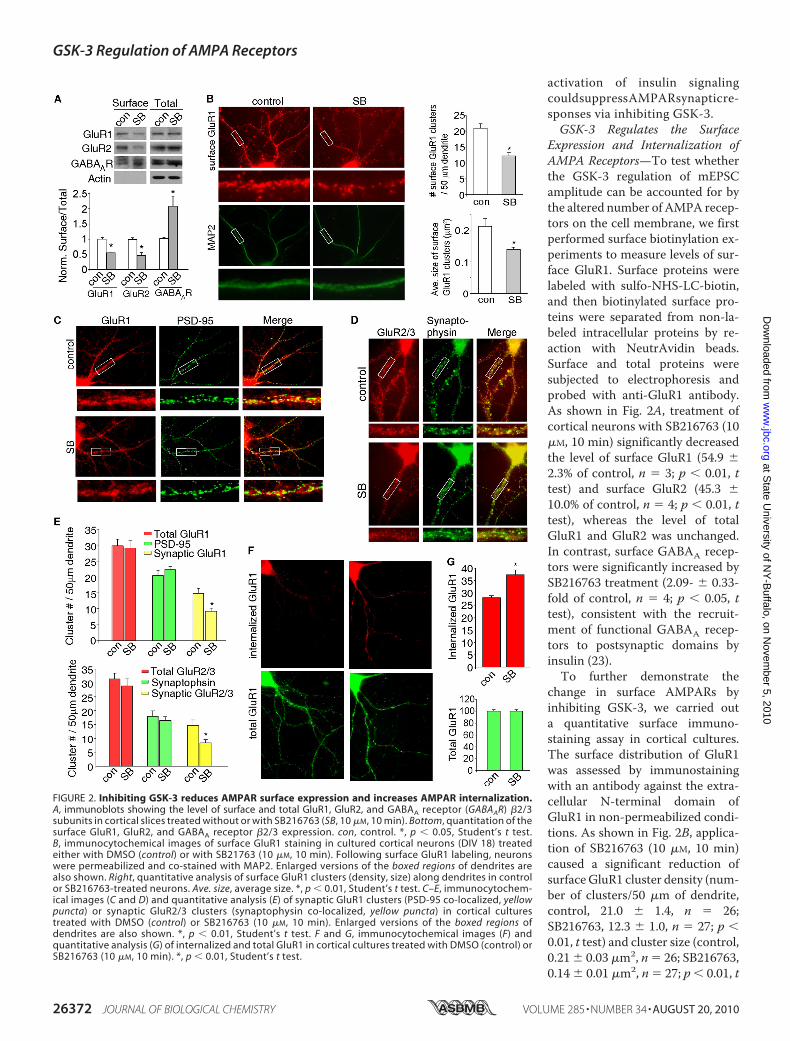
Expression and Internalization ofAMPA Receptors—To test whetherthe GSK-3 regulation of mEPSCamplitude can be accounted for bythe altered number of AMPA recep-tors on the cell membrane, we firstperformed surface biotinylation ex-periments to measure levels of sur-face GluR1. Surface proteins werelabeled with sulfo-NHS-LC-biotin,and then biotinylated surface pro-teins were separated from non-la-beled intracellular proteins by re-action with NeutrAvidin beads.Surface and total proteins weresubjected to electrophoresis andprobed with anti-GluR1 antibody.As shown in Fig. 2A, treatment ofcortical neurons with SB216763 (10�M, 10 min) significantly decreasedthe level of surface GluR1 (54.9 �2.3% of control, n � 3; p � 0.01, ttest) and surface GluR2 (45.3 �10.0% of control, n � 4; p � 0.01, ttest), whereas the level of totalGluR1 and GluR2 was unchanged.In contrast, surface GABAA recep-tors were significantly increased bySB216763 treatment (2.09- � 0.33-fold of control, n � 4; p � 0.05, ttest), consistent with the recruit-ment of functional GABAA recep-tors to postsynaptic domains byinsulin (23).To further demonstrate the
change in surface AMPARs byinhibiting GSK-3, we carried outa quantitative surface immuno-staining assay in cortical cultures.The surface distribution of GluR1was assessed by immunostainingwith an antibody against the extra-cellular N-terminal domain ofGluR1 in non-permeabilized condi-tions. As shown in Fig. 2B, applica-tion of SB216763 (10 �M, 10 min)caused a significant reduction ofsurfaceGluR1 cluster density (num-ber of clusters/50 �m of dendrite,control, 21.0 � 1.4, n � 26;SB216763, 12.3 � 1.0, n � 27; p �0.01, t test) and cluster size (control,0.21 � 0.03 �m2, n � 26; SB216763,0.14 � 0.01 �m2, n � 27; p � 0.01, t
FIGURE 2. Inhibiting GSK-3 reduces AMPAR surface expression and increases AMPAR internalization.A, immunoblots showing the level of surface and total GluR1, GluR2, and GABAA receptor (GABAAR) �2/3subunits in cortical slices treated without or with SB216763 (SB, 10 �M, 10 min). Bottom, quantitation of thesurface GluR1, GluR2, and GABAA receptor �2/3 expression. con, control. *, p � 0.05, Student’s t test.B, immunocytochemical images of surface GluR1 staining in cultured cortical neurons (DIV 18) treatedeither with DMSO (control) or with SB21763 (10 �M, 10 min). Following surface GluR1 labeling, neuronswere permeabilized and co-stained with MAP2. Enlarged versions of the boxed regions of dendrites arealso shown. Right, quantitative analysis of surface GluR1 clusters (density, size) along dendrites in controlor SB216763-treated neurons. Ave. size, average size. *, p � 0.01, Student’s t test. C–E, immunocytochem-ical images (C and D) and quantitative analysis (E) of synaptic GluR1 clusters (PSD-95 co-localized, yellowpuncta) or synaptic GluR2/3 clusters (synaptophysin co-localized, yellow puncta) in cortical culturestreated with DMSO (control) or SB216763 (10 �M, 10 min). Enlarged versions of the boxed regions ofdendrites are also shown. *, p � 0.01, Student’s t test. F and G, immunocytochemical images (F) andquantitative analysis (G) of internalized and total GluR1 in cortical cultures treated with DMSO (control) orSB216763 (10 �M, 10 min). *, p � 0.01, Student’s t test.
GSK-3 Regulation of AMPA Receptors
26372 JOURNAL OF BIOLOGICAL CHEMISTRY VOLUME 285 • NUMBER 34 • AUGUST 20, 2010
at State U
niversity of NY
-Buffalo, on N
ovember 5, 2010
ww
w.jbc.org
Dow
nloaded from

test). These data suggest that inhibiting GSK-3 reducesAMPAR surface expression.To provide more direct evidence on GSK-3 regulation of
AMPARs at synapses, we measured the synaptic AMPARclusters, as indicated by GluR1 co-localized with the synap-tic marker PSD-95. As shown in Fig. 2, C and E, a significantdecrease of synaptic GluR1 (co-localized with PSD-95) clus-ter density (number of clusters/50 �m of dendrite) wasobserved in SB216763 (10 �M, 10min)-treated cultures (con-trol, 14.9� 1.5, n� 26; SB216763, 9.2� 0.9, n� 25, p� 0.01,t test), whereas the total GluR1 or PSD95 clusters were notaltered by SB216763 treatment. Similarly, synaptic GluR2/3(co-localized with synaptophysin) cluster density was alsosignificantly decreased in SB216763-treated cultures (con-trol, 14.7� 2.0, n� 27; SB216763, 8.4� 1.1, n� 36, p� 0.01,t test), whereas the total GluR2/3 or synaptophysin clusterswere not altered (Fig. 2, D and E). It suggests that inhibitingGSK-3 reduces the number of AMPARs at synapses, whichmay account for the reduction of mEPSC amplitude byGSK-3 inhibitors.We also performed immunocytochemical experiments to
detect AMPAR internalization in cultured cortical neurons.Surface AMPARs were first stained with an antibody to theextracellular region of GluR1 subunit, and then following thetreatment with GSK-3 inhibitors, surface-bound antibodieswere stripped away so that only internalized AMPARs werevisualized. As shown in Fig. 2, F and G, SB216763 (10 �M, 10min) treatment caused a significant increase in the fluorescenceintensity of internalized GluR1 on neuronal dendrites (control,28.1 � 0.7, n � 28; SB216763, 37.3 � 2.1, n � 30; p � 0.01, ttest). It suggests that inhibiting GSK-3 increases the internal-ization of AMPARs, which may result in the reduction ofAMPARs at synaptic membrane.GSK-3 Regulation of AMPARs Involves the Stimulation of the
GDI-Rab5 Complex—To understand the potential mechanismunderlying GSK-3 regulation of AMPAR internalization, weexamined the role of Rab5, a key mediator of protein transportfrom plasma membrane to early endosomes during clathrin-dependent endocytosis (24). First, we examined whether
GSK-3 could regulate the activity ofthis small GTPase. Because Rabap-tin-5, a molecule identified as aRab5-interacting protein, binds toonly the GTP-bound, active form ofRab5 at its C terminus (25), wemeasured Rabaptin-5-bound Rab5by co-immunoprecipitation experi-ments to indicate its activity level.As shown in Fig. 3A, SB216763 (10�M, 10 min) treatment of corticalslices induced a significant increaseof Rab5 activity, as indicated by theelevated level of Rabaptin-5-boundRab5 (2.02- � 0.19-fold of control,n � 3, p � 0.01, t test). To test thespecificity of Rab5 involvement, wealso examined Rab4, which medi-ates receptor recycling between
early endosomes and the plasma membrane. Rabaptin-5 bindsto the active form of Rab4 at its N terminus (25). As shown inFig. 3A, the Rabaptin-5-bound (active) Rab4 was not signifi-cantly changed by GSK-3 inhibition.To test whether Rab5-dependent endocytosis of AMPA
receptors is involved in GSK-3 regulation of mEPSC, weknocked down Rab5 expression in cultured cortical neurons bytransfecting with an siRNA against Rab5 (GFP was co-trans-fected). As shown in Fig. 3, B and C, SB216763 (10 �M, 10 min)had little effect onmEPSC amplitude in neurons (GFP�) trans-fected with Rab5 siRNA (control, 20.9 � 0.4 pA, n � 11;SB216763, 20.6 � 0.4 pA, n � 11), whereas it produced a sig-nificant reduction of mEPSC amplitude in neurons transfectedwith GFP alone (control, 21.4 � 0.8 pA, n � 11; SB216763,17.6 � 0.6 pA, n � 11, p � 0.01, ANOVA). It suggests thatGSK-3 regulates AMPAR trafficking and function via a Rab5-dependent mechanism.Next, we sought to determine the mechanism underlying
GSK-3 regulation of Rab5-mediatedAMPAR internalization. Itis known that the recycling of Rab proteins between a mem-brane-bound and a cytosolic state is dependent on the GDPdissociation inhibitor (26). Previous studies have found that theformation of GDI-Rab complex can be altered by phosphory-lation of GDI (27, 28), leading to accelerated exocytosis orendocytosis. Thus, we tested the potential involvement of GDIin GSK-3 regulation of AMPARs.First, we examined whether GSK-3 alters the formation of
the GDI-Rab5 complex. As shown in the co-immunoprecipita-tion assay (Fig. 4A), SB216763 (10 �M, 10 min) treatment sig-nificantly increased the amount of Rab5 that binds to GDI(1.63- � 0.13-fold of control, n � 3, p � 0.01, t test). It suggeststhat inhibiting GSK-3 increases the formation of GDI-Rab5complex, which may account for the increased endocytic traf-ficking of surface AMPA receptors.To further test the role of GDI in GSK-3 regulation of
AMPARs, we dialyzed neuronswith an antibody against GDI toblock the function of endogenous GDI. As shown in Fig. 4B, theGDI antibody (4 �g/ml) produced a significant decrease ofmEPSC amplitude in cultured cortical neurons (control, 21.0�
FIGURE 3. GSK-3 regulation of synaptic AMPA activity requires Rab5 activation. A, top panel, co-immuno-precipitation (IP) blots showing the level of active (Rabaptin-5-bound) Rab5 or Rab4 in cortical slices without orwith SB216763 (SB) treatment (10 �M, 10 min). con, control; WB, Western blot. Lower panel, quantificationshowing the level of Rabaptin-5-bound (active) Rab5 or Rab4. *, p � 0.01, Student’s t test. B, cumulative plots ofmEPSC amplitude distribution before (control) and after SB216763 treatment (10 �M, 10 min) in a culturedcortical neuron transfected with Rab5 siRNA. Inset, representative mEPSC traces. Scale bar, 20 pA, 1 s. C, bargraphs showing the effect of SB216763 on mEPSC amplitude in neurons transfected with GFP or Rab5 siRNA. *,p � 0.01, ANOVA.
GSK-3 Regulation of AMPA Receptors
AUGUST 20, 2010 • VOLUME 285 • NUMBER 34 JOURNAL OF BIOLOGICAL CHEMISTRY 26373
at State U
niversity of NY
-Buffalo, on N
ovember 5, 2010
ww
w.jbc.org
Dow
nloaded from
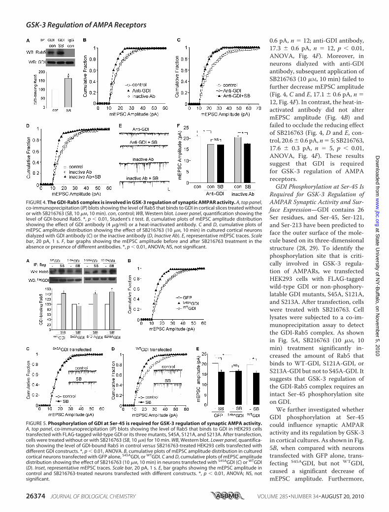
0.6 pA, n � 12; anti-GDI antibody,17.3 � 0.6 pA, n � 12, p � 0.01,ANOVA, Fig. 4F). Moreover, inneurons dialyzed with anti-GDIantibody, subsequent application ofSB216763 (10 �M, 10 min) failed tofurther decrease mEPSC amplitude(Fig. 4, C and E, 17.1 � 0.6 pA, n �12, Fig. 4F). In contrast, the heat-in-activated antibody did not altermEPSC amplitude (Fig. 4B) andfailed to occlude the reducing effectof SB216763 (Fig. 4, D and E, con-trol, 20.6� 0.6 pA,n� 5; SB216763,17.6 � 0.3 pA, n � 5, p � 0.01,ANOVA, Fig. 4F). These resultssuggest that GDI is requiredfor GSK-3 regulation of AMPAreceptors.GDI Phosphorylation at Ser-45 Is
Required for GSK-3 Regulation ofAMPAR Synaptic Activity and Sur-face Expression—GDI contains 26Ser residues, and Ser-45, Ser-121,and Ser-213 have been predicted toface the outer surface of the mole-cule based on its three-dimensionalstructure (28, 29). To identify thephosphorylation site that is criti-cally involved in GSK-3 regula-tion of AMPARs, we transfectedHEK293 cells with FLAG-taggedwild-type GDI or non-phosphory-latable GDI mutants, S45A, S121A,and S213A. After transfection, cellswere treated with SB216763. Celllysates were subjected to a co-im-munoprecipitation assay to detectthe GDI-Rab5 complex. As shownin Fig. 5A, SB216763 (10 �M, 10min) treatment significantly in-creased the amount of Rab5 thatbinds to WT-GDI, S121A-GDI, orS213A-GDI but not to S45A-GDI. Itsuggests that GSK-3 regulation ofthe GDI-Rab5 complex requires anintact Ser-45 phosphorylation siteon GDI.We further investigated whether
GDI phosphorylation at Ser-45could influence synaptic AMPARactivity and its regulation by GSK-3in cortical cultures. As shown in Fig.5B, when compared with neuronstransfected with GFP alone, trans-fecting S45AGDI, but not WTGDI,caused a significant decrease ofmEPSC amplitude. Furthermore,
FIGURE 4. The GDI-Rab5 complex is involved in GSK-3 regulation of synaptic AMPAR activity. A, top panel,co-immunoprecipitation (IP) blots showing the level of Rab5 that binds to GDI in cortical slices treated withoutor with SB216763 (SB, 10 �M, 10 min). con, control; WB, Western blot. Lower panel, quantification showing thelevel of GDI-bound Rab5. *, p � 0.01, Student’s t test. B, cumulative plots of mEPSC amplitude distributionshowing the effect of GDI antibody (4 �g/ml) or a heat-inactivated antibody. C and D, cumulative plots ofmEPSC amplitude distribution showing the effect of SB216763 (10 �M, 10 min) in cultured cortical neuronsdialyzed with GDI antibody (C) or the inactive antibody (D, Inactive Ab). E, representative mEPSC traces. Scalebar, 20 pA, 1 s. F, bar graphs showing the mEPSC amplitude before and after SB216763 treatment in theabsence or presence of different antibodies. *, p � 0.01, ANOVA; NS, not significant.
FIGURE 5. Phosphorylation of GDI at Ser-45 is required for GSK-3 regulation of synaptic AMPA activity.A, top panel, co-immunoprecipitation (IP) blots showing the level of Rab5 that binds to GDI in HEK293 cellstransfected with FLAG-tagged wild-type GDI or its three mutants, S45A, S121A, and S213A. After transfection,cells were treated without or with SB216763 (SB, 10 �M) for 10 min. WB, Western blot. Lower panel, quantifica-tion showing the level of GDI-bound Rab5 in control versus SB216763-treated HEK293 cells transfected withdifferent GDI constructs. *, p � 0.01, ANOVA. B, cumulative plots of mEPSC amplitude distribution in culturedcortical neurons transfected with GFP alone, S45AGDI, or WTGDI. C and D, cumulative plots of mEPSC amplitudedistribution showing the effect of SB216763 (10 �M, 10 min) in neurons transfected with S45AGDI (C) or WTGDI(D). Inset, representative mEPSC traces. Scale bar, 20 pA, 1 s. E, bar graphs showing the mEPSC amplitude incontrol and SB216763-treated neurons transfected with different constructs. *, p � 0.01, ANOVA; NS, notsignificant.
GSK-3 Regulation of AMPA Receptors
26374 JOURNAL OF BIOLOGICAL CHEMISTRY VOLUME 285 • NUMBER 34 • AUGUST 20, 2010
at State U
niversity of NY
-Buffalo, on N
ovember 5, 2010
ww
w.jbc.org
Dow
nloaded from
the reducing effect of SB216763 on mEPSC amplitude was lostin neurons transfected with S45AGDI (Fig. 5C, S45AGDI, 16.2 �0.6 pA, n � 14; S45AGDI � SB216763, 16.5 � 0.6, n � 14, Fig.5E) but not in those transfected with WTGDI (Fig. 5D, WTGDI,19.7 � 0.8 pA, n � 14; WTGDI � SB216763, 15.6 � 1.0 pA, n �14, p � 0.01, ANOVA, Fig. 5E) or GFP alone (GFP, 20.3 � 1.3pA, n � 13; GFP � SB216763, 16.1 � 0.8 pA, n � 13, p � 0.01,ANOVA, Fig. 5E). These data suggest that GSK-3 regulation ofsynaptic AMPAR currents requires an intact Ser-45 phosphor-ylation site on GDI.To confirm the role of GDI phosphorylation in GSK-3 regu-
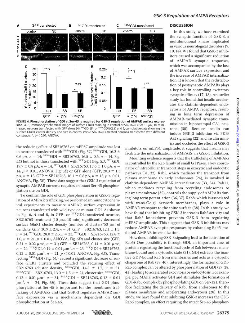
lation ofAMPAR trafficking, we performed immunocytochem-ical experiments to measure AMPAR surface expression inneurons transfected with wild-type or mutant GDI. As shownin Fig. 6, A and B, in GFP- or WTGDI-transfected neurons,SB216763 treatment (10 �M, 10 min) significantly decreasedsurface GluR1 cluster density (number of clusters/50 �m ofdendrite, GFP, 30.9� 2.4, n� 31; GFP� SB216763, 12.1� 1.3,n� 24;WTGDI, 28.0� 2.5, n� 23;WTGDI� SB216763, 12.8�1.0, n � 21, p � 0.01, ANOVA, Fig. 6D) and cluster size (GFP,0.21 � 0.02 �m2, n � 31; GFP � SB216763, 0.14 � 0.01 �m2,n � 24; WTGDI, 0.19 � 0.01 �m2, n � 23; WTGDI � SB216763,0.13 � 0.01 �m2, n � 21, p � 0.01, ANOVA, Fig. 6E). Trans-fecting S45AGDI (Fig. 6C) caused a significant decrease of sur-face GluR1 clusters and occluded the reducing effect ofSB216763 (cluster density, S45AGDI, 14.8 � 1.7, n � 31;S45AGDI � SB216763, 13.0 � 1.5, n � 24; cluster size, S45AGDI,0.13 � 0.01 �m2, n � 31; S45AGDI � SB216763, 0.13 � 0.01�m2, n � 24, Fig. 6E). These data suggest that GDI phos-phorylation at Ser-45 is important for the membrane traf-ficking of AMPARs and that GSK-3 regulates AMPAR sur-face expression via a mechanism dependent on GDIphosphorylation at Ser-45.
DISCUSSION
In this study, we have examinedthe synaptic function of GSK-3, amultifunctional kinase implicatedin various neurological disorders (9,10, 14).We found that GSK-3 inhib-itors caused a significant reductionof AMPAR synaptic responses,which was accompanied by the lossof AMPAR surface expression andthe increase of AMPAR internaliza-tion. It is known that the redistribu-tion of postsynaptic AMPARs playsa key role in controlling excitatorysynaptic efficacy (17, 18). An earlierstudy has found that insulin acceler-ates the clathrin-dependent endo-cytosis of AMPA receptors, result-ing in long term depression ofAMPAR-mediated synaptic trans-mission in hippocampal CA1 neu-rons (30). Because insulin caninduce GSK-3 inhibition via PKB/Akt signaling (22) and insulin mim-ics and occludes the effect of GSK-3
inhibitors on mEPSC amplitude, it suggests that insulin mayfacilitate the internalization of AMPARs via GSK-3 inhibition.Mounting evidence suggests that the trafficking of AMPARs
is controlled by the Rab family of small GTPases, a key coordi-nator of intracellular transport steps in exocytic and endocyticpathways (31, 32). Rab5, which mediates the transport fromplasma membrane to early endosomes (24), is involved inclathrin-dependent AMPAR internalization (33, 34). Rab11,which mediates recycling from recycling endosomes toplasma membrane (35), controls the supply of AMPARs dur-ing long term potentiation (36, 37). Rab8, which is associatedwith trans-Golgi network membranes, plays a role inAMPAR transport to the spine surface (38). In this study, wehave found that inhibiting GSK-3 increases Rab5 activity andthat Rab5 knockdown prevents GSK-3 from regulatingmEPSC amplitude. This suggests that GSK-3 inhibitors mayreduce AMPAR synaptic responses by enhancing Rab5-me-diated AMPAR internalization.Howdoes inhibitingGSK-3 signaling lead to the activation of
Rab5? One possibility is through GDI, an important class ofproteins regulating the functional cycle of Rab between amem-brane-bound and a cytosolic state (31). GDI extracts the inac-tive GDP-bound Rab from membranes and acts as a cytosolicchaperone of Rab (39, 40). Interestingly, the formation of GDI-Rab complex can be altered by phosphorylation of GDI (27, 28,41), leading to accelerated exocytosis or endocytosis. For exam-ple, p38 MAPK activates GDI and stimulates the formation ofGDI-Rab5 complex by phosphorylating GDI on Ser-121, there-fore facilitating the delivery of Rab5 from endosomes to theplasma membrane and accelerating endocytosis (28). In thisstudy, we have found that inhibiting GSK-3 increases the GDI-Rab5 complex, an effect requiring the intact Ser-45 phosphor-
FIGURE 6. Phosphorylation of GDI at Ser-45 is required for GSK-3 regulation of AMPAR surface expres-sion. A–C, immunocytochemical images of surface GluR1 staining in control or SB216763 (SB, 10 �M, 10 min)-treated neurons transfected with GFP alone (A), WTGDI (B), or S45AGDI (C). D and E, cumulative data showing thesurface GluR1 cluster density and size in control versus SB216763-treated neurons transfected with differentconstructs. *, p � 0.01, ANOVA.
GSK-3 Regulation of AMPA Receptors
AUGUST 20, 2010 • VOLUME 285 • NUMBER 34 JOURNAL OF BIOLOGICAL CHEMISTRY 26375
at State U
niversity of NY
-Buffalo, on N
ovember 5, 2010
ww
w.jbc.org
Dow
nloaded from
ylation site on GDI. The non-phosphorylatable S45AGDIdecreases AMPAR trafficking/function and occludes thereducing effect of GSK-3 inhibitors. It suggests that GSK-3 reg-ulation of AMPARs is through a mechanism involving GDIphosphorylation at Ser-45 and GDI-Rab5 complex formation.In summary, we have revealed a potential mechanism for
GSK-3 regulation of AMPARs. Our results suggest that consti-tutively active endogenous GSK-3 plays an important role inmaintaining AMPARs at the synaptic membrane. It is conceiv-able that dysregulation of glutamatergic transmission byimpaired GSK-3 signaling may be a key pathophysiologicalmechanism for those mental illnesses involving GSK-3.
Acknowledgments—We thank Xiaoqing Chen and Dr. Eunice Yuenfor excellent technical support.
REFERENCES1. Welsh, G. I., Wilson, C., and Proud, C. G. (1996) Trends Cell Biol. 6,
274–2792. Frame, S., and Cohen, P. (2001) Biochem. J. 359, 1–163. Jope, R. S., and Johnson, G. V. (2004) Trends Biochem. Sci. 29, 95–1024. Zhou, F. Q., and Snider, W. D. (2005) Science 308, 211–2145. Jiang, H., Guo, W., Liang, X., and Rao, Y. (2005) Cell 120, 123–1356. Kim,W. Y., Wang, X., Wu, Y., Doble, B. W., Patel, S., Woodgett, J. R., and
Snider, W. D. (2009) Nat. Neurosci. 12, 1390–13977. Woodgett, J. R. (1990) EMBO J. 9, 2431–24388. Cross, D. A., Alessi, D. R., Cohen, P., Andjelkovich, M., and Hemmings,
B. A. (1995) Nature 378, 785–7899. Coyle, J. T., and Duman, R. S. (2003) Neuron 38, 157–16010. Phiel, C. J., and Klein, P. S. (2001) Annu. Rev. Pharmacol. Toxicol. 41,
789–81311. Emamian, E. S., Hall, D., Birnbaum,M. J., Karayiorgou,M., andGogos, J. A.
(2004) Nat. Genet. 36, 131–13712. Phiel, C. J., Wilson, C. A., Lee, V. M., and Klein, P. S. (2003) Nature 423,
435–43913. Hong,M., Chen,D.C., Klein, P. S., and Lee, V.M. (1997) J. Biol. Chem.272,
25326–2533214. Meijer, L., Flajolet, M., and Greengard, P. (2004) Trends Pharmacol. Sci.
25, 471–48015. Chen, P., Gu, Z., Liu, W., and Yan, Z. (2007)Mol. Pharmacol. 72, 40–5116. Peineau, S., Taghibiglou, C., Bradley, C., Wong, T. P., Liu, L., Lu, J., Lo, E.,
Wu, D., Saule, E., Bouschet, T., Matthews, P., Isaac, J. T., Bortolotto, Z. A.,
Wang, Y. T., and Collingridge, G. L. (2007) Neuron 53, 703–71717. Malinow, R., andMalenka, R. C. (2002) Annu. Rev. Neurosci. 25, 103–12618. Collingridge, G. L., Isaac, J. T., andWang, Y. T. (2004)Nat. Rev. Neurosci.
5, 952–96219. Yuen, E. Y., Jiang, Q., Chen, P., Gu, Z., Feng, J., and Yan, Z. (2005) J. Neu-
rosci. 25, 5488–550120. Gu, Z., Liu, W., and Yan, Z. (2009) J. Biol. Chem. 284, 10639–1064921. Gu, Z., Jiang, Q., Fu, A. K., Ip, N. Y., and Yan, Z. (2005) J. Neurosci. 25,
4974–498422. Cohen, P., and Frame, S. (2001) Nat. Rev. Mol. Cell Biol. 2, 769–77623. Wan, Q., Xiong, Z. G., Man, H. Y., Ackerley, C. A., Braunton, J., Lu,W. Y.,
Becker, L. E., MacDonald, J. F., and Wang, Y. T. Nature (1997) 388,686–690
24. Bucci, C., Parton, R. G., Mather, I. H., Stunnenberg, H., Simons, K.,Hoflack, B., and Zerial, M. (1992) Cell 70, 715–728
25. Vitale, G., Rybin, V., Christoforidis, S., Thornqvist, P., McCaffrey, M.,Stenmark, H., and Zerial, M. (1998) EMBO J. 17, 1941–1951
26. Sasaki, T., Kikuchi, A., Araki, S., Hata, Y., Isomura, M., Kuroda, S., andTakai, Y. (1990) J. Biol. Chem. 265, 2333–2337
27. Shisheva, A., Chinni, S. R., and DeMarco, C. (1999) Biochemistry 38,11711–11721
28. Cavalli, V., Vilbois, F., Corti, M., Marcote, M. J., Tamura, K., Karin, M.,Arkinstall, S., and Gruenberg, J. (2001)Mol. Cell 7, 421–432
29. Schalk, I., Zeng, K., Wu, S. K., Stura, E. A., Matteson, J., Huang, M., Tan-don, A., Wilson, I. A., and Balch, W. E. (1996) Nature 381, 42–48
30. Man,H. Y., Lin, J.W., Ju,W.H., Ahmadian, G., Liu, L., Becker, L. E., Sheng,M., and Wang, Y. T. (2000) Neuron 25, 649–662
31. Zerial, M., and McBride, H. (2001) Nat. Rev. Mol. Cell Biol. 2, 107–11732. Pfeffer, S. R. (2001) Trends Cell Biol. 11, 487–49133. Brown, T. C., Tran, I. C., Backos, D. S., and Esteban, J. A. (2005)Neuron 45,
81–9434. Zhong, P., Liu, W., Gu, Z., and Yan, Z. (2008) J. Physiol. 586, 4465–447935. Ullrich, O., Reinsch, S., Urbe, S., Zerial, M., and Parton, R. G. (1996) J. Cell
Biol. 135, 913–92436. Park,M., Penick, E. C., Edwards, J. G., Kauer, J. A., and Ehlers,M. D. (2004)
Science 305, 1972–197537. Brown, T. C., Correia, S. S., Petrok, C. N., and Esteban, J. A. (2007) J. Neu-
rosci. 27, 13311–1331538. Gerges, N. Z., Backos, D. S., and Esteban, J. A. (2004) J. Biol. Chem. 279,
43870–4387839. Shisheva, A., Sudhof, T. C., and Czech, M. P. (1994) Mol. Cell Biol. 14,
3459–346840. Novick, P., and Zerial, M. (1997) Curr. Opin. Cell Biol. 9, 496–50441. Steele-Mortimer, O., Gruenberg, J., and Clague, M. J. (1993) FEBS Lett.
329, 313–318
GSK-3 Regulation of AMPA Receptors
26376 JOURNAL OF BIOLOGICAL CHEMISTRY VOLUME 285 • NUMBER 34 • AUGUST 20, 2010
at State U
niversity of NY
-Buffalo, on N
ovember 5, 2010
ww
w.jbc.org
Dow
nloaded from