relationship between prognostic score and thyrotropin receptor
TRANSCRIPT

British Joumal of Cancer (1997) 76(5), 594-599© 1997 Cancer Research Campaign
Relationship between prognostic score and thyrotropinreceptor (TSH-R) in papillary thyroid carcinoma:immunohistochemical detection of TSH-R
K Tanaka, H Inoue, H Miki, E Masuda, M Kitaichi, K Komaki, T Uyama and Y Monden
The Second Department of Surgery, School of Medicine, The University of Tokushima, Tokushima 770, Japan
Summary We have demonstrated the expression of thyrotropin receptor (TSH-R) in thyroid neoplasms (13 adenomas, 21 papillarycarcinomas, two follicular carcinomas) and adjacent normal thyroid using the monoclonal antibody against human TSH-R and have alsodemonstrated a relationship between prognostic scores and the expression of TSH-R. Among the adenomas, eight showed an intensitysimilar to that of normal thyroid and five showed a higher intensity than normal. Two tumours exhibited heterogeneous distribution of TSH-R.Among the papillary carcinomas, seven showed similar intensity to normal tissue and four showed higher intensity and ten showed weakerintensity. Eight tumours showed heterogeneous distribution of the stain. Among the follicular carcinomas, one showed similar intensity tonormal tissue and the other exhibited weaker intensity. Both cases showed homogeneous distribution of TSH-R. The adenomas nevershowed a weaker intensity than normal thyroid, but various intensities of TSH-R occurred in differentiated carcinomas. There was nosignificant relationship between the clinical data and the signal intensity in the adenomas. Among the papillary carcinomas, however, thegroup with weaker intensity had significantly poorer prognostic scores than the other two groups. Thus, we assume that low TSH-R may beexpressed by the clinically high-rsk group of patients with papillary thyroid carcinoma.
Keywords: thyroid neoplasia; thyrotropin receptor; prognostic score
Thyrotropin (TSH) is the major regulator of thyroid function andof thyrocyte growth (Vassart and Dumont, 1992). Previously, wedetermined the distribution ofTSH-R messenger RNA (mRNA) inthyroid neoplasms and adjacent normal thyroid tissues by in situhybridization (Tanaka et al, 1996). In normal thyroids andadenomas, TSH-R mRNA was distributed homogeneously, but insome papillary carcinomas it was distributed heterogeneously. Inother words, some papillary carcinoma cells showed no expressionof TSH-R mRNA. There have been few reports regarding TSH-Rusing the monoclonal antibody against it (Loosfelt et al, 1992). Ina recent study, it was reported that TSH-R protein was generallymore strongly expressed in papillary thyroid carcinomas than innormal thyroids (Mizukami et al, 1994). There have also beenseveral reports regarding responsiveness to TSH in papillary carci-nomas (Kimura et al, 1992; Namba et al, 1993). These authorsconcluded that impairment of the action of TSH on thyroid carci-noma cells was not due to reduction of the receptor number. Thus,the aim of this study was to determine the relationship between theexpression of TSH-R mRNA and TSH-R protein in normalthyroids and thyroid tumours. Using the monoclonal antibodyagainst TSH-R, we examined the expression levels of TSH-Rprotein in normal thyroid tissues and thyroid neoplasms immuno-histochemically. Our previous study showed that those papillarycarcinomas with a low expression of TSH-R mRNA had atendency to be in the advanced stages. Thus, in this study, we also
Received 2 December 1996Revised 17February 1997Accepted 24 February 1997
Correspondence to: K Tanaka, Kawasaki Medical School, Department ofBreast and Thyroid Surgery, 577 Matsushima, Kurashiki 701-01, Japan
discussed the relationship between prognostic score and the statusof TSH-R protein.
MATERIALS AND METHODS
Materials
We used human papillary thyroid carcinomas (n = 21), follicularthyroid carcinomas (n = 2), adenomas (n = 13) and their adjacentnormal thyroid tissue (n = 36) resected for surgical treatment. Thethyroid function of all patients was euthyroid. All adenomas werenon-functioning tumours. None of the patients had received anymedications that would have affected thyroid function beforesurgical treatment (i.e. thyroid hormone or anti-thyroid drugs). Allpatients underwent surgical treatment within 3 years and are alivewithout recurrence. Informed consent was obtained from allsubjects enrolled in this study.
Table 1 shows the background of the carcinomas under study.Only 3 of the 21 papillary carcinomas were poorly differentiated.The clinical stages of the papillary carcinomas and follicular carci-nomas were determined according to the UICC classification(Hermanek and Sobin, 1990). The total score of the patients wasdecided according to the EORTC prognostic index system (Byar etal, 1979). The total score of EORTC is calculated by summing age+ 12 (if male), +10 (if medullary or if the principal cell type is offollicular, less differentiated type and provided that the associatedcell type is not anaplastic), +45 (if anaplastic cell type), +10 (if theT-category is T3), +15 (if one distant metastasis) or +15 (if multipledistant metastases). We also calculated MACIS scores for thepapillary carcinomas according to the protocol of Hay et al (1993).The MACIS score was defined as 3.1 (if aged < 39 years) or0.08 x age (if aged . 40 years), +0.3 x tumour size (in centimetres),
594

Demonstration of TSH-R in thyroid neoplasms 595
B | W s l | , | fl | | l | | N.< ffi I , X . I si , ! B Is, g X_ | S S | X l E | N | | N _Ig B..E.gN_ a I | | | I | I | | , N=--__ _ a - n w 'w . g__Sl3R B illl | ; | |R____XE:iS=;ii _ _ l 1 1E 11S 3; | 15__ _ | C_iR_ I-| | | | | | | 11 ES_XY_8__ _ | | _ E i i | i i_w_IYI__ iJ - l 1|11 1|# Ri 11 11 11 i 1|1 _ _ __;;E _ l e 1s1 _! s N * -i l | Ei l __ | X1 I W1 W 30 | l 1N i 1 5 | I 11Dw__B6-' _ ffi ffi | r es | l | | l-
_SiS&Mag§°.MfE l i6 ! =1 * | | * | | 11 DE_a_;SlNElU0>eY;iS:9i _ les <§NF _ * | | sal 2 M§_f|Ce) l i -* | | i i ! | * | -e Alexai l _ }lS 11 ele Bl i! fi -S _ g,b"til b S.ps s ffi | " * | B -
are__ ^ l s s | | | X / 2 | _M_ XiS% Sfi-X z e ID E l E | | ___rgz dx s a a B e i s E |. X 8. b.,, 4 Y _N_ N 11 _ |xi; t R s u i _ ' ._fiu-Y gM aXWOf_ i- 1 i _ | _ _ ___wat X _ X X ! __la i-fl:,,D3s * a | S aS _ | | l S_csPg ge_ r_ E N __R gR___ R !__ __ ! .
z |X X _X | ! |_ * s _ -
11 | ____| | _ ===
* S _
s | |
* * _ ____S mil * s _ s_ s : ID}_Bl2 ..... :_lL>, .................. :6i_ ii ^6;bE .Ss 3 fi . .................................................... ._t.,2t,, , ;.,, ,'e i ; . s t° -.j,.,,,.! ; , ! o " !; ; o e_i8to>J B4 prou!_22g ........................................ !-.;°o<.W.! .. >_=_M;ga§,,.4:,vs: t::.,, .°. e .. .°.. 9. > u. ia
__088!5 k °.'^e°e'§t,AgEi.S sSe °_o>W ,J eI_L ................ i!n
|NXd= ..: o ..io:i°aSot&l!hlci ...:<:.::'.::::: .: :,s!:;:>:>e;!R.: e ;.e_G_ } ;t;-*sbsws Or .e. .......... £i .e.; . f. ... itsi;|s __ | .< >=.w__4r t _:§80ZiXS o s::.o::e: ....... :':'::.::.::: ::!::.:: :':,:ep ''}}':x; ................................................................................ ' ':RaN§0i'-°.< '.>: . i .. .: .: .: ,. >,, ___ ............................................. } .;; _X_ . e ................. . ., .SWe.. j.j,;., ., .,,..:,; ..i .:i ..sl :....,
s§:.L'.'....! i.°'@'
_x.x.w._ws_ _0 .* : _18 ;2 <_s,e,sg_SEZ , :,.;.i.t _ . . . - _s__r_wrXe N.! .SS.' _
-''t_A . _p; .... j -.:, 4w ;.id2.^.i^ ........................................................... .................. e!:E;- ,:' g;j 2B S.J--<x' ;.},.M L, ._
...E..; o S- lsCSi?_
.t. ;U..o.Y>; .@. °:6'i :@.c.' i? r ' :->iRiS M3 fj 44aJ oy > t§_|| _ _2 41
., .. Sj ..j ....s ....t,.t,£>. s-i >° >.,..!.<.<.n,;o.j a,j ;, .,,i.i, ....... !i.e,,'.,. EsI_
[i .................................... S iFigure 1 Demonstation of TSH-R. Dark purpGe-coloured staining appearedin the cytoplasm of thyrocytes. The edge of the cytoplasm was particularlystrongly stained. A Adenoma. B Adjacent normal thyroid. A and B show acase of adenoma that exhibited more intensity than norrnal thyroid andhomogeneous distnbution of positive staining (x 200). C shows a case ofadenoma that exhibited sxtrome!y hetergeneous distribution. In this case,positive staining appeared only along the basal surface (x 100). D Papillarycarcinoma. E Adjacent normal thyroid. D and E show a case of papillarycarcinoma that exhibited less intensity than nonnal thyroid and homogeneousdistnbution (x 200). F Follicular carcenoma. G Adjacent normal thyroid. F andG shows a case of follicular carcinoma that exhibited less intensity thannormal thyroid (x 200).
British Joumal of Cancer (1997) 76(5), 594-599
A
C
E
G
0 Cancer Research Campaign 1997

596 K Tanaka et al
Table 1 Backgrounds of differentiated carcinomas
No. Age (years) Sex Tumour size (cm) Clinical classificationa Differentiation Preoperative serumthyroglobulin (ng ml-')
Papillary carcinomas1 61 F 1.4 pT4NOMO Well 15.22 52 F 2.0 pT1N1aMO Poor Unknown3 64 F 1.3 pTl NOMO Well Unknown4 49 F 2.3 pT2N1bMO Well Unknown5 43 F 1.0 pT4N1aMO Well < 1.56 41 F 1.5 pT2N1aMO Well 60.77 50 F 0.7 pT1N1bMO Poor 11408 56 F 0.9 pTl NOMO Well Unknown9 65 F 1.6 pT4NOMO Poor Unknown10 72 M 2.0 pT4N1bMO Well 11411 43 M 2.1 pT2N1aMO Well 2212 40 F 1.2 pT3N1aMO Well 25.213 77 F 1.8 pT1N1aMO Well 68.514 50 F 1.1 pT4NOMO Well 21.315 71 F 2.2 pT4N1bMO Well 36.416 56 F 1.3 pT4N1aMO Well 56.917 30 F 1.1 pT1N1aMO Well 18.818 55 F 1.3 pT4NOMO Well 8.319 69 F 1.7 pT1NOMO Well < 1.520 72 F 1.2 pTl NOMO Well Unknown21 63 M 1.5 pT4N1bMO Well 16.3
Follicular carcinomas1 60 F 2.5 pT2NOMO Well Unknown2 58 M 4.6 pT2NOMO Well Unknown
aAccording to UICC classification. Histological differentiation was decided by haematoxylin-eosin staining. Normal range of serum thyroglobulin is under45 ng ml-1 in Japan.
+1 (if incompletely resected), +1 (if locally invasive) or +3 (ifdistant metastases). With these two scoring systems, the higher thescore, the poorer the prognosis (Byar et al, 1979; Hay et al, 1993).According to Hay et al (1993), the survival rates for patients withMACIS scores of < 6, 6-6.99, 7-7.99, and 8+ were 99%, 89%,56%, and 24% respectively.
Surgically resected specimens were fixed in cold 4% paraformal-dehyde for 4 h, and then placed in cold 30% sucrose solution untilthe tissues sank. The tissues were then embedded in OCT compound(Miles Laboratories, USA), and stored at -80°C until used.
Immunohistochemical evaluation of TSH-R
Frozen sections in OCT compound were cut into 6 jm-thicksections using a cryostat, fixed on aminopropyltrietoxysilane-coatedslide glasses (Matsunami, Japan) and dried. After rinsing in phos-phate-buffered saline (PBS) for 10 min, normal rabbit serum wasapplied and the sections were incubated for 30 min at room temper-ature to block non-specific binding of the antibody. The slides werethen incubated overnight at 40C with anti-TSH-R antibody(T3-495) at a concentration of 1.5 jig ml-'. This antibody is a mousemonoclonal immunoglobulin GI antibody (Transbio, France)directed against the C-terminal segment (between amino acids604-764) of the human TSH receptor (Loosfelt et al, 1992;Mizukami et al, 1994). After incubation, the slides were washed inPBS for 15 min, and a secondary antibody was applied using theAPAAP kit (Dako, USA) according to the manufacturer's protocol.After washing, the colour was developed using 5 gl of nitro bluetetrazolium chloride (Boehringer Mannheim Biochemica, USA) and3.75 jil of 5-bromo-4-chloro-3-indolyl-phosphate, 4-toluidine salt(Boehringer Mannheim Biochemica) in 1 ml of buffer (0.1 mol 1-1
Tris-hydrochloric acid, pH 9.5, 0.1 mol -' sodium chloride,50 mmol 1-1 magnesium chloride). Levamisole was added at aconcentration of 1 mmol 1-' to block endogenous alkaline phos-phatase. Then the slides were washed in distilled water for 5 min,counterstained with methyl green and mounted.We used normal mouse serum instead of primary antibody in a
control study. Control studies were performed for all slides.
Analysis of immunohistochemical results
Immunostaining was evaluated by more than two repeated stain-ings of the same specimens and by more than two observers. Theseobservers were blinded to the characteristics of patients, thetumour extent and prognostic scores. A cell of a tissue section wasevaluated as positive when it showed a distinct specific stain whencompared with cells of the negative control sections. Positive cellswere graded into three levels of intensity from + (slightly stained)to +++ (strongly stained). Three grades for the standard slides ascontrols were also decided by the following method. First, thestrongest stained slides and the weakest stained slides wereselected from among all the slides and then the median stainedslides were selected. The rate of discordance among observers indeciding the grade of each slide was about 6%, and any discor-dance was settled by discussion between the observers.
Next, we compared the intensity of positive stainings of normalthyroid tissues and thyroid neoplasms in each case. We also classifiedthe cases into three groups: a weaker group, in which the stainedintensity of the tumour was weaker than that of the normal thyroid ofthe same patients; a similar group, in which the tumour intensity wasalmost the same as that of the normal thyroid, and a higher group, inwhich the tumour intensity was higher than that of the normal thyroid.
British Journal of Cancer (1997) 76(5), 594-599 0 Cancer Research Campaign 1997

Demonstration of TSH-R in thyroid neoplasms 597
Table 2 Backgrounds of cases and the results of immunohistochemistry inadenoma
No. Age Sex Tumour Distribution(years) size (cm) of staining
'The higher group'1 59 F 4.5 Homo2 62 F 7.0 Homo3 75 M 1.5 Hetero4 66 F 5.0 Homo5 34 F 3.7 Homo
59.2 ± 15.34.34 ± 2.0 (mean ± sd)
'The similar group'6 36 F 3.0 Homo7 25 M 4.8 Homo8 62 F 6.0 Homo9 48 F 4.0 Homo10 71 M 5.0 Homo11 42 F 5.2 Homo12 34 F 2.7 Homo13 42 F 5.0 Hetero
45 ± 15.1a 4.46 1.14a
aNot significant. Homo, homogeneous; hetero, heterogeneous.
In addition, we examined whether the distribution of positivestained cells in the tissue was homogeneous or heterogeneous. Forthe heterogeneous cases, the grade of intensity of each slide wasbased on the grade of the positively stained cells having the largestpopulation.
STATISTICAL ANALYSIS
For statistical analysis, the Mann-Whitney U-test and Scheffe's testwere used as post hoc tests, and P < 0.05 was taken as significant.
RESULTS
A dark purple-coloured positive stain appeared in the cytoplasm ofthe thyrocytes, and was especially strong at the edge of the cyto-plasm. In some cases, the strongest stain appeared along the basalcell surface. Table 2 shows the background and the expressionlevel of TSH-R in the adenomas.
In the adenomas, the staining pattern was almost completelyhomogeneous (Figure IA). Regarding staining intensity, 8 of the13 adenomas belonged to the similar group and the other fivebelonged to the higher group. The adenoma of patient 13 showedan extremely heterogeneous distribution of TSH-R (Figure 1B). In
Table 3 Prognostic scores and the results of immunohistochemistry and TSH-R mRNA detected by in situ hybridizationa in papillary carcinoma
Case no. Stageb EORTC indexc MACIS scoresd Distribution of Comparison with normal Positivity of signal ofTotal score staining (TSH-R) thyroida (THS-R mRNA) tumour (TSH-R mRNA) (%)
'The weaker group'5 53 4.74 Homo Weaker 72.2
11 III 66 5.87 Hetero - -
13 III 71 6.3 Homo -
14 Il 85 6.49 Homo - -
15 64 5.51 Homo -
16 III 85 6.68 Hetero -
17 1 69 6.03 Homo -
18 III 81 7.34 Hetero -
19 Il 94 7.36 Homo -
21 Il 77 6.7 Hetero -
74.5 ± 12.2 6.30 ± 0.81 (mean ± s.d.)
'The similar group'1 1 30 3.43 Homo Weaker 94.14 1 55 4.07 Hetero Weaker 80.16 III 49 4.61 Homo Weaker 94.27 Il 60 4.21 Hetero Weaker 67.78 Il 60 4.33 Homo Similar 92.79 III 62 4.76 Hetero Weaker 63.210 III 65 5.79 Hetero -
54.4 ± 12.0*** 4.46 ± 0.73*
'The higher group2 1 40 3.56 Homo Similar 77.63 41 3.73 Homo Similar 95.612 56 4.75 Homo -20 1 72 6.12 Homo
52.3 15.1** 4.54 ± 1.18**
Homo, homogeneous; hetero, heterogeneous. *P < 0.005 vs weaker group; **P < 0.01 vs weaker group; ***P < 0.05 vs weaker group. arefer to our previousstudy (Tanaka et al, 1996); baccording to UICC classification; caccording to scoring system of EORTC thyroid cancer cooperative group; daccording to Hay'sprotocol.
British Journal of Cancer (1997) 76(5), 594-5990 Cancer Research Campaign 1997
598 K Tanaka et al
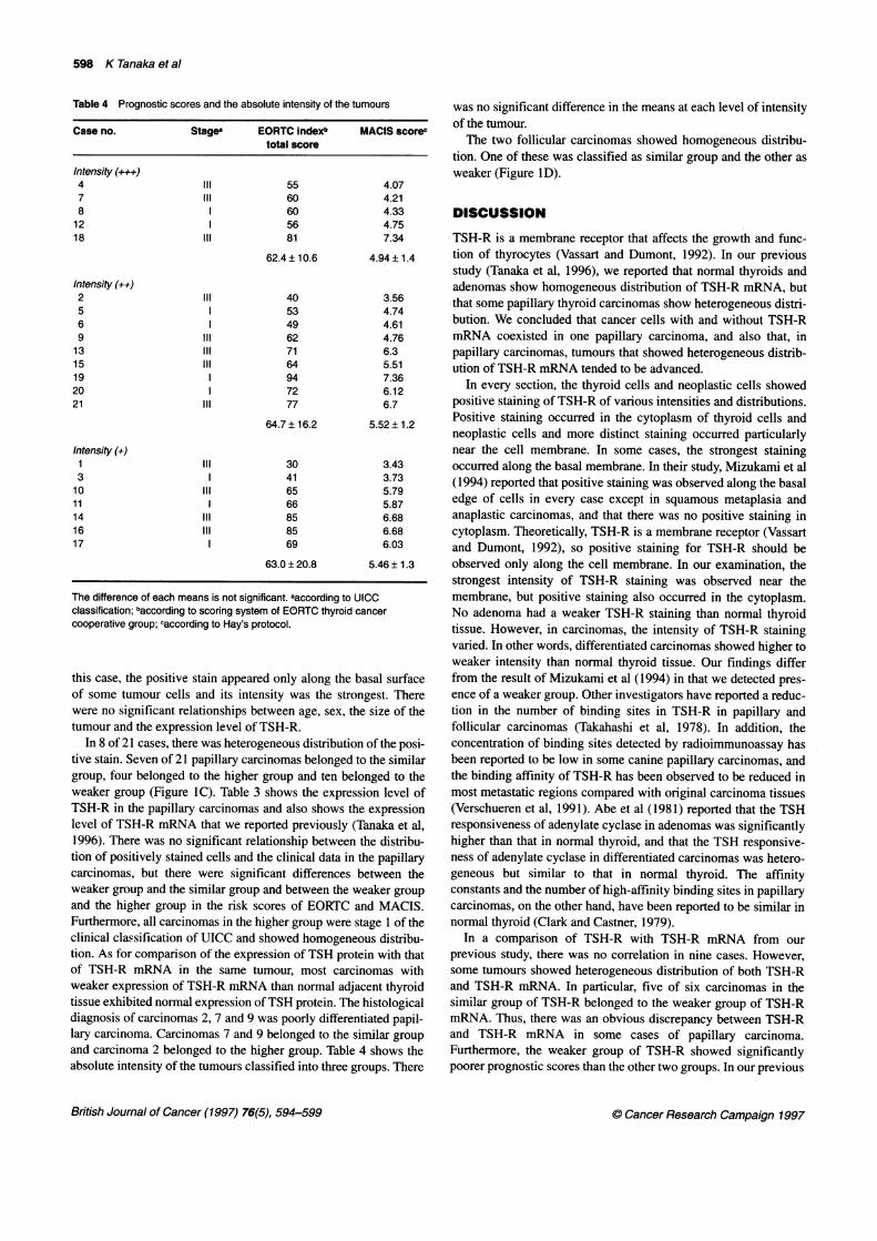
Table 4 Prognostic scores and the absolute intensity of the tumours
Case no. Stage' EORTC indexb MACIS scorectotal score
Intensity (+++)4 Il 55 4.077 Il 60 4.218 60 4.3312 1 56 4.7518 II 81 7.34
62.4 ± 10.6 4.94 ± 1.4
Intensity (++)2 Il 40 3.565 53 4.746 49 4.619 Il 62 4.7613 Il 71 6.315 II 64 5.5119 1 94 7.3620 72 6.1221 Il 77 6.7
64.7 ± 16.2 5.52 ± 1.2
Intensity (÷)1 Il 30 3.433 41 3.7310 II 65 5.7911 1 66 5.8714 ll 85 6.6816 ll 85 6.6817 69 6.03
63.0 ± 20.8 5.46 ± 1.3
The difference of each means is not significant. aaccording to UICCclassification; baccording to scoring system of EORTC thyroid cancercooperative group; caccording to Hay's protocol.
this case, the positive stain appeared only along the basal surfaceof some tumour cells and its intensity was the strongest. Therewere no significant relationships between age, sex, the size of thetumour and the expression level of TSH-R.
In 8 of 21 cases, there was heterogeneous distribution of the posi-tive stain. Seven of 21 papillary carcinomas belonged to the similargroup, four belonged to the higher group and ten belonged to theweaker group (Figure lC). Table 3 shows the expression level ofTSH-R in the papillary carcinomas and also shows the expressionlevel of TSH-R mRNA that we reported previously (Tanaka et al,1996). There was no significant relationship between the distribu-tion of positively stained cells and the clinical data in the papillarycarcinomas, but there were significant differences between theweaker group and the similar group and between the weaker groupand the higher group in the risk scores of EORTC and MACIS.Furthermore, all carcinomas in the higher group were stage 1 of theclinical classification of UICC and showed homogeneous distribu-tion. As for comparison of the expression of TSH protein with thatof TSH-R mRNA in the same tumour, most carcinomas withweaker expression of TSH-R mRNA than normal adjacent thyroidtissue exhibited normal expression of TSH protein. The histologicaldiagnosis of carcinomas 2, 7 and 9 was poorly differentiated papil-lary carcinoma. Carcinomas 7 and 9 belonged to the similar groupand carcinoma 2 belonged to the higher group. Table 4 shows theabsolute intensity of the tumours classified into three groups. There
was no significant difference in the means at each level of intensityof the tumour.The two follicular carcinomas showed homogeneous distribu-
tion. One of these was classified as similar group and the other asweaker (Figure ID).
DISCUSSION
TSH-R is a membrane receptor that affects the growth and func-tion of thyrocytes (Vassart and Dumont, 1992). In our previousstudy (Tanaka et al, 1996), we reported that normal thyroids andadenomas show homogeneous distribution of TSH-R mRNA, butthat some papillary thyroid carcinomas show heterogeneous distri-bution. We concluded that cancer cells with and without TSH-RmRNA coexisted in one papillary carcinoma, and also that, inpapillary carcinomas, tumours that showed heterogeneous distrib-ution of TSH-R mRNA tended to be advanced.
In every section, the thyroid cells and neoplastic cells showedpositive staining of TSH-R of various intensities and distributions.Positive staining occurred in the cytoplasm of thyroid cells andneoplastic cells and more distinct staining occurred particularlynear the cell membrane. In some cases, the strongest stainingoccurred along the basal membrane. In their study, Mizukami et al(1994) reported that positive staining was observed along the basaledge of cells in every case except in squamous metaplasia andanaplastic carcinomas, and that there was no positive staining incytoplasm. Theoretically, TSH-R is a membrane receptor (Vassartand Dumont, 1992), so positive staining for TSH-R should beobserved only along the cell membrane. In our examination, thestrongest intensity of TSH-R staining was observed near themembrane, but positive staining also occurred in the cytoplasm.No adenoma had a weaker TSH-R staining than normal thyroidtissue. However, in carcinomas, the intensity of TSH-R stainingvaried. In other words, differentiated carcinomas showed higher toweaker intensity than normal thyroid tissue. Our findings differfrom the result of Mizukami et al (1994) in that we detected pres-ence of a weaker group. Other investigators have reported a reduc-tion in the number of binding sites in TSH-R in papillary andfollicular carcinomas (Takahashi et al, 1978). In addition, theconcentration of binding sites detected by radioimmunoassay hasbeen reported to be low in some canine papillary carcinomas, andthe binding affinity of TSH-R has been observed to be reduced inmost metastatic regions compared with original carcinoma tissues(Verschueren et al, 1991). Abe et al (1981) reported that the TSHresponsiveness of adenylate cyclase in adenomas was significantlyhigher than that in normal thyroid, and that the TSH responsive-ness of adenylate cyclase in differentiated carcinomas was hetero-geneous but similar to that in normal thyroid. The affinityconstants and the number of high-affinity binding sites in papillarycarcinomas, on the other hand, have been reported to be similar innormal thyroid (Clark and Castner, 1979).
In a comparison of TSH-R with TSH-R mRNA from ourprevious study, there was no correlation in nine cases. However,some tumours showed heterogeneous distribution of both TSH-Rand TSH-R mRNA. In particular, five of six carcinomas in thesimilar group of TSH-R belonged to the weaker group of TSH-RmRNA. Thus, there was an obvious discrepancy between TSH-Rand TSH-R mRNA in some cases of papillary carcinoma.Furthermore, the weaker group of TSH-R showed significantlypoorer prognostic scores than the other two groups. In our previous
British Journal of Cancer (1997) 76(5), 594-599 . Cancer Research Campaign 1997

Demonstration of TSH-R in thyroid neoplasms 599
report (Tanaka et al, 1996), papillary carcinomas in the advancedclinical stage showed a tendency towards low expression andheterogeneous distribution of TSH-R mRNA, but distribution ofTSH-R did not show any relationship with either scoring system.We have no explanation for these results. This discrepancy shouldbe investigated.
In this report, we also investigated the relationship between theclinical prognostic score and the expression status of TSH-R inpapillary carcinomas. Some investigators have reported that the ageof the patient is an important prognostic factor in papillary andfollicular carcinomas (Crile and Hazard, 1953; Cady et al, 1979),whereas others (Franssila, 1975; Byar et al, 1979; Hay et al, 1993)have great emphasis on age, sex and tumour status in the scoringsystem. Shi and Farid (1993) reported a negative correlationbetween expression of TSH-R mRNA and tumour stage in mostpatients. Thyroglobulin (Tg) is also thought to be a marker of differ-entiated thyroid cancer (Schlumberger et al, 1980). Some investiga-tors have reported that cells of moderately differentiated thyroidcancers contain about two to three times less Tg mRNA than thoseof well-differentiated thyroid cancers detected by in situ hybridiza-tion (Berge-Lefranc et al, 1985). In addition, Ohta et al (1991)reported that mRNAs of TSH-R and Tg are expressed in relation totheir degree of differentiation. Based upon our findings, TSH-Rprotein also exhibited a significant relationship with the prognosticscore rather than the differentiation of cancer. In this relationship,the comparative intensity of the TSH-R protein of the tumouragainst adjacent tissue is important but the intensity of the TSH-Rprotein of the tumour itself is less important. In vitro FRTL-5 cellstransfected with the v-ras oncogene were reported to have acquiredcomplete malignancy or a transformed phenotype and to have lostTSH-R mRNA (Berlingieri et al, 1990). Cady et al (1983) reportedno significant improvement in survival times with TSH suppressiontherapy in patients with a poor prognostic score. However, manysurgeons and endocrinologists have performed thyroid hormonereplacement (DeGroot, 1994; Solomon et al, 1996).
Based on our findings, we assume that reduced expression ofTSH-R is associated with a poorer prognosis and that, as a result,TSH suppression therapy would have less effect in such patientsthan in patients whose tumours overexpress TSH-R.
ACKNOWLEDGEMENT
This work was supported in part by Grants-in-Aid for ScientificResearch from the Ministry of Education, Science, and Culture ofJapan.
REFERENCES
Abe Y, Ichikawa Y, Muraki T, Ito K and Homma M (1981) Thyrotropin (TSH)receptor and adenylate cyclase activity in human thyroid tumors: Absence ofhigh affinity receptor and loss of TSH responsiveness in undifferentiatedthyroid carcinoma. J Clin Endocrinol Metab 52: 23-28
Berge-Lefranc JL, Cartouzou G, Micco C, Fragu P and Lissitzky S (1985)Quantification of thyroglobulin messenger RNA by in situ hybridization indifferentiated thyroid cancers. Cancer 56: 345-350
Berlingieri MT, Akamizu T, Fusco A, Grieco M, Colletta G, Cirafici AM, IkuyamaS, Kohn LD and Vecchio G (1990) Thyrotropin receptor gene expression inoncogene-transfected rat thyroid cells: Correlation between transformation,loss of thyrotropin-dependent growth, and loss of thyrotropin receptor geneexpression. Biochem Biophys Res Commun 173: 172-178
Byar DP, Green SB, Dor P, Williams ED, Colon J, van Gilse HA, Mayer M,Sylvester RJ and Glabbeke MV (1979) A prognostic index for thyroidcarcinoma. a study of the E.O.R.T.C. thyroid cancer cooperative group. Eur JCancer 15: 1033-1041
Cady B, Sedgewick CE, Meissner WA, Wool MS, Salzman FA and Weber J (1979)Risk factor analysis in differentiated thyroid cancer. Cancer 43: 810-820
Cady B, Cohn K, Rossi RL, Sedgewick CE, Meissner WA, Weber J and Gelman RS(1983) The effect of thyroid hormone administration upon survival in patientswith differentiated thyroid carcinoma. Surgery 94: 978-983
Clark OH and Castner BJ (1979) Thyrotropin "receptors" in normal and neoplastichuman thyroid tissue. Surgery 85: 624-632
Crile G and Hazard JB ( 1953) Relationship of the age of the patient to the naturalhistory and prognosis of carcinoma of the thyroid. Ann Surg 138: 33-38
DeGroot LJ (1994) Long-term impact of initial and surgical therapy on papillary andfollicular thyroid cancer. Am J Med 97: 499-500
Franssila KO (1975) Prognosis in thyroid carcinoma. Cancer 36: 1138-1146Hay ID, Bergstralh EJ, Goellner JR, Ebersold JR and Grant CS (1993) Predicting
outcome in papillary thyroid carcinoma: development of a reliable prognosticscoring system in a cohort of 1779 patients surgically treated at one institutionduring 1940 through 1989. Surgery 114: 1050-1058
Hermanek P and Sobin LH (1990) Thyroid gland (ICD-O 193). International UnionAgainst Cancer TNM Classification ofMalignant Tumors, 4th fully revisededn, Japanese edn, pp 33-35, Springer: Berlin; Kanehara and Co: Tokyo
Kimura H, Yamashita S, Namba H, Usa T, Fujiyama K, Tsuruta M, Yokogawa N,Izumi M and Nagataki S (1992) Impairment of the TSH signal transductionsystem in human thyroid carcinoma cells. Exp Cell Res 203: 402-406
Loosfelt H, Pichon C, Jolivet A, Misraht M, Caillou B, Jamous M, Vannier B andMilgrom E (1992) Two-subunit structure of the human thyrotropin receptor.Proc Natl Acad Sci USA 89: 3765-3769
Mizukami Y, Hashimoto T, Nonomura A, Michigishi T, Nakamura S, Noguchi Mand Matsukawa S (1994) Immunohistochemical demonstration of thyrotropin(TSH)-receptor in normal and diseased human thyroid tissues usingmonoclonal antibody against recombinant human TSH-receptor protein. J ClinEndocrinol Metab 79: 616-619
Namba H, Yamashita S, Usa T, Kimura H, Yokogawa N, Izumi M and Nagataki S(1993) Overexpression of the thyrotropin receptor in a human thyroidcarcinoma cell line. Endocrinology 132: 839-845
Ohta K, Endo T and Onaya T ( 1991 ) The mRNA levels of thyrotropin receptor,thyroglobulin and thyroid peroxidase in neoplastic human thyroid tissues.Biochem Biophys Res Commun 174: 1148-1153
Schlumberger M, Charbord P, Fragu P, Lumbroso J and Parmentier C (1980)Circulating thyroglobulin and thyroid hormones in patients with metastases ofdifferentiated thyroid carcinoma: relationship to serum thyrotropin levels.J Clin Endocrinol Metab 51: 513-519
Shi Y and Farid NR (1993) Expression of thyrotropin receptor gene in thyroidcarcinoma is associated with a good prognosis. Clin Endocrinol 39: 269-274
Solomon BL, Wartofsky L and Burman KD (1996) Current trends in themanagement of well differentiated papillary thyroid carcinoma. J ClinEndocrinol Metab 81: 333-339
Takahashi H, Jiang NS, Gorman CA and Lee CY (1978) Thyrotropin receptors innormal and pathological human thyroid tissues. J Clin Endocrinol Metab 47:870-876
Tanaka K, Inoue H, Miki H, Komaki K and Monden Y (1996) Heterogeneousdistribution of thyrotropin receptor messenger ribonucleic acid (TSH-RmRNA) in papillary thyroid carcinomas detected by in situ hybridization. ClinEndocrinol 44: 259-267
Vassart G and Dumont JE (1992) Thyrotropin receptor and the regulation ofthyrocyte function and growth. Endocrine Rev 13: 596-611
Verschueren CP, Ruttenman GR, Vos JH, Van Dijik JE and de Brnin TWA (1991)Thyrotropin receptors in normal and neoplastic (primary and metastatic) caninethyroid tissue. J Endocrinol 132: 461-468
C Cancer Research Campaign 1997 British Journal of Cancer (1997) 76(5), 594-599