reproduction ratios on standardization … · the influence of medical photography’s westminster...
TRANSCRIPT

THE INFLUENCE OF MEDICAL
PHOTOGRAPHY’S WESTMINSTER
REPRODUCTION RATIOS ON
STANDARDIZATION IN FORENSIC
PHOTOGRAPHY – AN OPINION
Hoosain M EbrahimDepartment of Medical Illustration
and Audio Visual Services
University of Limpopo
South Africa

PHOTOGRAPHY
Forensic Medical
Different aims
Different objectives
Legal purpose

FORENSIC PHOTOGRAPHY
Final photographic product
(may only be the beginning)
Photographer’s involvement in a legal
system that might want to know
WHY, WHERE, HOW, WHEN and under
WHAT CONDITIONS the images were
taken

Forensic photographs must meet the
following:
• Technical qualities
• Sharpness of detail
• Clarity of image
• Use of lighting
• Perspective
• Accuracy of reproduction of both colour and form

The medical photograph…
• provides a precise record
• is comparable with others taken over a
period of time
• is obtained with the least inconvenience to
the patient
• meets the intention of the request
• is in accord with current methods of
presenting medical data
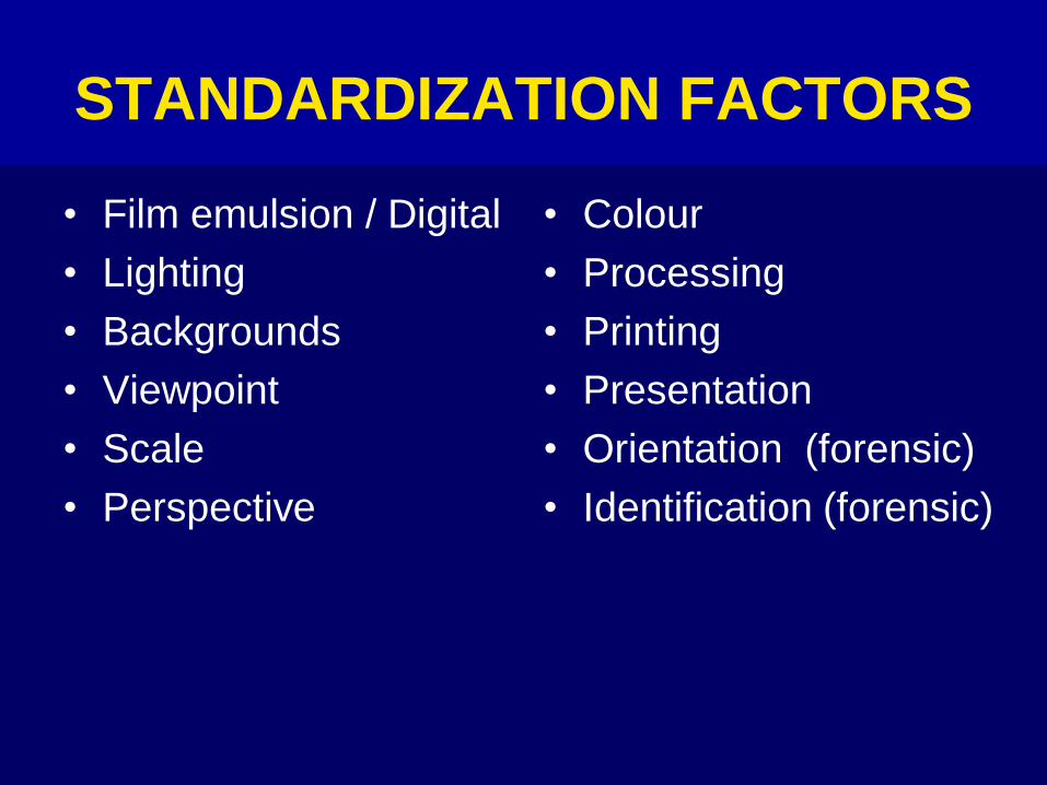
STANDARDIZATION FACTORS
• Film emulsion / Digital
• Lighting
• Backgrounds
• Viewpoint
• Scale
• Perspective
• Colour
• Processing
• Printing
• Presentation
• Orientation (forensic)
• Identification (forensic)

STANDARDIZATION REQUIRES WORKING TO PRE-DETERMINED
RULES

WORKING TO SCALE
The scale of reproduction must be constant for
any given area both at the recording and
printing stage

WESTMINSTER REPRODUCTION
RATIOS FOR STANDARD
ANATOMICAL REGIONS

REPRODUCTION RATIOS
Full length 1:50
Chest 1:15 1:15
Abdomen 1:15 1:15
Forearms 1:15 1:15
Both feet 1:10 1:10
Head and neck 1:10
Face 1:8
Both eyes 1:4
Genitalia 1:3
Teeth 1:2
Single eye 1:1
35mm formatmicro
55mm lens 105mm


View from above. Diagram explaining why photographs taken from different distances show different contours of the head. Short-distance photographs do not show coronal contours and portions of the head.
level of contour
camera camera
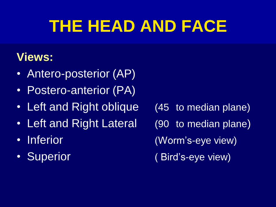
THE HEAD AND FACE
Views:
• Antero-posterior (AP)
• Postero-anterior (PA)
• Left and Right oblique (45 to median plane)
• Left and Right Lateral (90 to median plane)
• Inferior (Worm’s-eye view)
• Superior ( Bird’s-eye view)
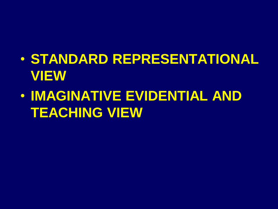
• STANDARD REPRESENTATIONAL
VIEW
• IMAGINATIVE EVIDENTIAL AND
TEACHING VIEW

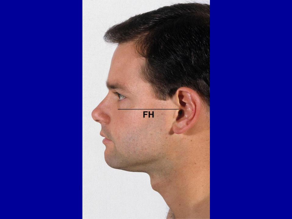
FRANKFURT HORIZONTAL /
PLANE
– A plane defined by three osteometric
points, the right and left porion points and
the left orbitale. These osteometric points
are at the top of each external auditory
meatus and the bottom of the orbital
margin
– It is used to systematically orient the skull.
(Convention held in Frankfurt in 1884)

MEASURING LINEAR
PROJECTIVE DISTANCES
The Head and Face should be photo-
graphed in the standard antero-posterior
and lateral positions. The position of the
head should be standard (in Frankfurt
Horizontal).


COMPARISONS BETWEEN
FACES MAY BE MADE TO:
• identify offenders
• identify missing persons
• confirm or refute claims of identity
• study the relationship between the skull
and the overlying soft tissue
• develop diagnostic methods for facial
syndromes

FACIAL LANDMARKS
Can be used:
• to establish points of reference
• to identify features defining the cranial-facial geometry of humans
• to perform reconstructive or correctivesurgery
• by forensic artists to identify the remains ofindividuals, or when they compose age-adjusted photographs of missing children
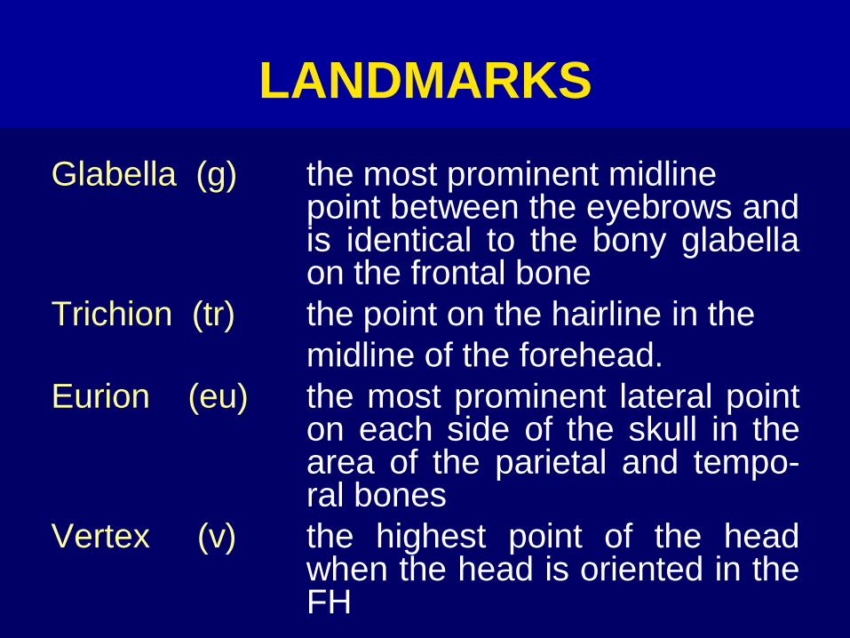
LANDMARKS
Glabella (g) the most prominent midlinepoint between the eyebrows andis identical to the bony glabellaon the frontal bone
Trichion (tr) the point on the hairline in the
midline of the forehead.
Eurion (eu) the most prominent lateral pointon each side of the skull in thearea of the parietal and tempo-ral bones
Vertex (v) the highest point of the headwhen the head is oriented in theFH

CRANIOFACIAL SURFACE /
SKELETON LANDMARKSv vertex
tr trichion
eu eurion
g glabella
zy zygion
go gonion
gn gnathion
pg pogonion
sl sublabiale
ch cheilion
cph crista philtri
sn subnasale
al alare
prn pronasale
or orbitale
ex ectocanthion
en endocanthion
ft frontotemporale
sci superciliare
gn
pg
slch
snal
or
exex
ft
tr
v
tr
geu
zy
go go
gn
zy
eu
sci
en en
g
alprn
gocph
ch


ORIENTATION
• Different angles can cause unnecessary confusion
• Consider:
(a) camera to the subject
(b) area of interest on the body
(c) orientation of individual body parts
(d) standardization rules

POSITIONING OF SKULL
• Skull holding jig
• Pan-and-tilt device
• Camera mount
• Supporting rails


STANDARDIZATION OF SKULL
PHOTOGRAPHS IN PERSONAL
IDENTIFICATIONS BY
PHOTOGRAPHIC SUPERIMPOSITION
• Relationship between the image of the unidentifiedsubject and the original photographic portrait
• Central projection (distance between camera andthe head or skull that resembles the distance of theoriginal photograph)
• Posture
• Magnification
• Perspective

BITEMARK EVIDENCE
• Standardized photography
• Scale of reference
• Teeth models and wax base
• Overlays
• Fluorescent image analysis
• Changes in the dermal and epidermal tissues
• Toneline bitemark photography
• Use of videotape
• Use of alternative light source – 450nm

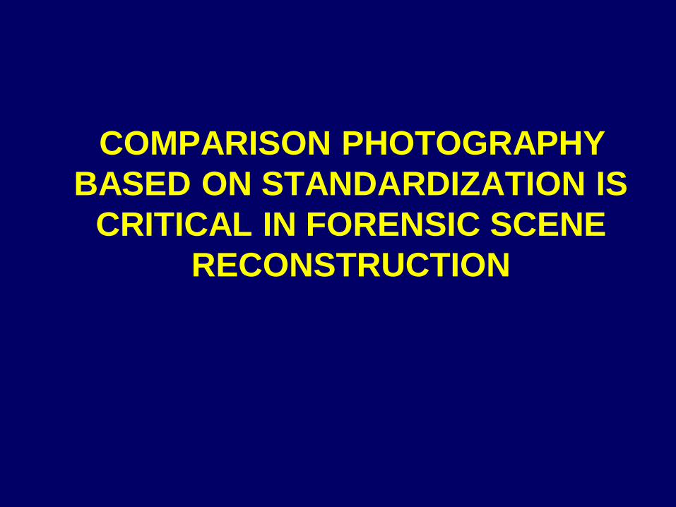
COMPARISON PHOTOGRAPHY
BASED ON STANDARDIZATION IS
CRITICAL IN FORENSIC SCENE
RECONSTRUCTION

• ACCURATE MEASUREMENTS
• COMPARISON PURPOSES

THE INFLUENCE OF MEDICAL
PHOTOGRAPHY’S WESTMINSTER
REPRODUCTION RATIOS ON
STANDARDIZATION IN FORENSIC
PHOTOGRAPHY – AN OPINION
Hoosain M EbrahimDepartment of Medical Illustration
and Audio Visual Services
University of Limpopo
South Africa