requirement of ras-gtp-raf complexes for activation of raf-1 by protein kinase c
TRANSCRIPT
18. A. M. Krezel et a/. , Prot. Sci. 4 1478 (1 995). assistance in preparat~on of the figures and helpful 19. A. Nlcholls, K. A. Sharp, B. Honig, Proteins 11, 281 discussions on the work. Supported by NIH
(1 991). GM43949. R.M. is an investigator in the Howard
20. T. A. Jones, J. Y. Zou, J. Y. Cowan, M. Kjeldgaard, Hughes Medical Institute.
Acta Crystallogr. A47, 1 10 (1 991). 21. We thank J. Gulbis, J. P. Imredy, and D. A. Doylefor 6 March 1998; accepted 13 March 1998
Requirement of Ras-GTP-Raf Complexes for Activation of Raf-I by Protein Kinase C
Richard Marais, Yvonne Light, Clive Mason, Hugh Paterson, Michael F. Olson, Christopher J. Marshall*
Receptor tyrosine kinase-mediated activation of the Raf-1 protein kinase is coupled to the small guanosine triphosphate (GTP)- binding protein Ras. By contrast, protein kinase C (PKCFmediated activation of Raf-1 is thought to be Ras independent. Nevertheless, stimulation of PKC in COS cells led to activation of Ras and formation of Ras-Raf-1 complexes containing active Raf-1. Raf-1 mutations that prevent its association with Ras blocked activation of Raf-1 by PKC. However, the activation of Raf-1 by PKC was not blocked by dominant negative Ras, indicating that PKC activates Ras by a mechanism distinct from that initiated by activation of receptor tyrosine kinases.
T h e PKC family of lipid-dependent Ser- phatidyl inositols. In the case of 7-trans- Thr kinases has at least 11 members (1). membrane (7-TM) receptors that are cou- The typical and atypical PKC isozymes are pled to the heterotrimeric guanosine nucle- activated by diacylglycerol (DAG), which otide-binding protein (G protein) Gaq, is produced by the metabolism of phos- DAG is generated by the activation of
phospholipase C-P (PLC-P) (2). Both the typical and atypical PKCs are activated by phorbol esters. Activation of PKC leads to short-term responses such as altered meta- bolic activity and to long-term responses such as differentiation or effects on prolif- eration and apoptosis (1 ). The extracellular signal-regulated kinases (ERKs) are mito- gen-activated protein kinases (MAPKs), which are activated by PKC (3-7) and ap- pear to mediate the effects of PKC on dif- ferentiation, secretion, proliferation, and hypertrophy (8). Signaling. from receptor tyrosine kinases to ERKs is dependent on Ras proteins and the protein kinase Raf-1 (9). However, the role of Ras in transducing signals from PKC to the ERKs is unclear, because expression of a dominant negative Ras in which amino acid 17 is changed to Asn (N17Ras) does not block ERK activa- tion by PKC in a number of cell types (4, 5). This Ras mutant, which is thought to function by inhibiting guanine nucleotide exchange factors ( lo) , blocks activation of ERKs in response to stimulation of receptor tyrosine kinases in many cell types (4-6).
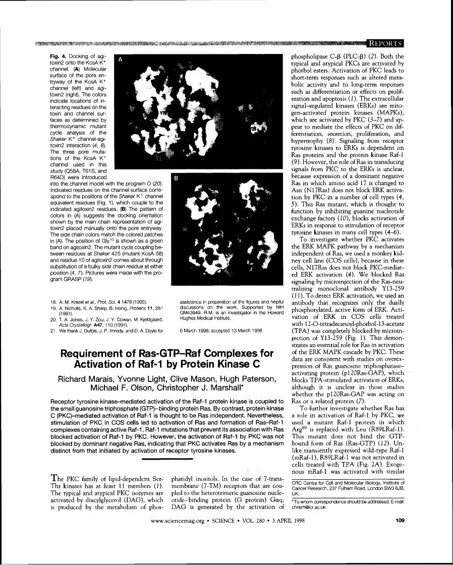
To investigate whether PKC activates the ERK MAPK pathway by a mechanism independent of Ras, we used a monkey kid- ney cell line (COS cells), because in these cells, N17Ras does not block PKC-mediat- ed ERK activation (4). We blocked Ras signaling by microinjection of the Ras-neu- tralizing monoclonal antibody Y13-259 (1 1 ). To detect ERK activation, we used an antibody that recognizes only the dually phosphorylated, active form of ERK. Acti- vation of ERK in COS cells treated with 12-0-tetradecanoyl-phorbol-13-acetate (TPA) was completely blocked by microin- jection of Y13-259 (Fig. 1). This demon- strates an essential role for Ras in activation of the ERK MAPK cascade by PKC. These data are consistent with studies on overex- pression of Ras guanosine triphosphatase- activating protein (pl 2ORas-GAP), which blocks TPA-stimulated activation of ERKs, although it is unclear in those studies whether the pl2ORas-GAP was acting on Ras or a related protein (7).
To further investigate whether Ras has a role in activation of Raf-1 by PKC, we used a mutant Raf-1 protein in which Arge9 is replaced with Leu (R89LRaf-1). This mutant does not bind the GTP- bound form of Ras (Ras-GTP) (12). Un- like transiently expressed wild-type Raf-1 (mRaf-1), R89LRaf-1 was not activated in cells treated with TPA (Fig. 2A). Exoge- nous mRaf-1 was activated with similar
CRC Centre for Cell and Molecular Biology, Institute of Cancer Research, 237 Fulham Road, London SW3 6JB, UK.
'To whom correspondence should be addressed. E-mail: [email protected]
www.sciencemag.org SCIENCE VOL. 280 3 APRl
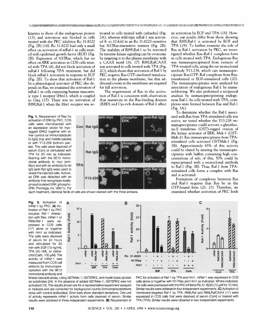
kinetics to those of the endogenous protein (13), and activation was blocked in cells treated with the PKC inhibitor Ro 31-8220 (Fig. 2B) (14). Ro 31-8220 had only a small effect on activation of mRaf-1 in cells treat- ed with epidermal growth factor (EGF) (Fig. 2B). Expression of N17Ras, which has no effect on ERK activation in COS cells treat- ed with TPA (4), did not block activation of mRaf-1 following TPA treatment, but did block mRaf-1 activation in response to EGF (Fig. 2B). To show that activation of Raf-1 by a physiological activator of PKC also de- pends on Ras, we examined the activation of mRaf-1 in cells expressing human muscarin- ic type 1 receptor (Hml), which is coupled to G a q (15). There was no activation of R89LRaf-1 when the Hml receptor was ac-
Fig. 1. Requirement of Ras for activation of ERK by PKC. COS cells were microinjected with an expression vector for myc- tagged ERK2 together with ei- ther control rat immunoblobulin G (IgG) (top and middle panels) or with Y13-259 (bottom pan- els). The cells were deprived of serum (Con) or stimulated with TPA for 20 min as indicated. Staining with the 9E10 mono- clonal antibody to myc (anti- Myc) and with an antibody to rat IgG (anti-Rat IgG) were used to reveal the injected cells. Activat- ed ERK was detected with an antibody that recognizes dually- phosphorylated ERK (phospho- ERK; Promega, no. V6671). For each treatment, identical fields of
Fig. 2. Activation of A mRaf-1 by PKC. (A) Ac- tivation of Raf-1 by PKC
tivated in cells treated with carbachol (Fig. 2A), whereas wild-type mRaf-1 was activat- ed 8- to 10-fold in an Ro 31-8220-sensitive but N 17Ras-insensitive manner (Fig. 2B). The inability of R89LRaf-1 to be activated by tyrosine kinase signaling can be overcome by targeting it to the plasma membrane with a CAAX motif (1 6, 17). R89LRafCAAX was activated in cells treated with TPA (Fig. 2C), which shows that activation of Raf-1 by PKC requires Ras-GTP-mediated transloca- tion to the plasma membrane, but that ad- ditional events at the membrane are required for full activation.
The requirement of Ras in the activa- tion of Raf-1 is consistent with observation that mutations in the Ras-binding domain (RBD) and Cys-rich domain of Raf-1 affect
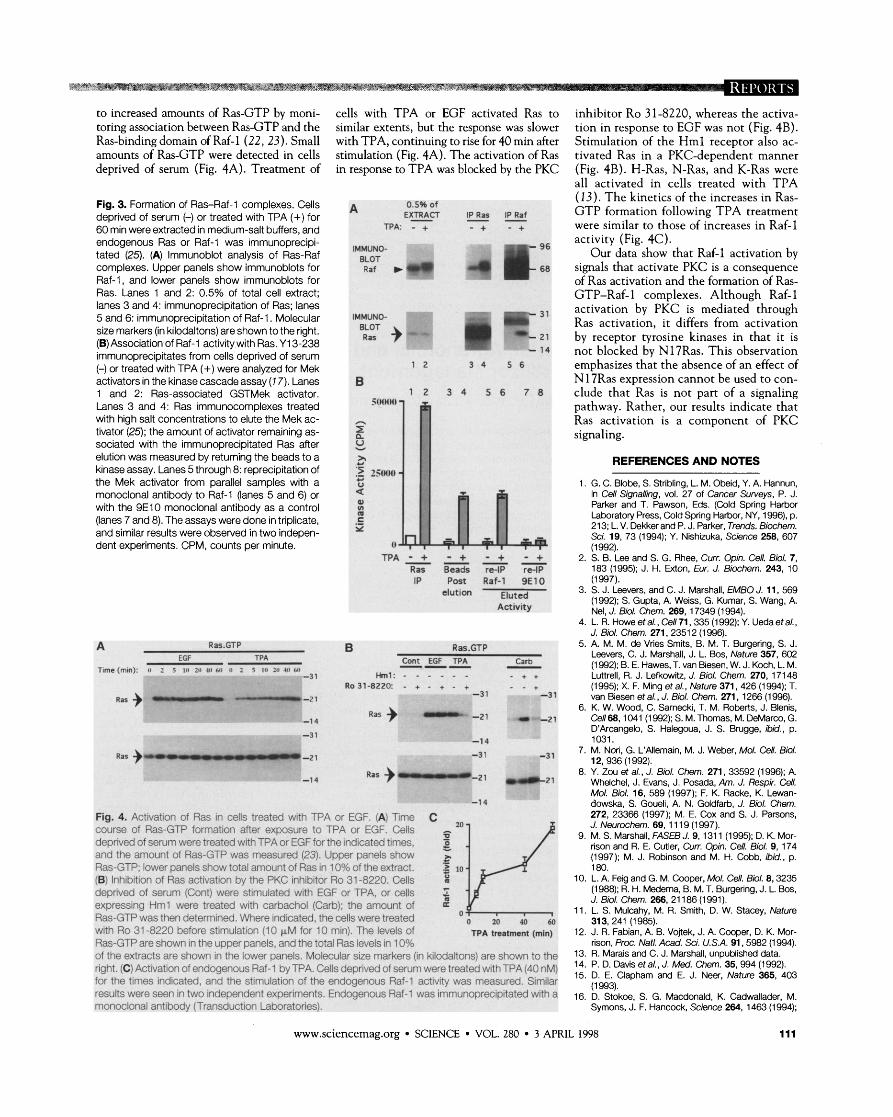
its activation by EGF and TPA (1 8). How- ever, our results differ from those showing that R89LRaf-1 is activated by EGF and TPA (1 9). To further examine the role of Ras in Raf-1 activation by PKC, we inves- tigated whether Ras-Raf-1 complexes form in cells treated with TPA. Endogenous Ras was immunoprecipitated from extracts of TPA-treated cells, using the rat monoclonal antibody Y 13-238, which can immunopre- cipitate Ras-GTP-Raf complexes from Ras- transformed or EGF-stimulated cells (20). The immunoprecipitates were analyzed for association of endogenous Raf-1 by immu- noblotting. We also performed a reciprocal analysis by immunoprecipitating endoge- nous Raf-1. In cells treated with TPA, com- plexes were formed between Ras and Raf-1 (Fig. 3A).
To determine whether the Raf-1 associ-
TPA
stimulated cells activated-GSTM~~-1 (Fig.
cipitates with buffers containing high con- centrations of salt; of this, 50% could be reprecipitated with a monoclonal antibody
'cells are shown stained with the three antisera. examined whether activation of PKC leads
requires Raf-1 interac- A c.
2 - tion with Ras mRaf-1 or % - 0 15 B - 0 R89LRaf-1 were ex- ; 10
2 tf.
pressed in COS cells 5 .- a > L .- .- (7 7) alone or together = .; l o .h .- 4 with Hml as indicated. 2 m 9 The cells were deprived z - of serum for 24 hours L 2 5 and stimulated for 20 E
min with EGF (1 0 ng/ml), 0 0 TPA (40 nM), or carba- X Z z % - 3 2 E E 0 chol (Carb, 100 pM). The E 19 E g E d R031-aZ20 - - + - . + . - + -
activity of mRaf-1 was ci co ci OD rn u Nl7Ras - - - + - - + - - + measured from COS cell ~ m l - - - - - + + --- Hml - - - . . - - + + + m extracts by immunopre- P
ES TPA Carb --- 2 E
cipitation with the 9E10 g;F TPA Carb -- Cont TPA monoclonal antibody and kinase cascade assay, using GSTMek-1, GSTERK2, and myelin basic protein PKC for activation of Raf-1 by TPA and Hml . mRaf-1 was expressed in COS as substrates (24). In the absence of added GSTMek-1, GSTERK2 was not cells alone or together with N17Ras and Hml as indicated. Where indicated, activated (73). The results shown are for a representative experiment assayed the cells were pretreated with the PKC inhibitor Ro 31 -8220 (1 0 pM for 10 rnin). in triplicate and are corrected for background counts (immunoprecipitations Similar results were obtained in four independent experiments. (C) Activation of done with control antibodies). Error bars show standard deviations. One unit membrane-targeted Raf-1 by TPA. R89LRaf and R89LRafCAAX (1 7) were of activity represents mRaf-1 activity from cells deprived of serum. Similar expressed in COS cells that were deprived of serum (Cont) or treated with results were obtained in three independent experiments. (B) Requirement of TPA (TPA). Similar results were obtained in two independent experiments.
110 SCIENCE VOL. 280 3 APRIL 1998 www.sciencemag.org
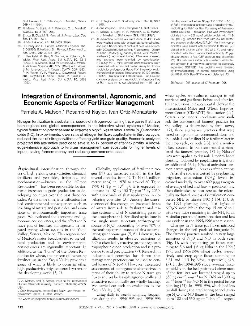
to increased amounts of Ras-GTP by moni- cells wi th TPA or EGF activated Ras to inhibitor Ro 31-8220, whereas the activa- toring association between Ras-GTP and the similar extents, but the response was slower t ion in response to EGF was not (Fig. 4B). Ras-binding domain of Raf-1 (22, 23). Small wi th TPA, continuing to rise for 40 m in after Stimulation of the H m l receptor also ac- amounts of Ras-GTP were detected in cells stimulation (Fig. 4A). The activation of Ras tivated Ras in a PKC-dependent manner deprived of serum (Fig. 4A). Treatment of in response to T P A was blocked by the PKC (Fig. 4B). H-Ras, N-Ras, and K-Ras were
a l l activated in cells treated w i th T P A
Fig. 3. Formation of Ras-Raf-1 complexes. Cells A 0.5% of (13). The kinetics o f the increases in Ras-
deprived of serum (-) or treated with TPA (+) for E ~ ~ C T IPS z f G T P formation following T P A treatment
60 min were extracted in medium-salt buffers, and TPA: - + - + - + were similar to those o f increases in Raf-1
endogenous Ras or Raf-1 was immunoprecipi- IMMUNO- activity (Fig. 4C). tated (25). (A) lmmunoblot analysis of Ras-Raf BLOT
Our data show that Raf-1 activation by complexes. Upper panels show immunoblots for Raf w ')(b 4 1: signals that activate PKC is a consequence Raf-1, and lower panels show immunoblots for of Ras activation and the formation of Ras- Ras. Lanes 1 and 2: 0.5% of total cell extract; lanes 3 and 4: immunoprecipitation of Ras; lanes 5 and 6: immunoprecipitation of Raf-1 . Molecular size markers (in kilodaltons) are shown to the right. (B) Association of Raf-1 activitywith Ras. Y13-238 immunoprecipitates from cells deprived of serum (-) or treated with TPA (+) were analyzed for Mek activators in the kinase cascade assay (7 7). Lanes 1 and 2: Ras-associated GSTMek activator. Lanes 3 and 4: Ras immunocomplexes treated with high salt concentrations to elute the Mek ac- tivator (25); the amount of activator remaining as- sociated with the immunoprecipitated Ras after elution was measured by returning the beads to a kinase assay. Lanes 5 through 8: reprecipitation of the Mek activator from parallel samples with a monoclonal antibody to Raf-1 (lanes 5 and 6) or with the 9E10 monoclonal antibody as a control (lanes 7 and 8). The assays were done in triplicate, and similar results were observed in two indepen- dent experiments. CPM, counts per minute.
IMMUNO- '- w =31
-- 21
F A - + - + - + - + Ras Beads re-IP re-IP iP Post Raf-1 9E10
=Iution Eluted Activity
, . -
Time (rnin): I I 1 5 111 211 41) 611 11 2 5 1,) :<I 411 &I) -31
COnf tGF I P A --- Carb - Hml: - - - - - - - + +
-21
-1 4 Ras -) r
Fig. 4. Activation of Ras in cells treated with TPA or EGF. (A) Time C 20 course of Ras-GTP formation after exposure to TPA or EGF. Cells =
deprived of serum were treated with TPA or EGFfor the indicated times, : and the amount of Ras-GTP was measured (23). Upper panels show Ras-GTP; lower panels show total amount of Ras in 1 OO/o of the extract. 2 10 (6) Inhibition of Ras activation by the PKC inhibitor Ro 31-8220. Cells 8 deprived of serum (Cont) were stimulated with EGF or TPA, or cells 5 expressing Hml were treated with carbachol (Carb); the amount of a
Ras-GTP was then determined. Where indicated, the cells were treated o I-/ o 20 40 60
with Ro 31 -8220 before stimulation (10 KM for 10 min). The levels of TPA tmatrnant (rnln) Ras-GTP are shown in the upper panels, and the total Ras levels in 1 OO/o of the extracts are shown in the lower panels. Molecular size markers (in kilodaltons) are shown to the right. (C) Activation of endogenous Raf-1 by TPA. Cells deprived of serum were treated with TPA (40 nM) for the times indicated, and the stimulation of the endogenous Raf-1 activity was measured. Similar results were seen in two independent experiments. Endogenous Raf-1 was immunoprecipitated with a monoclonal antibody (Transduction Laboratories).
GTP-Raf-1 complexes. Although Raf-1 activation by PKC is mediated through Ras activation, it differs from activation by receptor tyrosine kinases in that it is no t blocked by N17Ras. This observation emphasizes that the absence of an effect o f N17Ras ex~ression cannot be used to con- clude that Ras is no t part o f a signaling pathway. Rather, our results indicate that Ras activation is a component o f PKC signaling.
REFERENCES AND NOTES
1. G. C. Blobe. S. Stribling, L. M. Obeid, Y. A. Hannun, in Cell Signalling, vol. 27 of Cancer Surveys, P. J. Parker and T. Pawson, Eds. (Cold Spring Harbor Laboratoty Press, Cold Spring Harbor. NY. 1996), p. 213; L. V. Dekker and P. J. Parker, Trends. Biochem. Sci. 19, 73 (1994); Y. Nishizuka, Science 258, 607 (1 992).
2. S. B. Lee and S. G. Rhee, Curr. Opin. Cell. Biol. 7, 183 (1995); J. H. Exton, Eur. J. Biochem. 243, 10 (1997).
3. S. J. Leevers, and C. J. Marshall, EMBO J. 11, 569 (1992); S. Gupta, A. Weiss, G. Kumar, S. Wang, A. Nel, J. Biol. Chem. 269, 17349 (1994).
4. L. R. Howe et al. , Cell 71 , 335 (1 992); Y. Ueda et a/. , J. Biol. Chem. 271,23512 (1996).
5. A. M. M. de Vries Smits, B. M. T. Burgering, S. J. Leevers, C. J. Marshall, J. L. Bos, Nature 357, 602 (1992); B. E. Hawes, T. van Biesen, W. J. Koch, L. M. Luttrell, R. J. Lefkowitz, J. Biol. Chem. 270, 17148 (1 995); X. F. Ming et al., Nature 371 , 426 (1 994); T. van Biesen etal., J. Biol. Chem. 271. 1266 (1996).
6. K. W. Wood, C. Samecki, T. M. Roberts, J. Blenis, Ce1168,1041 (1992); S. M. Thomas, M. DeMarco, G. D'Arcangelo, S. Halegoua, J. S. Brugge, ibid., p. 1031.
7. M. Nori, G. L'Allemain, M. J. Weber, Mol. Cell. Biol. 12, 936 (1 992).
8. Y. Zou et a/., J. Biol. Chem. 271, 33592 (1996); A. Whelchel, J. Evans, J. Posada, Am. J. Respir. Cell. Mol. Biol. 16, 589 (1997); F. K. Racke, K. Lewan- dowska, S. Goueli, A. N. Goldfarb, J. Biol. Chem. 272, 23366 (1997); M. E. Cox and S. J. Parsons, J. Neurochem. 69, 1 1 19 (1997).
9. M. S. Marshall, FASEB J. 9, 131 1 (1995); D. K. Mor- rison and R. E. Cutler, Curr. Opin. Cell. Biol. 9, 174 (1997); M. J. Robinson and M. H. Cobb, ibid., p. 180.
10. L. A. Feig and G. M. Cooper, Mol. Cell. Biol. 8,3235 (1 988); R. H. Medema, B. M. T. Burgering, J. L. Bos, J. Biol. Chem. 266, 21 186 (1991).
11. L. S. Mulcahy, M. R. Smith, D. W. Stacey, Nature 313, 241 (I 985).
12. J. R. Fabian, A. B. Vojtek, J. A. Cooper, D. K. Mor- rison, Proc. Natl. Acad. Sci. U.S.A. 91,5982 (1994).
13. R. Marais and C. J. Marshall, unpublished data. 14. P. D. Davis et a/. , J. Med. Chem. 35,994 (1 992). 15. D. E. Clapham and E. J. Neer, Nature 365, 403
(1 993). 16. D. Stokoe, S. G. Macdonald, K. Cadwallader, M.
Syrnons, J. F. Hancock, Science 264, 1463 (1 994);
www.sciencemag.org SCIENCE VOL. 280 3 APRIL 1998 111
S. J. Leevers H. F. Paterson C. J. Marsha. Nature 369, 41 1 (1 994).
17. R. Marals Y. Llght, H. F. Paterson C. J. Marshall. EM50 J. 14 3136 (1 995).
18. 2. Lou B. Diaz M. S. Marsha, J. Avruch Mol. Cell. 5/01. 17, 46 (1 997).
19. H. Mschak et a/. . ib~d. 16 5409 (1 996). 20. R. Flnney and D. Herrera, Methods Enzymol. 255
310 (1995); B. Haberg S. I. Rayter. J. Downward, J. Biol. Chem. 269 391 3 (1 994).
21. L. Van Aelst. M. Barr. S. Marcus. A. Polver~no. M. Wlgler, Proc Nail. Acad. SCI. U.S.A. 90, 6213 (1993); S. A. Moode, B. M. Wiumsen, M. J. Weber, A. Wofman. Sclence 260. 1658 (1 993): A. B. Vojtek S. M. Holenberg, J. A. Cooper. Cell 74 205 (1993); P. H. Warne, P. R. Vlclana, J. Downward, Nature 364,352 (1993). H. Kode, T. Satoh, M. Nakafuku, Y. Kazlro Proc. Nail. Acad. SCI. U.S.A. 90, 8683 (1 993).
22. S. J. Taylor and D. Shaloway. Curr. 5101. 6 1621 (1 996).
23. J de ROOIJ and J. Bos Oncogene 14, 623 (1997). 24. R. Marals, Y. L~ght H. F. Paterson, C. S. Mason,
C. J. Marsha J. 5101. Chem. 272. 4378 (1997). 25. A extractlon procedures were done at 4°C. Cells
were washed twce wlth phosphate-buffered sane, and each 10-cm dsh of confluent cells was extract- ed In 300 p of d u t o n buffer (1 7) contanng I 0 0 mM K C and 5 mM MgCl, but only 0.05% v/v 2-mercap- toethanol (med~um-salt buffer). DNA was sheared and extracts were clarlfled by centr~fugat~on (13,000g for 2 mn). proten concentratons were measured wlth a BIO-Rad protein assay kit. mmu- noblotsfor endogenous Ras or Raf-1 were done w th monoclonal antbodes (products no. 021 20 and no. R19120, Transduct~on Laboratories). For Ras-Raf co~npex analysis by ~mmunoblot. endogenous Ras or Raf-1 was mrnunoprec~p~tated from -15 mg of
Integration of Environmental, Agronomic, and Economic Aspects of Fertilizer Management
Pamela A. Matson," Rosamond Naylor, Ivan Ortiz-Monasterio
Nitrogen fertilization is a substantial source of nitrogen-containing trace gases that have both regional and global consequences. In the intensive wheat systems of Mexico, typical fertilization practices lead to extremely high fluxes of nitrous oxide (N,O) and nitric oxide (NO). In experiments, lower rates of nitrogen fertilizer, applied later in the crop cycle, reduced the loss of nitrogen without affecting yield and grain quality. Economic analyses projected this alternative practice to save 12 to 17 percent of after-tax profits. A knowl- edge-intensive approach to fertilizer management can substitute for higher levels of inputs, saving farmers money and reducing environmental costs.
Agricultural intensification through the use of high-yielding crop varieties, chemical fertilizers and pesticides, irrigation, and mecha~lization-known as the "Green Revolution"-has bee11 responsible for dra- matic increases in grain production in de- veloping countries over the past three de- cades. A t the same time, inte~lsification has had en \~ i ronme~l ta l consequences such as leaching of nitrate and pesticides, and einis- slons of environmentally important trace gases. W e el~aluated the economlc and ag- ronomic consequences, and the effects o n N trace gas, of fertilizer management in irri- gated spring wheat systems in the Yailui Vallev. Sonora. Mexico. This reeion is one , , " of Mexico's major breadbaskets, so agricul- tural vroduction and its environine~ltal consequences are regionally important. In addition, as the "home" of the Green Rev- o l u t i o ~ ~ for wheat, the pattern of increasing fertilizer use in the Yaqui Valley prol~ides a gauge of what is likely to occur in other high-productivity irrigated cereal systems of the del~eloping world (1 , 2).
P. A. Matson and R. Naylor. nstltute for International Studes. Stanford University. Stanford. CA 94305-6055 USA. I. Orilz-Monasterio International Maize and Wheat m - provement Center. El Batan Mexlco.
*To whom correspondence should be addressed
Globally, applicatio~l of fertilizer nitro- gen ( N ) has increased rapidly in the last several decades, from 32 Tg N (32 million metric tons) in 1970 to around 80 Tg in 1990 ( 1 Tg = 10" g); it is expected to increase to 130 to 150 T g yearp1 by 2050, with two-thirds of that application in de- veloping countries (3). Among the conse- quences of this change are increased losses of nitrate from soils to freshwater and ma- rine systems and of N-containing gases to the atmosphere (4). Fertilized agriculture is the single most important anthropogenic source of N 2 0 , accounting for o17er 70% of the anthropogenic sources of this accuinu- lating greenhouse gas (5, 6) . Likewise, fer- tilizatio~l results in elevated emissions of N O , a cheinically reactive gas that regulates tropospheric ozone production and is a pre- cursor to acid precipitation (7). Research in industrialized cou~ltries has shown that management practices can be used to con- trol losses of N (6-9). However, integrated assessments of management alternatives in terms of their abilitv to reduce N trace eas fluxes and yet be feakible agronomically a71d attractive econoinically are wholly lacking. W e carried out such a n evaluation in the Yaqui Valley ( 10).
Using daily to weekly sampling frequen- cies during the 199411995 and 199511996
cellular proten wlth elther 10 p g of Yl3-238 or 10 p g of Raf-I monoclonal antbody and probed by mmu- nobottng for Raf-I or Ras. For assay of Ras-asso- clated GSTivlek-1 actlvatlon Ras was mmunopre- cpltated from -3.5 mg of cellular proten wlth Y13- 238 (10 pg), washed four tmes with low-salt buffer, and assayed as descrbed (1 7,241 Ras mmunopre- clptates were eluted with extractlon buffer (40 pi), du ted with d u t ~ o n buffer (160 pl) (171, and repre- clpltated wlth Raf-1 monoclonal antlbody (2 kg). Measurements of Ras-GTP were done as descrbed (23). The cells were extracted In medum-salt buffer. and protens (-3 mg) were absorbed to bacteraly expressed GSTRBD. Ras protelns were revealed by mmunoblott~ng. In control experiments uslng GSTR89LRBD Ras-GTP was not detected (13).
28 August 1997. accepted 17 February 1998
wheat cycles, we evaluated changes in soil nutrients and eas fluxes before and after fer- - tllizer additions in experimental plots at the International Maize and \Xiheat Improve- ment Center (CIMMYT) field station (1 1) . Sel~eral exverilnental conditions were stud- led, the con\~entional farmers' practlce for the vallev, as determined by farm sur17ev , , (12); three alternative practices that werb based o n agronomist recommendations and that added less fertilizer N or fertilizer later in the crop cycle, or both (13); and a nonfer- tilized control. 111 our treatment that siinu- lated the farmers' practice, 187 kg N/ha of urea were applied to dry soils 1 month beore planting, followed by preplanting irrigation; a n additional 63 kg N/ha of anhydrous am- m a ~ l i a were applied -6 weeks after planting.
After the soil was wetted by preplanting irrigation, ammonium (NH,) levels in- creased markedly to over 600 kg/g (weight- ed average of bed and furrow positions) and then diminished to near zero as the micro- bially mediated process of ~litrification con- l~er ted N H 4 to nitrate (NO,) (14, 15) . By the 1994 planting date, 116 kglha of NO, -N were left in the ton 15 cm of soil. with very little remaining in the NH, form. A similar pattern of transformation and loss was e l~ident in the 199511996 wheat season.
Changes in N trace gas fluxes mirrored changes in the soil pools of inorganic N . T h e farmers' practice resulted in 17ery large elllissions of N 2 0 and N O in both years (Fig. I ) , with preplanting gas fluxes sum- ming to 5.6 and 4.6 kg N/ha in the 1994/ 1995 and 199511996 wheat cycles, resuec- t i ~ e l y , and crop cycle fluxes summing to 6.61 and 11.3 kg Niha, respectively (16, 17) . In the 199411995 study, a17erage fluxes at midday in the bed positions (where most of the fertilizer was located) ranged up to 650 ng cmp2 hourp1 for N,O-N and 300 ng cin-2 hour-' for NO-N i n i h e period before planting (15). 111 199511996, which had less rainfall during the preplanting period, aver- age N,O and N O fluxes in the beds ranged up to 100 and 550 ng cm-' hourp1, respec-
112 SCIENCE \'OL 280 3 APRIL 1998 www.sciencemag.org