research article a study of oxygenation status and hrct
TRANSCRIPT
International Academic Research Journal of Internal Medicine & Public Health
ISSN Print : 2709-3301 | ISSN Online : 2709-331X Frequency : Bi-Monthly Language : English Origin : Kenya Website : https://www.iarconsortium.org/journal-info/iarjimph
Available: https://www.iarconsortium.org/journal-info/iarjimph
56
A Study of Oxygenation Status and HRCT Thorax in “Asymptomatic
& Mildly Symptomatic RT-PCR Positive COVID 19” Subjects Abstract: Background: Coronavirus disease 2019 (COVID-19) is an
infectious disease caused by severe acute respiratory syndrome coronavirus 2 (SARS-CoV-2). The virus is primarily spread between people during close contact, often via small droplets produced by
coughing, sneezing, or talking. 80% people with COVID-19 develop mild illness, 14% develop severe disease with lower respiratory involvement
that requires hospitalisation and oxygen support. 5% require ICU admission. A minority of cases (20%) do not have symptoms even with oxygen saturation <94% (“Happy Hypoxia”). They are asymptomatic on presentation and may collapse suddenly. Aims & Objectives: To evaluate
the oxygenation status in asymptomatic and mildly symptomatic RT-PCR positive COVID -19 subjects admitted at Government Medical College,
Nagpur. To assess the Lungs in these patients by imaging with HRCT Thorax and to study the in-hospital outcome of these patients. Methodology: This prospective observational study was performed at a
tertiary care hospital in May 2020 and June 2020 after taking approval from the Institutional Ethics Committee. 62 (35M/ 27F) asymptomatic
SARS CoV2 positive by RTPCR were included in the study. The subjects were divided into two groups - Group A - who had a Oxygen Saturation of < 94% by Pulse oximetry on admission and Group B having Saturation
> 94% on admission. Radiological evaluation with Xray Chest and CT Thorax was done in all subjects. Results:- There were 19 (30.64 %)
patients in Group A and 43 (69.35 %) patients in Group B. The Respiratory Rate in patients with hypoxia (Group A) was significantly more in patients with Hypoxia as compared to those without hypoxia
(Group B), however they were not symptomatic with complaints of breathlessness. On imaging of all patients - 15 (24.19%) patients had findings on X Ray Chest as compared to 43 (69.35%) on CT Thorax. 16
(84.21%) of Group A as compared to 27 (62.79%) of Group B patients had CT finding consistent with COVID 19 Infection. Ground Glass
opacities(GGO), GGO with Consolidation & Consolidation with Air Bronchogram in 26 (60.4%), 4 (9.3%), 4 (9.3%) respectively were the most common findings on CT Thorax in the study subjects. X-Ray chest
was found to have poor sensitivity and specificity to diagnose COVID -19 infection as compared to HRCT Thorax. Conclusions: 30% of
asymptomatic SARS CoV2 positive patients had “Happy Hypoxia”.
Ground glass opacities and consolidation were commonly found on CT Thorax of these patients. Xray Chest had poor sensitivity and specificity
to diagnose the condition as compared to CT Thorax.
Keywords: Happy Hypoxia, Ground Glass Opacities(GGO).
BACKGROUND Coronavirus disease 2019 (COVID-19) is an infectious disease caused by severe acute respiratory syndrome
coronavirus 2 (SARS-CoV-2). The disease was first identified in December 2019 in Wuhan, the capital of China's Hubei province, and has since spread globally, resulting in the ongoing 2019–20 coronavirus pandemic (Huang, C. et al., 2020; & Zhu, N. et al., 2020).
The virus is primarily spread between people during close contact, often via small droplets produced by coughing,
sneezing, or talking. Common symptoms include fever, cough, and shortness of breath. Other symptoms may include
Research Article
Article History
Received: 16.03.2021
Revision: 23.03.2021
Accepted: 30.03.2021
Published: 10.04.2021
Author Details Dr Sajal Mitra
1, Dr Dipti Chand
2, Dr
Sanjeev Kumar3, Dr Jawahar Rathod
4 &
Dr Sagar Khandare5
Author’s Affiliations 1Dr Sajal Mitra - Hon Dean, Government
Medical College, Nagpur.
2Dr Dipti Chand - Associate Professor in
Medicine, Government Medical College,
Nagpur. 3Dr Sanjeev Kumar - Divisional
Commissioner, Nagpur. 4Dr Jawahar Rathod - Associate Professor
inRadiology, Government Medical College, Nagpur
5Dr Sagar Khandare - Assistant Professor
in Medicine, Government Medical
College, Nagpur.
Corresponding Author* Dr Sajal Mitra
How to Cite the Article: Sajal Mitra, et al; (2021): A Study of
Oxygenation Status and HRCT Thorax in
“Asymptomatic & Mildly Symptomatic RT -
PCR Positive COVID 19” Subjects. Int Aca.
Res. J Int. Med. Pub. Hlth. 2(2), 56-62
Copyright @ 2021: This is an open-access article distributed under the terms of the Creative Commons Attribution license which permits unrestricted use, distribution, and reproduction in any medium for non commercial use (NonCommercial, or CC-BY-NC) provided the original author and source are credited.
Dr Sajal Mitra, et al; Int Aca. Res. J Int . Med. Pub. Hlth; Vol-2, Iss- 2 (Mar-Apr, 2021): 56-62
57
fatigue, muscle pain, diarrhoea, sore throat, loss of
smell, taste, and abdominal pain. The time from exposure to onset of symptoms is typically around five days but may range from two to fourteen days. While
most people with COVID-19 develop only mild or uncomplicated illness, approximately 14% develop severe disease with lower respiratory involvement that
requires hospitalisation and oxygen support. 5% require admission to an intensive care unit for viral pneumonia,
ARDS and multi-organ failure (Rothe, C. et al., 2020). A minority of cases (20%) do not develop
noticeable symptoms at any point in time. These asymptomatic carriers tend not to get tested and they may contribute to the spread of the disease. Some of
these patients despite being COVID-19 positive are comfortable and asymptomatic, but have an oxygen
saturation <94%. A medical term “Happy Hypoxia” has been coined for it. A state where the body’s oxygen concentration gets low (to about 60%), but they
continue to be lucid and clear, behave normally till they deteriorate rapidly and collapse. These patients come to hospital with low oxygen levels but are not in distress.
These patients have low lung elastance and high compliance, are asymptomatic on presentation and may
collapse suddenly. These patients respond well to high FiO2, Noninvasive ventilation or oxygen through High Flow nasal Cannula, as compared to patients presenting
with Breathlessness and the classical ARDS like picture with high elastance and low compliance requiring management with invasive ventilation as per ARDS Net
protocol. Thus all patients with COVID 19 diagnosis warrant an estimation of the Oxygen Saturation by
Pulse Oximetry, even if they are asymptomatic (Gattinoni, L. et al., 2014).
The diagnosis and treatment program (6th version) published by the National Health Commission of the People's Republic of China had defined the diagnosis of
viral pneumonia based on radiologic features by radiologists as one of the diagnostic criteria for
COVID-19. The CT features of COVID-19 consist of predominantly peripheral (subpleural) ground glass opacities confined to the middle and lower zones of the
lung. As the disease advances, consolidation and coalescing infiltrates pervades the upper lobes. Appearance of fibrosis and resolution of GGO or
consolidation indicates improvement. Ground-glass opacification was defined as hazy increased lung attenuation with preservation of bronchial and vascular
margins, whereas consolidation was defined as opacification with obscuration of margins of vessels
and airway walls (Zhu, N. et al., 2020; & Rothe, C. et al., 2020).
STUDY OBJECTIVE
Primary To evaluate the oxygenation status by Pulse
Oximetry ( SPO2) in asymptomatic and mildly
symptomatic COVID -19 positive (by RT-PCR)
patients admitted at Government Medical College, Nagpur. Secondary
To structurally assess the Lungs in these patients by imaging - Xray Chest PA View and CT Thorax
To study the in-hospital outcome of these patients.
STUDY DESIGN
Prospective Observational Study.
STUDY SETTING Wards and COVID ICU of Government Medical College, Nagpur.
STUDY TIME PERIOD
May 2020 – June 2020
INCLUSION CRITERIA All SARS-Co V-2 - RTPCR Positive on
Nasopharyngeal or Oropharyngeal swab
irrespective of gender > 12 years of age. Patients who are Asymptomatic or mildly
symptomatic.
Lucid on admission.
EXCLUSION CRITERIA 1. Patients with previous cognitive deficit, aphasia,
neurological deficit were not included in the study . 2. Moderate and Severe Covid -19 patients.
METHODOLOGY After approval by the Institutional Ethics
Committee, and written informed consent from patients
or their relatives, this study included asymptomatic patients or mildly symptomatic - i.e. cough, sore throat and low grade fever, more than 12 years of age, who
tested positive for SARS-CoV-2 (RT-qPCR) on Nasopharyngeal or Oropharyngeal swab and were
admitted in COVID ward of Government Medical College, Nagpur. Patients with complaints of breathlessness, altered sensorium were not included in
the study.
The following data was collected: Age, Gender,
Demographic Data, SPO2 was measured all subjects hospitalised.
The subjects were divided into two groups - Group
A - who had a Oxygen Saturation of < 94% by Pulse oximetry on admission and Group B having Saturation
> 94% on admission. Various parameters of asymptomatic subjects with Happy Hypoxia( Group A) were compared with those without Happy Hypoxia (
Group B).

Dr Sajal Mitra, et al; Int Aca. Res. J Int . Med. Pub. Hlth; Vol-2, Iss- 2 (Mar-Apr, 2021): 56-62
58
Radiological evaluation with Xray Chest and CT
Thorax was done in all subjects. For each patient, the chest HRCT scan was evaluated for the following characteristics: (1) presence of ground-glass opacities,
(2) presence of consolidation, (3) number of lobes affected where either ground-glass or consolidative opacities were present, (4) degree of involvement of
each lung lobe with interlobular and intralobular septal thickening. (5) presence of nodules, (6) presence of a
pleural effusion, (7) presence of thoracic lymphadenopathy (defined as lymph node size of ≥10 mm in short-axis dimension), (9) airways abnormalities
(including airway wall thickening, bronchiectasis, and endoluminal secretions). Other abnormalities, including linear opacities, opacities with a rounded morphology,
opacities with a “reverse halo” sign, opacities with a “crazy-paving” pattern, and opacities with intralesional
cavitation, were noted. Each of the five lung lobes was assessed for degree of involvement and classified as none (0%), minimal (1 - 25%), mild (26 - 50%),
moderate (51 - 75%),
lobe score of 5. An overall lung “total severity
score” was reached by summing the five lobe scores (range of possible scores, 0 - 25) (Gattinoni, L. et al.,
2020; Li, Y., & Xia, L. 2020; & Francone, M. et al., 2020).
Patients were administered treatment as per the
hospital protocol laid down by the State Government and the clinical status of the patient subjects enrolled were either asymptomatic 37 (59.7 %) or mildly
symptomatic 25 (40.3 %) with features of upper respiratory complaints. None of the patients had breathlessness or fatigue as their presenting complaints.
57 (91.9%) belonged to Nagpur. 5 patients (8.06%)
were from out of Nagpur. These patients were brought to the hospital by the district health authorities with either history of contact with a positive patient - 22
(35.4%) or were from the containment area and were under quarantine - 34 ( 54.83 %) or had history of travel 6 (9.6 %).
12 patients had co-morbidities like diabetes 4 (6.4
%), Hypertension 6 (9.6 %), Hypothyroidism in 4 (6.4 %), 1 patient had COPD. or severe (76 - 100%). No involvement corresponded to
a lobe score of 0, minimal to a lobe score of 1, mild to a lobe score of 2, moderate to a lobe score of 3, and moderate to severe to a lobe score of 4, Severe to a .
Patients were evaluated during hospital stay and at discharge.
OBSERVATIONS & RESULTS
Total 62 patients were enrolled in the study among them 35 (56.45%) were Male and 27 (43.55%) were female patients. The
Table -1. Demographic Profile, Clinical Parameters & Investigations in Study Subjects
Total No of Subjects (n=62)
GROUP A (n= 19) GROUP B (n=43 ) P value
No of Subjects 62 19 43
Mean Age + SD 41.16 14.17 (13-80) 42.89 16.45(14-80) 40.36 13.18(17-65) 0.5267,NS Male/female 13/49 2/17 11/32 0.179,NS Domicile -Nagpur 57 17 40
Co-Morbidiites 12 2 10 0.313,NS H/O contact 56 17 39
0.881, NS H/O travel 6 2 4
Mean SPO2 on Adm 96.61 + 27490-99) 92.73 + 1.36(90-94) 98.32 + 0.56(97-99) <0.0001, HS
Mean Pulse Rate 84.95 + 5.92(70-
104)
87.68 + 5.97(76-
100)
83.74 + 5.55(70-
104) 0.0145,S
Mean SBP 118.54 + 9.02(110-160)
118.84 + 8.75(110-140)
118.37 + 9.24(110-160)
0.8192,NS
Mean DBP 118.54 + 9.02(70-160)
75.78 + 6.06 (70-90) 77.18 + 6.24(70-90) 0.4165,NS
Mean RR 16.06 + 2.42(12-24) 17.26 + 2.35(14-24) 15.53 + 2.29(12-24) 0.0086,HS
Mean Temp 97.5 + 0.2 97.6+-0.2 97.4+-0.3 NSChest Exam Crepitations
52 11 41 10 8 2
CVS, P/A, CNS WNL WNL WNL
Hb 11.87 + 1.55(6-15) 11.73 + 1.27(9.4-
14.2) 11.93 + 1.67(6-15) 0.6473,NS
WBC Count 3402.34 + 9946.54 (2.6-7500)
4557.8 + 1715.26 (3.2 – 7500)
2866.89 + 3546.61 (2.6-12100)
0.0411, S
Neutrophils 3392.62 + 1603.48 3389.47 + 1444.10 3394.04 + 1687.33 0.9317,NS

Dr Sajal Mitra, et al; Int Aca. Res. J Int . Med. Pub. Hlth; Vol-2, Iss- 2 (Mar-Apr, 2021): 56-62
59
(200-7100) (300-5800) (200-7100)
Lymphocytes 2511.47 + 1173.04 (700-8600)
2284.21 + 692.22 (1200-3600)
2614.28 + 1329.87 (700-8600)
0.5328,NS
N/L ratio 1.59 + 1.02(0.04-6.71)
1.61 + 0.76(0.10-2.68)
1.58 + 1.13(0.04-6.71)
0.4361,NS
ECG/ Qtc 405.62 +
24.74(3165-458)
398.94 + 33.63(316-
458)
408.64 + 19.22
(349-438) 0.1724,NS
Urea 20.50 + 6.64(10-45) 23.15 + 8.53(12-45) 19.30 + 5.28(10-33) 0.0961,NS
Serum Creatinine 0.86 + 0.15(0.40-
1.2)
0.92 + 0.14(0.70-
1.2) 0.82 + 0.15(0.4-1.1) 0.0272,S
Serum Bilirubin 0.54 + 0.26(0.1-1.4) 0.60 + 0.3(0.1-1.3) 0.52 + 0.23(0.2-1.4) 0.3414,NS
Xray Chest 14 9 5 CT Thorax 43 16 27 Heparin 3 1 2
Methyl Prednisolone 5 2 3 O2 support 3 1 2 NIV 0 0 0
Inv ventilation 0 0 0 Vasopressors 0 0 0
Table 1 shows the Demographic profile, Clinical characteristics & Investigations of the studied population. The asymptomatic subjects were divided into 2 groups - GROUP A - Those with
CT Thorax Showing Ground Glass Opacities and Thickened Intra and interlobular Septa
deoxygenation - Happy Hypoxia on room air 19
(30.64 %) on admission, and GROUP B - those with normal oxygen saturation on admission 43 (69.35 %).
All patients were able to maintain oxygenation on nasal oxygen supplementation. The mean Pulse rate in Group A was 87.68 + 5.97(76-100) and Group B was 83.74 +
5.55(70-104) with p value of 0.0145,S. The mean
Respiratory Rate of Group A was 17.26 + 2.35 (14-24) as compared to Group B 15.53 + 2.29(12-24) in Group
B (p value - 0.0086,HS) . The blood pressure of the two groups on admission was comparable (p NS).
Dr Sajal Mitra, et al; Int Aca. Res. J Int . Med. Pub. Hlth; Vol-2, Iss- 2 (Mar-Apr, 2021): 56-62
60
10 patients had adventitious sounds on chest
examination. Of the 19 patients with deoxygenation status on admission i.e. Group A, only in 8 (42.1%) patients clinical examination revealed adventitious
sounds. 2 (4.67%) patients in Group B also had crepitation on clinical examination. In 52 (83.8% ) patients the chest examination was clinically normal.
Haemoglobin concentration in the two Groups was
11.73 + 1.27(9.4-14.2) vs 11.93 + 1.67(6-15) was comparable (p value - 0.6473,NS).
The mean Neutrophil-lymphocyte ratio (NLR) in the study subjects was 1.59 + 1.02 (0.04-6.71) with 1.61 + 0.76 (0.10-2.68 ) in Group A vs 1.58 1.13(0.04-6.71)
in Group B (p value 0.4361,NS ).
The mean QTc in the study subjects was 405.62 +
24.74 (316.5-458) with 398.94 + 33.63 (316-458) in Group A vs 408.64 19.22 (349-438) in Group B (p value 0.1724,NS). 2 patients had ST- T changes on
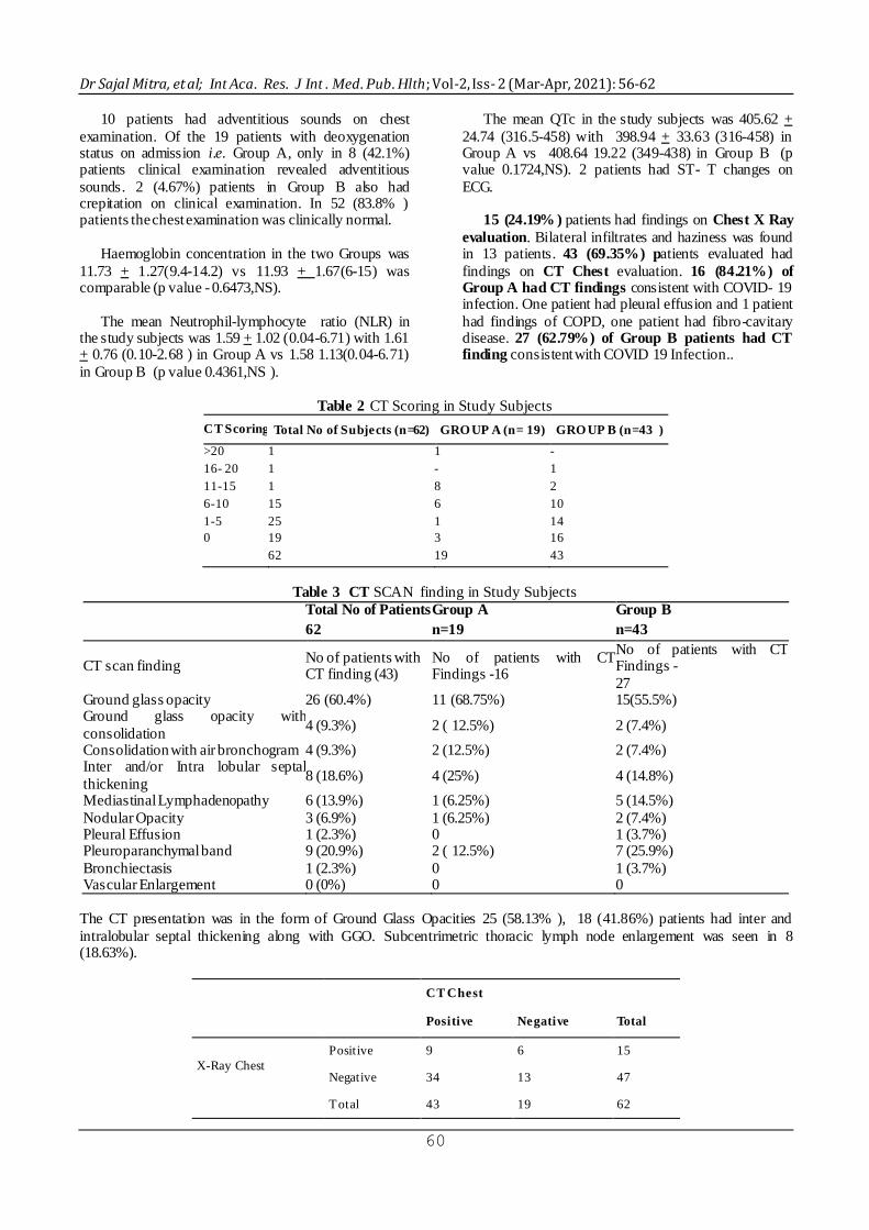
ECG. 15 (24.19% ) patients had findings on Chest X Ray
evaluation. Bilateral infiltrates and haziness was found in 13 patients. 43 (69.35%) patients evaluated had
findings on CT Chest evaluation. 16 (84.21%) of Group A had CT findings consistent with COVID- 19 infection. One patient had pleural effusion and 1 patient
had findings of COPD, one patient had fibro-cavitary disease. 27 (62.79%) of Group B patients had CT finding consistent with COVID 19 Infection..
Table 2 CT Scoring in Study Subjects
CT Scoring Total No of Subjects (n=62) GRO UP A (n= 19) GRO UP B (n=43 )
>20 1 1 -
16- 20 1 - 1
11-15 1 8 2
6-10 15 6 10
1-5 25 1 14
0 19 3 16
62 19 43
Table 3 CT SCAN finding in Study Subjects
Total No of Patients
62
Group A
n=19
Group B
n=43
CT scan finding No of patients with CT finding (43)
No of patients with CT Findings -16
No of patients with CT Findings -
27 Ground glass opacity 26 (60.4%) 11 (68.75%) 15(55.5%) Ground glass opacity with
consolidation 4 (9.3%) 2 ( 12.5%) 2 (7.4%)
Consolidation with air bronchogram 4 (9.3%) 2 (12.5%) 2 (7.4%) Inter and/or Intra lobular septal
thickening 8 (18.6%) 4 (25%) 4 (14.8%)
Mediastinal Lymphadenopathy 6 (13.9%) 1 (6.25%) 5 (14.5%)
Nodular Opacity 3 (6.9%) 1 (6.25%) 2 (7.4%) Pleural Effusion 1 (2.3%) 0 1 (3.7%) Pleuroparanchymal band 9 (20.9%) 2 ( 12.5%) 7 (25.9%)
Bronchiectasis 1 (2.3%) 0 1 (3.7%) Vascular Enlargement 0 (0%) 0 0
The CT presentation was in the form of Ground Glass Opacities 25 (58.13% ), 18 (41.86%) patients had inter and
intralobular septal thickening along with GGO. Subcentrimetric thoracic lymph node enlargement was seen in 8 (18.63%).
CT Chest
Positive Negative Total
X-Ray Chest
Positive 9 6 15
Negative 34 13 47
Total 43 19 62

Dr Sajal Mitra, et al; Int Aca. Res. J Int . Med. Pub. Hlth; Vol-2, Iss- 2 (Mar-Apr, 2021): 56-62
61
Diagnostic Evaluation of X-Ray Chest of
Asymptomatic COVID-19 Positive SUBJECTS as compared to CT Chest.
Xray Chest PA view has a Sensitivity = 20.93%, Specificity = 68.42% , PPV = 60.0% , NPV = 27.66% ,
Diagnostic accuracy=35.48% as compared to gold
standard of CT Chest.
All patients as a part of the standard of care received HCQS, Vitamin C & Zinc. Those with Lung infiltrates were given Favipiravir, Oseltamavir and Antibiotics in
addition. All patients with Hypoxia on Room air on admission were able to maintain their SPO2 levels on nasal oxygen therapy. Patients with Hypoxia and lung
GGO received Methyl Prednisolone and those with raised D- Dimer were given Heparin.
None of the patients deteriorated in the hospital and
had to be put on Assisted Ventilation. All patients
survived the hospital admission and were successfully discharged after being hospitalised for 10- 14 days as per government protocol.
DISCUSSION
Respiratory dysfunction is the principal source of
morbidity and mortality in COVID -19 as the disease advances. Pulse oximetry (SPO2) estimates arterial oxygen saturation by illuminating the skin and
measuring changes in light absorption of oxyhemoglobin and reduced hemoglobin.
Asymptomatic carriers contribute to the spread of the disease. Some of these patients despite being
COVID-19 positive have no complaints and are lucid asymptomatic despite having an oxygen saturation <94%. (“Happy Hypoxia /Silent Hypoxia)”. These
patients are in danger of rapid deterioration and collapse. It is intriguing that why despite hypoxia these patients are initially comfortable and asymptomatic.
The lung is inflating in these patients and defending their CO2, and their low oxygen levels are not always
coupled with obvious respiratory difficulty. C02 levels may be normal and breathing deeply is comfortable. The respiratory centres are exquisitely sensitive to CO2.
Small increases in PaCO2 usually rapidly evoke large
increases in minute ventilation; an increase in PaCO2 of
10 mmHg produces a level of respiratory discomfort that cannot be tolerated for even a few minutes. Abnormal lung mechanics also provokes dyspnea, but
considerably less than with hypercapnia. A fall in end-tidal PO2 below 60 mmHg elicit a strong increase in
dyspnea in only half of COVID-19 subjects (Tobin, M.
J. et al., 2020). The ventilatory and dyspnea responses to hypoxia are heavily influenced by prevailing PaCO2.
Severe hypoxia elicits an effective increase in ventilation only when background PaCO2 exceeds 39
mmHg.
Martin J. Tobin MD, et al., also mention that
COVID-19 patients exhibit several unusual findings. The explanation put forth is that - it is possible the virus has an idiosyncratic effect on the respiratory control
system. Angiotensin - Converting enzyme 2 (ACE2) the cell receptor of severe acute respiratory syndrome corona virus -2 (SARS -CoV-2) the virus responsible
for COVID -19, is expressed in the carotid body, the site at which chemoreceptor sense oxygen. These
attacked ACE-2 receptor play role in the depressed dyspnoea response in COVID -19. Hypoxemia produces dyspnea through stimulation of the carotid
bodies, which send signals to the medulla oblongata. The resulting increase in respiratory centre output is transmitted down to the phrenic nerves and diaphragm
causing increased minute ventilation. Heightened medullary centre activity is concurrently transmitted up
to the cerebral cortex. It is this cortical projection (corollary discharge) that produces the unpleasant sensation of dyspnea (Li, Y., & Xia, L. 2020). Hence it
seems that in COVID -19 the brain’s response to hypoxia is blunted and it has been baffling the physicians in light of the long established principles of
respiratory physiology (Tobin, M. J. et al., 2020).
Thoracic imaging with chest radiography and CT Thorax are key tools for pulmonary disease diagnosis and management. Imaging of these asymptomatic or
mildly symptomatic patients with X Ray Chest and CT Thorax was done. Only 15 (24.19%) patients had findings on X Ray Chest evaluation as compared to 43
(69.35%) on CT Chest. Of these 16 (84.21%) of Group A had CT findings as compared to 27 (62.79%) of
Group B patients consistent with COVID 19 Infection. The Sensitivity and specificity of X-Ray Chest was found to be poor as compared to CT Thorax for
diagnosing Chest pathology. The CT abnormalities found were peripherally
distributed Ground-glass opacities (GGOs), consolidation with air bronchogram, with intra and
interlobular septal, pulmonary nodules, and fissural thickening. The signs were distributed more in the lower lobes. The “reversed halo” sign, pulmonary artery
dilatation as described in COVID -19 patients was not seen in any patients. in the study done on 51 patients with COVID -19 infection by Yan Li and Liming Xia
published in American Journal of Roentgenology June 2020, found that the initial chest CT showed that disease affected all five lobes in 38 (74.5%) patients,
both lower lobes in eight (15.7%) patients, the right lower lobe in three patients (5.9%), the left upper lobe
and right lower lobe in one patient (2.0%), and the left upper lobe and right middle lobe in one patient (2.0%). The lesions were predominantly peripheral and
subpleural in 49 (96.1%) patients, and there were fewer lesions along the bronchovascular bundles. GGO and consolidation are two main signs of COVID-19 lesions
on CT images. CT showed singular or multiple irregular areas of GGO or consolidation or both in 49 of the 51
Dr Sajal Mitra, et al; Int Aca. Res. J Int . Med. Pub. Hlth; Vol-2, Iss- 2 (Mar-Apr, 2021): 56-62
62
(96.1%) patients. In the remaining two (3.9%) patients, neither GGO nor consolidation was seen on CT. The
The “reversed halo” sign and pulmonary nodules with a halo sign are uncommon CT features. Similar findings were also observed in our study.
As initially asymptomatic patients were being
admitted and isolated at Government Medical College,
Nagpur till the Covid Care Centre were opened in the city, this study could be carried out. These
asymptomatic COVID-19 subjects were evaluated for the presence of desaturation by Pulse oximetry (“Happy Hypoxia”). The wide spread presence of CT findings
may help to diagnose COVID-19 in patients who have an initial RT PCR test negative. The sensitivity of RTPCR by nasopharyngeal / oropharyngeal swab being
37-71%. These patients with CT findings of COVID-19 may eventually test positive with serial sampling. The
incidence of CT findings was 54% in the Diamond Princess ship in asymptomatic patients (Tobin, M. J. et al., 2020; & Inui, S. et al., 2020). Imaging may thus
direct towards the diagnosis in those who turn out to be false negative on RT PCR testing. Although Ultrasound has been suggested as a potential triage and diagnostic
tool for COVID-19 given the predilection for the disease in subpleural regions, there is limited
experience and expertise available in this field at this time. Hypoxic patients demand special care and attention despite being asymptomatic to prevent sudden
deterioration and and adverse outcome (Inui, S. et al., 2020).
CONCLUSIONS
The study included 62 asymptomatic or mildly Symptomatic patients. Nineteen (30.64 %) patients
were found to have Hypoxia on admission. Forty three (69.35 %) patients had normal oxygen saturation on admission. The Respiratory Rate in patients with
hypoxia (Group A) was more in patients with Hypoxia as compared to those without hypoxia (Group B) however they were not symptomatic with complaints of
breathlessness. 15 (24.19%) patients had findings on X -Ray Chest. 43 (69.35%) patients had findings on CT
Chest. Ground-Glass Opacities (GGOs), Consolidation with Air Bronchogram, with intra and interlobular septal and fissural thickening were the common
findings on HRCT Thorax. The signs were distributed more in the lower lobes. The “reversed halo” sign, pulmonary artery dilatation as described in COVID -19
patients was not seen in any patients. Patients with hypoxia on admission had more marked changes as compared to those without hypoxia. All patients
received standard line of care and could be discharged successfully.
Based on these findings, local administration in
Nagpur has formulated a policy to screen all patients
who are asymptomatic or mildly symptomatic (Category A,B & C) to find out those with Silent Hypoxia and Pneumonitis on Imaging so that they can
be followed and monitored closely in COVID Hospital settings.
LIMITATIONS OF THE STUDY
The study has some limitations like, first; limited sample size, second; inflammatory biomarkers
including Procalcitonin and D- Dimer could not be done in all subjects and hence not included for comparison in final results. Further studies are recommended to look
into the ABGs, Inflammatory biomarkers, and vascular flow of the pulmonary circulation to understand the
pathophysiology of the process.
REFERENCES
1. Francone, M., Iafrate, F., Masci, G. M., Coco, S.,
Cilia, F., Manganaro, L., ... & Catalano, C. (2020). Chest CT score in COVID-19 patients: correlation with disease severity and short-term
prognosis. European radiology, 30(12), 6808-6817. 2. Gattinoni, L., Chiumello, D., Caironi, P., Busana,
M., Romitti, F., Brazzi, L., & Camporota, L. (2020). COVID-19 pneumonia: different respiratory treatments for different phenotypes?.
(2020) Intensive Care Medicine; DOI: 10.1007/s00134-020-06033-2
3. Huang, C., Wang, Y., Li, X., Ren, L., Zhao, J., Hu,
Y., ... & Cao, B. (2020). Clinical features of patients infected with 2019 novel coronavirus in
Wuhan, China. The lancet, 395(10223), 497-506. 4. Inui, S., Fujikawa, A., Jitsu, M., Kunishima, N.,
Watanabe, S., Suzuki, Y., ... & Uwabe, Y. (2020).
Chest CT findings in cases from the cruise ship diamond princess with Coronavirus disease (COVID-19). Radiology: Cardiothoracic
Imaging, 2(2), e200110. 5. Li, Y., & Xia, L. (2020). Coronavirus disease 2019
(COVID-19): role of chest CT in diagnosis and management. American Journal of Roentgenology, 214(6), 1280-1286.
6. Rothe, C., Schunk, M., Sothmann, P., Bretzel, G., Froeschl, G., Wallrauch, C., ... & Hoelscher, M. (2020). Transmission of 2019-nCoV infection from
an asymptomatic contact in Germany. New England journal of medicine, 382(10), 970-971.
7. Tobin, M. J., Laghi, F., & Jubran, A. (2020). Why COVID-19 silent hypoxemia is baffling to physicians. American journal of respiratory and
critical care medicine, 202(3), 356-360. 8. Zhu, N., Zhang, D., Wang, W., Li, X., Yang, B.,
Song, J., ... & Tan, W. (2020). A novel coronavirus
from patients with pneumonia in China, 2019. New England journal of medicine.382:727-733.