research article effects of ppar agonist...
TRANSCRIPT

Hindawi Publishing CorporationPPAR ResearchVolume 2013, Article ID 541871, 11 pageshttp://dx.doi.org/10.1155/2013/541871
Research ArticleEffects of PPAR𝛾 Agonist Pioglitazone on Redox-SensitiveCellular Signaling in Young Spontaneously Hypertensive Rats
Ima Dovinová,1 Miroslav Barancik,2 Miroslava Majzunova,1 Stefan Zorad,3
Lucia Gajdosechová,3 Linda Gresová,1 Sona Cacanyiova,1 Frantisek Kristek,1
Peter Balis,1 and Julie Y. H. Chan4
1 Institute of Normal and Pathological Physiology, SAS, Sienkiewiczova 1, 813 71 Bratislava, Slovakia2 Institute for Heart Research, SAS, Dubravska cesta 9, 840 05 Bratislava, Slovakia3 Institute of Experimental Endocrinology, SAS, Vlarska 3, 833 06 Bratislava, Slovakia4Center for Translational Research in Biomedical Science, Kaohsiung Chang Gang Memorial Hospital, 123 Ta Pei Road, Kaohsiung83301, Taiwan
Correspondence should be addressed to Ima Dovinova; [email protected]
Received 26 July 2013; Revised 17 October 2013; Accepted 6 November 2013
Academic Editor: Nanping Wang
Copyright © 2013 Ima Dovinova et al. This is an open access article distributed under the Creative Commons Attribution License,which permits unrestricted use, distribution, and reproduction in any medium, provided the original work is properly cited.
PPAR𝛾 receptor plays an important role in oxidative stress response. Its agonists can influence vascular contractility in experimentalhypertension. Our study was focused on the effects of a PPAR𝛾 agonist pioglitazone (PIO) on blood pressure regulation,vasoactivity of vessels, and redox-sensitive signaling at the central (brainstem, BS) and peripheral (left ventricle, LV) levels in youngprehypertensive rats. 5-week-old SHR were treated either with PIO (10mg/kg/day, 2 weeks) or with saline using gastric gavage.Administration of PIO significantly slowed down blood pressure increase and improved lipid profile and aortic relaxation afterinsulin stimulation. A significant increase in PPAR𝛾 expression was found only in BS, not in LV. PIO treatment did not influenceNOS changes, but had tissue-dependent effect on SOD regulation and increased SOD activity, observed in LV. The treatment withPIO differentially affected also the levels of other intracellular signaling components: Akt kinase increased in the the BS, while𝛽-catenin level was down-regulated in the BS and up-regulated in the LV. We found that the lowering of blood pressure in youngSHR can be connected with insulin sensitivity of vessels and that 𝛽-catenin and SOD levels are important agents mediating PIOeffects in the BS and LV.
1. Introduction
Blood pressure is regulated by several internal systems.Power-spectral analysis identifies oscillations in heart rateand blood pressure that are modulated by inputs from therenin-angiotensin system, sympathetic and parasympatheticneurons, and locally released vasoactive factors such as nitricoxide (NO). Current findings support the view that thereactive oxygen species (ROS) and antioxidant enzymes suchas superoxide dismutase (SOD) expressed in the central ner-vous system (CNS) play an important role in the regulationof blood pressure, and perturbations in redox homeostasiscontribute to pathogenesis of hypertension [1]. Activity ofROS sources (such as NADPH oxidases), stimulated throughangiotensin II system and AT1 receptors are involved in
redox-sensitive intracellular signaling modulated by kinasecascades and transcriptional factors in cardiovascular system[2]. Antioxidant response and the potential sites of internalantioxidants of different SOD isoforms, could modulateblood pressure in the vasculature, the brain, and the kidney[1, 2].
The peroxisome proliferator-activated receptor 𝛾(PPAR𝛾) is a nuclear receptor that takes part in the regulationof lipid metabolism and in cellular signaling. Dysregulationin the PPAR𝛾 activity may underlie diseases connected withthe metabolic syndrome and hypertension. As a nuclearreceptor, PPAR𝛾 acts together with another nuclear receptor,the retinoid X receptor, to dissociate corepressor andrecruitment of coactivator protein, which, in turn, promotestranscription of the downstream target genes involved

2 PPAR Research
in adipocyte differentiation, glucose homeostasis, lipidtrafficking, or anti-inflammatory response [3]. The activityof PPAR𝛾 is influenced by a variety of extracellular ligands:glitazones (rosiglitazone, pioglitazone, and troglitazone)and intracellular ligands: prostaglandins, leukotrienes, and𝛼-lipoic acid [4].
Studies of PPAR𝛾 document a larger increase in bloodpressure in endothelial PPAR𝛾, null mice in response toangiotensin II infusion [5]. This effect is connected withimpaired vascular relaxation in response to acetylcholinecombined with unaffected relaxation in response to sodiumnitroprusside.These findings indicate that endothelial PPAR𝛾regulates vascular NO production and that the disruptionof endothelial PPAR𝛾 contributes to endothelial dysfunc-tion in vivo. Oral intake of rosiglitazone (RSG) resulted invasodepression and reduction of the augmented sympatheticvasomotor activity in the spontaneously hypertensive rat[6]. Overexpression of PPAR𝛾 and amelioration of oxidativestress in the brainstem rostral ventrolateral medulla (RVLM),where sympathetic promotor neurons reside, underlies thecardiovascular protective action of RSG. A recent studyshowed different effects of RSG application in young ver-sus adult SHR. While in young SHR this PPAR𝛾 agonistinfluences PI3K/Akt/NO signaling in blood vessels throughdeactivation of the insulin resistance pathway, this effect wasnot evident in adult animals [7]. These observations openthe possibility of differential effects of the PPAR𝛾 agonist incellular signaling involved in development of hypertension inSHR of different ages.
Application of the PPAR𝛾 agonist pioglitazone (PIO) wasfound to influence the vascular contractility of blood vesselsin SHR [8]. Several recent studies also demonstrated theimplication of PPAR𝛾 in oxidative stress responses and in theimbalances between pro-oxidant and antioxidant responsesthat influence apoptotic or necrotic cell death [3]. PPAR𝛾may directly modulate activation of several antioxidantsinvolved in oxidative stress, such as the mitochondrialmanganese SOD (MnSOD) [9], mitochondrial uncouplingprotein (UCP2) [6], or catalase [10, 11]. This modulation wasobserved in several kinds of cells including cardiomyocytes[9], neuronal cells [6], adipocytes, and endothelial cells [10,11]. The PPAR𝛾 modulation also plays a role in the isoform-specific modulation of expression of NO synthases (NOSs).PPAR𝛾 ligands promoted expression of endothelial NOS(eNOS) in vivo [5], but down-regulated the inducible NOS(iNOS) isoform [12, 13]. Moreover, the effects of PPAR𝛾ligands were found to be connected with downregulation ofthe inducible cyclooxygenase-2 (COX-2) [12].
In the regulation of cellular response, the modulationof these enzyme systems is associated with several signal-ing pathways, such as Nrf2 (antioxidant pathway), WnT/𝛽-catenin (antioxidant pathway), or NF-𝜅B (prooxidant path-way) [3]. Several studies have shown the modulation of Aktkinase phosphorylation by PIO [14–17]. PIO was found toincrease Akt phosphorylation (activation) [14, 15], but insome cell types also the inhibition or reduction of Akt-1 activity by PPAR𝛾 agonists has been observed [16, 17].Recently, it was reported that the protein whose function
could be regulated by Akt kinase-mediated phosphorylationat Ser552 is 𝛽-catenin [18]. Under normal cellular conditions,𝛽-catenin acting through Wnt signaling is involved in cellproliferation and differentiation, but under changed ROSconditions, its function can shift to regulate the transcriptionfactors that support cell survival through increased stressresistance and ROS clearance [19]. This suggests that 𝛽-catenin can play a pivotal role in the re-programming of thetranscriptional activity in response to changes in ROS [19].
Arterial blood pressure can be influenced through severalmain regulatory systems located in different organs andtissues: brain (mainly in brainstem), heart (ventricles), largeand small vessels (such as aorta and mesentery artery),and kidney (juxtaglomerular apparatus). In several studies,the effects of PPAR gamma on blood pressure have beenstudied. However, these studies did not focus on redoxcellular signaling in the heart and only few results wereobtained in the brain [6]. Therefore, our study was focusedon the investigation of PPAR𝛾 agonist PIO on blood pressureregulation in young prehypertensive rats (SHR) and onredox-sensitive cellular signaling within systems of central(brainstem) and peripheral (left ventricle) regulation of bloodpressure.
The concrete aims of this study included the determina-tion of PIO effects on
(1) blood pressure modulation, lipid profile, adipocytesRAS components, and vessel responses,
(2) different components of redox-sensitive intracellularsignaling (SOD, NOS, Akt kinase, and 𝛽-catenin) inthe brainstem and in the left ventricle.
2. Materials and Methods
2.1. Experimental Model and Treatment Protocol. Youngmalespontaneously hypertensive rats (SHR; 5 weeks old, 𝑛 =7) were treated with PPAR𝛾 agonist pioglitazone (PIO), inthe dose of 10mg/kg/day. PIO was dissolved in saline andadministered orally by gavage as a suspension during 2weeks.Animals of the control group (𝑛 = 7) were gavaged dailywith physiological solution (saline). The body weight, dailyfood intake, and tap water intake were measured before,during, and after the two week treatment period. All animalexperiments were performed in accordance with the rules ofthe State Veterinary Administration of the Slovak Republicand with the guidelines of the Animal Research and CareCommittee of the Institute of Normal and PathologicalPhysiology of the Slovak Academy of Sciences.
2.2. Blood Pressure Determination. Systolic blood pressurewas measured noninvasively by tail cuff plethysmographyin both the control and PIO-treated groups of rats. Bloodpressure measurements were performed on days 1, 5, 9, and12 of the treatment period.
2.3. Collection of Samples. At the end of the experiment,the animals were sacrificed and hearts and brain wererapidly excised. Aorta was also isolated and used for further

PPAR Research 3
Table 1: Primer pairs used for amplification of selected genes.
Gene Forward (sense) primer Reverse (antisense) primer Ampliconsize (bp)
Temp(∘C)
SOD1 CAC TCT AAG AAA CAT GGC G CTG AGA GTG AGA TCA CAC G 124 54SOD2 TTC AGC CTG CAC TGA AG GTC ACG CTT GAT AGC CTC 122 54SOD3 CTT GAC CTG GTT GAG AAG ATA G GAT CTG TGG CTG ATC GG 154 58NOS3 CCC ACA GTC TGG TTG CT TCA CCG TGC CCA TGA GT 124 57NOS1 CGC TAC GCG GGC TAC AAG CA GCA CGT CGA AGC GGC CTC TT 118 60GAPDH TGG AGG TGC TGG AAG AGT T TCA CGC CAC AGC TTT CCA 103 57p22phox CAG GCA TAT ACC CGC TAC CT TCT GTC ACC CTG TGC TTG AC 120 60ACE ATG GTA CAG AAG GGC TGG AA TTG TAG AAG TCC CAC GCA GA 170 60AGT CAT GAG TTC TGG GTG GAC AA AAG TTG TTC TGG GCG TCA CT 95 60AT1R TCT CAG CAT CGA TCG CTA CCT AGG CGA GAC TTC ATT GGG TC 51 60aP2 (FABP4) AGC GTA GAA GGG GAC TTG GT ATG GTG GTC GAC TTT CCA TC 185 60PPAR𝛾 AGG ATT CAT GAC CAG GGA GTT AGC AAA CTC AAA CTT AGC CTC CAT 79 60RPS29 GCT GAA CAT GTG CCG ACA GT GGT CGC TTA GTC CAA CTT AAT GAA G 74 60UCP-1 GCC TCT ACG ATA CGG TCC TGC ATT CTG ACC TTC ACC AC 145 60
functional studies. Excised hearts were weighed, and left andright ventricles were separated. The whole heart and ven-tricular weights were registered. Further processing of tissuesamples depended on the specific assay. For measurement ofsuperoxide levels, tissues were collected into ice-cold Krebsbuffer. For measurements of NOS and SOD activities, thetissue samples were cooled in Tris-HCl supplemented with aprotease inhibitor cocktail and further processed accordingto individual methods. The tissue samples for molecular-biological (qPCR) and biochemical (Western blot) analyseswere frozen in liquid nitrogen and stored at −80∘C untilfurther use.
Plasma samples were prepared from whole blood col-lected into Na-EDTA solution. After centrifugation for 5minat 1200×g, the plasma was collected. Prepared plasma sam-ples were stored at −80∘C until further analysis.
2.4. Functional Studies In Vitro. Thoracic aorta was isolated,cleaned of connective tissue, and cut into rings (4mm inlength). The rings were fixed between two stainless wiretriangles in an incubation organ bath containing Krebs solu-tion (NaCl 118mM, KCl 5mM, NaHCO
325mM, MgSO
4
1.2mM, KH2PO41.2mM, CaCl
22.5mM, glucose 11mM,
CaNa2EDTA 0.032mM). The solution was oxygenated with
pneumoxide (95% O2, 5% CO
2) at 37∘C. The upper wire
triangles were connected to electromechanical transducers(Experimetria) and the changes in isometric tension wereregistered using AD converter and Dewetron software. Theresting tension was adjusted to 1 g and applied to each ring.
Subsequently, preparations were equilibrated for 60 min-utes and the presence of functional endotheliumwas assessedin all preparations. Contractile responses induced by KCl(100mmol/L) were used to achieve receptor-independentmaximal contraction. The relaxant responses were followedon rings precontracted with noradrenaline (10−6mol/L) toproduce a stabile plateau of contraction. The rings werethen exposed to acetylcholine (10−5mol/L) or insuline
(10−6mol/L) to induce a maximal response. Relaxation wasexpressed as a percentage of noradrenaline-induced contrac-tion.
2.5. Gene Expression Determination. Total RNA from thebrainstem and left ventricle tissue samples was isolated withTRIsure reagent (Bioline) according to the manufacturer’sprotocol. Total RNA from adipose tissue was isolated byRNeasy Universal Plus Mini Kit (Qiagen). The isolated totalRNA was quantified spectrophotometrically at 260/280 nmusing Nanodrop. Reverse transcription (RT) reaction wasperformed using TetrocDNA kit (Bioline) on EppendorfMastercycler.
Real-time polymerase chain reaction (qPCR) by ampli-fication of cDNA was performed on a Biorad CFX96 Real-time systemusing the SensiFAST SYBRNoROXkit (Bioline).PCR reaction for each sample was carried out in duplicate forall cDNA and for the housekeeping gene of glyceraldehyde-3-phosphate dehydrogenase (GAPDH).
The primer pairs used for amplification of studied genes(PPAR𝛾, SOD1, SOD2, SOD3, NOS1, NOS3, p22phox, AT1R,UCP-1, AGT, ACE, aP2, GAPDH, and RPS29) are listed inTable 1.
2.6. Determination of Total Superoxide Dismutase and NitricOxide Synthase Activities. The total superoxide dismutase(SOD) activity was analyzed using the SOD Assay kit (Fluka)in tissue samples. The rate of WST-1 reduction with super-oxide is linearly related to the xanthine oxidase activity, andis inhibited by SOD. The SOD activity was determined asan inhibition activity by measuring the color decrease ofWST-1-formazan production at 450 nm (Thermo ScientificMultiscan FC).
Total activity of nitric oxide synthases (NOSs) was deter-mined using measurement of the conversion of radioactive3H-L-Arginine to 3H-L-Citrulline [20] and the product was
4 PPAR Research
detected by liquid scintillator Tri-Carb 2910 TR (PerkinElmer).
2.7. Electrophoresis and Immunochemical Western Blot Anal-ysis. Samples of protein fractions containing equivalentamounts of proteins per lane were separated by sodiumdodecyl sulfate-polyacrylamide gel electrophoresis (SDS-PAGE). After electrophoretic separation, proteins were trans-ferred to a nitrocellulose membrane. The quality of thetransfer was controlled by Ponceau S staining of nitrocel-lulose membranes after the transfer and protein loading byusing glyceraldehyde-3-phosphate dehydrogenase (GAPDH)as a housekeeper. Specific anti-SOD1, anti-SOD2, anti-Aktkinase, anti-𝛽-catenin, anti-GAPDH (all from Santa CruzBiotechnology), and anti-phospho-Akt kinase (Ser473 andThr308) (from Cell Signaling Technology) antibodies wereused for the primary immunodetection. Peroxidase-labelledanti-rabbit immunoglobulin (Cell Signaling Technology) wasused as the secondary antibody. Bound antibodies weredetected by the enhanced chemiluminescence (ECL) detec-tion method.
2.8. Lipid Profile, Glucose and Insulin Level Determination.Plasma insulin levels were evaluated by RIA kits (Milli-pore) following the manufacturer’s protocol. Lipid profileand blood glucose determinations were done at Synlab(Bratislava, Slovakia) using the COBAS Integra 800 multian-alyzer (Roche).
2.9. Statistical Evaluation. The data for blood pressure mea-surements were evaluated using a two-way ANOVA and forin vitro functional studies using a one-way ANOVA withBonferroni adjustment in post hoc tests. Data from othermeasurements were analyzed either by the Student’s 𝑡-testor the Wilcoxon-Mann-Whitney 𝑈-test. Differences wereconsidered significant at 𝑃 < 0.05 in all tests.
3. Results
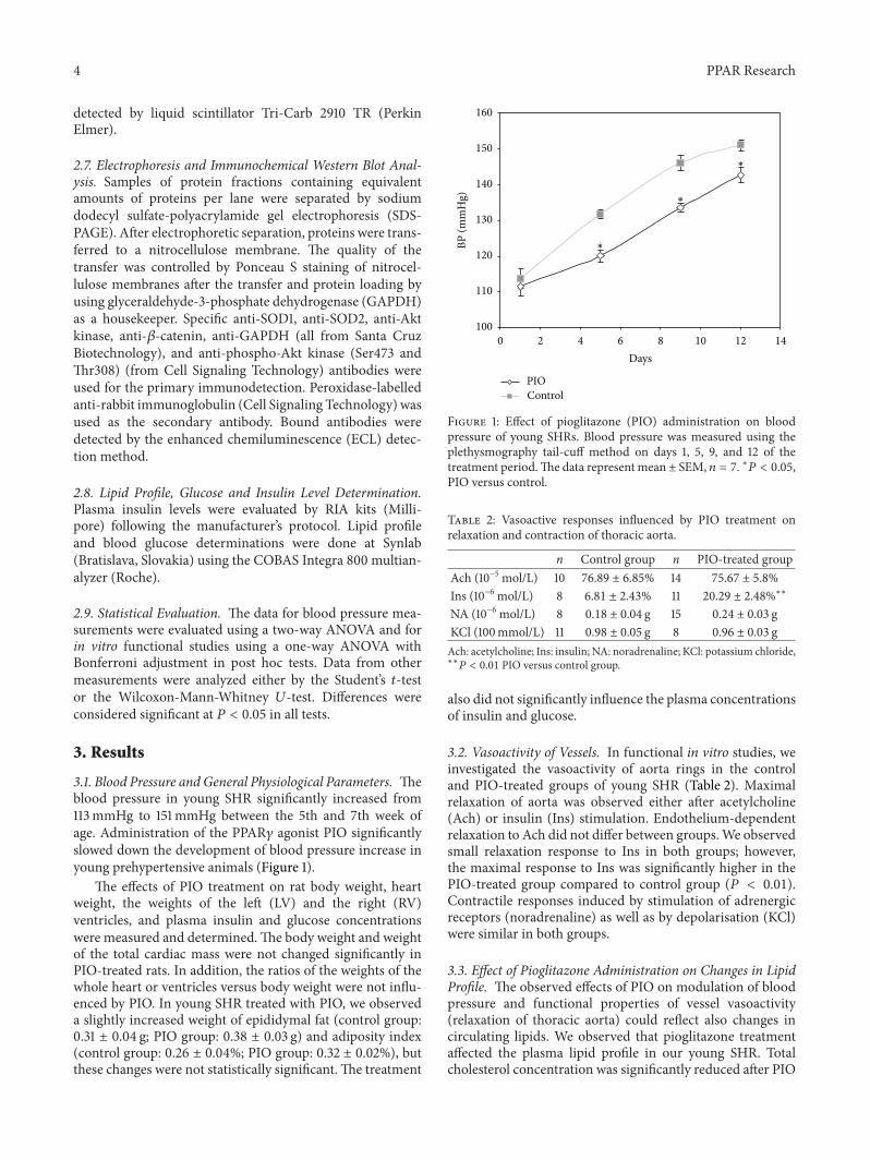
3.1. Blood Pressure and General Physiological Parameters. Theblood pressure in young SHR significantly increased from113mmHg to 151mmHg between the 5th and 7th week ofage. Administration of the PPAR𝛾 agonist PIO significantlyslowed down the development of blood pressure increase inyoung prehypertensive animals (Figure 1).
The effects of PIO treatment on rat body weight, heartweight, the weights of the left (LV) and the right (RV)ventricles, and plasma insulin and glucose concentrationswere measured and determined.The body weight and weightof the total cardiac mass were not changed significantly inPIO-treated rats. In addition, the ratios of the weights of thewhole heart or ventricles versus body weight were not influ-enced by PIO. In young SHR treated with PIO, we observeda slightly increased weight of epididymal fat (control group:0.31 ± 0.04 g; PIO group: 0.38 ± 0.03 g) and adiposity index(control group: 0.26 ± 0.04%; PIO group: 0.32 ± 0.02%), butthese changes were not statistically significant.The treatment
100
110
120
130
140
150
160
0 2 4 6 8 10 12 14Days
BP (m
mH
g)
PIOControl
∗
∗
∗
Figure 1: Effect of pioglitazone (PIO) administration on bloodpressure of young SHRs. Blood pressure was measured using theplethysmography tail-cuff method on days 1, 5, 9, and 12 of thetreatment period.The data represent mean ± SEM, 𝑛 = 7. ∗𝑃 < 0.05,PIO versus control.
Table 2: Vasoactive responses influenced by PIO treatment onrelaxation and contraction of thoracic aorta.
n Control group n PIO-treated groupAch (10−5 mol/L) 10 76.89 ± 6.85% 14 75.67 ± 5.8%Ins (10−6 mol/L) 8 6.81 ± 2.43% 11 20.29 ± 2.48%∗∗
NA (10−6 mol/L) 8 0.18 ± 0.04 g 15 0.24 ± 0.03 gKCl (100mmol/L) 11 0.98 ± 0.05 g 8 0.96 ± 0.03 gAch: acetylcholine; Ins: insulin; NA: noradrenaline; KCl: potassium chloride,∗∗
𝑃 < 0.01 PIO versus control group.
also did not significantly influence the plasma concentrationsof insulin and glucose.
3.2. Vasoactivity of Vessels. In functional in vitro studies, weinvestigated the vasoactivity of aorta rings in the controland PIO-treated groups of young SHR (Table 2). Maximalrelaxation of aorta was observed either after acetylcholine(Ach) or insulin (Ins) stimulation. Endothelium-dependentrelaxation to Ach did not differ between groups.We observedsmall relaxation response to Ins in both groups; however,the maximal response to Ins was significantly higher in thePIO-treated group compared to control group (𝑃 < 0.01).Contractile responses induced by stimulation of adrenergicreceptors (noradrenaline) as well as by depolarisation (KCl)were similar in both groups.
3.3. Effect of Pioglitazone Administration on Changes in LipidProfile. The observed effects of PIO on modulation of bloodpressure and functional properties of vessel vasoactivity(relaxation of thoracic aorta) could reflect also changes incirculating lipids. We observed that pioglitazone treatmentaffected the plasma lipid profile in our young SHR. Totalcholesterol concentration was significantly reduced after PIO

PPAR Research 5
ControlPIO
(mm
ol/L
)
3.0
2.5
2.0
1.5
1.0
0.5
0.0CHOL HDL LDLm TGL VLDLc
∗
∗
∗
Figure 2: Effect of pioglitazone administration on plasma lipidprofile in young SHRs. CHOL: cholesterol; HDL: high densitylipoprotein; LDLm: low density lipoprotein (measured value); TGL:triglycerides; and VLDLc: very low-density lipoprotein (calculatedvalue). Data represent mean ± SEM, 𝑛 = 7. ∗𝑃 < 0.05, PIO versuscontrol.
treatment (Figure 2) and, due to the total cholesterol reduc-tion, also HDL and LDL were significantly reduced (actually,LDL/HDL and cholesterol/HDL ratios were unchanged).Plasma triglycerides and VLDL were not influenced by thepioglitazone treatment.
3.4. Gene Expression in Adipose Tissues. mRNA expression ofPPAR𝛾 and AP2, markers of adipogenesis, was not changedby pioglitazone. When analyzing the mRNA of selectedcomponents of local adipose tissue RAS (AGT, ACE, andAT1 receptors), only the AT1 receptor was found significantlyelevated. Gene expression of UCP1 tended to increase underpioglitazone treatment (Figure 3).
PPAR𝛾: peroxisome proliferator-activated receptor;AT1R: angiotensin-1 receptor; aP2: adipose fatty acid-bindingprotein; UCP1: uncoupling protein-1; AGT: angiotensinogen;ACE: angiotensin converting enzyme. Data representmean ±SEM, 𝑛 = 7. ∗𝑃 < 0.05, PIO versus control.
3.5. Effect of Pioglitazone on PPAR𝛾 Expression in the LeftVentricle and Brainstem. In the brainstem of the PIO-treatedanimals, we observed a significant increase in mRNA encod-ing PPAR𝛾. In the LV, we observed no change relative tocontrol (Figure 4).
3.6. ROS Activation, Antioxidant Response, and SOD/NOSBalance in the Left Ventricle and Brainstem. Our study wasfocused also on the effects of PIO on enzymes involved in theregulation of ROS levels (p22phox subunit of NADPH oxi-dase system and SOD), and we observed different responsesin the brainstem and the left ventricle. We found tissue-specific changes in the expression of genes involved in radicalsignaling, AT1R, and p22hphox. PIO treatment modulated
ControlPIO
Nor
mal
izat
ion
on rp
s29
expr
essio
n
3.0
2.5
2.0
1.5
1.0
0.5
0.0PPAR𝛾 AT1R aP2 UCP1 AGT ACE
∗
Figure 3: Effect of pioglitazone administration on expression ofseveral genes in epididymal fat (adipose) tissue of young SHRs.Expression was normalized to the expression of rsp29.
ControlPIO
PPA
R𝛾/G
APD
H m
RNA
ratio
Left ventricle Brainstem
2.0
1.5
1.0
0.5
0.0
∗
Figure 4: Effect of pioglitazone administration on PPAR𝛾 expres-sion in different tissues of young SHRs. PPAR𝛾 expression wasnormalized to expression of GAPDH as a housekeeper gene. Dataare shown for left ventricle and brainstem and bars representmean ±SEM, 𝑛 = 7. ∗𝑃 < 0.05, PIO versus control.
the expression of AT1R and p22hphox only in the brainstemand inducedmoderate increase in theirmRNA levels (by 34%for AT1R and by 20% for p22hphox).
Antioxidant response of SOD genes and SOD/NOS bal-ance in the left ventricle and brainstem were investigatedusing a study of expression of individual SOD or NOSisoforms and by determination of their total activities. In theleft ventricle, we observed no change in the expression of theSOD1 (Table 3) or SOD3 (not shown) isoforms. Significantchanges were found only for the expression of mRNAencoding SOD2. In the brainstem, the increase of PPAR𝛾mRNAwas accompanied with the upregulation of SOD2 anddownregulation of SOD1mRNAexpression (Table 3). Similarto the left ventricle, the expression of SOD3 was unchanged(not shown).

6 PPAR Research
Table 3: mRNA expression of SOD1 and SOD2 isoforms after PIO treatment.
mRNA Left ventricle BrainstemControl PIO Control PIO
SOD1/GAPDH 1.46 ± 0.115 1.30 ± 0.087 1.39 ± 0.247 0.75 ± 0.099∗
SOD2/GAPDH 1.20 ± 0.107 0.85 ± 0.075∗
0.97 ± 0.067 1.24 ± 0.078∗
Data represent mean ± SEM, ∗𝑃 < 0.05, PIO versus control.
PIO treatment revealed different, tissue-specific effects onSOD activities. In the left ventricle, PIO induced a significantincrease of total SOD activity, while no change was observedin the brainstem (Figure 5(d)). The observed increase in thetotal SOD activities in the left ventricle has been found tobe connected with partial (nonsignificant) upregulation ofSOD2 protein levels (Figures 5(a) and 5(c)). Similar changesin SOD2 protein levels were found in the brainstem (Figures5(b) and 5(c)). Levels of the SOD1 isoform were not changedneither in LV nor in BS (Figures 5(a) and 5(b)).
For NOS isoforms, the only significant effect of PIOtreatment was observed for NOS1 (nNOS), a decrease in thebrainstem (Figure 6(a)). The treatment did not influence thetotal NOS activities in the left ventricle or in the brainstem(Figure 6(b)).
3.7. Effect of Pioglitazone on Regulatory Proteins Involved inRedox Signaling. The influence of PIO on Akt kinase and 𝛽-catenin, components of two distinct redox-sensitive signalingkinase pathways (PI3K/Akt andWnt/𝛽-catenin), was studiedin tissue samples of the left ventricle and brainstem. Ourdata show that PIO induced increase in protein levels of Aktkinase in brainstem (Figures 7(b) and 7(c)), but not in the leftventricle (Figures 7(a) and 7(c)). Using antibodies specific fordetection of Akt kinase phosphorylated specifically at Ser473or Thr308, we looked also for changes in activation of theenzyme. However, the actions of PIO observed in the brain-stem were not connected with modulation of specific Ser473(Figure 7(b)) orThr308 (data not shown) phosphorylation ofthis enzyme. We found opposite effects of PIO on 𝛽-cateninlevels in the left ventricle and brainstem: in the left ventricle,the levels significantly increased (Figures 7(a) and 7(d)),while in the brainstem, we observed a significant decrease(Figures 7(b) and 7(d)). Protein loading was controlled byusing glyceraldehyde-3-phosphate dehydrogenase (GAPDH)and we did not observe any differences.
4. Discussion
The development of hypertension can be induced by variousstimuli and is associated with several changes in the radicaland antioxidant responses as well as in cellular signaling.In the present study, we investigated the effects of PPAR𝛾agonist pioglitazone during hypertension development inyoung SHR. Our study focused on PIO-induced changes inthe brainstem (CNS level) and the left ventricle of the heart(peripheral level) on blood pressure regulation.Moreover, theeffects of PIO on changes in overall lipids and adipocytesduring hypertension development in young SHR were deter-mined.
PPAR𝛾 agonists are substances used as insulin sensitiz-ers in diabetic patients with or without hypertension [21].Administration of PIO to young SHR in our study retardedthe blood pressure development. Several studies using RSGconfirmed the blood pressure lowering effect of this PPAR𝛾agonist in experimental hypertension [6, 7]. The effects ofRSG observed in these studies were usually associated withchanges in insulin sensitivity but without effects on glucoselevel. Our experimental data with PIO, another PPAR𝛾 ago-nist, partially contradict the findings with RSG. We observedno significant change in glucose or in plasma insulin levelsafter PIO treatment in young rats.
In our study, SHRs were used at the age of 7 weeks,which is a period before insulin resistance is developing[22]. This was confirmed by normal glucose and insulinplasma levels in both control and PIO-treated SHR groups. Inaddition, plasma lipid parameters seem to be also in normalrange in young SHR. However, PIO significantly reducedtotal plasma cholesterol by decreasing both HDL and LDLfractions. To our knowledge this is the first demonstration ofPIO lipid-lowering activity in rats without dyslipidemia [23].An explanation of the underlying mechanisms will require astudy of the liver pathways of cholesterol synthesis as well asits inflammatory status in young SHR. Our in vitro studies ofvessel vasoactivity showed improved vasodilatation responseafter insulin stimulation in the young SHR treated with PIO.These observations suggest increased insulin sensitivity inthe aorta of treated animals and may have an impact on thedelay of blood pressure increase. Similar to our data, alsoanother PPAR gamma agonist rosiglitazone has been foundto improve aortic vasodilatory response to insulin in SHR [7].
PPAR𝛾 mRNA expression in LV and BS was different.While in the brainstemwe obtained an increase in the gene, inthe left ventricle we did not observe any changes. Therefore,we suppose that the responses in the brainstem can be directlymodulated through PPAR𝛾, while in the left ventricle theregulation can be independent from PPAR𝛾 and affectedby pioglitazone only. Direct and indirect effects of PPAR𝛾agonists to the cardiac metabolism have been observed alsoon model of PPAR𝛾, knockout mice [24]. It was found thatat baseline conditions, PPAR𝛾 does not play a crucial role inregulating cardiacmetabolism inmice.The authors suggestedthat it is likely due to low myocardial PPAR𝛾 expression andthat agonists may protect myocardium indirectly.
Some studies documented that application of RSG canactivate PI3K/Akt/NOS signaling pathway in endothelium ofyoung SHR [7]. In our study, we investigated whether PIOcan affect this signaling pathway. However, in the brainstemand the left ventricle, we did not observe significant influenceof PIO on Akt kinase activation. Similar to our data obtained

PPAR Research 7
SOD1
Left ventricle
SOD2
C PIO
GAPDH
(a)
Brainstem
SOD1
SOD2
C PIO
GAPDH
(b)
ControlPIO
1.4
1.2
1.0
0.8
0.6
0.4
0.2
0.0
SOD
2/G
APD
H p
rote
in ra
tio
Left ventricle Brainstem
(c)
10
8
6
4
2
0
SOD
activ
ities
(U/m
g pr
otei
n)
∗
ControlPIO
Left ventricle Brainstem
(d)
Figure 5: Effect of pioglitazone administration on SOD1 and SOD2 protein levels and total SOD activities in the left ventricle and brainstemof young SHRs. (a) Western blot records documenting SOD1, SOD2, and GAPDH protein levels in fraction isolated from the tissues of theleft ventricle of control and PIO-treated group. (b)Western blot records documenting SOD1, SOD2, and GAPDH protein levels in brainstem.(c) Quantification of SOD2 protein levels normalized to the GAPDH protein levels. Data were obtained fromWestern blot records and eachbar represents mean ± SEM of 7 tissue samples per group. ∗𝑃 < 0.05, PIO versus control. (d) The SOD activities were analyzed using theSOD Assay kit (Fluka) in tissue samples of left ventricle and brainstem. The specific activities are expressed in units per mg of proteins andare presented as mean ± SEM, 𝑛 = 7. ∗𝑃 < 0.05, PIO versus control.
in the left ventricle are the findings that pretreatment withPIO does not significantly increase myocardial Akt kinasephosphorylation in the rat [25]. Akt kinase expression inthe brain was reported to modify food intake [26]. However,in our study, the increase in Akt kinase expression in thebrainstemwas not associated with significant changes in foodintake and there were no differences in body weight betweencontrol and PIO-treated rats.
We also found that activities of NOS, as another com-ponent of this PI3K/Akt/NOS pathway, were not influencedby PIO. Similarly, another study showed that in an in vivo
rat model, PIO alone did not cause significant increase inmyocardial phospho-Akt or phospho-eNOS [25]. Severalworks investigated the association between PIO treatmentand NO level. The cardioprotective effects of PIO duringischemia and reperfusion inmyocardial ischemic injury wereobserved in rabbits and may depend on NO [27]. In patientswith diabetes mellitus, also the effects of PIO on eNOS andiNOS were studied. In patients with insulin resistance, asignificant decrease in the levels of eNOS and iNOS wasobserved [28]. On the other hand, these changes in NOS iso-forms were not found in patients without developed insulin

8 PPAR Research
ControlPIO
ControlPIO
Left ventricle BrainstemNOS1 NOS3 NOS1 NOS3
2.0
1.5
1.0
0.5
0.0
NO
S/G
APD
H m
RNA
ratio
∗
(a)
ControlPIO
Left ventricle Brainstem
2.0
1.5
1.0
0.5
0.0
NO
S ac
tiviti
es (p
mol
/min
/mg
prot
ein)
(b)
Figure 6: Effect of pioglitazone administration on nitric oxide synthase (NOS) expression and activities in the left ventricle and brainstem ofyoung SHRs. (a) Expression of NOS1 and NOS3 isoforms was normalized to the expression of GAPDH. Data are shown for the left ventricleand brainstem. Bars represent mean ± SEM, 𝑛 = 7. ∗𝑃 < 0.05, PIO versus control. (b) The NOS activities were determined using conversionof radioactive 3H-Arginine to 3H-Citrulline. The specific activities are expressed in pmols of citrulline produced in one min and related to1mg of proteins. Data are presented as mean ± SEM, 𝑛 = 7.
resistance. Our results show that NOS isoforms were notchanged in the LV, while only the nNOS isoform was down-regulated in the brainstem. Moreover, PIO did not induceany change in NOS activities in young SHR.This observationmay be related to the fact that no significant differences inNOS activities were observed in young hypertensive SHR andyoung normotensive WKY rats below 9 weeks of age. Onlya tendency of a decrease in NOS activities was observed inyoung SHRs [29].
PIO, known as an antidiabetic agent with antioxidant andanti-inflammatory effects against oxidative stress conditions[30, 31], is also suggested to reduce, directly and/or indirectly,the overproduction of ROS. Our study was focused also onthe effects of PIO on the enzymes involved in the regulationof ROS levels (p22phox subunit of NADPH oxidase system,SOD) and observed different responses in the brainstemand the left ventricle. In the brainstem, where the effects ofPIO were connected to upregulation of PPAR𝛾, we foundalso a borderline increase in AT1 receptor, p22phox, andsignificant upregulation of the SOD2 isoform. The observedSOD2 upregulation is in agreement with promotor analysisshowing that gene of this mitochondrial SOD isoform can bea direct target of PPAR𝛾 [9]. However, total SOD activitieswere not influenced in the brainstem. On the other hand,there were no observable changes in AT1 receptor andp22phox in the left ventricle, and the effects of PIO werenot joined directly with PPAR𝛾 overexpression. In contrastto the brainstem, we observed antioxidant effects of PIOtreatment in the left ventricle realized through an increase intotal SOD activities. Tissue-specific differences in responseto pioglitazone were observed also for 𝛽-catenin. Severalstudies have demonstrated a direct connection betweenPPAR𝛾 signaling and 𝛽-catenin pathway [32–35]. Moreover,
it has been shown that interaction between PPAR𝛾 and 𝛽-catenin promotes regulation of genes that confer normalfunction and homeostasis to vascular cells [33]. 𝛽-catenin isa protein acting through Wnt signaling and under changedROS conditions acting as a regulator of the transcriptionfactors that support cell survival through increased stressresistance and ROS clearance [19]. The observed differenteffects of PIO on proteins involved in redox signaling atcentral and peripheral level should be clarified by furtherinvestigation.
Pioglitazone induces adipogenesis in vitro on 3T3L1adipocyte culture [36]. In vivo RSG was shown to induceadipogenesis in subcutaneous adipose tissue [37] and thiseffect seems to be depot-specific [38]. In our experiment, PIOdid not influence expression of PPAR𝛾 and AP2, markersof adipogenesis, in epididymal adipose tissue. Thus, weconclude that stimulation of PPAR𝛾 in young SHR underour experimental conditions does not lead to alternationof adipose tissue structure and mass. It has been shownthat RSG decreases both angiotensinogen protein expressionand angiotensin II release in isolated human subcutaneousadipocytes [39]. In addition, it was reported that PIOtreatment decreases serum angiotensin II level in type 2diabetes patients [40]. The above facts lead us to study theexpression of adipose tissue RAS components under PIOtreatment. Angiotensinogen and ACE mRNA in epididymaladipose tissue were not influenced by PIO, but AT1 receptorexpression was significantly up-regulated. Since PIO retardsthe development of high blood pressure in young SHR, wehypothesize that the reduced serum angiotensin II might beinvolved in the mechanisms of this blood-reducing effect.Despite the absence of data on serum angiotensin II, we

PPAR Research 9
Akt
𝛽-Catenin
C PIO
GAPDH
P-Akt
Left ventricle
(a)
Akt
𝛽-Catenin
C PIO
GAPDH
P-Akt
Brainstem
(b)
ControlPIO
1.0
0.8
0.6
0.4
0.2
0.0
∗
Left ventricle Brainstem
Akt
/GA
PDH
pro
tein
ratio
Akt kinase
(c)
𝛽-Catenin
ControlPIO
Left ventricle Brainstem
1.4
1.2
1.0
0.8
0.6
0.4
0.2
0.0
∗
∗
𝛽-c
aten
in/G
APD
H ra
tio
(d)
Figure 7: Effect of pioglitazone administration on Akt kinase and 𝛽-catenin in the left ventricle and brainstem of young SHRs. (a) Westernblot records documenting Akt kinase, 𝛽-catenin and GAPDH protein levels, and levels of Akt kinase phosphorylated specifically at Ser473(P-Akt) in protein fractions isolated from the tissues of the left ventricle. (b) Western blot records documenting Akt kinase, 𝛽-catenin andGAPDH protein levels, and P-Akt in brainstem. (c) Quantification of Akt kinase protein levels normalized to the GAPDH protein levelsin left ventricle and brainstem. (d) Quantification of 𝛽-catenin protein levels normalized to the GAPDH protein levels in left ventricle andbrainstem. Data were obtained from Western blot records and each bar represents mean ± SEM, of 7 tissue samples per group. ∗𝑃 < 0.05,PIO versus control.
speculate that the upregulation of AT1 receptor expression isa physiological response to decreased serum angiotensin II.
5. Conclusion
Our results show that the treatment of young SHRs withPIO retards high blood pressure development, and this effectis associated with an improvement of lipid profile (CHOL,LDL) and vessel vasoactivity, without changes in glucose andinsulin plasma levels, and with some changes in adipocyteRAS components. The treatment also differentially affectsthe redox-sensitive intracellular signaling (SOD, NOS, Akt
kinase, and𝛽-catenin) in the brainstemand in the heart. Low-ering of blood pressure in young SHR can be directly affectedby vessel vasorelaxation stimulated by insulin, and our datasuggest that𝛽-catenin and antioxidant SOD response, but notNOS, can be important agents of PIO effects in the brainstemand the left ventricle of young prehypertensive rats.
Acknowledgments
The authors would like to thank Maria Fogarassyova, Anto-nia Koplikova, and Lucia Kovacovicova for their excel-lent technical assistance and Peter Kvasnicka for assistance

10 PPAR Research
in manuscript preparation. This study was supported byGrants SAS-NSC JRP 2010/01, NSC100-2923-B075B-001-MY3 (JYHC), APVV-0348-12, VEGA SR 2/0169/12, andVEGA SR 2/0089/11.
References
[1] H. E. Lob, P. J. Marvar, T. J. Guzik et al., “Induction of hyper-tension and peripheral inflammation by reduction of extra-cellular superoxide dismutase in the central nervous system,”Hypertension, vol. 55, no. 2, pp. 277–283, 2010.
[2] M. Majzunova, I. Dovinova, M. Barancik, and J. Y. Chan,“Redox signaling in pathophysiology of hypertension,” Journalof Biomedical Science, vol. 20, no. 1, p. 69, 2013.
[3] S. Polvani, M. Tarocchi, and A. Galli, “PPAR and oxidativestress: Con(𝛽) catenating NRF2 and FOXO,” PPAR Research,vol. 2012, Article ID 641087, 15 pages, 2012.
[4] K. L. Houseknecht, B.M. Cole, and P. J. Steele, “Peroxisome pro-liferator-activated receptor gamma (PPAR𝛾) and its ligands: areview,” Domestic Animal Endocrinology, vol. 22, no. 1, pp. 1–23,2002.
[5] J. M. Kleinhenz, D. J. Kleinhenz, S. You et al., “Disruptionof endothelial peroxisome proliferator-activated receptor-𝛾reduces vascular nitric oxide production,” American Journal ofPhysiology, vol. 297, no. 5, pp. H1647–H1654, 2009.
[6] S.H.H. Chan, K. L.H.Wu, P. S. S. Kung, and J. Y.H. Chan, “Oralintake of rosiglitazone promotes a central antihypertensiveeffect via upregulation of peroxisome proliferator-activatedreceptor-𝛾 and alleviation of oxidative stress in rostral ventrolat-eral medulla of spontaneously hypertensive rats,”Hypertension,vol. 55, no. 6, pp. 1444–1453, 2010.
[7] R. Li, H. Zhang, W. Wang et al., “Vascular insulin resistance inprehypertensive rats: role of PI3-kinase/Akt/eNOS signaling,”European Journal of Pharmacology, vol. 628, no. 1–3, pp. 140–147, 2010.
[8] D. Gao, N. Ning, G. Hao, and X. Niu, “Pioglitazone attenuatesvascular fibrosis in spontaneously hypertensive rats,” PPARResearch, vol. 2012, Article ID 856426, 7 pages, 2012.
[9] G. Ding, M. Fu, Q. Qin et al., “Cardiac peroxisome proliferator-activated receptor 𝛾 is essential in protecting cardiomyocytesfrom oxidative damage,” Cardiovascular Research, vol. 76, no.2, pp. 269–279, 2007.
[10] Y. Okuno,M.Matsuda, Y.Miyata et al., “Human catalase gene isregulated by peroxisome proliferator activated receptor-gammathrough a response element distinct from that of mouse,”Endocrine Journal, vol. 57, no. 4, pp. 303–309, 2010.
[11] G. D. Girnun, F. E. Domann, S. A. Moore, and M. E. C. Rob-bins, “Identification of a functional peroxisome proliferator-activated receptor response element in the rat catalase pro-moter,”Molecular Endocrinology, vol. 16, no. 12, pp. 2793–2801,2002.
[12] Y. Zhao, A. Patzer, T. Herdegen, P. Gohlke, and J. Culman, “Acti-vation of cerebral peroxisome proliferator-activated receptorsgamma promotes neuroprotection by attenuation of neuronalcyclooxygenase-2 overexpression after focal cerebral ischemiain rats,”The FASEB Journal, vol. 20, no. 8, pp. 1162–1175, 2006.
[13] M. Collino, M. Aragno, R. Mastrocola et al., “Modulation of theoxidative stress and inflammatory response by PPAR-𝛾 agonistsin the hippocampus of rats exposed to cerebral ischemia/reperfusion,” European Journal of Pharmacology, vol. 530, no.1-2, pp. 70–80, 2006.
[14] Y. Birnbaum, B. Long, J. Qian, J. R. Perez-Polo, and Y. Ye,“Pioglitazone limits myocardial infarct size, activates Akt,and upregulates cPLA2 and COX-2 in a PPAR-𝛾-independentmanner,” Basic Research in Cardiology, vol. 106, no. 3, pp. 431–446, 2011.
[15] S. Yasuda, H. Kobayashi, M. Iwasa et al., “Antidiabetic drugpioglitazone protects the heart via activation of PPAR-𝛾 recep-tors, PI3-kinase, Akt, and eNOS pathway in a rabbit model ofmyocardial infarction,”American Journal of Physiology, vol. 296,no. 5, pp. H1558–H1565, 2009.
[16] S. Goetze, F. Eilers, A. Bungenstock et al., “PPAR activatorsinhibit endothelial cell migration by targeting Akt,” Biochemicaland Biophysical Research Communications, vol. 293, no. 5, pp.1431–1437, 2002.
[17] Y. Xiao, T. Yuan, W. Yao, and K. Liao, “3T3-L1 adipocyte apop-tosis induced by thiazolidinediones is peroxisome proliferator-activated receptor-𝛾-dependent andmediated by the caspase-3-dependent apoptotic pathway,” FEBS Journal, vol. 277, no. 3, pp.687–696, 2010.
[18] D. Fang, D. Hawke, Y. Zheng et al., “Phosphorylation of 𝛽-catenin by AKT promotes 𝛽-catenin transcriptional activity,”Journal of Biological Chemistry, vol. 282, no. 15, pp. 11221–11229,2007.
[19] D. Hoogeboom and B.M. T. Burgering, “Should I stay or shouldI go: 𝛽-catenin decides under stress,” Biochimica et BiophysicaActa, vol. 1796, no. 2, pp. 63–74, 2009.
[20] J. Kopincova, A. Puzserova, and I. Bernatova, “Chronic low-dose L-NAME treatment effect on cardiovascular system ofborderline hypertensive rats: feedback regulation?” Neuroen-docrinology Letters, vol. 29, no. 5, pp. 784–789, 2008.
[21] M. Shargorodsky, K. Michaelova, M. Boaz, D. Gavish, and R.Zimlichman, “Effect of long-term treatment with rosiglitazoneon arterial elasticity and metabolic parameters in patients withtype 2 diabetes mellitus: a 2-year follow-up study,” DiabeticMedicine, vol. 24, no. 11, pp. 1254–1260, 2007.
[22] M. Pravenec, V. Zıdek, V. Landa et al., “Age-related autocrinediabetogenic effects of transgenic resistin in spontaneouslyhypertensive rats: gene expression profile analysis,” Physiolog-ical Genomics, vol. 43, no. 7, pp. 372–379, 2011.
[23] M. Collino, M. Aragno, S. Castiglia et al., “Pioglitazoneimproves lipid and insulin levels in overweight rats on a highcholesterol and fructose diet by decreasing hepatic inflamma-tion,” British Journal of Pharmacology, vol. 160, no. 8, pp. 1892–1902, 2010.
[24] M. Barbieri, C. Di Filippo, A. Esposito et al., “Effects of PPARsagonists on cardiac metabolism in littermate and cardiomyo-cyte-specific PPAR-𝛾 -knockout (CM-PGKO) mice,” PLoSONE, vol. 7, no. 4, Article ID e35999, 2012.
[25] Y. Ye, Y. Lin, S. Atar et al., “Myocardial protection by piogli-tazone, atorvastatin, and their combination: mechanisms andpossible interactions,” American Journal of Physiology, vol. 291,no. 3, pp. H1158–H1169, 2006.
[26] D. H. Kim, S. C. Woods, and R. J. Seeley, “HypothalamicAkt/PKB signaling in regulation of food intake,” Fronteer inBioscience, vol. 4, pp. 953–966, 2012.
[27] H. Kawabata and K. Ishikawa, “Cardioprotection with pioglita-zone is abolished by nitric oxide synthase inhibitor in ischemicrabbit hearts—comparison of the effects of pioglitazone andmetformin,”Diabetes/Metabolism Research and Reviews, vol. 19,no. 4, pp. 299–305, 2003.
[28] K. Nishio, M. Shigemitsu, Y. Kodama et al., “The effect ofpioglitazone on nitric oxide synthase in patients with type 2

PPAR Research 11
diabetes mellitus,” Journal of the cardiometabolic syndrome, vol.3, no. 4, pp. 200–204, 2008.
[29] A. Kondrashov, S. Vrankova, I. Dovinova et al., “The effectsof new Alibernet red wine extract on nitric oxide and reactiveoxygen species production in spontaneously hypertensive rats,”Oxidative Medicine and Cellular Longevity, vol. 2012, Article ID806285, 8 pages, 2012.
[30] A. H. Dorafshar, K. Moodley, M. Khoe, C. Lyon, and M. Bryer-Ash, “Pioglitazone improves superoxide dismutase mediatedvascular reactivity in the obese Zucker rat,” Diabetes andVascular Disease Research, vol. 7, no. 1, pp. 20–27, 2010.
[31] A. Gumieniczek, Ł. Komsta, and M. R. Chehab, “Effects of twooral antidiabetics, pioglitazone and repaglinide, on aconitaseinactivation, inflammation and oxidative/nitrosative stress intissues under alloxan-induced hyperglycemia,” European Jour-nal of Pharmacology, vol. 659, no. 1, pp. 89–93, 2011.
[32] G. D. Girnun, W. M. Smith, S. Drori et al., “APC-dependentsuppression of colon carcinogenesis by PPAR𝛾,” Proceedings ofthe National Academy of Sciences of the United States of America,vol. 99, no. 21, pp. 13771–13776, 2002.
[33] T.-P. Alastalo, M. Li, V. De Jesus Perez et al., “Disruption ofPPAR𝛾/𝛽-catenin-mediated regulation of apelin impairs BMP-induced mouse and human pulmonary arterial EC survival,”The Journal of Clinical Investigation, vol. 121, no. 9, pp. 3735–3746, 2011.
[34] D. Lu and D. A. Carson, “Repression of 𝛽-catenin signaling byPPAR𝛾 ligands,”European Journal of Pharmacology, vol. 636, no.1–3, pp. 198–202, 2010.
[35] C. Sharma, A. Pradeep, L. Wong, A. Rana, and B. Rana, “Perox-isome proliferator-activated receptor 𝛾 activation can regulate𝛽-catenin levels via a proteasome-mediated and adenoma-tous polyposis coli-independent pathway,” Journal of BiologicalChemistry, vol. 279, no. 34, pp. 35583–35594, 2004.
[36] Y. Kanda, M. Matsuda, K. Tawaramoto et al., “Effects ofsulfonylurea drugs on adiponectin production from 3T3-L1adipocytes: implication of different mechanism from pioglita-zone,” Diabetes Research and Clinical Practice, vol. 81, no. 1, pp.13–18, 2008.
[37] J.MacKellar, S.W.Cushman, andV. Periwal, “Differential effectsof thiazolidinediones on adipocyte growth and recruitment inZucker fatty rats,” PLoS ONE, vol. 4, no. 12, Article ID e8196,2009.
[38] J. G. Kang, C.-Y. Park, S.-H. Ihm et al., “Mechanisms of adiposetissue redistribution with rosiglitazone treatment in variousadipose depots,”Metabolism, vol. 59, no. 1, pp. 46–53, 2010.
[39] A. Harte, P. McTernan, R. Chetty et al., “Insulin-mediatedupregulation of the renin angiotensin system in human subcu-taneous adipocytes is reduced by rosiglitazone,”Circulation, vol.111, no. 15, pp. 1954–1961, 2005.
[40] A. Saiki, M. Ohira, K. Endo et al., “Pioglitazone decreasesplasma angiotensin II concentration in type 2 diabetes,” Journalof Atherosclerosis and Thrombosis, vol. 17, no. 6, pp. 651–657,2010.

Submit your manuscripts athttp://www.hindawi.com
Stem CellsInternational
Hindawi Publishing Corporationhttp://www.hindawi.com Volume 2014
Hindawi Publishing Corporationhttp://www.hindawi.com Volume 2014
MEDIATORSINFLAMMATION
of
Hindawi Publishing Corporationhttp://www.hindawi.com Volume 2014
Behavioural Neurology
EndocrinologyInternational Journal of
Hindawi Publishing Corporationhttp://www.hindawi.com Volume 2014
Hindawi Publishing Corporationhttp://www.hindawi.com Volume 2014
Disease Markers
Hindawi Publishing Corporationhttp://www.hindawi.com Volume 2014
BioMed Research International
OncologyJournal of
Hindawi Publishing Corporationhttp://www.hindawi.com Volume 2014
Hindawi Publishing Corporationhttp://www.hindawi.com Volume 2014
Oxidative Medicine and Cellular Longevity
Hindawi Publishing Corporationhttp://www.hindawi.com Volume 2014
PPAR Research
The Scientific World JournalHindawi Publishing Corporation http://www.hindawi.com Volume 2014
Immunology ResearchHindawi Publishing Corporationhttp://www.hindawi.com Volume 2014
Journal of
ObesityJournal of
Hindawi Publishing Corporationhttp://www.hindawi.com Volume 2014
Hindawi Publishing Corporationhttp://www.hindawi.com Volume 2014
Computational and Mathematical Methods in Medicine
OphthalmologyJournal of
Hindawi Publishing Corporationhttp://www.hindawi.com Volume 2014
Diabetes ResearchJournal of
Hindawi Publishing Corporationhttp://www.hindawi.com Volume 2014
Hindawi Publishing Corporationhttp://www.hindawi.com Volume 2014
Research and TreatmentAIDS
Hindawi Publishing Corporationhttp://www.hindawi.com Volume 2014
Gastroenterology Research and Practice
Hindawi Publishing Corporationhttp://www.hindawi.com Volume 2014
Parkinson’s Disease
Evidence-Based Complementary and Alternative Medicine
Volume 2014Hindawi Publishing Corporationhttp://www.hindawi.com