research article open access outer membrane protein a … · mediated by the tlr4 signaling pathway...
TRANSCRIPT
RESEARCH ARTICLE Open Access
Outer membrane protein a of Salmonella entericaserovar Typhimurium activates dendritic cells andenhances Th1 polarizationJun Sik Lee1, In Duk Jung2, Chang-Min Lee2, Jin Wook Park2, Sung Hak Chun2, Soo Kyung Jeong2, Tae kwun Ha3,Yong Kyoo Shin4, Dae Jin Kim5, Yeong-Min Park2*
Abstract
Background: Typhoid, which is caused by Salmonella enterica serovar Typhimurium, remains a major healthconcern worldwide. Multidrug-resistant strains of Salmonella have emerged which exhibit increased survivabilityand virulence, thus leading to increased morbidity. However, little is known about the protective immune responseagainst this microorganism. The outer membrane protein (Omp)A of bacteria plays an important role inpathogenesis.
Results: We purified OmpA from S. enterica serovar Typhimurium (OmpA-sal) and characterized the role of OmpA-sal in promoting adaptive and innate immune responses. OmpA-sal functionally activated bone marrow-deriveddendritic cells by augmenting expression of CD80, CD86, and major histocompatibility complex classes I and II.Interestingly, OmpA-sal induced production of interferon-g from T cells in mixed lymphocyte reactions, thusindicating Th1-polarizing capacity. The expression of surface markers and cytokine production in dendritic cells wasmediated by the TLR4 signaling pathway in a TLR4 Knock-out system.
Conclusions: Our findings suggest that OmpA-sal modulates the adaptive immune responses to S. enterica serovarTyphimurium by activating dendritic cells and driving Th1 polarization, which are important properties to considerin the development of effective S. enterica serovar Typhimurium vaccines and immunotherapy adjuvant.
BackgroundDendritic cells (DCs) are professional antigen-presentingcells (APCs) that play key roles in the regulation ofimmune responses to a variety of antigens and immunesentinels as initiators of T cell responses against microbialpathogens [1-3]. In addition, during inflammation or infec-tion, DCs are mobilized in and out of the peripheral tis-sues. Activated DCs are targeted to secondary lymphoidorgans and toward T cell activation by antigen presenta-tion [4,5]. DCs can capture degraded bacteria or protein ofbacteria and present their antigens on major histocompat-ibility complex (MHC) class molecules to T cells [6]. As aresult, an adaptive immune response that specifically tar-gets bacteria-derived antigens is initiated. Maturing DCs
then migrate to the lymphoid organs, where they activatenaïve T cells by stimulating antigenic peptide-presentingMHC type I and II receptors and their co-stimulatorymolecules [7]. Therefore, DCs provide a link betweeninnate and adaptive immune responses.Salmonella species cause typhoid fever and gastroen-
teritis in humans and pose a global threat to humanhealth [8]. Salmonella also infect broad array of animals,resulting in diseases ranging from gastroenteritis to life-threatening systemic infections [9,10]. A recent reporthas shown that Salmonella enterica serovar Typhimur-ium is a bacterial pathogen capable of interfering withDC functions, and causes a typhoid-like disease in mice[11]. It has also been reported that the effect of selec-tively reduced intracellular proliferation of S. enteria ser-ovar Typhimurium within APCs limits both antigenpresentation and development of a rapid CD8 T cellresponse [12]. Outer membrane protein (Omp) from
* Correspondence: [email protected] of Microbiology and Immunology & National ResearchLaboratory of Dendritic Cell Differentiation & Regulation, Pusan NationalUniversity School of Medicine, Yang-san 626-770, South KoreaFull list of author information is available at the end of the article
Lee et al. BMC Microbiology 2010, 10:263http://www.biomedcentral.com/1471-2180/10/263
© 2010 Lee et al; licensee BioMed Central Ltd. This is an Open Access article distributed under the terms of the Creative CommonsAttribution License (http://creativecommons.org/licenses/by/2.0), which permits unrestricted use, distribution, and reproduction inany medium, provided the original work is properly cited.
S. enteria serovar Typhimurium was shown to contri-bute to confers protection against typhod.However, it is still not known if hosts mount protective
immune responses against S. enterica serovar Typhimur-ium, thus understanding how the immune systemresponds to these bacteria is essential for the developmentof an effective S. enterica serovar Typhimurium vaccine.In this study, we determined the effects of a non-
cytotoxic concentration of purified outer membraneprotein A from S. enterica serovar Typhimurium(OmpA-sal) on the maturation and function of DCs.Our findings suggest, for the first time, that exposure toOmpA-sal induces phenotypic and functional matura-tion of DCs. Interestingly, exposure to OmpA-salinduced the activation of ERK1/2 and p38 MAPK viaTLR4. The findings presented herein suggest thatOmpA-sal induces activation of DCs and initiates anadaptive immune response by polarizing T-cell develop-ment to a Th1 response, information which will provecrucial in the development of a S. enterica serovarTyphimurium vaccine.
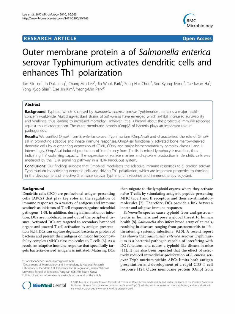
ResultsOmpA-sal induces DC maturationWe purified OmpA-sal from E. coli and assessed its cyto-toxicity on DCs because the purified OmpA-sal wasderived from S. enterica serovar Typhimurim. DCs weretreated with various concentrations of OmpA-sal for24 h. There were no statistically significant differences inthe percentages of dead cells in DC cultures exposed toas much as 800 ng/ml of OmpA-sal, the concentration atwhich cell death was detected by annexin V/PI staining(Fig. 1A). This indicated that our recombinant OmpA-salwas not cytotoxic to DCs and did not contain amounts ofendotoxin that would interfere with our studies usingconcentrations < 400 ng/ml. To determine the effects ofOmpA-sal on the maturation of sentinel DCs into effec-tor DCs, BM-derived DCs were cultured with GM-CSFand IL-4 for 6 days under standard conditions, followedby 1 day in the presence of 100, 200, and 400 ng/ml ofOmpA-sal. LPS was used as a positive control. Theresulting populations of DCs were analyzed by flow cyto-metry for expression of co-stimulatory moleculesinvolved in T cell activation. OmpA-sal-treated DCs hadincreased expression of DC maturation co-stimulatorymarkers (DC80, CD86, MHC class I, and MHC class II;Fig 1B). Interestingly, the expression of CD86 and MHCclass II by OmpA-sal-treated DCs was higher than LPS-treated DCs. These results indicated that OmpA-salinduces DC maturation in a dose-dependent manner.
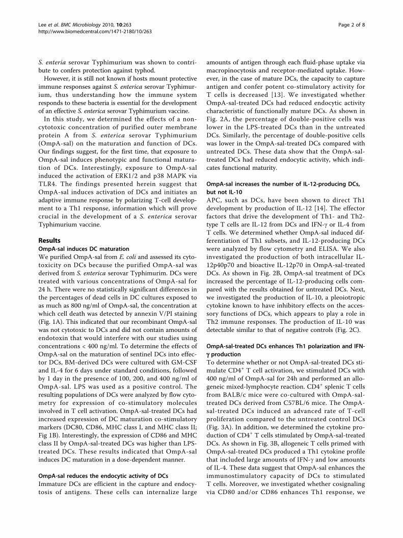
OmpA-sal reduces the endocytic activity of DCsImmature DCs are efficient in the capture and endocy-tosis of antigens. These cells can internalize large
amounts of antigen through each fluid-phase uptake viamacropinocytosis and receptor-mediated uptake. How-ever, in the case of mature DCs, the capacity to captureantigen and confer potent co-stimulatory activity forT cells is decreased [13]. We investigated whetherOmpA-sal-treated DCs had reduced endocytic activitycharacteristic of functionally mature DCs. As shown inFig. 2A, the percentage of double-positive cells waslower in the LPS-treated DCs than in the untreatedDCs. Similarly, the percentage of double-positive cellswas lower in the OmpA-sal-treated DCs compared withuntreated DCs. These data show that the OmpA-sal-treated DCs had reduced endocytic activity, which indi-cates functional maturity.
OmpA-sal increases the number of IL-12-producing DCs,but not IL-10APC, such as DCs, have been shown to direct Th1development by production of IL-12 [14]. The effectorfactors that drive the development of Th1- and Th2-type T cells are IL-12 from DCs and IFN-g or IL-4 fromT cells. We determined whether OmpA-sal induced dif-ferentiation of Th1 subsets, and IL-12-producing DCswere analyzed by flow cytometry and ELISA. We alsoinvestigated the production of both intracellular IL-12p40p70 and bioactive IL-12p70 in OmpA-sal-treatedDCs. As shown in Fig. 2B, OmpA-sal treatment of DCsincreased the percentage of IL-12-producing cells com-pared with the results obtained for untreated DCs. Next,we investigated the production of IL-10, a pleoiotropiccytokine known to have inhibitory effects on the acces-sory functions of DCs, which appears to play a role inTh2 immune responses. The production of IL-10 wasdetectable similar to that of negative controls (Fig. 2C).
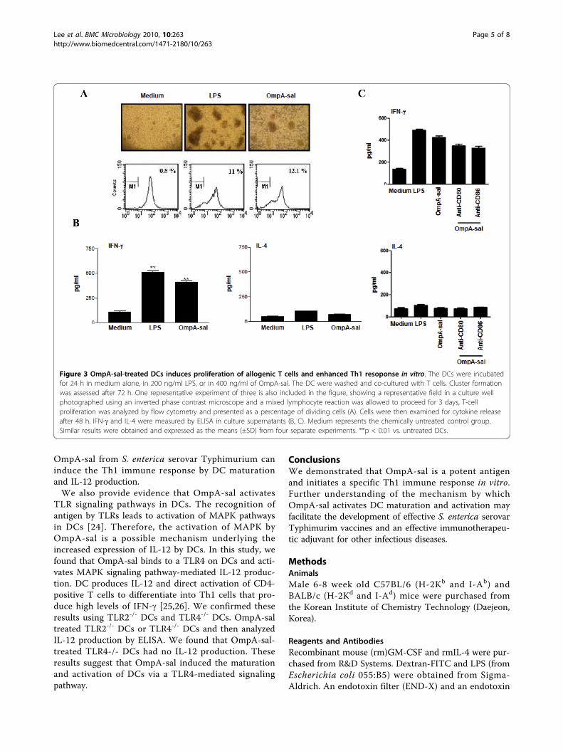
OmpA-sal-treated DCs enhances Th1 polarization and IFN-g productionTo determine whether or not OmpA-sal-treated DCs sti-mulate CD4+ T cell activation, we stimulated DCs with400 ng/ml of OmpA-sal for 24h and performed an allo-geneic mixed-lymphocyte reaction. CD4+ splenic T cellsfrom BALB/c mice were co-cultured with OmpA-sal-treated DCs derived from C57BL/6 mice. The OmpA-sal-treated DCs induced an advanced rate of T-cellproliferation compared to the untreated control DCs(Fig. 3A). In addition, we determined the cytokine pro-duction of CD4+ T cells stimulated by OmpA-sal-treatedDCs. As shown in Fig. 3B, allogeneic T cells primed withOmpA-sal-treated DCs produced a Th1 cytokine profilethat included large amounts of IFN-g and low amountsof IL-4. These data suggest that OmpA-sal enhances theimmunostimulatory capacity of DCs to stimulatedT cells. Moreover, we investigated whether cosignalingvia CD80 and/or CD86 enhances Th1 response, we
Lee et al. BMC Microbiology 2010, 10:263http://www.biomedcentral.com/1471-2180/10/263
Page 2 of 8
found that blockage of CD80 and CD86 decreased IFN-gproduction. These data suggested that both CD80 andCD86 are essential for the Th1 response of OmpA-saltreated DCs.
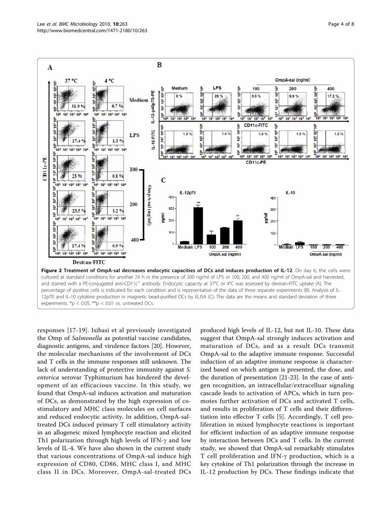
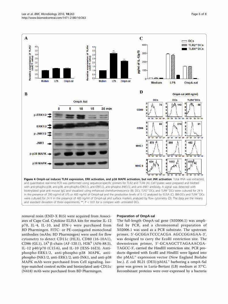
OmpA-sal induces DC maturation by TLR4 signalingToll-like receptors (TLRs) link innate and adaptiveimmune responses [15]. The DC response to TLR ligandsdepends on the activation of mitogen-activated proteinkinases (MAPKs), including ERK1/2, JNK1/2, and p38MAPK [16]. We determined the effects of OmpA-sal onTLRs and the MAPK signaling pathway. DCs were trea-ted with 400 ng/ml of OmpA-sal and TLR activation wasmeasured by real-time quantitative reverse transcription-PCR and phophorylation-specific Western blotting. Thelevel of TLR4 mRNA was significantly higher in OmpA-sal-treated DCs than in untreated control DCs, but therewas no change in TLR2 mRNA (Fig. 4A). Moreover,OmpA-sal enhanced the phosphorylation of ERK1/2 andp38 MAPK in DCs, but not JNK1/2 (Fig. 4B). To confirm
whether or not the maturation of DCs by OmpA-sal wasmediated by a TLR4-related signaling pathway, we iso-lated DCs from TLR2 and TLR4 knock-out mice, thenmeasured IL-12 production in DCs by OmpA-sal treat-ment. The inducing effect of OmpA-sal on IL-12 produc-tion was completely inhibited by TLR4-/- DCs, but it hadno effect on TLR2-/- DCs (Fig. 4C). Moreover, wedemonstrated that OmpA-sal-treated TLR4-/-DCs had noincreased expression of DC maturation co-stimulatorymarkers (DC80, CD86, MHC class I, and MHC class II;Fig 4D). These results indicate that the activation andmaturation of DCs by OmpA-sal is involved in TLR4signaling.
DiscussionWe have shown that OmpA-sal, a major virulence factorof S. enterica serovar Typhimurium, is a highly immu-nogenic protein that induces Th1 polarization of T cellsby DC maturation. Some of the Omps from bacteriainduce DC maturation and regulate Th1/Th2 immune
Figure 1 OmpA-sal is not cytotoxic and induces the expression of co-stimulatory molecules in DCs. BM-DCs were cultured for 24 h in thepresence of 200 ng/ml of LPS or 100, 200, 400, and 800 ng/ml of OmpA-sal and analyzed by flow cytometry. The DCs were stained with annexinV and PI. The percentage of positive cells is indicated (A). The cells were gated to exclude CD11c+ cells. Medium, untreated control; LPS, positivecontrol. DCs were stained with anti-CD80, anti-CD86, anti-MHC class I, and anti-MHC class II molecules (B). The data are representative of threeexperiments that yielded similar results.
Lee et al. BMC Microbiology 2010, 10:263http://www.biomedcentral.com/1471-2180/10/263
Page 3 of 8
responses [17-19]. Isibasi et al previously investigatedthe Omp of Salmonella as potential vaccine candidates,diagnostic antigens, and virulence factors [20]. However,the molecular mechanisms of the involvement of DCsand T cells in the immune responses still unknown. Thelack of understanding of protective immunity against S.enterica serovar Typhimurium has hindered the devel-opment of an efficacious vaccine. In this study, wefound that OmpA-sal induces activation and maturationof DCs, as demonstrated by the high expression of co-stimulatory and MHC class molecules on cell surfacesand reduced endocytic activity. In addition, OmpA-sal-treated DCs induced primary T cell stimulatory activityin an allogeneic mixed lymphocyte reaction and elicitedTh1 polarization through high levels of IFN-g and lowlevels of IL-4. We have also shown in the current studythat various concentrations of OmpA-sal induce highexpression of CD80, CD86, MHC class I, and MHCclass II in DCs. Moreover, OmpA-sal-treated DCs
produced high levels of IL-12, but not IL-10. These datasuggest that OmpA-sal strongly induces activation andmaturation of DCs, and as a result DCs transmitOmpA-sal to the adaptive immune response. Successfulinduction of an adaptive immune response is character-ized based on which antigen is presented, the dose, andthe duration of presentation [21-23]. In the case of anti-gen recognition, an intracellular/extracelluar signalingcascade leads to activation of APCs, which in turn pro-motes further activation of DCs and activated T cells,and results in proliferation of T cells and their differen-tiation into effector T cells [5]. Accordingly, T cell pro-liferation in mixed lymphocyte reactions is importantfor efficient induction of an adaptive immune responseby interaction between DCs and T cells. In the currentstudy, we showed that OmpA-sal remarkably stimulatesT cell proliferation and IFN-g production, which is akey cytokine of Th1 polarization through the increase inIL-12 production by DCs. These findings indicate that
Figure 2 Treatment of OmpA-sal decreases endocytic capacities of DCs and induces production of IL-12. On day 6, the cells werecultured at standard conditions for another 24 h in the presence of 200 ng/ml of LPS or 100, 200, and 400 ng/ml of OmpA-sal and harvested,and stained with a PE-conjugated anti-CD11c+ antibody. Endocytic capacity at 37°C or 4°C was assessed by dextran-FITC uptake (A). Thepercentage of positive cells is indicated for each condition and is representative of the data of three separate experiments (B). Analysis of IL-12p70 and IL-10 cytokine production in magnetic bead-purified DCs by ELISA (C). The data are the means and standard deviation of threeexperiments. *p < 0.05, **p < 0.01 vs. untreated DCs.
Lee et al. BMC Microbiology 2010, 10:263http://www.biomedcentral.com/1471-2180/10/263
Page 4 of 8
OmpA-sal from S. enterica serovar Typhimurium caninduce the Th1 immune response by DC maturationand IL-12 production.We also provide evidence that OmpA-sal activates
TLR signaling pathways in DCs. The recognition ofantigen by TLRs leads to activation of MAPK pathwaysin DCs [24]. Therefore, the activation of MAPK byOmpA-sal is a possible mechanism underlying theincreased expression of IL-12 by DCs. In this study, wefound that OmpA-sal binds to a TLR4 on DCs and acti-vates MAPK signaling pathway-mediated IL-12 produc-tion. DC produces IL-12 and direct activation of CD4-positive T cells to differentiate into Th1 cells that pro-duce high levels of IFN-g [25,26]. We confirmed theseresults using TLR2-/- DCs and TLR4-/- DCs. OmpA-saltreated TLR2-/- DCs or TLR4-/- DCs and then analyzedIL-12 production by ELISA. We found that OmpA-sal-treated TLR4-/- DCs had no IL-12 production. Theseresults suggest that OmpA-sal induced the maturationand activation of DCs via a TLR4-mediated signalingpathway.
ConclusionsWe demonstrated that OmpA-sal is a potent antigenand initiates a specific Th1 immune response in vitro.Further understanding of the mechanism by whichOmpA-sal activates DC maturation and activation mayfacilitate the development of effective S. enterica serovarTyphimurim vaccines and an effective immunotherapeu-tic adjuvant for other infectious diseases.
MethodsAnimalsMale 6-8 week old C57BL/6 (H-2Kb and I-Ab) andBALB/c (H-2Kd and I-Ad) mice were purchased fromthe Korean Institute of Chemistry Technology (Daejeon,Korea).
Reagents and AntibodiesRecombinant mouse (rm)GM-CSF and rmIL-4 were pur-chased from R&D Systems. Dextran-FITC and LPS (fromEscherichia coli 055:B5) were obtained from Sigma-Aldrich. An endotoxin filter (END-X) and an endotoxin
Figure 3 OmpA-sal-treated DCs induces proliferation of allogenic T cells and enhanced Th1 resoponse in vitro. The DCs were incubatedfor 24 h in medium alone, in 200 ng/ml LPS, or in 400 ng/ml of OmpA-sal. The DC were washed and co-cultured with T cells. Cluster formationwas assessed after 72 h. One representative experiment of three is also included in the figure, showing a representative field in a culture wellphotographed using an inverted phase contrast microscope and a mixed lymphocyte reaction was allowed to proceed for 3 days, T-cellproliferation was analyzed by flow cytometry and presented as a percentage of dividing cells (A). Cells were then examined for cytokine releaseafter 48 h. IFN-g and IL-4 were measured by ELISA in culture supernatants (B, C). Medium represents the chemically untreated control group.Similar results were obtained and expressed as the means (±SD) from four separate experiments. **p < 0.01 vs. untreated DCs.
Lee et al. BMC Microbiology 2010, 10:263http://www.biomedcentral.com/1471-2180/10/263
Page 5 of 8
removal resin (END-X B15) were acquired from Associ-ates of Cape Cod. Cytokine ELISA kits for murine IL-12p70, IL-4, IL-10, and IFN-g were purchased fromBD Pharmingen. FITC- or PE-conjugated monoclonalantibodies (mAbs; BD Pharmingen) were used for flowcytometry to detect CD11c (HL3), CD80 (16-10A1),CD86 (GL1), IAb b-chain (AF-120.1), H2Kb (AF6-88.5),IL-12 p40/p70 (C15.6), and IL-10 (JESS-16E3). Anti-phospho-ERK1/2, anti-phospho-p38 MAPK, anti-phospho-JNK1/2, anti-ERK1/2, anti-JNK1, and anti-p38MAPK mAb were purchased from Cell signaling. Iso-type-matched control mAbs and biotinylated anti-CD11c(N418) mAb were purchased from BD Pharmingen.
Preparation of OmpA-salThe full-length OmpA-sal gene (X02006.1) was ampli-fied by PCR, and a chromosomal preparation ofX02006.1 was used as a PCR substrate. The upstreamprimer, 5’-GCGGATCCCACGA AGCCGGAGAA-3’,was designed to carry the EcoRI restriction site. Thedownstream primer, 5’-GCAAGCTTAGAAACGA-TAGCC-3’, carried the HindIII restriction site. PCR pro-ducts digested with EcoRI and HindIII were ligated intothe pMAL™ expression vector (New England BiolabsInc.). E. coli BL21 (DE3)/pMAL™ harboring a ompA-Salgene was grown in Luria-Bertani (LB) medium at 37°C.Recombinant proteins were over-expressed by a bacteria
Figure 4 OmpA-sal induces TLR4 expression, ERK activation, and p38 MAPK activation, but not JNK activation. Total RNA was extracted,and quantitative real-time PCR was performed using sequence-specific primers for TLR2 and TLR4 (A). Cell lysates were prepared and blottedwith anti-phopho-p38, anti-p38, anti-phopho-ERK1/2, anti-ERK1/2, anti-phopho-JNK1/2, and anti-JNK1 antibody. A signal was detected withbiotinylated goat-anti mouse IgG and visualized using enhanced chemiluminescence (B). DCs, TLR2-/-DCs, and TLR4-/-DCs were cultured for 24 hin the presence of 200 ng/ml of LPS or 400 ng/ml of OmpA-sal and the production levels of IL-12 analyzed by ELISA (C). BM-DCs and TLR4-/-DCswere cultured for 24 h in the presence of 400 ng/ml of OmpA-sal and surface markers analyzed by flow cytometry (D). The data are the meansand standard deviation of three experiments. **, P < 0.01 for a compare with untreated DCs.
Lee et al. BMC Microbiology 2010, 10:263http://www.biomedcentral.com/1471-2180/10/263
Page 6 of 8
protein expression system [27]. The quantity of OmpAendotoxin was ≤0.01 ng/mg.
Generation and culture of DCsDCs were generated from murine whole bone marrow(BM) cells. Briefly, the BM was flushed from the tibiaeand femurs of BALB/c mice and depleted of red bloodcells with ammonium chloride. The cells were platedin 6-well culture plates (106 cells/ml) and cultured at37°C in 5% CO2 and OptiMEM (Invitrogen Life Tech-nologies) supplemented with 10% heat-inactivated fetalbovine serum (FBS), 2 mM L-glutamine, 100 U/mlpenicillin, 100 μg/ml streptomycin, 5 × 10⁻⁵ M b-mer-captoethanol, 10 mM HEPES (pH 7.4), 20 ng/mlrmGM-CSF, and rmIL-4. On day 3 of culture, floatingcells were gently removed and fresh medium wasadded. On day 6 or 7 of culture, non-adherent cellsand loosely adherent proliferating DC aggregates wereharvested for analysis or stimulation, or in someexperiments, replated into 60 mm dishes.
Quantitation of antigen uptakeIn brief, DCs were equilibrated at 37°C or 4°C for 45min, then pulsed with fluorescein-conjugated dextran ata concentration of 1 mg/ml. Cold staining buffer wasadded to stop the reaction. The cells were washed threetimes and stained with PE-conjugated anti-CD11c Abs,then analyzed with the FACSCalibur. Non-specific bind-ing of dextran to DCs was determined by incubation ofDCs with FITC-conjugated dextran at 4°C and sub-tracted as background. The medium used in the cultureswith OmpA-sal stimulation was supplemented withGM-CSF, which is required for the ability of DCs tocapture antigen.
Cytokine assaysCells were first blocked with 10% (v/v) normal goatserum for 15 min at 4°C, then stained with FITC-conjugated CD11c+ antibody for 30 min at 4°C. Cellsstained with the appropriate isotype-matched Ig wereused as negative controls. The cells were fixed and per-meabilized with the Cytofix/Cytoperm kit (PharMingen)according to the manufacturer’s instructions. Intracellu-lar IL-12p40/p70 and IL-10 were detected with fluores-cein PE-conjugated antibodies (PharMingen) in apermeation buffer. The presence of murine IL-12p70,IL-10, IL-4, and IFN-g in DCs was measured using anELISA kit (R&D systems) according to the manufac-turer’s instructions.
Cytoplasmic extracts and Western blotThe cells were exposed to LPS (200 ng/ml) with orwithout OmpA-sal pre-treatment (400 ng/ml). Following5, 10, 15, or 30 min of incubation at 37°C, cells were
washed twice with cold PBS and lysed with modifiedRIPA buffer for 15 min at 4°C. The protein content ofcell lysates was determined using the Micro BCA assaykit (Pierce, Rockford, IL, USA). Equivalent amounts ofproteins were separated by 10% or 12% SDS-PAGE andanalyzed by Western blotting using anti-phospho-ERK1/2, anti-phospho-p38 MAPK, anti-phospho-JNK1/2, anti-ERK1/2, anti-JNK1, and anti-p38 MAPK mAb for 3 h,as described by the manufacturers.
Mixed lymphocyte reactionResponder T cells, which participate in allogeneic T-cellreactions, were isolated from spleens of BALB/c miceusing a MACS column (positive selection sorting).Staining with FITC-conjugated anti-CD4 Abs revealedthat the recovered cells consisted mainly of CD4+ cells.The lymphocyte population was then washed twice inPBS and labeled with CFSE, as previously described[28]. The cells were washed once in pure FBS and twicein PBS with 10% FBS. DCs (1×10⁴), or DCs exposed toOmpA-sal or LPS for 24 h, were co-cultured with 1×10⁵
allogeneic CFSE-labeled T lymphocytes in 96-well U-bottom plates. After 3 days, the CFSE dilution optically-gated lymphocytes were assessed.
Evaluation of gene expression by real time PCRTLR 2 and 4 PCR primers were used. Quantitativeamounts of each gene were standardized against theGAPDH housekeeping gene. Real-time PCR was per-formed using a BioRad MiniOpticon System (BioRadLaboratories, Ltd.) with a SYBR green fluorophore.Reactions were performed in a total volume of 20 μl,including 10 μl of 2x SYBR Green PCR Master Mix, 1μl of each primer at 10 ng, and 1 μl of the previouslyreverse-transcribed cDNA template. The protocolsused were as follows: denaturation (95°C for 10 min),and amplification repeated 40 times (95°C for 30 s, 52°C for 30 s, 72°C for 30 s, and acquisition temperaturefor 15 s).
Statistical analysisAll data are expressed as the mean ± standard devia-tion (SD) and were representative of at least two dif-ferent experiments. Comparisons between individualdata points were made using the Student’s t-test andperformed using one-way ANOVA analysis (Least Sig-nificant Difference (LSD) as post-hoc test). Throughoutthe figures and legends, the following terminology wasused to denote statistical significance:**, p < 0.01, *,p < 0.05.
AcknowledgementsThis work was supported by a National Research Foundation of Korea (NRF)grant funded by the Korea government [(MEST)-314-2008-1-E00195].
Lee et al. BMC Microbiology 2010, 10:263http://www.biomedcentral.com/1471-2180/10/263
Page 7 of 8
Author details1Department of Microbiology and Immunology, Albert Einstein College ofMedicine, Bronx, NY 10461, USA. 2Department of Microbiology andImmunology & National Research Laboratory of Dendritic Cell Differentiation& Regulation, Pusan National University School of Medicine, Yang-san 626-770, South Korea. 3Department of Surgery, Busan Paik Hospital, InjeUniversity, College of Medicine, Busan, 614-735, South Korea. 4College ofMedicine, Chung-Ang University, 221 Heukseok-Dong, Dongjak-Gu, Seoul,Korea. 5Department of Anatomy, College of Medicine, Chung-Ang University,221 Heukseok-Dong, Dongjak-Gu, Seoul, Korea.
Authors’ contributionsContribution: JSL performed research, analyzed data and wrote the paper;DJ and CML, JWP, and SHC performed research; TKH performed statisticalanalysis: SKJ, YKS and DJ K analyzed and interpreted data; JSL and YMPdesigned research, interpreted data and wrote the paper. All authors readand approved the final manuscript.
Received: 8 April 2010 Accepted: 15 October 2010Published: 15 October 2010
References1. Steinman RM: The dendritic cell system and its role in immunogenicity.
Annu Rev Immunol 1991, 9:271-296.2. Granucci F, Zanoni I, Feau S, Ricciardi-Castagnoli P: Dendritic cell
regulation of immune responses: a new role for interleukin 2 at theintersection of innate and adaptive immunity. Embo J 2003,22(11):2546-2551.
3. Nagl M, Kacani L, Mullauer B, Lemberger EM, Stoiber H, Sprinzl GM,Schennach H, Dierich MP: Phagocytosis and killing of bacteria byprofessional phagocytes and dendritic cells. Clin Diagn Lab Immunol 2002,9(6):1165-1168.
4. Kelsall BL, Rescigno M: Mucosal dendritic cells in immunity andinflammation. Nat Immunol 2004, 5(11):1091-1095.
5. Guermonprez P, Valladeau J, Zitvogel L, Thery C, Amigorena S: Antigenpresentation and T cell stimulation by dendritic cells. Annu Rev Immunol2002, 20:621-667.
6. MacDonald TT, Vossenkamper A, Di Sabatino A: Antigen presenting cellsand T cell interactions in the gastrointestinal tract. Mol Nutr Food Res2009, 53(8):947-951.
7. Meyer zum Bueschenfelde CO, Unternaehrer J, Mellman I, Bottomly K:Regulated recruitment of MHC class II and costimulatory molecules tolipid rafts in dendritic cells. J Immunol 2004, 173(10):6119-6124.
8. Valdez Y, Ferreira RB, Finlay BB: Molecular mechanisms of Salmonellavirulence and host resistance. Curr Top Microbiol Immunol 2009,337:93-127.
9. Chiu CH, Su LH, Chu C: Salmonella enterica serotype Choleraesuis:epidemiology, pathogenesis, clinical disease, and treatment. ClinMicrobiol Rev 2004, 17(2):311-322.
10. McClelland M, Sanderson KE, Spieth J, Clifton SW, Latreille P, Courtney L,Porwollik S, Ali J, Dante M, Du F, et al: Complete genome sequence ofSalmonella enterica serovar Typhimurium LT2. Nature 2001,413(6858):852-856.
11. Bueno SM, Tobar JA, Iruretagoyena MI, Kalergis AM: Molecular interactionsbetween dendritic cells and Salmonella: escape from adaptive immunityand implications on pathogenesis. Crit Rev Immunol 2005, 25(5):389-403.
12. Alaniz RC, Deatherage BL, Lara JC, Cookson BT: Membrane vesicles areimmunogenic facsimiles of Salmonella typhimurium that potentlyactivate dendritic cells, prime B and T cell responses, and stimulateprotective immunity in vivo. J Immunol 2007, 179(11):7692-7701.
13. Piemonti L, Monti P, Allavena P, Leone BE, Caputo A, Di Carlo V:Glucocorticoids increase the endocytic activity of human dendritic cells.Int Immunol 1999, 11(9):1519-1526.
14. Macatonia SE, Hosken NA, Litton M, Vieira P, Hsieh CS, Culpepper JA,Wysocka M, Trinchieri G, Murphy KM, O’Garra A: Dendritic cells produce IL-12 and direct the development of Th1 cells from naive CD4+ T cells. JImmunol 1995, 154(10):5071-5079.
15. Michelsen KS, Doherty TM, Shah PK, Arditi M: TLR signaling: an emergingbridge from innate immunity to atherogenesis. J Immunol 2004,173(10):5901-5907.
16. Zaru R, Ronkina N, Gaestel M, Arthur JS, Watts C: The MAPK-activatedkinase Rsk controls an acute Toll-like receptor signaling response indendritic cells and is activated through two distinct pathways. NatImmunol 2007, 8(11):1227-1235.
17. Shaw J, Grund V, Durling L, Crane D, Caldwell HD: Dendritic cells pulsedwith a recombinant chlamydial major outer membrane protein antigenelicit a CD4(+) type 2 rather than type 1 immune response that is notprotective. Infect Immun 2002, 70(3):1097-1105.
18. Lee JS, Lee JC, Lee CM, Jung ID, Jeong YI, Seong EY, Chung HY, Park YM:Outer membrane protein A of Acinetobacter baumannii inducesdifferentiation of CD4+ T cells toward a Th1 polarizing phenotypethrough the activation of dendritic cells. Biochem Pharmacol 2007,74(1):86-97.
19. Jeannin P, Magistrelli G, Herbault N, Goetsch L, Godefroy S, Charbonnier P,Gonzalez A, Delneste Y: Outer membrane protein A renders dendriticcells and macrophages responsive to CCL21 and triggers dendritic cellmigration to secondary lymphoid organs. Eur J Immunol 2003,33(2):326-333.
20. Isibasi A, Ortiz V, Vargas M, Paniagua J, Gonzalez C, Moreno J, Kumate J:Protection against Salmonella typhi infection in mice after immunizationwith outer membrane proteins isolated from Salmonella typhi 9,12,d, Vi.Infect Immun 1988, 56(11):2953-2959.
21. Zinkernagel RM, Moskophidis D, Kundig T, Oehen S, Pircher H,Hengartner H: Effector T-cell induction and T-cell memory versusperipheral deletion of T cells. Immunol Rev 1993, 133:199-223.
22. Fuller MJ, Khanolkar A, Tebo AE, Zajac AJ: Maintenance, loss, andresurgence of T cell responses during acute, protracted, and chronicviral infections. J Immunol 2004, 172(7):4204-4214.
23. Ostrand-Rosenberg S, Baskar S, Patterson N, Clements VK: Expression ofMHC Class II and B7-1 and B7-2 costimulatory molecules accompaniestumor rejection and reduces the metastatic potential of tumor cells.Tissue Antigens 1996, 47(5):414-421.
24. Re F, Strominger JL: Toll-like receptor 2 (TLR2) and TLR4 differentiallyactivate human dendritic cells. J Biol Chem 2001, 276(40):37692-37699.
25. O’Garra A, Hosken N, Macatonia S, Wenner CA, Murphy K: The role ofmacrophage- and dendritic cell-derived IL12 in Th1 phenotypedevelopment. Res Immunol 1995, 146(7-8):466-472.
26. Jego G, Palucka AK, Blanck JP, Chalouni C, Pascual V, Banchereau J:Plasmacytoid dendritic cells induce plasma cell differentiation throughtype I interferon and interleukin 6. Immunity 2003, 19(2):225-234.
27. Choi CH, Hyun SH, Lee JY, Lee JS, Lee YS, Kim SA, Chae JP, Yoo SM, Lee JC:Acinetobacter baumannii outer membrane protein A targets the nucleusand induces cytotoxicity. Cell Microbiol 2008, 10(2):309-319.
28. Lyons AB: Analysing cell division in vivo and in vitro using flowcytometric measurement of CFSE dye dilution. J Immunol Methods 2000,243(1-2):147-154.
doi:10.1186/1471-2180-10-263Cite this article as: Lee et al.: Outer membrane protein a of Salmonellaenterica serovar Typhimurium activates dendritic cells and enhancesTh1 polarization. BMC Microbiology 2010 10:263.
Submit your next manuscript to BioMed Centraland take full advantage of:
• Convenient online submission
• Thorough peer review
• No space constraints or color figure charges
• Immediate publication on acceptance
• Inclusion in PubMed, CAS, Scopus and Google Scholar
• Research which is freely available for redistribution
Submit your manuscript at www.biomedcentral.com/submit
Lee et al. BMC Microbiology 2010, 10:263http://www.biomedcentral.com/1471-2180/10/263
Page 8 of 8