research article performance of monocyte activation test
TRANSCRIPT

ALTEX 38(2), 2021 307
Received August 26, 2020; Accepted October 26, 2020; Epub October 28, 2020; © The Authors, 2021.
ALTEX 38(2), 307-315. doi:10.14573/altex.2008261
Correspondence: Anja ten Brinke, PhD Sanquin Plesmanlaan 125 1066 CX Amsterdam The Netherlands ([email protected])
siveness between humans and rabbits to pyrogens differs: Both species show a high reactivity to endotoxin, but humans usually show a higher reactivity to Gram-positive pyrogens than rabbits (Schindler et al., 2003).
The monocyte activation test (MAT) is an in vitro-based alter-native to the RPT based on the capacity of human immune cells such as monocytes to respond to endotoxins and NEPs by pro-duction of pro-inflammatory cytokines like interleukin (IL)-6, IL-1β or tumor necrosis factor (TNF) (Dinarello, 2004; Kik-kert et al., 2008). The current European Pharmacopeia (Ph. Eur.) chapter 2.6.30 (07/2017) states that “MAT is suitable, after prod-uct specific validation, as a replacement for the rabbit pyrogen test” (EDQM, 2020).
According to Ph. Eur. chapter 2.6.30, multiple cell sources can be used for the MAT assay, namely whole blood, peripher-
1 Introduction
Pharmaceutical products intended for parenteral use must be shown to have pyrogenicity below a specified level to protect humans from “injection fever”, a marked rise in body temperature caused by in-jection/infusion of a drug product contaminated with pyrogens. Py-rogens are subdivided into endotoxins (lipopolysaccharides; LPS) derived from Gram-negative bacteria and non-endotoxin pyrogens (NEPs) such as peptidoglycans (PGN), flagellin and β-glucans de-rived from Gram-positive bacteria and fungi.
Historically, the rabbit pyrogen test (RPT) was used to de-termine pyrogenicity of injectable drugs for humans, including plasma-derived products and implantable medical devices. How-ever, the RPT assay requires the use of animals, which should be avoided for ethical reasons where possible. Also, the respon-
Research Article
Performance of Monocyte Activation Test Supplemented with Human Serum Compared to Fetal Bovine SerumMarijke W. A. Molenaar-de Backer1, Eelo Gitz2, Miranda Dieker3, Paulien Doodeman1 and Anja ten Brinke3,41Sanquin Diagnostics, Dept Virology and MAT services, Amsterdam, The Netherlands; 2Sanquin Reagents, Amsterdam, The Netherlands; 3Sanquin Research, Dept Immunopathology, Amsterdam, The Netherlands; 4Landsteiner Laboratory, Amsterdam UMC, University of Amsterdam, Amsterdam, The Netherlands
AbstractThe monocyte activation test (MAT) is used to detect pyrogens in pharmaceutical products and serves as replacement of the rabbit pyrogen test. The peripheral blood mononuclear cell-based MAT assay requires the addition of serum to the medium and is performed with either fetal bovine serum (FBS) or human serum (HS). Since the capacity to detect non-endotoxin pyrogens (NEPs) in a sensitive manner is an important strength of MAT compared to the bacterial endo-toxin test, the performance of the MAT using FBS and HS was compared using endotoxin and several NEPs. The MAT was more sensitive for endotoxin when FBS was used, however for most NEPs the MAT was more sensitive when per-formed in HS. Furthermore, heat-inactivation of FBS affected the performance of the MAT for endotoxin to some extent but not for the NEPs. Interestingly, heat-inactivation of HS led to an almost complete loss of reactivity towards endotoxin, reduced the response towards heat-killed Staphylococcus aureus and peptidoglycan, but had minor or no effects on the responses towards R848, flagellin, and Pam3CSK4. Product testing of a human blood-derived product in MAT using HS was beneficial since endotoxin spike recoveries were improved. This product is therefore currently batch released with the HS-based MAT assay. Overall, to guarantee optimal performance of MAT, heat-inactivated serum should be avoided. The HS-based MAT appears to be the first choice to replace the rabbit pyrogen test, while in some cases the FBS-based MAT may be favored.
This is an Open Access article distributed under the terms of the Creative Commons Attribution 4.0 International license (http://creativecommons.org/licenses/by/4.0/), which permits unrestricted use, distribution and reproduction in any medium, provi-ded the original work is appropriately cited.
Molenaar-de Backer et al.
ALTEX 38(2), 2021 308
2 Materials and methods
PyrogensEndotoxin Standard Biological Reference Preparation (BRP) batch 5 (EDQM, Council of Europe, Strasbourg, France) was used to prepare the endotoxin standard curve. A stock solution of 2000 EU/mL was prepared in endotoxin-free water, aliquoted (50 µL), and stored at -80°C. The endotoxin stock solution was diluted further in complete medium to the required endotoxin concentrations. Heat-killed S. aureus (HKSA), flagellin (S. typh-imurium), Pam3CSK4, R848 and PGN (S. aureus) were all ob-tained from Invivogen ( San Diego, CA, USA) and aliquoted and stored according to the manufacturer’s instructions. The absence of endotoxins in these NEP preparations was claimed by the man-ufacturer and reconfirmed in-house for HKSA, flagellin and PGN using the Limulus Amebocyte Lysate (LAL) test (Pam3CSK4 and R848 are not suitable for testing in the LAL).
Of note, the endotoxin and NEP concentrations stated through-out this study are final concentrations in the well (after addition of cells to the well). Concentration in the sample can be calculat-ed by multiplying the stated concentrations by 2 (since samples and cells are mixed 1:1).
MAT execution: Cell cultureThe MAT was performed using the MAT Cell Set (Sanquin Reagents, Amsterdam, The Netherlands) following the manufac-turer’s instructions with adaptations as described: In short, a vial containing 5x10⁶ (± 1x10⁶) cryopreserved PBMCs (1 mL) was thawed in a water bath at 37°C until only a little clump of ice remained. The cell suspension was subsequently transferred to a new tube and diluted with 10 mL Iscove’s Modified Dulbec-co’s Medium (IMDM; Lonza, Verviers, Belgium) supplemented with either 5% FBS or HS (both from Biowest, Nuaillé, France). Where indicated, FBS or HS was heat-inactivated at 56°C for 45 min before use. In the MAT cell culture plate, 100 µL cell suspension was added to 100 µL sample (tested in triplicate or quadruplicate where indicated). In case of spiking experiments, 5 mL supplemented IMDM was added to the thawed cell suspen-sion, and 50 µL cell suspension was added to 100 µL sample plus 50 µL endotoxin spike solution or 50 µL supplemented IMDM (for unspiked samples). In both cases, each well contained approximately 45,000 cells. Subsequently, the 96-well cell cul-ture plate was incubated in a humidified incubator at 37°C in the presence of 5% CO2 for 18-24 h.
MAT execution: IL-6 ELISACell culture supernatants were harvested and analyzed in 1:5 dilution, except if indicated otherwise, for the presence of IL-6 using a commercially available enzyme-linked immuno-sorbent assay (ELISA) kit (PeliKine Compact™ human ELISA kit, Sanquin, Amsterdam, The Netherlands) following the manufacturer’s instructions. The ODs at 450 nm after subtraction of the background OD at 540 nm are depicted.
Drug product testing with the MATTo exclude interference between the test and the product being
al blood mononuclear cells (PBMCs), or a monocytic cell line as reviewed by Poole and colleagues (2003). The monocytic cell line has the disadvantages that it represents just one genet-ic background, is derived from a tumor cell line, and function-al stability over passaging has to be ensured. Whole blood or PBMCs may be either freshly isolated or thawed from cryopre-served material. Whole blood or PBMCs may be derived from a single donor or employed as pools from at least four donors (Solati et al., 2015; Koryakina et al., 2014). The requirement of four donors for pools is intended to include genetic variation in humans to the responsiveness to NEPs (Jaeger et al., 2015), while the responsiveness to endotoxins is conserved among humans (Copeland et al., 2005).
In this study, the MAT was performed with cryopreserved pooled PBMCs from four donors combined with an IL-6 ELISA as cytokine readout. Previous results from our and other labo-ratories have shown that a PBMC-based MAT assay has high sensitivity and high specificity (Hoffmann et al., 2005; Solati et al., 2015; Vipond et al., 2019; Etna et al., 2020).
According to the Ph. Eur. (EDQM, 2020), each new prod-uct has to go through a product-specific validation to determine that it does not interfere with the MAT test. This product-spe-cific validation consists of 1) tests showing that the MAT de-tects endotoxins and NEPs when spiked in the product and 2) a test showing that the product does not interfere with the detection system (ELISA). Subsequently, release testing can be performed on the production process qualification (PPQ) batches (for registration) and/or on each production batch. During release testing according to method A (quantitative test) or method B (semi-quantitative test), as described in Ph. Eur. chapter 2.6.30 (EDQM, 2020), three dilutions of the product, which were determined during the product-specific validation, are tested with and without an endotoxin spike. The lowest di-lution with a valid endotoxin spike recovery, between 50 and 200%, is used to calculate the amount of endotoxin equivalent units per milliliter (EEU/mL) for method A. If the responses to dilutions of a product are not comparable to the responses of standard endotoxin dilutions, then method B should be used. Method B is a pass/fail test, e.g., the contaminant concentration of the preparation should be below the contaminant limit con-centration (CLC) to pass.
A PBMC-based MAT assay requires supplementation of the cell culture medium with serum. Serum contains proteins that aid the immune reaction triggered by pyrogens. The MAT can be performed with either fetal bovine serum (FBS) or human serum (HS) as serum source. Since the capacity to detect NEPs in a sensitive manner is an important asset of the MAT, we compared the performance of the MAT in FBS and HS to gain more insight into the detection of endotoxins and NEPs by the PBMC-based MAT using these different serum sources. This is especially relevant since results might support a MAT format without use of animal-derived materials. In addition, we stud-ied the effect of heat-inactivation of the serum on the response to endotoxin and NEPs and the effect of serum source on the recovery of the endotoxin spikes in a specific human blood- derived product.

Molenaar-de Backer et al.
ALTEX 38(2), 2021 309
Fig. 1: Comparison of MAT performed in FBS and HSPBMCs were stimulated overnight with endotoxin (LPS), HKSA, Pam3CSK4, PGN, flagellin and R848 in a 2-fold concentration range using either FBS or HS as serum source. An IL-6 ELISA was performed on the culture supernatants; supernatants of R848 were diluted 1:25 for IL-6 determination; for all other conditions, supernatants were tested in 1:5 dilution. (A) Representative graphs are depicted; results are shown as mean OD ± SD of triplicate measurements. (B) Area under the curve (AUC) of fitted pyrogen curves of all experiments is calculated and depicted as mean ± SD. Endotoxin (LPS) (n = 4), HKSA (n = 4), Pam3CSK4 (n = 4), PGN (n = 3), flagellin (n = 5) and R848 (n = 4). Paired t-test was performed. *p < 0.05, *** p < 0.001.

Molenaar-de Backer et al.
ALTEX 38(2), 2021 310
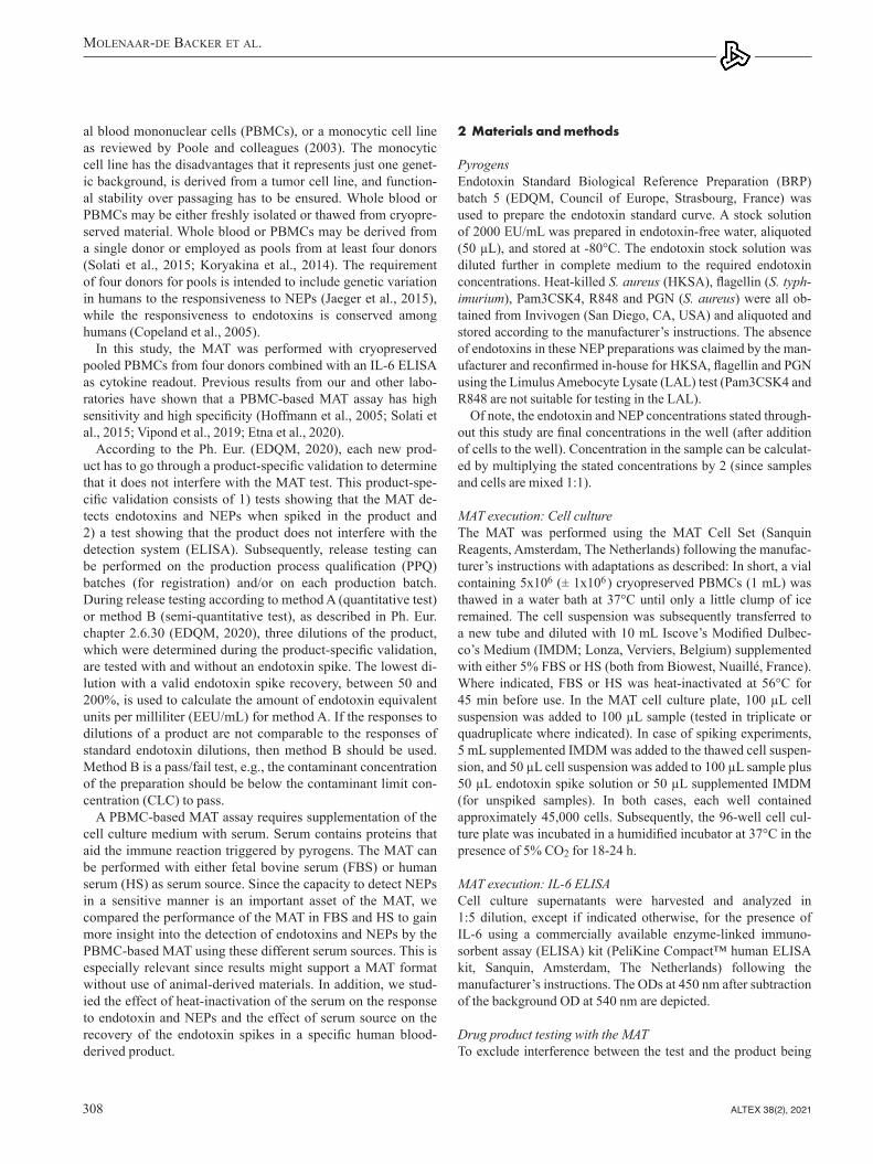
without evidence of a beneficial effect (Rahman et al., 2011). To study if the reactivity of the MAT is affected by heat-inactivation of the serum, we compared the responses in MAT using untreated or heat-activated FBS or HS.
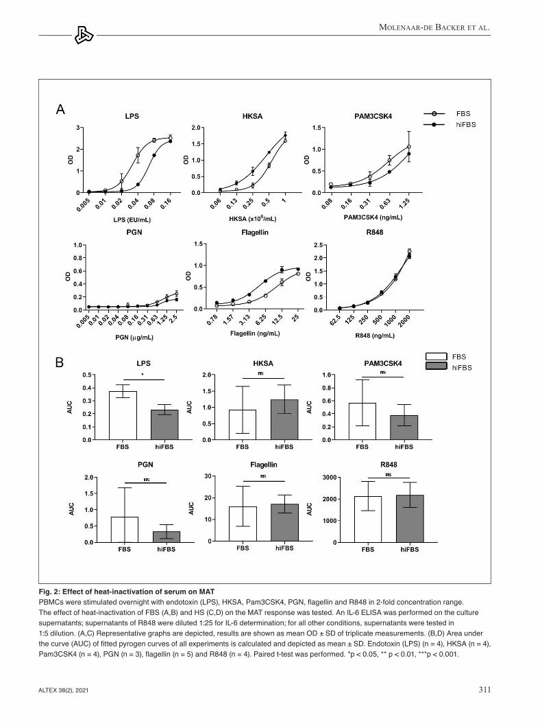
Heat-inactivation of FBS reduced the reactivity of the MAT towards endotoxin (Fig. 2A,B) but not towards NEPs. Heat- inactivation of HS had a more pronounced effect compared to heat-inactivation of FBS (Fig. 2C,D). It resulted in an almost complete loss of reactivity of MAT towards endotoxin and had varying effects on reactivity towards NEPs. The reactivity of the MAT to Pam3CSK4 was not affected by heat-inactivation of HS, the reactivity to R848 and flagellin was significantly higher, but the response towards HKSA and PGN was significantly lower using heat-inactivated HS. Overall, heat-inactivation of HS had differential effects on the responses of the MAT to pyrogens.
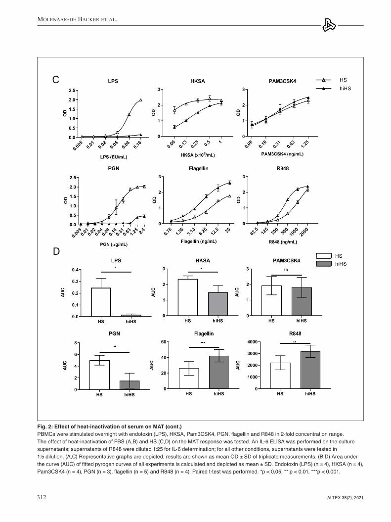
3.3 Interference of HS in an FBS-based MATSince the reactivity towards endotoxin was lower in the HS-based MAT compared to the FBS-based MAT, we wondered if HS contains either lower amounts of stimulatory constituents or higher amounts of inhibitory constituents affecting the endo-toxin response. Therefore, we performed an FBS-based MAT in IMDM with 5% FBS using a fixed amount of endotoxin (0.08 EU/mL) and added increasing amounts of HS to investigate if this affected the response towards endotoxin. To correct for the higher total concentration of serum, we added the same amount of additional FBS to controls containing the same amount of endotoxin. The experiments were performed with serum that had not been heat-inactivated.
The addition of HS caused a significant inhibition of the re-sponse towards endotoxin, while this was not the case for ad-dition of FBS (Fig. 3). The inhibition was already observed at 0.16 % HS, but was more pronounced at higher HS concentra-tions. This indicates that HS contains constituents that can inhibit the response to endotoxin in an FBS-based MAT.
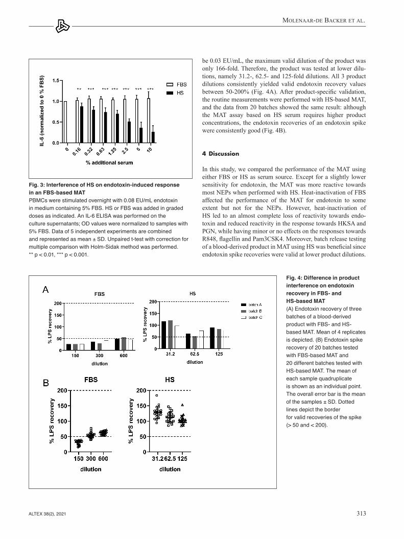
3.4 Effect of serum source during drug product testingIn previous studies, it was observed that certain plasma-de-rived products yielded invalid recoveries of the endotoxin spike that was added to the products, making it complex to test these products in the MAT (method A or B of Ph. Eur. 2.6.30) (Perdomo- Morales et al., 2011). Here a blood-derived product was tested in the MAT, using either FBS or HS as serum source, to assess the effects on endotoxin recovery. The maximum valid dilution of a product is determined by dividing the contaminant limit by the LOD.
With the FBS-based MAT, the LOD was 0.008 EU/mL, and this resulted in a maximum valid dilution (MVD) for the product of 625-fold. The product was tested at 150-, 300- and 600-fold dilutions; however, the 150-fold dilution did not yield a valid en-dotoxin recovery (< 50%) (Fig. 4). The 300-fold product dilution yielded recoveries around 50% and the 600-fold around 60%. This resulted in many repeats of the routine MAT assay due to invalid endotoxin spike recoveries.
The same blood-derived product was also tested with the HS-based MAT. Since the LOD of this MAT was determined to
tested, the MAT has to be validated for that product according to the pharmacopeia guidelines. For this, endotoxin is spiked into uncontaminated batches of the product and tested in the MAT. The product is considered free from interfering factors if the mean recovery of the added endotoxin is in the range of 50-200%. During these experiments, all samples were tested in quadruplicate and 50 µL cell suspension was added to 100 µL sample and 50 µL endotoxin spike or 50 µL complete medium (for unspiked samples). The endotoxin spike was 0.02 EU/mL for the FBS-based MAT or 0.16 EU/mL for the HS-based MAT. In addition, an endotoxin standard curve was included on the same plate in quadruplicate, ranging from 0.005 to 0.08 EU/mL for FBS-based MAT and 0.02 to 0.32 EU/mL for the HS-based MAT.
Statistical analysisGraphPad Prism 8.0.2. was used for the analysis. Area under the curve (AUC) of nonlinear fitted regression lines ([agonist] vs. re-sponse-variable slope (four parameters) were calculated. Paired t-test was used to compare the AUC of the two serum sources. Unpaired t-test with correction for multiple comparison with Holm-Sidak method was performed to analyze the effect of HS in FBS-based MAT on detection of endotoxin.
3 Results
3.1 Comparison of MAT responses to endotoxin and NEPs using FBS and HS as serum sourceTo investigate if the reactivity of the MAT is different for endo-toxin and a selection of NEPs using FBS or HS as serum source, we performed the MAT with medium containing either 5% FBS or 5% HS.
Sensitivity towards endotoxin was lower in the HS-based MAT compared to the FBS-based MAT. The limit of detection (LOD) for endotoxin using FBS was ≤ 0.01 and for HS ≤ 0.03 EU/mL per well (Fig. 1A). The HS-based MAT was more responsive towards HKSA (a ligand for toll-like receptor-2 (TLR-2)), Pam3CSK4 (a synthetic mimic of bacterial lipopeptides (TLR-2/1 ligand)), and peptidoglycan (PGN; a NOD1/2 ligand from S. aureus) in comparison to the FBS-based MAT. In case of flagellin (TLR5 ligand from gram-negative Salmonella typhimurium) and imidaz-oquinoline compound R848 (TLR7/8 ligand), we did not observe a significant difference in reactivity between MAT using FBS or HS. The observed differences between performing MAT using FBS or HS were not dependent on the PBMC pool, batch of HS or batch of FBS (data not shown). Overall, we observed that the MAT is more sensitive for endotoxin when performed with FBS instead of HS, while for the NEPs the sensitivity was either lower or the same when performed with FBS compared to HS.
3.2 Effect of heat-inactivation of serum on detection of endotoxin and NEPs by MAT Heat-inactivation of serum is usually performed to inactivate complement (Soltis et al., 1979). However, heating of serum also reduces or modifies serum growth factors and is often performed
Molenaar-de Backer et al.
ALTEX 38(2), 2021 311
Fig. 2: Effect of heat-inactivation of serum on MAT PBMCs were stimulated overnight with endotoxin (LPS), HKSA, Pam3CSK4, PGN, flagellin and R848 in 2-fold concentration range. The effect of heat-inactivation of FBS (A,B) and HS (C,D) on the MAT response was tested. An IL-6 ELISA was performed on the culture supernatants; supernatants of R848 were diluted 1:25 for IL-6 determination; for all other conditions, supernatants were tested in 1:5 dilution. (A,C) Representative graphs are depicted, results are shown as mean OD ± SD of triplicate measurements. (B,D) Area under the curve (AUC) of fitted pyrogen curves of all experiments is calculated and depicted as mean ± SD. Endotoxin (LPS) (n = 4), HKSA (n = 4), Pam3CSK4 (n = 4), PGN (n = 3), flagellin (n = 5) and R848 (n = 4). Paired t-test was performed. *p < 0.05, ** p < 0.01, ***p < 0.001.
Molenaar-de Backer et al.
ALTEX 38(2), 2021 312
Fig. 2: Effect of heat-inactivation of serum on MAT (cont.)PBMCs were stimulated overnight with endotoxin (LPS), HKSA, Pam3CSK4, PGN, flagellin and R848 in 2-fold concentration range. The effect of heat-inactivation of FBS (A,B) and HS (C,D) on the MAT response was tested. An IL-6 ELISA was performed on the culture supernatants; supernatants of R848 were diluted 1:25 for IL-6 determination; for all other conditions, supernatants were tested in 1:5 dilution. (A,C) Representative graphs are depicted, results are shown as mean OD ± SD of triplicate measurements. (B,D) Area under the curve (AUC) of fitted pyrogen curves of all experiments is calculated and depicted as mean ± SD. Endotoxin (LPS) (n = 4), HKSA (n = 4), Pam3CSK4 (n = 4), PGN (n = 3), flagellin (n = 5) and R848 (n = 4). Paired t-test was performed. *p < 0.05, ** p < 0.01, ***p < 0.001.
Molenaar-de Backer et al.
ALTEX 38(2), 2021 313
be 0.03 EU/mL, the maximum valid dilution of the product was only 166-fold. Therefore, the product was tested at lower dilu-tions, namely 31.2-, 62.5- and 125-fold dilutions. All 3 product dilutions consistently yielded valid endotoxin recovery values between 50-200% (Fig. 4A). After product-specific validation, the routine measurements were performed with HS-based MAT, and the data from 20 batches showed the same result: although the MAT assay based on HS serum requires higher product concentrations, the endotoxin recoveries of an endotoxin spike were consistently good (Fig. 4B).
4 Discussion
In this study, we compared the performance of the MAT using either FBS or HS as serum source. Except for a slightly lower sensitivity for endotoxin, the MAT was more reactive towards most NEPs when performed with HS. Heat-inactivation of FBS affected the performance of the MAT for endotoxin to some extent but not for the NEPs. However, heat-inactivation of HS led to an almost complete loss of reactivity towards endo-toxin and reduced reactivity in the response towards HKSA and PGN, while having minor or no effects on the responses towards R848, flagellin and Pam3CSK4. Moreover, batch release testing of a blood-derived product in MAT using HS was beneficial since endotoxin spike recoveries were valid at lower product dilutions.
Fig. 3: Interference of HS on endotoxin-induced response in an FBS-based MATPBMCs were stimulated overnight with 0.08 EU/mL endotoxin in medium containing 5% FBS. HS or FBS was added in graded doses as indicated. An IL-6 ELISA was performed on the culture supernatants; OD values were normalized to samples with 5% FBS. Data of 5 independent experiments are combined and represented as mean ± SD. Unpaired t-test with correction for multiple comparison with Holm-Sidak method was performed. ** p < 0.01, *** p < 0.001.
Fig. 4: Difference in product interference on endotoxin recovery in FBS- and HS-based MAT(A) Endotoxin recovery of three batches of a blood-derived product with FBS- and HS-based MAT. Mean of 4 replicates is depicted. (B) Endotoxin spike recovery of 20 batches tested with FBS-based MAT and 20 different batches tested with HS-based MAT. The mean of each sample quadruplicate is shown as an individual point. The overall error bar is the mean of the samples ± SD. Dotted lines depict the border for valid recoveries of the spike (> 50 and < 200).
Molenaar-de Backer et al.
ALTEX 38(2), 2021 314
assay resulted in valid spike recovery at 10-20-fold lower prod-uct dilutions compared to the FBS-based MAT (31.2-fold vs 300-600-fold diluted). A possible explanation for obtaining val-id spike recoveries at lower product dilutions using the HS-based MAT compared to the FBS-based MAT could be that an inhibito-ry component of HS provides a form of basal inhibition towards endotoxin in the assay (as indicated by a higher LOD). This may limit further inhibition of any possible inhibiting component in the drug product, resulting in improved spike recovery. This is supported by our finding that adding HS to the FBS-based MAT results in interference. The HS-based MAT has been routinely used for releasing batches of this specific blood-derived medici-nal product since 2018.
In conclusion, the MAT is more sensitive for endotox-in detection when FBS is used as serum source; however, the MAT is more sensitive for most NEPs when performed in HS. Further, we show that the HS-based MAT is preferred for certain blood-derived products. The HS-based MAT is therefore con-sidered to be the first choice to replace the rabbit pyrogen test, since the detection of certain NEPs is superior compared to FBS. HS also better reflects the human immune system and, impor-tantly, avoids the use of animal-derived FBS, which is under debate as a cell culture supplement owing to animal welfare concerns and compliance with the 3R principle (van der Valk et al., 2018).
Recently, several papers have described the use of MAT for py-rogen testing in vaccines (Vipond et al., 2019; Etna et al., 2020; Rossi et al., 2020). Vaccines are complex products since they dif-fer in formulations (e.g., type of antigens, adjuvant, excipient and stabilizer), and some vaccines have inherent pyrogenic activi-ties while others do not. For vaccines with pyrogenic activity, the amount of pyrogenicity should be balanced: too much immune activation will lead to more severe side-effects (e.g., fever), too little will not provide protection. Especially for vaccines with pyrogenic activity, the MAT can serve as a release test to deter-mine the consistency between batches. The FBS-based MAT how-ever could be more suitable for testing certain vaccines, especial-ly those against diseases for which the HS donors may already have antibodies, which could interfere in the MAT assay, e.g., by binding to the vaccine. Finally, to guarantee optimal performance of the MAT, heat-inactivation of serum should be avoided.
ReferencesBerbee, J. F., Havekes, L. M. and Rensen, P. C. (2005). Apolipopro-
teins modulate the inflammatory response to lipopolysaccharide. J Endotoxin Res 11, 97-103. doi:10.1179/096805105X35215
Copeland, S., Warren, H. S., Lowry, S. F. et al. (2005). Acute inflammatory response to endotoxin in mice and humans. Clin Diagn Lab Immunol 12, 60-67. doi:10.1128/CDLI.12.1.60-67. 2005
EDQM – European Directorate for the Quality of Medicines and HealthCare (2020). Monocyte Activation Test Chapter 2.6.30 European Pharmacopoeia (Version 07/2017), 10th edition.
Dinarello, C. A. (2004). Infection, fever, and exogenous and en-dogenous pyrogens: Some concepts have changed. J Endotoxin Res 10, 201-222. doi:10.1179/096805104225006129
Several co-factors are required for the stimulation of TLR4 by endotoxin including LPS-binding protein (LBP), CD14 and MD-2 (as reviewed in Raetz and Whitfield, 2002; Park and Lee, 2013). MD-2 forms a receptor complex with TLR4 at the mem-brane and is required for the recognition of LPS molecules by the TLR4/MD-2 complex and subsequent signaling events. LBP is a soluble protein present in serum and is necessary for the extraction of LPS from the bacterial surface or from LPS aggre-gates. LBP transfers the LPS molecule to CD14, which presents the LPS molecule to the TLR4/MD-2 complex. CD14 is present as a GPI-anchored membrane protein on monocytes and also in soluble form in serum.
Endotoxin was the only pyrogen tested that gave a higher reac-tivity in FBS compared to HS. Differences in LBP concentration between FBS and HS might explain the difference in reactivity. However, since additional HS in an FBS-based MAT inhibited the response to endotoxin, this indicates that (in addition) inhibitory components are present in HS which might (partly) lower the re-sponse to endotoxin in HS-based MAT. It has been described that serum lipoproteins in HS, e.g., LDL and VLDL, which can inhib-it the response to endotoxin (as reviewed in Berbee et al., 2005; Wendel et al., 2007), are absent or present in lower amounts in FBS (Haylett and Moore, 2002; Forte et al., 1981). Furthermore, the presence of anti-LPS antibodies in human serum, possibly blocking the binding to the TLR4/MD-2 complex, can also not be ruled out (Fomsgaard et al., 1987; Nys et al., 1996).
Heat-inactivation of FBS led to a reduced response towards endotoxin, while heat-inactivation of HS almost completely blocked the response towards endotoxin, demonstrating that heat-inactivation of serum is neither beneficial nor advisable for detection of endotoxin in the MAT. The effect of the heat- inactivation might be explained by the heat-sensitivity of LBP (Meszaros et al., 1995).
The reactivity to the different NEPs in the MAT was much higher or comparable when performed with HS compared to FBS. Especially the response towards HKSA and PGN increased substantially. The response towards Pam3CSK4, a synthetic lipopeptide that mimics the acylated amino terminus of bacte-rial lipoproteins (a constituent of the cell wall of Gram-positive and Gram-negative bacteria), was also enhanced in the HS-based MAT.
Heat-inactivation of HS had differential effects for the differ-ent NEPs tested. The most profound effect was the lowered re-sponse towards HKSA and PGN. Altogether, heat-inactivation of HS on responses towards endotoxin, HKSA and PGN im-pacted the overall performance of the MAT in HS far more than the slight positive effect of heat-inactivation of HS observed for R848 and flagellin.
Drug product testing in the MAT requires that endotoxin spike recoveries in the product are between 50% and 200%. Some plasma-derived products show high interference in the MAT, rendering the assay unsuitable as a pyrogen test due to invalid spike recovery (Perdomo-Morales et al., 2011). In some cases, especially with a highly sensitive MAT (low LOD), interference can be overcome by dilution of the product close to the MVD. Although our HS-based MAT had a lower MVD due to a higher LOD for endotoxin, testing of a blood-derived product in this

Molenaar-de Backer et al.
ALTEX 38(2), 2021 315
Poole, S., Mistry, Y., Ball, C. et al. (2003). A rapid ‘one-plate’ in vitro test for pyrogens. J Immunol Methods 274, 209-220. doi:10.1016/s0022-1759(02)00519-7
Raetz, C. R. and Whitfield, C. (2002). Lipopolysaccharide endo-toxins. Annu Rev Biochem 71, 635-700. doi:10.1146/annurev.biochem.71.110601.135414
Rahman, H., Qasim, M., Schultze, F. C. et al. (2011). Fetal calf serum heat inactivation and lipopolysaccharide contamination influence the human T lymphoblast proteome and phosphopro-teome. Proteome Sci 9, 71. doi:10.1186/1477-5956-9-71
Rossi, O., Citiulo, F. and Mancini, F. (2020). Outer membrane ves-icles: Moving within the intricate labyrinth of assays that can predict risks of reactogenicity in humans. Hum Vaccin Immuno-ther 17, 601-613. doi:10.1080/21645515.2020.1780092
Schindler, S., Bristow, A., Cartmell, T. et al. (2003). Comparison of the reactivity of human and rabbit blood towards pyrogenic stimuli. ALTEX 20, 59-63. https://www.altex.org/index.php/ altex/article/view/1045
Solati, S., Aarden, L., Zeerleder, S. et al. (2015). An improved monocyte activation test using cryopreserved pooled hu-man mononuclear cells. Innate Immun 21, 677-684. doi: 10.1177/1753425915583365
Soltis, R. D., Hasz, D., Morris, M. J. et al. (1979). The effect of heat inactivation of serum on aggregation of immunoglobulins. Immunology 36, 37-45.
van der Valk, J., Bieback, K., Buta, C. et al. (2018). Fetal bo-vine serum (FBS): Past – Present – Future. ALTEX 35, 99-118. doi:10.14573/altex.1705101
Vipond, C., Sutherland, J., Nordgren, K. et al. (2019). Develop-ment and validation of a monocyte activation test for the con-trol/safety testing of an OMV-based meningococcal B vaccine. Vaccine 37, 3747-3753. doi:10.1016/j.vaccine.2018.06.038
Wendel, M., Paul, R. and Heller, A. R. (2007). Lipoproteins in in-flammation and sepsis. II. Clinical aspects. Intensive Care Med 33, 25-35. doi:10.1007/s00134-006-0433-x
Conflict of interestSanquin Diagnostics performs MAT testing for customers. San-quin Reagents produces and distributes MAT cell kits.
AcknowledgementsWe thank Cyrill Zwakke and Gijs van Schijndel for practical as-sistance.
Etna, M. P., Giacomini, E., Rizzo, F. et al. (2020). Optimization of the monocyte activation test for evaluating pyrogenicity of tick-borne encephalitis virus vaccine. ALTEX 37, 532-544. doi: 10.14573/altex.2002252
Fomsgaard, A., Dinesen, B. and Baek, L. (1987). Anti-lipopolysac-charide antibodies measured by enzyme-immunoassay in Dan-ish blood donors. Acta Pathol Microbiol Immunol Scand C 95, 9-13. doi:10.1111/j.1699-0463.1987.tb00002.x
Forte, T. M., Bell-Quint, J. J. and Cheng, F. (1981). Lipoproteins of fetal and newborn calves and adult steer: A study of devel-opmental changes. Lipids 16, 240-245. doi:10.1007/bf02535023
Haylett, A. K. and Moore, J. V. (2002). Comparative analysis of foetal calf and human low density lipoprotein: Relevance for pharmacodynamics of photosensitizers. J Photochem Photobiol B 66, 171-178. doi:10.1016/s1011-1344(02)00241-5
Hoffmann, S., Peterbauer, A., Schindler, S. et al. (2005). Interna-tional validation of novel pyrogen tests based on human mono-cytoid cells. J Immunol Methods 298, 161-173. doi:10.1016/j.jim.2005.01.010
Jaeger, M., Stappers, M. H., Joosten, L. A. et al. (2015). Genetic variation in pattern recognition receptors: Functional conse-quences and susceptibility to infectious disease. Future Micro- biol 10, 989-1008. doi:10.2217/fmb.15.37
Kikkert, R., de Groot, E. R. and Aarden, L. A. (2008). Cytokine in-duction by pyrogens: Comparison of whole blood, mononuclear cells, and TLR-transfectants. J Immunol Methods 336, 45-55. doi:10.1016/j.jim.2008.03.010
Koryakina, A., Frey, E. and Bruegger, P. (2014). Cryopreserva-tion of human monocytes for pharmacopeial monocyte acti-vation test. J Immunol Methods 405, 181-191. doi:10.1016/j.jim.2014.01.005
Meszaros, K., Aberle, S., White, M. et al. (1995). Immunoreac-tivity and bioactivity of lipopolysaccharide-binding protein in normal and heat-inactivated sera. Infect Immun 63, 363-365. doi:10.1128/IAI.63.1.363-365.1995
Nys, M., Laub, R., Damas, P. et al. (1996). Screening and char-acterization of specific anti-lipopolysaccharide antibodies in Belgian blood donors by enzyme-linked immunosorbent as-says. Eur J Clin Invest 26, 1134-1142. doi:10.1046/j.1365-2362.1996.500591.x
Park, B. S. and Lee, J. O. (2013). Recognition of lipopolysac-charide pattern by TLR4 complexes. Exp Mol Med 45, e66. doi:10.1038/emm.2013.97
Perdomo-Morales, R., Pardo-Ruiz, Z., Spreitzer, I. et al. (2011). Monocyte activation test (MAT) reliably detects pyrogens in parenteral formulations of human serum albumin. ALTEX 28, 227-235. doi:10.14573/altex.2011.3.227