research article monocyte-derived macrophages are impaired in myelodysplastic...
TRANSCRIPT

Research ArticleMonocyte-Derived Macrophages Are Impaired inMyelodysplastic Syndrome
Yu Han, Huaquan Wang, and Zonghong Shao
Department of Hematology, General Hospital, Tianjin Medical University, Tianjin 300052, China
Correspondence should be addressed to Huaquan Wang; [email protected]
Received 7 October 2016; Accepted 23 November 2016
Academic Editor: Andreia M. Cardoso
Copyright © 2016 Yu Han et al. This is an open access article distributed under the Creative Commons Attribution License, whichpermits unrestricted use, distribution, and reproduction in any medium, provided the original work is properly cited.
Background. The myelodysplastic syndrome (MDS) comprises a group of clonal hematopoietic stem cell diseases characterized bycytopenia, dysplasia in one or more of the major myeloid lineages, ineffective hematopoiesis, and increased risk of developmentof acute myeloid leukemia (AML). Macrophages are innate immune cells that ingest and degrade abnormal cells, debris, andforeign material and orchestrate inflammatory processes. We analyzed the role of macrophages from MDS patients in vitro.Methods. Macrophages were induced from peripheral blood of patients with MDS via granulocyte macrophage colony-stimulatingfactor (GM-CSF). Phagocytic capacity of macrophages was measured with carboxyfluorescein succinimidyl ester and fluorescentmicrospheres. CD206 and signal regulatory protein alpha (SIRP𝛼) on macrophages were detected by flow cytometry. Induciblenitric oxide synthase (iNOS) was measured by ELISA method. Results. Compared with normal control group, the number ofmonocytes increased inMDS patients. However, themonocytes showed impaired ability to inducemacrophages and the number ofmacrophages induced fromMDS samples was lower. Further, we demonstrated that the ex vivo phagocytic function ofmacrophagesfrom MDS patients was impaired and levels of reorganization receptors CD206 and SIRP𝛼 were lower. Levels of iNOS secreted bymacrophages in MDS were increased. Conclusions. Monocyte-derived macrophages are impaired in myelodysplastic syndromes.
1. Background
Myelodysplastic syndrome (MDS) is an incurable hema-tological malignancy in which clonal hematopoietic stemcells proliferate and expand within bone marrow, leading tocytopenia, dysplasia in one or more of the myeloid lineages,ineffective hematopoiesis, and increased risk of developmentof acutemyeloid leukemia (AML). Clinical studies and exper-imental mouse models indicate that the bone marrowmicro-environment and immune system play important roles inpathogenesis of MDS [1, 2].
Macrophages are innate immune cells that are positionedthroughout the body tissues, where they ingest and degradeabnormal cells, debris, and foreign material and orchestrateinflammatory processes. When monocytes migrate from thecirculation and extravasate through the endothelium, theydifferentiate into macrophages. Monocytes and macrophagesare professional phagocytic cells. The various macrophagesubsets play either a protective or a pathogenic role inantimicrobial defense, allergy and asthma, autoimmunity,
antitumor immune responses, tumorigenesis, metabolismand obesity, atherosclerosis, fibrosis, and wound healing [3,4].
The role of macrophages in the pathophysiology ofhumanmalignancies has received increasing interest. In solidtumors, 5%–40% of tumor mass consists of tumor-associatedmacrophages (TAMs). The TAMs are now known to beimportant for development and progression of malignantdiseases, owing to suppression of antitumor immunity. Fur-thermore, infiltration by TAMs is related to poor outcome inmost human malignancies [5–8].
In this article, we have investigated the role of monocyte-induced macrophages in the pathogenesis of MDS.
2. Methods
2.1. Patients. Twenty-four patients diagnosedwithMDSwereenrolled in this study, as per the criteria of World HealthOrganization (WHO) (2008). The study was carried out at
Hindawi Publishing CorporationJournal of Immunology ResearchVolume 2016, Article ID 5479013, 7 pageshttp://dx.doi.org/10.1155/2016/5479013
2 Journal of Immunology Research
Table 1: Clinical characteristics of MDS patients.
Case Sex/age Diagnosis Cytogenetics IPSS1 Female/61 5q− Good Int-12 Female/79 RAEB-2 Good Int-23 Female/68 RAS Good Int-14 Female/49 RAEB-2 Good Int-15 Male/25 RCMD Good Int-16 Female/62 RAEB-2 Good Int-27 Male/57 RAEB-2 Good Int-28 Male/42 RAS Good Low9 Female/70 RAEB-2 Good Int-210 Male/30 RAEB-2 Int High11 Female/29 RAEB-2 Poor High12 Male/58 RAEB-1 Poor Int-213 Male/58 RAEB-2 Good Int-214 Female/69 RAEB-2 Good Int-215 Male/76 RAEB-2 Good Int-216 Male/59 RAEB-2 Good Int-217 Male/61 RAEB-2 Good Int-218 Female/60 RAEB-2 Good Int-219 Male/49 RA Good Low20 Male/16 RCMD Int Int-121 Male/50 RCMD Good Int-122 Female/41 RCMD Int Int-123 Female/64 RCMD Good Low24 Male/69 RCMD Int Int-1
the Hematology Department of General Hospital, TianjinMedical University, Tianjin, China, from September 2014to December 2015. Basic characteristics of the patients aredescribed in Table 1. Briefly, 1 case of refractory anemia (RA),2 cases of RA with ringed sideroblasts (RARS), 6 cases ofRA with multilineage dysplasia (RCMD), 1 case of RA withexcess blasts (RAEB)1, 13 cases of RAEB2, and 1 case of5q− syndrome were included in the study. According to theInternational Prognostic Scoring System (IPSS), there were3 cases with low-, 8 cases with intermediate 1-, 11 cases withintermediate 2-, and 2 cases with high-risk MDS. There were13males and 11 females withmedian age of 58.5 (range: 16–79)years. Fifteen healthy blood donors were selected as controls,including 10males and 5 females (median age of 46; age range:25–56). The study was approved by the Ethics Committee ofthe General Hospital, Tianjin Medical University. Informedwritten consent was obtained from all patients or theirguardians in accordance with the Declaration of Helsinki.
2.2. Cell Culture:Morphology andCounting. Peripheral bloodmononuclear cells (PBMCs) were separated from fresh hep-arinized blood samples (5mL).The PBMCswere seeded at 3–5million cells/mL in sterile RPMI 1640 (Invitrogen, Carlsbad,CA, USA) and cultured for 7 days with the addition of gran-ulocyte macrophage colony-stimulating factor (GM-CSF)(Huabei Pharmacy, Shijiazhuang, China). The macrophagesbecame attached to the bottom of the culture dishes during
the course of the culture. On day 7, the cells were observedunder the microscope and collected for counting.
2.3. In Vitro Phagocytosis Assays. Normal PBMCs were labe-ledwith 0.5 𝜇Mcarboxyfluorescein succinimidyl ester (CFSE;Molecular Probes, Leiden, Netherlands) and incubated witheither MDS or normal human derived macrophages for2 hours with gentle shaking at 37∘C. The cells were thenanalyzed by fluorescence microscopy to determine thephagocytic index (PI, number of cells ingested per 100macrophages).
2.4. Phagocytic Capacity of Cultured Macrophages for Detect-ing Fluorescent Microspheres. The fluorescent microspheres(80 𝜇L) were first incubated with 8mL of 1% fetal bovineserum (FBS) at 37∘C for 30min and then added to a 6-well plate containing preprocessed macrophages. Each wellcontained 4–6 × 10⁵ macrophages and 1 × 10⁷ preconditionedfluorescent microspheres. The macrophages were incubatedwith microspheres at 37∘C in the dark for 1.5 hours and werethen harvested for flow cytometric analysis. Samples wereacquired on a FACSCalibur and analyzed using CellQuestsoftware version 3.1 (Becton Dickinson, Franklin Lakes, NJ,USA).
2.5. Enzyme-Linked Immunosorbent Assay (ELISA). The levelof inducible nitric oxide synthase (iNOS) in the supernatantof macrophage cultures was measured by human ELISA kit(Elabscience Biotechnology, Wuhan, China). In addition, theexpression of iNOS was stimulated by lipopolysaccharide(LPS) and IFN-𝛾.
2.6. Measurement of Effector Proteins in Macrophages. Theeffector proteins of macrophages were measured in periph-eral blood samples from patients with MDS and normalcontrols.The cells were stainedwithCD14, CD68, andCD206or signal regulatory protein alpha (SIRP𝛼) antibodies at 4∘Cfor 20min. The stained cells were then analyzed by flowcytometry.TheCD206 and SIRP𝛼 expressed onmacrophageswere analyzed.The fluorophore-conjugatedmonoclonal anti-bodies (mAb) including CD14-FITC, CD68-PE, CD206-APC, and SIRP𝛼-APC and relevant human isotype controlswere purchased (BectonDickinson, Franklin Lakes,NJ,USA)and used in the assays.
2.7. Statistical Analysis. All analyses were performed usingSPSS 18.0 software (SPSS Science). Data were presented asmean ± SE. Student’s 𝑡-test was used for two independentgroups. Mann–Whitney test was used for two groups ofpaired data. A 𝑝 value < 0.05 was considered statisticallysignificant.
3. Results
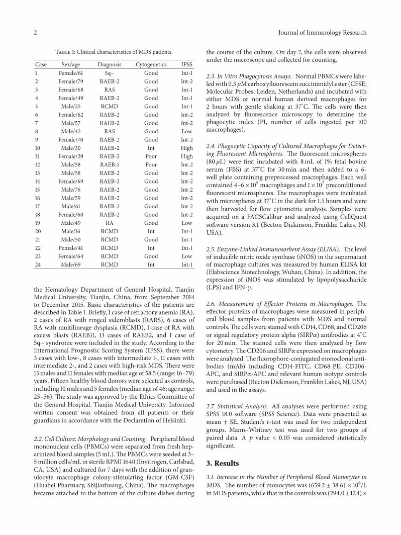
3.1. Increase in the Number of Peripheral Blood Monocytes inMDS. The number of monocytes was (659.2 ± 38.6) × 106/LinMDSpatients, while that in the controlswas (294.0± 17.4)×
Journal of Immunology Research 3
p < 0.01
ControlMDS0
500
1000
1500
Mon
ocyt
es (×
106/L
)
Figure 1: Quantity of monocytes increased in MDS patients (𝑝 <0.01).
106/L.The quantity of monocytes inMDS patients was higherthan that in the controls (𝑝 < 0.01) (Figure 1).
3.2. Reduction in the Number of Monocyte-Induced Macroph-ages inMDS. ThePBMCs fromMDSgroup showed impairedcapacity to induce macrophages. The macrophages wereobserved under the microscope and collected for subsequentexperiments. The induced macrophages (CD14+CD68+) inthe MDS group and normal controls were 10.06% ± 2.04%and 75.29% ± 5.94%, respectively (𝑝 < 0.05) (Figure 2).
3.3. Impairment of Macrophage Phagocytosis in MDS. Themonocyte-differentiated macrophages in the MDS groupshowed lower phagocytic capacity than those from thenormal controls by fluorescent microspheres.
To determine the role of macrophages, the monocyte-differentiated macrophages from patients with MDS andnormal controls were evaluated. The phagocytic percentage(PP, the count of macrophages engulfing fluorescent micro-spheres/total macrophage cell number × 100%) of monocyte-differentiated macrophages (23.69% ± 3.22%) was signifi-cantly decreased in the MDS group compared to that innormal controls (42.75% ± 2.13%, 𝑝 < 0.05).The PI (the totalnumber of swallowed fluorescent microspheres/total macro-phage number) was also dramatically decreased in the MDSgroup (0.45 ± 0.08 versus 0.92 ± 0.07, 𝑝 < 0.05) (Figure 3).
The ability of macrophages to engulf CFSE-labeled nor-mal PBMCs was decreased in the MDS group compared tothe normal controls, as evidenced by immunofluorescencemicroscopy.
We applied another method to confirm the impairedphagocytosis of macrophages in MDS patients. The CFSE-labeled normal PBMCs were incubated with macrophagesfrom MDS patients or normal controls and then assessed forphagocytosis by immunofluorescence microscopy. The PI ofmacrophages in the MDS patients (0.24 ± 0.04) was signif-icantly lower than that in the normal controls (0.48 ± 0.06,𝑝 < 0.05) (Figure 4).
3.4. Reduction of CD206 Expression on Macrophages inMDS. The expression of the macrophage mannose receptor(CD206) on macrophages in MDS patients was significantlyreduced compared to that in normal controls (9.73% ± 2.59%versus 51.15% ± 10.82%, respectively; 𝑝 < 0.05) (Figure 5).
3.5. Reduction of SIRP𝛼 Expression on Macrophages in MDS.The expression of SIRP𝛼 on macrophages in MDS patientswas significantly reduced compared to that in normal con-trols (0.51% ± 0.09% versus 0.77% ± 0.06%, respectively; 𝑝 <0.05) (Figure 6).
3.6. Increased iNOS Secretion by Macrophages in MDS. Thelevel of iNOS in the supernatant ofmacrophage cultures fromMDS patients was increased compared to that in normalcontrols (35.87 ± 6.25 pg/mL versus 22.05 ± 3.67 pg/mL,respectively; 𝑝 < 0.05) (Figure 7).
4. Discussion
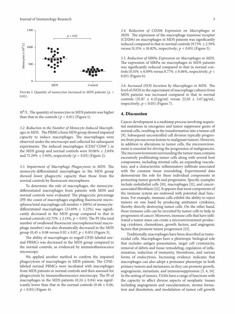
Cancer development is a multistep process involving sequen-tial mutations in oncogenes and tumor suppressor genes ofnormal cells, resulting in the transformation into a tumor cell[9]. Subsequent uncontrolled cell division typically progres-ses fromprecancerous lesions tomalignant tumors. However,in addition to alterations in tumor cells, the microenviron-ment is essential for driving the progression of malignancies.Themicroenvironment surrounding the tumormass containsexcessively proliferating tumor cells along with several hostcomponents, including stromal cells, an expanding vascula-ture, and a characteristic inflammatory infiltrate associatedwith the constant tissue remodeling. Experimental datademonstrate the role for these individual components inpromoting tumor growth and progression. Specific examplesinclude endothelial cells [10], macrophages [11], and cancer-associated fibroblasts [12]. It appears thatmost components ofthe immune system are endowed with potential dual func-tions. For example, immune cells exhibit the ability to rejecttumors on one hand by producing antitumor cytokines,thereby directly destroying tumor cells. On the other hand,these immune cells can be recruited by tumor cells to help inprogression of cancer.Moreover, immune cells that have infil-trated a tumor mass can create a microenvironment produc-ing cytokines, chemokines, growth factors, and angiogenicfactors that promote tumor progression [13].
Traditionally, macrophages have been described as tumo-ricidal cells. Macrophages have a pleiotropic biological rolethat includes antigen presentation, target cell cytotoxicity,removal of debris and tissue remodeling, regulation of infla-mmation, induction of immunity, thrombosis, and variousforms of endocytosis. Increasing evidence indicates thatmacrophages can also adopt a protumor phenotype in bothprimary tumors andmetastases, as they can promote growth,angiogenesis, metastasis, and immunosuppression [3, 4, 14].In the setting of tumors, TAMs have a range of functions withthe capacity to affect diverse aspects of neoplastic tissuesincluding angiogenesis and vascularization, stroma forma-tion and dissolution, and modulation of tumor cell growth

4 Journal of Immunology Research
101
102
103
10410
0
CD14 FITC
0
100SS
C he
ight
(a)
p < 0.01
ControlMDS0
20
40
60
80
100
CD14
+CD
68+
cells
(%)
(b)
Figure 2: Ability of monocytes to induce macrophages was lower in MDS patients. (a) Monocyte-induced macrophages (CD14+) derivedfrom peripheral blood of patients with MDS and normal controls were measured by flow cytometry. (b) Quantity of CD14+CD68+ cellsdecreased in MDS patients (𝑝 < 0.01).
101
102
103
10410
0
FL2-H10
110
210
310
4100
FL2-H
M1
R2 R3 R4 R5 R6
2-3.0052-3.005
0
20
40
60
80
100
Cou
nts
0
200
400
600
800
1000
SSC
heig
ht
(a)
p < 0.01
p < 0.01
ControlMDS ControlMDS
Phag
ocyt
ic p
erce
ntag
e (%
)
0.0
0.5
1.0
1.5
Phag
ocyt
ic in
dex
0
10
20
30
40
50
(b)
Figure 3: Phagocytosis of monocyte-induced macrophages as demonstrated by fluorescent microspheres. (a) Phagocytic capacity ofdifferentiated macrophages derived from peripheral blood from patients with MDS and normal controls was tested with fluorescentmicrospheres by flow cytometry. In the picture, the left represents the macrophages not engulfing the fluorescent microspheres; theright represents the macrophages engulfing the fluorescent microspheres. R3 suggests that the macrophages are swallowing a fluorescentmicrosphere; R4 suggests that the macrophages are swallowing two fluorescent microspheres; R5 suggests that the macrophages areswallowing three fluorescent microspheres; R6 suggests that the macrophages are swallowing four fluorescent microspheres. (b) The PI andPP of monocyte-induced macrophages fromMDS and normal controls are shown, respectively (𝑝 < 0.01).

Journal of Immunology Research 5
(a) (b)
p < 0.01
ControlMDS0.0
0.2
0.4
0.6
0.8
Phag
ocyt
ic in
dex
(c)
Figure 4: Phagocytosis of monocytes-induced macrophage as demonstrated by CFSE. CFSE-labeled normal PBMCs were incubated withmonocyte-inducedmacrophages from either normal controls (a) or patients withMDS (b).These cells were assessed by immunofluorescencemicroscopy for the presence of fluorescently labeled normal PBMCs within the macrophages (indicated by arrows). (c) Phagocytic capacityof differentiated macrophages from patients with MDS and normal controls was tested by immunofluorescence microscopy. The PI ofdifferentiated macrophages fromMDS and normal controls are shown (𝑝 < 0.01).
(enhancement and inhibition). When activated, they caninduce neoplastic cell death (cytotoxicity and apoptosis) and/or elicit tumor destructive reactions through alterations of thetumor microvasculature [5, 6].
In our study, we observed that although the number ofmonocytes in majority of the MDS patients was increased,the macrophages derived from MDS monocytes were fewerand exhibited impaired phagocytosis. This suggested thatthe ability of abnormal monocytes to develop into normalmacrophages was inhibited inMDSpatients.Moreover, whenabnormal MDS clonal cells arise, the macrophages fail tophagocytose them. In addition, macrophages and monocytescould be partly progenies of MDS clone in most cases, whichleads to bone marrow protumor microenvironment.
We observed that the levels of CD206 and SIRP𝛼 weredecreased in MDS patients compared with those in normalcontrols. CD206, which is a macrophage mannose receptor,enables themacrophage to bind tomicroorganisms and inter-nalize themduring the process of phagocytosis [15, 16]. SIRP𝛼is an immunoglobulin superfamily protein that binds to theprotein tyrosine phosphatases SHP-1 and SHP-2 through itscytoplasmic region. CD47, another immunoglobulin super-family protein, is a ligand for SIRP𝛼, with the two pro-teins constituting a cell-cell communication system (the
CD47-SIRP𝛼 signaling system) [17, 18]. This might explainthe observation that macrophages originating from MDSpatients could not bind to or phagocytose “tumor cells” ascompetently as those from normal controls. Therefore, wemay speculate that the impaired macrophages from MDSpatients might be a result of the biological behavior of tumorgrowth and progression.
In this study, we found that iNOS expression was upregu-lated inMDS patients, compared to normal controls. Humancarcinomas are associated with upregulation of iNOS, whichis otherwise generally not expressed in normal (noncancer-ous) tissues, with the exception of the kidney, brain, and pla-centa [19]. Human carcinomas exhibiting high levels of iNOSexpression include those in stomach, liver, and lung. Thissynthase is involved in many physiological and pathologicalprocesses. Moreover, its expression is closely related to thebiological behavior of tumor growth, progression, metastasis,and prognosis. A key function of iNOS is the enzymaticconversion of arginine to generate a locally high concentra-tion of nitric oxide (NO). From a tumorigenic perspective,the iNOS-mediated increase in NO supports cancer devel-opment [20]. The above evidence indicates that iNOS con-tributes to the development of MDS.

6 Journal of Immunology Research
0
100SS
C he
ight
100
101
102
103
104
CD20
6 A
PC
101
102
103
10410
0
CD14 FITC10
110
210
310
4100
CD68 PE
p < 0.01
ControlMDS
CD20
6+/C
D14
+CD
68+
cells
(%)
0
20
40
60
80
100
Figure 5: Expression of CD206 (CD206+/CD14+CD68+) on macrophages from peripheral blood from patients with MDS and normalcontrols was tested by flow cytometry (𝑝 < 0.01).
p < 0.01
ControlMDS0
20
40
60
80
100
SIRP
+/C
D14
+CD
68+
cells
(%)
Figure 6: Expression of SIRP𝛼 (SIRP𝛼+/CD14+CD68+) on macro-phages from peripheral blood from patients with MDS and normalcontrols was tested by flow cytometry (𝑝 < 0.01).
5. Conclusions
In this study, we explored the role of macrophages in thepathogenesis of MDS by inducing the monocytes to become
iNO
S (p
g/m
L)
0
10
20
30
40
50 p < 0.05
ControlMDS
Figure 7: Comparison of levels of iNOS in the supernatant ofmacrophage cultures from either MDS patients or normal controls,as measured by ELISA (𝑝 < 0.05).
macrophages. Compared with normal controls, the macro-phage phagocytosis activity in MDS patients was abnormal.The expressions of recognized receptors CD206 and SIRP𝛼were lower in macrophages in MDS patients, but the level of
Journal of Immunology Research 7
iNOS was increased. These results suggested that macroph-ages in MDS patients could not recognize, phagocytose, andkill the MDS clonal cells. Our study provides a new insightfor the research ofmacrophages inMDS patients, while it alsooffers a new therapeutic strategy targeting macrophages.
Data Access
The author should be contacted for data requests.
Ethical Approval
The study was approved by the Ethics Committee of theGeneral Hospital, Tianjin Medical University.
Consent
Informed written consent was obtained from all patientsor their guardians in accordance with the Declaration ofHelsinki.
Competing Interests
The authors declare that they have no competing interests.
Authors’ Contributions
YuHan performed the research and analyzed the data. Huaq-uanWang designed the study, ensured correct analysis of thedata, and wrote the manuscript. Zonghong Shao assisted indesigning the experiments, oversaw the collection of the data,and contributed to the writing of the manuscript. All authorscritically revised and approved the manuscript.
Acknowledgments
This project is partly supported by the National NaturalScience Foundation of China (nos. 81170472, 81400088, and81570111) and Application Bases and Advanced TechnologyResearch Program of Tianjin (nos. 14JCYBJC27200 and09JCYBJC11200). These funders have no role in the design ofthe study and collection, analysis, and interpretation of dataand in writing the manuscript.
References
[1] R. Bejar and D. P. Steensma, “Recent developments in myelody-splastic syndromes,” Blood, vol. 124, no. 18, pp. 2793–2803, 2014.
[2] L. Ades, R. Itzykson, and P. Fenaux, “Myelodysplastic syndro-mes,”The Lancet, vol. 383, no. 9936, pp. 2239–2252, 2014.
[3] T. A. Wynn, A. Chawla, and J. W. Pollard, “Macrophage biologyin development, homeostasis and disease,” Nature, vol. 496, no.7446, pp. 445–455, 2013.
[4] F. Ginhoux, J. L. Schultze, P. J. Murray, J. Ochando, and S. K.Biswas, “New insights into the multidimensional concept ofmacrophage ontogeny, activation and function,” Nature Immu-nology, vol. 17, no. 1, pp. 34–40, 2016.
[5] R.Noy and J.W. Pollard, “Tumor-associatedmacrophages: frommechanisms to therapy,” Immunity, vol. 41, no. 1, pp. 49–61, 2014.
[6] A. Mantovani, S. Sozzani, M. Locati, P. Allavena, and A. Sica,“Macrophage polarization: tumor-associated macrophages as aparadigm for polarizedM2mononuclear phagocytes,”Trends inImmunology, vol. 23, no. 11, pp. 549–555, 2002.
[7] J. W. Pollard, “Macrophages define the invasive microenviron-ment in breast cancer,” Journal of Leukocyte Biology, vol. 84, no.3, pp. 623–630, 2008.
[8] E. Suyanı, G. T. Sucak, N. Akyurek et al., “Tumor-associatedmacrophages as a prognostic parameter in multiple myeloma,”Annals of Hematology, vol. 92, no. 5, pp. 669–677, 2013.
[9] D. Hanahan and R. A.Weinberg, “Hallmarks of cancer: the nextgeneration,” Cell, vol. 144, no. 5, pp. 646–674, 2011.
[10] J. Folkman, “Angiogenesis: an organizing principle for drugdiscovery?” Nature Reviews Drug Discovery, vol. 6, no. 4, pp.273–286, 2007.
[11] F. Balkwill, K. A. Charles, and A. Mantovani, “Smolderingand polarized inflammation in the initiation and promotion ofmalignant disease,” Cancer Cell, vol. 7, no. 3, pp. 211–217, 2005.
[12] A. Orimo and R. A. Weinberg, “Stromal fibroblasts in cancer: anovel tumor-promoting cell type,” Cell Cycle, vol. 5, no. 15, pp.1597–1601, 2006.
[13] M. T. Chow, A. Moller, and M. J. Smyth, “Inflammation andimmune surveillance in cancer,” Seminars inCancer Biology, vol.22, no. 1, pp. 23–32, 2012.
[14] S. K. Biswas and A. Mantovani, “Macrophage plasticity andinteraction with lymphocyte subsets: cancer as a paradigm,”Nature Immunology, vol. 11, no. 10, pp. 889–896, 2010.
[15] A. Wollenberg, M. Mommaas, T. Oppel, E.-M. Schottdorf, S.Gunther, and M. Moderer, “Expression and function of themannose receptor CD206 on epidermal dendritic cells in infla-mmatory skin diseases,” Journal of Investigative Dermatology,vol. 118, no. 2, pp. 327–334, 2002.
[16] R. A. Paveley, S. A. Aynsley, J. D. Turner et al., “The MannoseReceptor (CD206) is an important pattern recognition receptor(PRR) in the detection of the infective stage of the helminthSchistosoma mansoni and modulates IFN𝛾 production,” Inter-national Journal for Parasitology, vol. 41, no. 13-14, pp. 1335–1345,2011.
[17] A. N. Barclay and T. K. Van den Berg, “The interaction betweensignal regulatory protein alpha (SIRP𝛼) and CD47: structure,function, and therapeutic target,” Annual Review of Immunol-ogy, vol. 32, pp. 25–50, 2014.
[18] S. Jaiswal, M. P. Chao, R. Majeti, and I. L. Weissman, “Macro-phages as mediators of tumor immunosurveillance,” Trends inImmunology, vol. 31, no. 6, pp. 212–219, 2010.
[19] C. Bogdan, “Nitric oxide synthase in innate and adaptive immu-nity: an update,” Trends in Immunology, vol. 36, no. 3, pp. 161–178, 2015.
[20] A. J. Burke, F. J. Sullivan, F. J. Giles, and S. A.Glynn, “The yin andyang of nitric oxide in cancer progression,” Carcinogenesis, vol.34, no. 3, pp. 503–512, 2013.

Submit your manuscripts athttp://www.hindawi.com
Stem CellsInternational
Hindawi Publishing Corporationhttp://www.hindawi.com Volume 2014
Hindawi Publishing Corporationhttp://www.hindawi.com Volume 2014
MEDIATORSINFLAMMATION
of
Hindawi Publishing Corporationhttp://www.hindawi.com Volume 2014
Behavioural Neurology
EndocrinologyInternational Journal of
Hindawi Publishing Corporationhttp://www.hindawi.com Volume 2014
Hindawi Publishing Corporationhttp://www.hindawi.com Volume 2014
Disease Markers
Hindawi Publishing Corporationhttp://www.hindawi.com Volume 2014
BioMed Research International
OncologyJournal of
Hindawi Publishing Corporationhttp://www.hindawi.com Volume 2014
Hindawi Publishing Corporationhttp://www.hindawi.com Volume 2014
Oxidative Medicine and Cellular Longevity
Hindawi Publishing Corporationhttp://www.hindawi.com Volume 2014
PPAR Research
The Scientific World JournalHindawi Publishing Corporation http://www.hindawi.com Volume 2014
Immunology ResearchHindawi Publishing Corporationhttp://www.hindawi.com Volume 2014
Journal of
ObesityJournal of
Hindawi Publishing Corporationhttp://www.hindawi.com Volume 2014
Hindawi Publishing Corporationhttp://www.hindawi.com Volume 2014
Computational and Mathematical Methods in Medicine
OphthalmologyJournal of
Hindawi Publishing Corporationhttp://www.hindawi.com Volume 2014
Diabetes ResearchJournal of
Hindawi Publishing Corporationhttp://www.hindawi.com Volume 2014
Hindawi Publishing Corporationhttp://www.hindawi.com Volume 2014
Research and TreatmentAIDS
Hindawi Publishing Corporationhttp://www.hindawi.com Volume 2014
Gastroenterology Research and Practice
Hindawi Publishing Corporationhttp://www.hindawi.com Volume 2014
Parkinson’s Disease
Evidence-Based Complementary and Alternative Medicine
Volume 2014Hindawi Publishing Corporationhttp://www.hindawi.com