research article phytoconstituents analysis by gc-ms ... i/ijpsr vol i issue i... · research...
TRANSCRIPT
International Journal of Pharmaceutical Studies and Research E-ISSN 2229-4619
IJPSR/Vol. I/ Issue I/July-September,2010/Pg.34-48
Research Article
PHYTOCONSTITUENTS ANALYSIS BY GC-MS,
CARDIOPROTECTIVE AND ANTIOXIDANT
ACTIVITY OF BUCHANANIA AXILLARIS AGAINST
DOXORUBICIN-INDUCED CARDIO TOXICITY IN
ALBINO RATS K. Sakthivel
1*
S.Palani
2, R. Santhosh Kalash
2, K. Devi
3, B. Senthil
Kumar1
Address for Correspondence 1 PG Research, Dept of Zoology, C Abdul Hakeem College, Melvisharam, Tamil Nadu, India
2Dept of Biotechnology, Anna Bioresearch Foundation, Arunai Engineering College,
Tiruvannamalai, Tamil Nadu, India. 3PG Research, Dept of Zoology, DKM College for women, Vellore, Tamil Nadu, India
Email- [email protected]
ABSTRACT:
Buchanania axillaries(Anacardiaceae) is a traditional herbal medicine , the leaves are used to treat many
diseases including cardiotoxicity.’The present study was designed to scientifically evaluate the
cardioprotective potential of the ethanol extract of Buchanania axillaries(BA), on Doxorubicin (DOX)
induced cardiotoxicity, in albino rats. DOX is one of the most effective chemotherapeutic drugs in cancer;
however, its incidence of cardiotoxicity compromises its therapeutic index. DOX-induced heart failure is
thought to be caused by reduction/oxidation cycling of DOX to generate oxidative stress and
cardiomyocyte cell death. A Doxorubicin dose of 20 mg/kg was selected for the present study as this dose
offered significant alteration in biochemical parameters and moderate necrosis in heart. Effect of BA oral
treatment for 14 days at two doses (250 mg and 500 mg/kg body weight) was evaluated against DOX-
induced cardiotoxicity. Significant cardiotoxicity, depletion of endogenous antioxidants and biochemical
parameters were observed in DOX-treated animals when compared with the normal animals. The
Pretreatment of DOX- induced rats with BA significantly prevented the altered biochemical variation such
as marker enzymes (SGPT, SGOT, CPK, ALP and LDH), lipid profile (LDL, VLDL, TGs, HDL and Total
cholesterol), and antioxidant parameters (SOD, GSH, CAT, GPx, MDA, and GR) to near normal status.
Serum urea, and uric acid which increased on DOX administration, registered near normal values on
pretreatment with BA. Histology of Dox-induced heart of rats pretreated with BA showed a significant
recovery from cell damage. The present findings have demonstrated that the cardioprotective effects of BA
in DOX-induced oxidative damage may be due to an augmentation of the endogenous antioxidants and
inhibition of lipid peroxidation of cell membrane.
KEY WORDS: cardioprotective, antioxidant, Buchanania axillaris, Doxorubicin, myocardial infarction.
INTRODUCTION:
Myocardial infarction (MI) is an acute
condition of necrosis of the myocardium that
occurs as a result of imbalance between
coronary blood supply and myocardial
demand (1). This is most commonly due to
occlusion of a coronary artery following the
rupture of a vulnerable atherosclerotic
plaque, which is an unstable collection of
lipids and white blood cells in the wall of an
artery. An increased risk of MI is associated
with high levels of serum total cholesterol
(2) low density lipoprotein (LDL) (3) and
decreased levels of high density lipoprotein
(HDL) (4). Oxidative stress produced by
free radicals or reactive oxygen species
(ROS), as evidenced by marked increase in
production of lipid peroxidative products
and transient inhibition of endogenous
antioxidant defense such as superoxide
dismutase (SOD), catalase (CAT) and
reduced glutathione (GSH) has been shown
to underlie myocardial damage during MI
(5,6,7). Minimizing myocardial necrosis

International Journal of Pharmaceutical Studies and Research E-ISSN 2229-4619
IJPSR/Vol. I/ Issue I/July-September,2010/Pg.34-48
and improving heart function have been
proved to be effective strategies to reduce
the morbidity and mortality from myocardial
infarction (8). Accordingly, antioxidants
may decrease cellular injury and apoptosis
through a radical-scavenging mechanism
(9). Therapeutic intervention via suppression
of free radical generation and/or augment
action of endogenous antioxidant enzymes
may attenuate myocardial dysfunction. An
anthracycline anticancer drug, doxorubicin
(DOX) is effective against malignancies
such as leukemias, lymphomas and several
solid tumors. However, dose-dependent
cardiotoxic effects limit its practical
therapeutic use. Thus DOX is reported to
increase oxygen free radical activity (10) as
well as induces the peroxidation of
unsaturated lipids within the membranes
(11). Although modern drugs are effective in
preventing cardiovascular disorders, their
use is often limited because of their side
effects (12). Herbal drugs are prescribed
widely, even when their biologically active
compounds are unknown, because of their
effectiveness, lesser side effects and
relatively low cost (13). Now-a- days the
usage of herbal drugs is gaining wider
acceptance by the medical professional due
to their positive contribution and influence
on health and qualify of life. Therefore the
search for indigenous cardioprotective
herbal drugs is still continuing as part of
scientific research. Buchanania axillaries
(Anacardiaceae) is a traditional medicinal
plant distributed in India and other Asian
countries. Its leaf extract has been reported
to possess anti-inflammatory and
cardioprotective activity.(14). It is also
reported to possess, the aerial parts is used
to cure itch of the skin and to remove
blemishes from the face. The kernels are
used in Indian medicine as a brain tonic. The
gum is antidiarhoeal used internally
rheumatism (15). In addition the ethanolic
extract of the aerial part showed CNS
depressant activity in mice. Further the
leaves are reported to be cooling digestive
expectorant purgative depurative and
aphrodisiac and are useful in hyperdipsia,
burning sensation cough braonchitis,
dyspepsia, leprosy and constipation. (16).
Extensive phytochemical investigations
carried out on BA revealed the presence of
many chemical constituents including
palmitic and linoleic acid such as n-
hexadecanoic acid , 9,12-octadecadienoic
acid (Z,Z)-, and oleic acid, which are
considered significant for its
Hypocholesterolemic property (17,18,19)
However, no data is available on the
cardioprotective and antioxidant properties
of Buchanania axillaries(BA). Therefore,
this study was designed to investigate the
protective effects of the ethanol extract of
Buchanania axillaris against DOX-induced
cardiotoxicity in rats.
MATERIALS AND METHODS
Plant material
Leaves of Buchanania axillaries were
collected identified and authenticated by a
Botanist, Dr.C.Madhavachetty, Tirupathi
university, Tirupathi, India. Voucher
specimen (CAHC- 10/2010) was retained in
the C.Abdul hakeem College, Melvisharam,
Tamil Nadu, India.
Extraction

International Journal of Pharmaceutical Studies and Research E-ISSN 2229-4619
IJPSR/Vol. I/ Issue I/July-September,2010/Pg.34-48
Leaves were cleaned with water and dried in
the Buchanania axillaries until a constant
weight was obtained. Then it was powdered
using a mechanical grinder to obtain a
coarse powder. Equal quantity of powder
was passed through 40 mesh sieve and
extracted with ethanol (90% v/v) in Soxhlet
apparatus at 60°C (20). The solvent was
completely removed by rotary vacuum
evaporator. The extract was freeze-dried and
stored in a vacuum desiccator.
GC–MS analyses of ethanol extract of
Buchanania axillaries for the identification
of chemical composition
The identification of chemical composition
of ethanol extract of BA was performed
using a GC–MS spectrograph (Agilent
6890/Hewlett–Packard 5975) fitted with
electron impact (EI) mode. The ethanol
extract (2.0 mL) of BA was injected with a
Hamilton syringe to the GC–MS manually
for total ion chromatographic analysis in
split mode. In quantitative analysis, selected
ion monitoring (SIM) mode was employed
during the GC MS analysis. SIM plot of the
ion current resulting from very small mass
range with only compounds of the selected
mass were detected and plotted.
Experimental animals
Studies were carried out using Wistar albino
rats (150–200 g), obtained from Institute of
Veterinary Preventive Medicine (IVPM),
Ranipet, Tamil Nadu, India. The animals
were housed in polyacrylic cages (38 cm, 23
cm, 10 cm) and maintained under standard
laboratory conditions (temperature 25-20°C)
with dark/light cycle (12/12 h). The animals
were fed with standard pellet diet (supplied
by poultry research station, Nandhanam,
India) and fresh water ad libitum. All the
animals were acclimatized to lab conditions
for a week before commencement of the
experiment. All the procedures described
were reviewed and approved by the
University Animal Ethics Committee.
Experimental procedure
Animals were randomized and divided into
five groups (1–5) of six animals each. Group
1 served as normal and the rats fed orally
with normal saline (0.75 ml/animal, 5mL kg-
1) body weight daily for 14 days. Group II
rats were treated similar to those in group 1.
Rats of group 3 were treated with 500mg kg-
1 body weight of the ethanol extract of BA
for 14 days, respectively. Rats of group 4
and 5 are pretreated with 250 mg kg-1
and
500mg kg-1
of ethanol extract of BA
body
weight respectively.
Induction of experimental myocardial
infarction
Doxorubicin was dissolved in sterile double
distilled water and injected subcutaneously
to rats (20 kg/kg) in group 2, 4 and 5
respectively after the last dose of the extract
to induce cardiotoxicity
Isolation of working heart preparation:
The animals were anesthetized with
chloroform after 72 h of DOX
administration, then the heart was punctured
with sterile syringe and blood was stored
with EDTA is an anticoagulant agent and
later excised. Cardiac muscle from lower
third of the ventricle was visualized under
light microscope and the remaining heart
tissue was snap frozen in liquid nitrogen
Histopathological studies:
The hearts were removed, washed
immediately with saline and then fixed in

International Journal of Pharmaceutical Studies and Research E-ISSN 2229-4619
IJPSR/Vol. I/ Issue I/July-September,2010/Pg.34-48
10% buffered formalin. The hearts stored in
10% buffered formalin were embedded in
paraffin, sections cut at 5 mm and stained
with hematoxylin and eosin. These sections
were then examined under a light
microscope for histoarchitectural changes.
Biochemical analysis
Blood samples were collected into tubes pre-
coated with EDTA by vein puncture at
baseline and post intervention. Serum was
separated by centrifuging for 10min 3000×g
at 4 °C. The serum used for the assay of
urea, and uric acid which were was
estimated by the methods of [21]
respectively. The activities of serum
glutamate-pyruvate transaminase (SGPT)
and serum glutamate oxaloacetate
transaminase (SGOT) were determined
spectrophotometrically by the method of
[22]. The lactate dehydrogenase (LDH),
creatine phosphokinase (CPK) and alkaline
phosphatase (ALP) were determined by the
methods of [23]. The levels of total
cholesterol and triglycerides (TGs) were
estimated by the methods of [24,25]. Serum
high density lipoprotein (HDL) was
determined according to the method of [26].
Serum low density lipoproteins (LDL) and
very low density lipoproteins (VLDL) were
calculated as VLDL=triglycerides/5 and
LDL = total cholesterol − (HDL cholesterol
+ VLDL cholesterol) respectively.
Antioxidant assay
The heart was dissected, immediately
washed in ice-cold saline and a homogenate
was prepared in 0.1 M Tris-HCl buffer (pH
7.4). The homogenate was centrifuged and
the supernatant was used for the assay of
antioxidant parameters. MDA content was
measured according to an earlier method
[27]. Superoxide dismutase (SOD) activity
was determined according to [28]. CAT
activity was determined from the rate of
decomposition of H2O2 according to [29].
Glutathione peroxidase (GPx) activity was
determined by measuring the decrease in
GSH content after incubating the sample in
the presence of H2O2 and NaN3 according to
[30]. Myocardial GSH content was
estimated by the method of [31]. GR activity
was determined according to the method
described by [32].
Statistical analysis
The obtained results were obtained analyzed
for statistical significance using one way
ANOVA followed by Dunnet test statistical
software for comparison with control group
and DOX treated group. P < 0.05 was
considered as significant.
RESULTS:
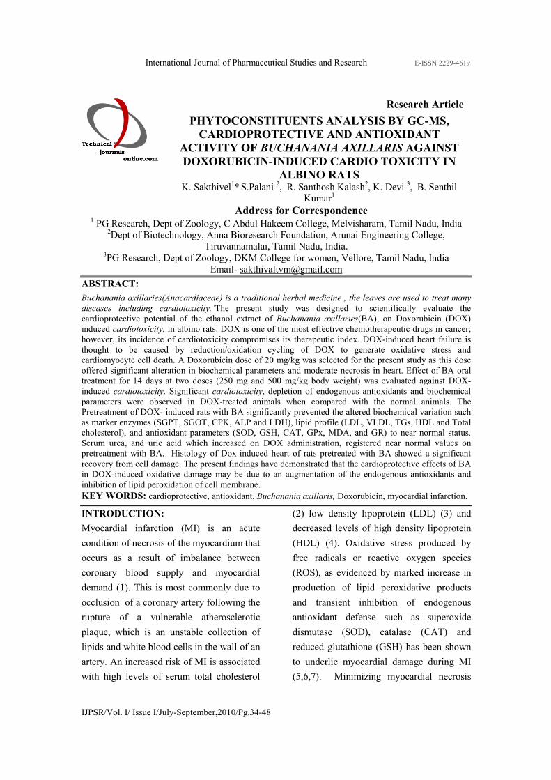
Phytochemical analysis
The ethanol extract of BA was a complex
mixture of many constituents and
compounds which were identified in this
plant by GC–MS (Table1). Phyto-
constituents such as 1-Amino-2,6-
dimethylpiperidine(1.52), 2-Octenoic acid,
4,5,7-trhydroxy(1.9), à-Methyl-D-manno-
pyranoside (2.12) 4H-Pyran-4-one, 2,3-
dihydro-3,5-dihydroxy-6-methyl (0.38)
Naphthalene, 1,2,3,4-tetrahydro-1,1,6-
trimethyl- [Synonyms: à-Ionene] (2.94), D-
Galactose, 6-deoxy-[Synonyms: D-Fucose]
(6.06), 1,6-Anhydro-á-D-glucopyranose
(Synonyms: levoglucosan) (5.85),
Dodecanoic acid (0.83), Phosphonofluoridic
acid, (1-methylethyl)-, cyclohexyl ester
(0.50), (1R,3R,4R,5R)-(-)-Quinic

International Journal of Pharmaceutical Studies and Research E-ISSN 2229-4619
IJPSR/Vol. I/ Issue I/July-September,2010/Pg.34-48
acid(26.04), Tetradecanoic acid (15.69),
3,7,11,15-Tetramethyl-2-hexadecen-1-
zl(2.55), n-Hexadecanoic acid (11.44),
Hexadecanoic acid, Octadecanoic acid ethyl
ester(0.32), Phytol( 1.85), 9,12-
Octadecadienoic acid (Z,Z)-(7.70), Oleic
Acid(1.89),4,8,12,16-etramethylheptadecan-
4-olide ( 0.65), 1,2-Benzenedicarboxylic
acid, diisooctyl ester (0.93), Phenol, 3-
pentadecyl-(2.12), ç-Sitosterol (6.43), were
identified in the ethanol extract of BA by
relating to the corresponding peak area
through coupled GC–MS (Fig 1).
Table 1.Phyto-Components identified in the Buchannania axillaris -365 [GC MS study]
No. RT Name of the compound Molecular MW Peak
Area %
Compound
Nature
1 4.31 1-Amino-2,6-dimethylpiperidine C7H16N2 128 1.52 Alkaloid
2 4.65 2-Octenoic acid, 4,5,7-
trhydroxy C8H14O5 190 1.9
Hydroxy
compound
3 5.14 à-Methyl-D-mannopyranoside C7H14O6 194 2.12 Sugar
compound
4 5.75 4H-Pyran-4-one, 2,3-dihydro-
3,5-dihydroxy-6-methyl- C6H8O4 144 0.38
Flavonoid
compound
5 8.92
Naphthalene, 1,2,3,4-
tetrahydro-1,1,6-trimethyl-
[Synonyms: à-Ionene]
C13H18 174 2.94
Naphthalene
compound
6 9.21 D-Galactose, 6-deoxy-
[Synonyms: D-Fucose] C6H12O5 164 6.06
Sugar
compound
7 10.26
1,6-Anhydro-á-D-
glucopyranose
(Synonyms: levoglucosan)
C6H10O5 162 5.85
Sugar moiety
8 10.91 Dodecanoic acid C12H24O2 200 0.83 Lauric acid
9 11.02 Phosphonofluoridic acid, (1-
methylethyl)-, cyclohexyl ester C9H18FO2P 208 0.50
Fluro
compound
10 12.14 (1R,3R,4R,5R)-(-)-Quinic acid C7H12O6 192 26.04 Quinic acid
11 13.38 Tetradecanoic acid C14H28O2 228 15.69 Myristic acid
12 14.45 3,7,11,15-Tetramethyl-2-
hexadecen-1-ol C20H40O 296 2.55
Terpene
alcohol
13 16.22 n-Hexadecanoic acid C16H32O2 256 11.44 Palmitic acid
14 16.49 Hexadecanoic acid, ethyl ester C18H36O2 284 0.88 Ester
compound
15 18.44 Phytol C20H40O 296 0.32 Diterpene
16 18.77 9,12-Octadecadienoic acid
(Z,Z)- C18H32O2 280 1.85
Linoleic acid
17 18.87 Oleic Acid C18H34O2 282 7.70 Oleic acid
18 19.17 Octadecanoic acid C18H36O2 284 1.89 Stearic acid
19 21.96 4,8,12,16-
Tetramethylheptadecan-4-olide C21H40O2 324 0.65
Methyl
compound
20 24.68 1,2-Benzenedicarboxylic acid,
diisooctyl ester C24H38O4 390 0.93
Plasticizer
compound
21 27.11 Phenol, 3-pentadecyl- C21H36O 304 2.12 Phenolic
compound
22 29.55 ç-Sitosterol C29H50O 414 6.43 Steroid
International Journal of Pharmaceutical Studies and Research E-ISSN 2229-4619
IJPSR/Vol. I/ Issue I/July-September,2010/Pg.34-48
Fig 1: The chromatogram showing n-Hexadecanoic acid (16.49), 9, 12-Octadecadienoic acid
(Z,Z)- (18.77), 4,8,12,16-Tetramethylheptadecan-4-olide(21.96),and ç-Sitosterol (29.55)
peaks detected by GC-MS.
Effects of BA extract on serum urea and
uric acid concentrations:
Serum urea, uric acid and alkaline
phosphatase concentrations were
significantly increased in DOX-treated
animals (group 2) compared to normal
(group1), indicating the induction of severe
cardiotoxicity. Treatment of DOX-
administered rats (Groups 4 and 5) with BA
significantly lowered concentrations of
serum urea, uric acid, and alkaline
phosphatase compared with treatment with
DOX alone (Fig 2). BA (500mg/kg) extract
alone treated rats did not show any
significant effect on serum urea, uric acid
and alkaline phosphatase concentrations
(group 3).
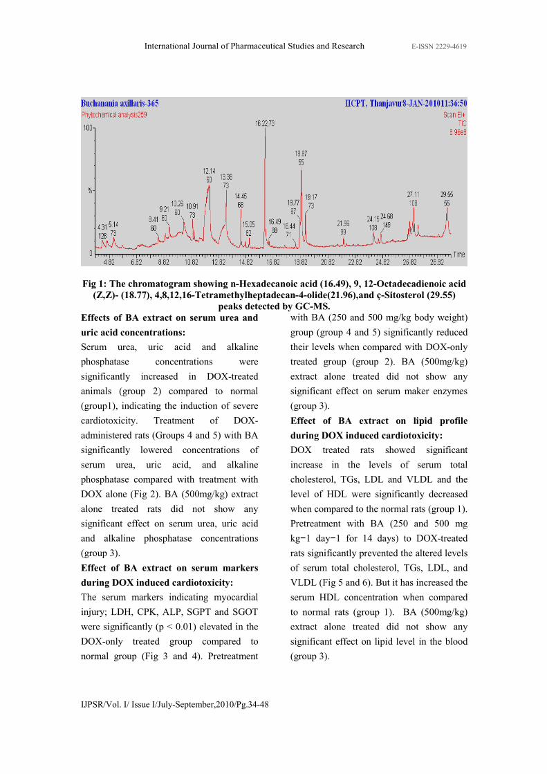
Effect of BA extract on serum markers
during DOX induced cardiotoxicity:
The serum markers indicating myocardial
injury; LDH, CPK, ALP, SGPT and SGOT
were significantly (p < 0.01) elevated in the
DOX-only treated group compared to
normal group (Fig 3 and 4). Pretreatment
with BA (250 and 500 mg/kg body weight)
group (group 4 and 5) significantly reduced
their levels when compared with DOX-only
treated group (group 2). BA (500mg/kg)
extract alone treated did not show any
significant effect on serum maker enzymes
(group 3).
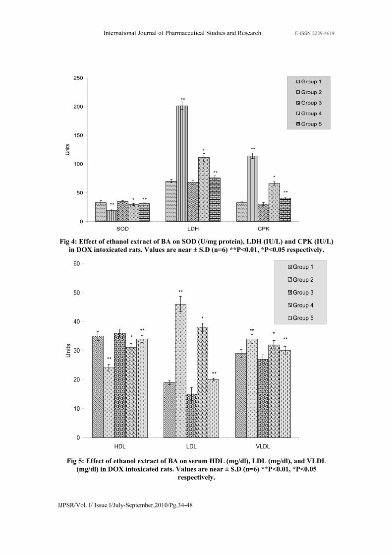
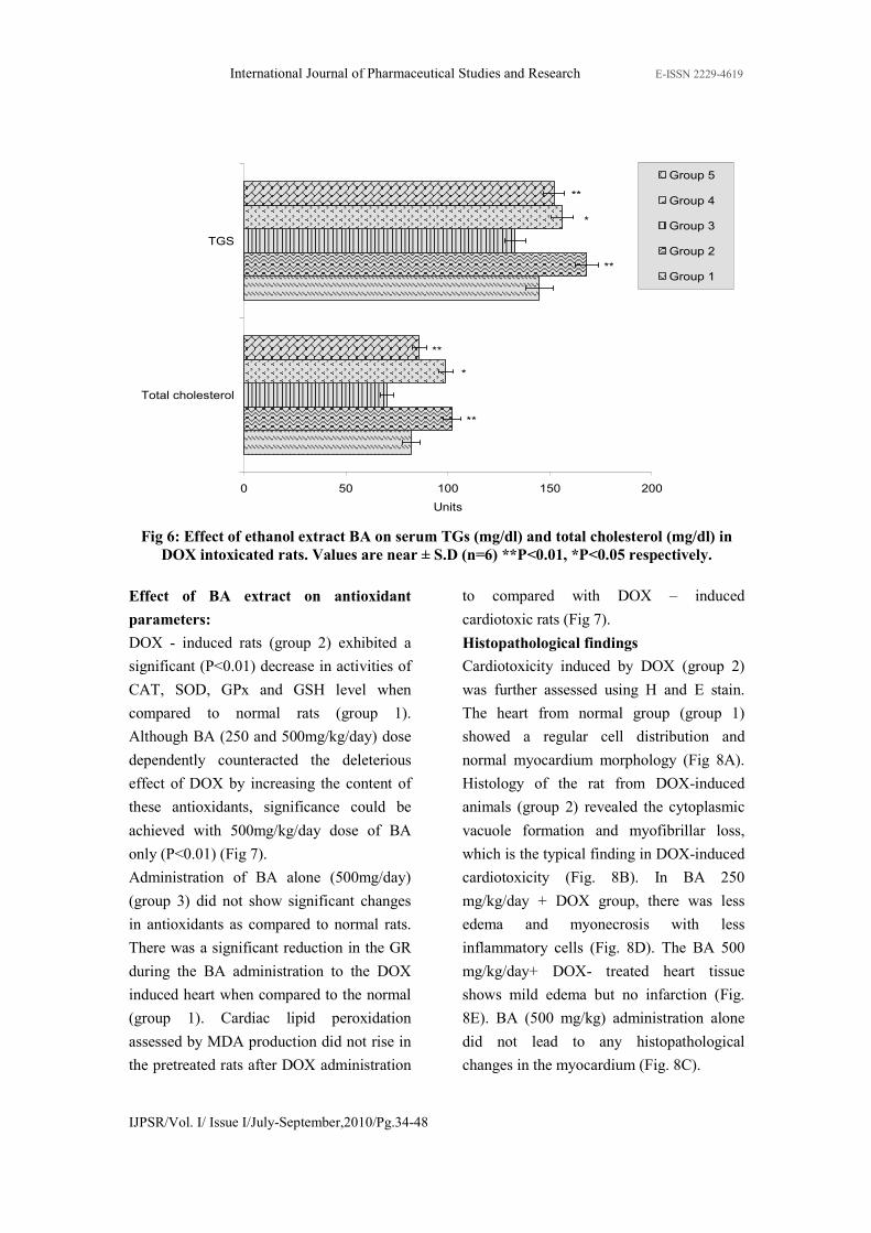
Effect of BA extract on lipid profile
during DOX induced cardiotoxicity:
DOX treated rats showed significant
increase in the levels of serum total
cholesterol, TGs, LDL and VLDL and the
level of HDL were significantly decreased
when compared to the normal rats (group 1).
Pretreatment with BA (250 and 500 mg
kg−1 day−1 for 14 days) to DOX-treated
rats significantly prevented the altered levels
of serum total cholesterol, TGs, LDL, and
VLDL (Fig 5 and 6). But it has increased the
serum HDL concentration when compared
to normal rats (group 1). BA (500mg/kg)
extract alone treated did not show any
significant effect on lipid level in the blood
(group 3).

International Journal of Pharmaceutical Studies and Research E-ISSN 2229-4619
IJPSR/Vol. I/ Issue I/July-September,2010/Pg.34-48
0
20
40
60
80
100
120
140
160
Urea Uric acid Creatinine Alkaline phosphate
Units
Group 1
Group 2
Group 3
Group 4
Group 5
**
**
**
**
**
**
**
**
*
*
*
*
Fig 2: Effect of ethanol extract of BA on urea (mg/dl), uric acid (mg/dl), GR (nmol of
NADPH oxidized/min/100mg protein), and alkaline phosphatase (mg/dl) in DOX
intoxicated rats. Values are near ± S.D (n=6) **P<0.01, *P<0.05 respectively.
0 5 10 15 20 25
SGOT
SGPT
Units
Group 5
Group 4
Group 3
Group 2
Group 1
**
**
**
**
*
*
Fig 3: Effect of ethanol extract of BA on SGOT (IU/L) and SGPT (IU/L) in DOX
intoxicated rats. Values are near ± S.D (n=6) **P<0.01, *P<0.05 respectively.
International Journal of Pharmaceutical Studies and Research E-ISSN 2229-4619
IJPSR/Vol. I/ Issue I/July-September,2010/Pg.34-48
0
50
100
150
200
250
SOD LDH CPK
Units
Group 1
Group 2
Group 3
Group 4
Group 5
****
**
**
**
**
*
*
*
Fig 4: Effect of ethanol extract of BA on SOD (U/mg protein), LDH (IU/L) and CPK (IU/L)
in DOX intoxicated rats. Values are near ± S.D (n=6) **P<0.01, *P<0.05 respectively.
0
10
20
30
40
50
60
HDL LDL VLDL
Units
Group 1
Group 2
Group 3
Group 4
Group 5
**
***
**
*
**
***
**
Fig 5: Effect of ethanol extract of BA on serum HDL (mg/dl), LDL (mg/dl), and VLDL
(mg/dl) in DOX intoxicated rats. Values are near ± S.D (n=6) **P<0.01, *P<0.05
respectively.
International Journal of Pharmaceutical Studies and Research E-ISSN 2229-4619
IJPSR/Vol. I/ Issue I/July-September,2010/Pg.34-48
0 50 100 150 200
Total cholesterol
TGS
Units
Group 5
Group 4
Group 3
Group 2
Group 1
**
**
**
**
*
*
Fig 6: Effect of ethanol extract BA on serum TGs (mg/dl) and total cholesterol (mg/dl) in
DOX intoxicated rats. Values are near ± S.D (n=6) **P<0.01, *P<0.05 respectively.
Effect of BA extract on antioxidant
parameters:
DOX - induced rats (group 2) exhibited a
significant (P<0.01) decrease in activities of
CAT, SOD, GPx and GSH level when
compared to normal rats (group 1).
Although BA (250 and 500mg/kg/day) dose
dependently counteracted the deleterious
effect of DOX by increasing the content of
these antioxidants, significance could be
achieved with 500mg/kg/day dose of BA
only (P<0.01) (Fig 7).
Administration of BA alone (500mg/day)
(group 3) did not show significant changes
in antioxidants as compared to normal rats.
There was a significant reduction in the GR
during the BA administration to the DOX
induced heart when compared to the normal
(group 1). Cardiac lipid peroxidation
assessed by MDA production did not rise in
the pretreated rats after DOX administration
to compared with DOX – induced
cardiotoxic rats (Fig 7).
Histopathological findings
Cardiotoxicity induced by DOX (group 2)
was further assessed using H and E stain.
The heart from normal group (group 1)
showed a regular cell distribution and
normal myocardium morphology (Fig 8A).
Histology of the rat from DOX-induced
animals (group 2) revealed the cytoplasmic
vacuole formation and myofibrillar loss,
which is the typical finding in DOX-induced
cardiotoxicity (Fig. 8B). In BA 250
mg/kg/day + DOX group, there was less
edema and myonecrosis with less
inflammatory cells (Fig. 8D). The BA 500
mg/kg/day+ DOX- treated heart tissue
shows mild edema but no infarction (Fig.
8E). BA (500 mg/kg) administration alone
did not lead to any histopathological
changes in the myocardium (Fig. 8C).

International Journal of Pharmaceutical Studies and Research E-ISSN 2229-4619
IJPSR/Vol. I/ Issue I/July-September,2010/Pg.34-48
(A)
(B)
(C)
(D)
(E)
Fig 8: Cardioprotective effect of Buchanania axillaris extract. Histopathological
observations (heart sections stained with Hematoxylin-Eosin, magnification-100x)
(A) Normal, (B) DOX, (C)BA alone (500mg/kg), (D) Extract 250 mg/kg + DOX
(E) Extract 500 mg/kg + DOX
International Journal of Pharmaceutical Studies and Research E-ISSN 2229-4619
IJPSR/Vol. I/ Issue I/July-September,2010/Pg.34-48
DISCUSSION
Cardiotoxicity caused by treatment with
Doxorubicin can be life-threatening and may
occur even years after completion of therapy
[33]. The current study assesses the
cardioprotective potential of the ethanol
extract of BA against DOX-induced
cardiotoxicity. Uric acid is considered to be
a risk factor in the development of
cardiotoxicity [34]. We observed a
significant increase in the level of plasma
uricacid in DOX induced rats which could
be due to increased free radical production
by DOX. In hypoxic tissue, ATP depletion
occurs which leads to accumulation of
hypoxanthine when tissues are disturbed, the
enzyme Xanthine Dehydrogenase is
converted to Xanthine Oxidase by the
oxidation of essential SH groups. Xanthine
Oxidase catalyzes the conversion of
Hypoxanthine to Xanthine, Uric acid to
super oxide [35]. This could be one of the
reasons for the elevated levels of plasma
uric acid in DOX induced rats. It is found
that urea inhibits the lactate dehydrogenase
activities of the crystalline ox-heart, rabbit-
muscle enzymes and of human heart and
human liver extracts to a much greater
extent with 2-oxobutyrate as substrate than
with pyruvate. It also said to be reducing the
serum LDH activity to about 50% (36).
Cardiotoxicity is also associated with altered
lipid metabolism. The increased
concentration of cholesterol could be due to
a decrease in HDL, since HDL is known to
be involved in the transport of cholesterol
from tissues to the liver for its catabolism
[37]. In this context, we have observed
decreased levels of HDL in DOX-treated
rats. The observed increase in TGs might be
due to a decrease in the activity of
lipoprotein lipase, resulting in decreased
uptake of TGs from the circulation [38].
Pretreatment with BA decreases the
concentration of total cholesterol, TGs,
VLDL and increases the concentration of
HDL in heart of DOX-induced rats. These
changes in lipid levels might be due to
enhanced lipid biosynthesis by cardiac
cyclic adenosine monophosphate[39].
Studies have shown that high levels of LDL
cholesterol have a positive correlation with
cardiotoxicity, whereas high levels of HDL
cholesterol have a negative correlation with
cardiotoxicity,[40]. Serum CPK, SGPT,
SGOT, ALP and LDH are well known
markers of myocardial infarction. When
myocardial cells are damaged or destroyed
due to deficient oxygen supply or glucose,
the cardiac membrane becomes permeable
or may rupture which results in leakage of
enzymes. These enzymes enter into the
blood stream thus increasing their
concentration in the serum[40]. Activities of
these enzymes in serum decreased in the BA
pretreated DOX induced group probably due
to the protective effect of BA on
myocardium, which would have reduced the
extent of myocardial damage induced by
DOX and thereby restriced the leakage of
these enzymes from myocardium. It is
widely accepted that oxygen-free radicals
generated during Doxorubicin redox cycling
are responsible for the damage that
doxorubicin causes to the heart
[41,42,43,44]. Oxygen radical generation
affects the heart because doxorubicin and its
toxic metabolite doxorubicinol accumulate

International Journal of Pharmaceutical Studies and Research E-ISSN 2229-4619
IJPSR/Vol. I/ Issue I/July-September,2010/Pg.34-48
in cardiac tissue that has low antioxidant
levels [45].
Cardioprotective activity of BA is supported
by increased myocardial antioxidant enzyme
activity and decreased extent of lipid
peroxidation. The most abundant ROS
generated in living cells are superoxide
anion and its derivatives, particularly highly
reactive and damaging hydroxyl radical,
which induces peroxidation of cell
membrane lipids [46]. Lipid peroxidation is
known to cause cellular damage and is
primarily responsible for ROS-induced
organ damage [47]. Our studies have shown
that in DOX-induced cardiotoxicity,there
was considerable increased in lipid
peroxidation, which was significantly
prevented by BA pretreatment.
Redox cycling of DOX generates superoxide
free radicals [48] due to conversion of
quinone to semi-quinone moiety, whereas
SOD enzyme dismutates this free radical to
hydrogen peroxide. In this respect, any
increase in SOD activity of the organ
appears to be beneficial in the event of
increased free-radical generation. Our
studies showed that the activity of SOD was
significantly decreased in DOX-treated
animals and the pretreatment with BA
reversed the SOD activity in a dose-
dependent manner. However, it has been
reported that a rise in SOD activity, without
a concomitant rise in the activity of
catalase/GSH might be detrimental [49].
This is due to the fact that SOD generates
hydrogen peroxide as a metabolite, which is
cytotoxic and needs to be scavenged by
catalase/GSH. Thus a simultaneous increase
in catalase/GSH activity is essential for an
overall beneficial effect of increase in SOD
activity (50). Inhibition of DOX-induced
oxidative stress and tissue injury might be
due to an increase in GSH, myocardial SOD
and catalase activities, following the
pretreatment of BA. The observed increase
in catalase activity in DOX-treated animals
supports the above hypothesis that this
increase is possibly required to overcome
excessive oxidative stress [51]. GSH levels
were also lowered significantly in DOX-
treated animals, while pretreatment with BA
showed significant increase in GSH levels in
DOX-treated animals at doses of 250 mg/kg
and 500 mg/kg in rats. Catalase activity was
increased after DOX treatment and
pretreatment with BA further increased its
activity significantly at 250 mg/kg and
500mg/kg dose levels. The increase in
catalase activity in DOX-treated animals
could be indicative of enhanced oxidative
stress due to an adaptive myocardial
mechanism. These findings indicate the
promising role of BA as a cardioprotective
agent against DOX-induced cardiotoxicity.
CONCLUSION
The present study shows that the
administration of ethanol extract of BA has
cardioprotective potential against DOX-
induced cardiotoxicity. It provides
experimental evidence that BA augmented
the myocardial antioxidant enzymes level,
preserved histoarchitecture and improved
cardiac performance following DOX
administration. This cardioprotective
activity of BA might be due to the
synergetic effect of chemical compounds
present in them making them good sources
for the production of a cardioprotective

International Journal of Pharmaceutical Studies and Research E-ISSN 2229-4619
IJPSR/Vol. I/ Issue I/July-September,2010/Pg.34-48
herbal medicine. The identification of
molecules with cardioprotective potential
from this ethanol extract of BA may provide
new directions for identification of
cardioprotectives, which could be given
concomitantly during Dox treatment.
REFERENCE
[1] De Bono, D.P., Boon, N.A. 1992. Diseases of
the cardiovascular system. In Edwards CRW,
Boucheir IAS, editors. David- son’s principles
and practice and medicine. Hong Kong:
Churchill Livingstone. p. 249–340.
[2] Grundy, S.M.1986. Cholesterol and heart
disease: a new era. J Am Med Assoc. 256:2849-
2858.
[3]Brown, M.S., Goldstein, J.L. 1986. A
receptor-mediated pathway for cholesterol
homeostasis. Science; 232: 34-47
[4]Castelli, W.P., Garrison, R.J., Wilson, P.W.F.,
Abott, R.D., Kalousidan, S., Kannel, W.B. 1986.
Incidence of coronary heart disease and
lipoprotein cholesterol levels. The Framingham
Study. J Am Med Assoc; 256:2835-2838.
[5]Loper, J., Goy, J., Rozensztajn, L., Bedu, O.,
Moisson, P. 1961. Lipid peroxidation and
protective enzymes during myocardial infarction.
Clin Chim Acta. 196:119–26.
[6] Padmanabhan, M., Stanely Mainzen Prince,
P. 2006. Preventive effect of S-allylcysteine on
lipid peroxides and antioxidants in normal and
isoproterenol-induced cardiotoxicity in rats: a
histopathological study.Toxicology.224: 128–37.
[7] Zhou, R., Xu, Q., Zheng, P., Yan, L., Zheng,
J., Dai, G. 2008. Cardioprotective effect of
fluvastatin on isoproterenol- induced myocardial
infarction in rat. Eur J Pharmacol .586:244–50.
[8] Kloner, R.A., Rezkalla, S.H., 2004. Cardiac
protection during acute myocardial infarction:
where do we stand in 2004? J. Am. Coll.
Cardiol. 22: 276–286.
[9] Angeloni, C., Spencer, J.P.E., Leoncini, E.,
Biagi, P.L., Hrelia, S. 2007. Role of
quercetinandits in vivo metabolites in protecting
H9c2 cells against oxidative stress. Biochimie.
89: 73–82.
[10] Lee, V., Randhawa, A.K., Singhal, P.K.,
1991. Adriamycin induced myocardial
dysfunction in vitro is mediated by free radicals.
Am. J. Physiol. 261: H989–H995.
[11] Gurvinder Singh., Anu Singh, T., Aji
Abrahama., Beena Bhat., Ashok Mukherjee.,
Ritu Verma., Shiv Agarwal, K., Shivesh Jha .,
Rama Mukherjee., Anand Burmana, C. 2008.
Protective effects of Terminalia arjuna against
doxorubicin-induced cardiotoxicity. J
Ethnopharmacology. 117:123–129.
[12] Rajadurai, M., Prince, P.S.M.2005.
Comparative effects of Aegle marmelos extract
and alpha-tocopherol on serum lipids, lipid
peroxides and cardiac enzyme levels in rats with
isoprote renol-induced myocardial infarction.
Singapore Med J. 46: 78-81.
[13] Kumar, K.E., Mastan, S.K., Sreekanth, N.,
Chaitanya, G., Sumalatha, G., Krishna, P.V.
2009. Cardioprotective effect of methanolic
extract of Syzygium cumini seeds on
isoproterenol-induced myocardial infarction in
rats Pharmacology online. 3: 250-256.
[14] Madhavachetty, K., Shivaraj, K.,
Thulasirao, K. 2008. Flowering plants of chittoor
district, Andhra Pradesh. Tirupathi students
offset printers. 1st edition. 358
[15]Khare, C.P.2004.Indian medicinal plant
illustrated dictionary.Springer verlag
Heidelberg.p.104-105.
[16] Pullaiah.T.2006. Encyclopedia of world
medicinal plants.Vol 1. Regency publication
New Delhi.P. 366-367.
[17] Kurian, G.A, Srivats, R.S.S., Gomathi, R.,
BAbi, M.M., and Paddikkala, J., 2010.
Interpretation of inotropic Effect Exhibited by
Desmodium gangeticum Chloroform Root
Extract through GSMS and Atomic Mass
Spectroscopy: Evaluation of its Anti Ischemia
Reperfusion Property in Isolated Rat Heart.
Asian J biochem 5(1): 23-32.
[18] Hyo Ku Lee., Yang Mun Choi., Dong Ouk
Noh and Hyung Joo Suh. 2005. Antioxidant
Effect of Korean Traditional Lotus Liquor
(Yunyupju) Inter. J Food Sci & Tech. 40
(7):709 -715.
[19] Hajji Mohamed , Masmoudi Ons, Ellouz-
Triki Yosra, Siala Rayda, Gharsallah Neji, Nasri
Moncef. 2009. Chemical composition and
antioxidant and radical-scavenging activities of
Periploca laevigata root bark extracts. J. Sci
Food & Agri 89(5): 897 - 905
[20]Chattopadhyay, R.R. 2003. Possible
mechanism of hepatoprotective activity of
Azadirachta indica leaf extract: Part II. J.
Ethnopharmacology 89: 217–9.
[21] Caraway, W.T. 1963. In Standard Methods
of Clinical Chemistry. 4th ed., Academic Press,
London: pp. 239-247
[22] Mohun, A., Cook, I.J. 1957 Simple methods
for measuring serum level of glutamic
oxaloacetic and glutamic pyruvic transaminases
in routine laboratories.J Clin Pathol. 10: 394-
399.

International Journal of Pharmaceutical Studies and Research E-ISSN 2229-4619
IJPSR/Vol. I/ Issue I/July-September,2010/Pg.34-48
[23] King, J., 1965. In Prac Clini Enzymology.
Van D. ed., Nostrand Co, London, 83-93.
[24] Zlatkis, A., Zak, B., Boyle, G.J. 1953. A
new method for the direct determination
of serum cholesterol. J Clin Med. 41: 486-92.
[25] Foster, L.V., Dunn, R.T. 1973. Stable
reagents for determination of serum triglycerides
by a colorimetric Hatzsch condensation method.
Clin Chem. 19:338-340.
[26] Wilson, D.E., Spiger, M.J. 1973. A dual
precipitation method for quantitative plasma
lipoprotein measurement without ultra-
centrifugation. J Lab Clin Med , 82:473.
[27] Zhang, X.Z. 1992. Crop physiology
research methods. Beijing: China Agricultural
Press.
[28] Rai, S., A. Wahile, K. Mukherjee, B.P.
Saha, and P.K. Mukherjee. 2006. Antioxidant
activity of Nelumbo nucifera (sacred lotus)
seeds. J Ethnopharmacology. 104: 322–7.
[29] Bergmeyer, H.U., Gowehn, K and Grassel.
H. 1974. Enzymes as biochemical reagents. In
Methods of Enzymatic Analysis, ed. H.U.
Bergmeyer, 438–9. Weinheim: Verlag Chemine.
[30] Hafemann, D.G., Sunde, R.A., and
Houestra, W.G. 1974. Effect of dietary selenium
on erythrocyte and liver glutathione peroxidase
in the rat. J. Nutrition. 104: 580–4.
[31]Moron, M.S., Depierre, J.W., Mannervik, B.
1979. Levels of glutathione, glutathione
reductase and glutathione S-transferase activities
in rat lung and liver. Biochem Biophys Acta.
582:67–78.
[32] Staal, G.E., Visser, J., Veeger, C., 1969.
Purification and properties of glutathione
reductase of human erythrocytes. Biochim.
Biophys. Acta. 185: 39–48.
[33] Kapusta, L., Thijssen, J.M., Groot-Loonen,
J., Antonius, T., Mulder, J., Danie¨ls, O., 2000.
Tissue Doppler imaging in detection of
myocardial dysfunction in survivors of
childhood cancer treated with anthracyclines.
Ultrasound Med. Biol. 26 :1099– 1108.
[34] Upston, J.M., Terentis, A.C., Stocker, R.,
1999 Tocopherol-mediated peroxidation of
lipoproteins: implications for vitamin E as a
potential antiatherogenic supplement, FASEB J.
13:977–994.
[35] Weir, C.J., Muir, S.W., Walters, M.R., Lees,
K.R, 2003. Serum urate as an independent
predictor of poor outcome and future vascular
events after acute stroke, Stroke, 34 :1951–1956.
[36] Pauline, M. E and Wilkinson, J.H. 1965.
Urea and oxalate inhibition of the serum lactate
dehydrogenase. J. clin. Path. 18: 803.
[37] Mathew S, Menon VP, Kurup PA. Changes
in myocardial and aortic lipids, lipolytic activity
and fecal excretion of sterols and bile acids in
isoproterenol-induced myocardial infarction in
rats Indian J Biochem Biophys 1981; 18:131.
[38] SuBAmakumari, S., Varghese, A.,
Muraleedharan, D., Menon, V.P.1990. Protective
Action of Aspirin- in, Experimental Myocardial
infraction Induced by Isoproterenol in Rats and
its Effect on Lipid Peroxidation Indian J Exp
Biol. 28:480.
[39] Paritha, I.A., Devi, C.S.1997. Effect of α-
tocopherol on isoproterenol-induced changes in
lipids and lipoprotein profile in rats. Indian J
Pharmacol. 29:399.
[40] Buring, J.E., O' Connor., G.T., Goldhaber,
S.Z., Rosner, B., Herbert, P.N., Blum, C.B.
1992. Decreased HDL2 and HDL3 Cholesterol,
Apo A-I and Apo A-II, and Increased Risk of
Myocardial Infarction. Circulation. 85:22-29.
[41] Julicher, R.H., van der Laarse, A.,
Sterrenberg, L., Bloys van Treslong, C.H., Bast,
A., Noordhoek, J., 1985. The involvement of an
oxidative mechanism in the adriamycin induced
toxicity in neonatal rat heart cell cultures. Res.
Commun. Chem. Pathol. Pharmacol. 47: 35–47.
[42] Singal, P.K., Iliskovic, N., 1998.
Doxorubicin-induced cardiomyopathy. N. Engl.
J. Med. 339: 900–905.
[43] van Acker, S.A., Boven, E., Kuiper, K., van
den Berg, D.J., Grimbergen, J.A., Kramer, K.,
Bast, A., van der Vijgh, W.J.F. 1997.
Monohydroxyethylrutoside, a dose-dependent
cardioprotective agent, does not affect the
antitumor activity of doxorubicin. Clin. Cancer
Res. 3: 1747– 1754.
[44] Venditti, P., Balestrieri, M., De Leo, T., Di
Meo, S. 1998. Free radical involvement in
doxorubicin-induced electrophysiological
alterations in rat papillary muscle fibres.
Cardiovasc. Res. 38: 695–702.
[45] De Jong, J., Gue´rand, W.S., Schoofs, P.R.,
Bast, A., van der Vijgh, W.J.F., 1991. Simple
and sensitive quantification of anthracyclines in
mouse atrial tissue using high-performance
liquid chromatography and fluorescence
detection. J. Chromatogr., B: Biomed. Appl. 570:
209–216.
[46] Hemnani, T., Parihar, M.S., 1998. Reactive
oxygen species and oxidative DNA damage. Ind.
J Physol & Pharmacol. 42: 440–452.
[47] Halliwell, B., Gutteridge, J.M.C., 1989.
Free Radicals in Biology and Medicine, 2nd ed.
Clarendon Press, London
[48] Hardina, R., Gersl, V., Klimtova, I.,
Simunek, T., Machackova, J., Adamcova, M.

International Journal of Pharmaceutical Studies and Research E-ISSN 2229-4619
IJPSR/Vol. I/ Issue I/July-September,2010/Pg.34-48
2000. Anthracycline induced cardiotoxicity. Acta
Medica. 43: 75–82.
[49] Herman, D., 1991. The aging process major
risk factor for disease and death. Proceedings of
the National Academy of Sciences. 88: 5360–
5363.
[50] Mukherjee, S., Banerjee, S.K., Maulik, M.,
Dinda, A.K., Talwar, K.K., Maulik, S.K., 2003.
Protection against acute doxorubicin-induced
cardiotoxicity by garlic: role of endogenous
antioxidants and inhibition of TNF-α expression.
BioMed Central Pharmacology 3: 16.
[51] Li, T., Singhal, P.K., 2000. Doxorubicin-
induced early changes in myocardial antioxidant
enzymes and their modulation by probucol.
Circulation. 102: 2105–2110.