research open access caspase 3 activity in isolated … smoke (cs) extract activates caspase-3 in...
TRANSCRIPT
Ahmed et al. Tobacco Induced Diseases 2013, 11:25http://www.tobaccoinduceddiseases.com/content/11/1/25
RESEARCH Open Access
Caspase 3 activity in isolated fetal rat lungfibroblasts and rat periodontal ligamentfibroblasts: cigarette smoke induced alterationsAsra Ahmed1, James A Thliveris2, Anthony Shaw1,3, Michael Sowa1,3, James Gilchrist1 and James E Scott1,2,4,5*
Abstract
Background: Cigarette smoking is the leading cause of preventable death and has been implicated inpathogenesis of pulmonary, oral and systemic diseases. Smoking during pregnancy is a risk factor for thedeveloping fetus and may be a major cause of infant mortality. Moreover, the oral cavity, and all cells within are thefirst to be exposed to cigarette smoke and may be a possible source for the spread of toxins to other organs of thebody. Fibroblasts in general are morphologically heterogeneous connective tissue cells with diverse functions.Apoptosis or programmed cell death is a crucial process during embryogenesis and for the maintenance ofhomeostasis throughout life. Deregulation of apoptosis has been implicated in abnormal lung development in thefetus and disease progression in adults. Caspases are proteases which belong to the family of cysteine aspartic acidproteases and are key components for downstream amplification of intracellular apoptotic signals. Of 14 knowncaspases, caspase-3 is the key executioner of apoptosis. In the present study we explored the hypothesis thatcigarette smoke (CS) extract activates caspase-3 in two types of fibroblasts, both of which would be exposeddirectly to cigarette smoke, isolated fetal rat lung fibroblasts and adult rat periodontal ligament (PDL) fibroblasts.
Methods: Isolated fetal rat lung fibroblasts and adult PDLs were used. Cells were exposed to differentconcentrations of CS for 60 min. Caspase-3 activity and its inhibition by Z-VAD-fmk were measured by caspase-3fluorometric assay. The effect of CSE on cellular viability was measured using the MTT formazan assay. Caspase-3expression was detected by western blot analysis and cellular localization of caspase-3 was determined byimmunofluorescence using fluorescence microscopy.
Results: It was observed in fetal rat lung fibroblast cells that CSE extract significantly (p<0.05) increased caspase-3activity and decrease cell proliferation. However, no significant changes in activity or viability were observed inPDLs.
Conclusions: This indicates CS activates caspase-3 the key regulatory point in apoptosis in fetal rat lung fibroblastcells suggesting that smoking during pregnancy may alter the developmental program of fetal lung, jeopardizingthe establishment of critical cellular mechanisms necessary to expedite pulmonary maturation at birth.of criticalcellular mechanisms necessary to expedite pulmonary maturation at birth.
Keywords: Cigarette smoke extract, Fetal rat lung fibroblasts, Protease, Caspase-3, Lung development,Developmental toxicity, Periodontal ligament fibroblast, Periodontitis
* Correspondence: [email protected] of Oral Biology, University of Manitoba and Manitoba Institutefor Child Health, Winnipeg, Canada2Human Anatomy and Cell Science, University of Manitoba and ManitobaInstitute for Child Health, Winnipeg, CanadaFull list of author information is available at the end of the article
© 2013 Ahmed et al.; licensee BioMed Central Ltd. This is an open access article distributed under the terms of the CreativeCommons Attribution License (http://creativecommons.org/licenses/by/2.0), which permits unrestricted use, distribution, andreproduction in any medium, provided the original work is properly cited.

Ahmed et al. Tobacco Induced Diseases 2013, 11:25 Page 2 of 10http://www.tobaccoinduceddiseases.com/content/11/1/25
BackgroundCigarette smoking leads to exposure to more than 4000known chemicals many of which are carcinogenic and isthe leading cause of preventable premature death in theworld. There is strong evidence that smoking duringpregnancy is strongly associated with growth retardation[1], low birth weight, sudden infant death syndrome(SIDS), preterm delivery and higher incidence of stillbirth[2]. Moreover, cigarette smoke absorption begins in theoral cavity and many studies have provided convincingevidence that CS exposure is a risk factor for oral patho-logical conditions ranging from staining of teeth, discolor-ation of the gingival to degradation of tooth supportingstructures [3]. The oral cavity and the respiratory systemare the primary regions for exposure to toxic substancesin cigarette smoke. The mechanisms by which changesoccur in these regions due to cigarette smoke exposureare not clearly elucidated.The respiratory system is one of the most complex
systems of the body which originates from two primarygerm cell layers, the endoderm and the mesoderm [4].Electron microscopic studies have identified that devel-oping lungs undergo dramatic tissue growth and remod-eling to achieve a mature architectural structure andapoptosis plays an important role during these stages[5]. Synchronization of cell proliferation, differentiationand apoptosis is the basis of organogenesis. Any defectin the apoptotic processes during embryogenesis maylead to developmental abnormalities and damage to cellsdue to metabolic stress [6]. It is well known that thelungs have to be sufficiently mature at birth in order toacquire normal gas exchange of oxygen and carbondioxide for survival in the extra-uterine environment.The immature lung is susceptible to toxic substances incigarette smoke primarily because of the fewer numberof macrophages leading to a poor immune response [7]and also due to a decrease in surfactant quantity, whichis considered as the first line of defense against pathogens[8]. Fetal growth and development is influenced by theintrauterine environment and governed by physical, envir-onmental, genetic and hormonal factors [9]. It has beenreported that maternal smoking of more than 20 cigarettesper day is associated with high risk for fetal hypoxia [10].Previous studies noted that chronic fetal smoke exposureis a major predisposing factor for SIDS [11]. At the cellularlevel tobacco smoke induces apoptosis and reduces cellviability in human fetal lung fibroblast-derived cells [12].Moreover, cigarette smoking appeared to induce apoptosisvia a caspase-dependent pathway in isolated human type Icells [13].The periodontal ligament (PDL) is a unique and dy-
namic soft connective tissue which is a highly cellularconnective tissue with fibroblasts as the predominant celltype. The PDL forms a fibrous joint, primarily responsible
for anchorage of tooth to alveolar bone. Degradation ofthis connection between the tooth and bone may lead toloss of attachment, increased tooth mobility eventuallyleading to tooth loss [14]. PDL fibroblasts are responsiblefor the high turn over rate, rapid remodeling capacityduring injury and repair. This necessitates continuousinvolvement of the apoptotic process for maintenance ofPDL integrity. Since the greater part of the PDL is formedby fibroblasts, exposure to toxic substances in tobaccosmoke is a potential risk factor for these cells.Caspases are proteases which belong to the family of
cysteine-aspartic acid endo-peptidases. Caspases areprimarily localized in the cytoplasm and are synthesizedas inactive enzyme precursors or zymogens [15]. Theactivation of caspases leads to irreversible biochemicaland morphological changes in cells. Caspases can bebroadly classified into two groups; one which is thoughtto play a central role in apoptosis (caspases -2, -3, -6, -7,-8, -9, -10, and -12) and another group which are pri-marily involved in cytokine processing during inflamma-tion (caspases -1, -4, and -5) [16] Caspase-3 has beenreported to contribute mainly to the characteristic mor-phologic changes in apoptotic cells including membraneblebbing, chromatin condensation and DNA fragmenta-tion (19) functional role in the initiation and executionof apoptosis. Recent studies suggest that activation ofinitiator caspases is by autolytic cleavage within a linkersegment separating the large and small subunits due toan intrinsic proteolytic activity of the caspase zymogen(salvesen and riedl, 2008). Once activated the initiatorcaspases activate effector caspases [17]. The effectorcaspases lack the ability of selfactivation because oftheir small pro-domains. Activation of initiator caspasesdoes not always result in apoptosis, due to the fact thatmembers of anti-apoptotic family (Bcl-2) can preventthe activation of effector caspases [18]. Caspase-3 is thefirst of all the effector caspases to be activated for amp-lifying downstream apoptotic process. Caspase-3 can beactivated through caspase-8 and caspase-9 by extrinsicor intrinsic signaling, respectively [19], suggesting, thatthe apoptotic signal from either extrinsic or intrinsicpathways converge for the activation of caspase-3. Fur-thermore, activation of caspase-3 is a very rapid processin the cell death process [20] which is associated withmitochondrial membrane permeabilization [21].The aim of the present study was to examine the effects
of cigarette smoke extract on fetal lung fibroblasts andadult rat PDL fibroblasts. We hypothesized that CS extractinduces activation in these cells through caspase-3.
MethodsPreparation of cigarette smoke extractCigarette smoke extract (CSE) was prepared according tomethod designed by [22]. Unfiltered research cigarettes 2R1

Ahmed et al. Tobacco Induced Diseases 2013, 11:25 Page 3 of 10http://www.tobaccoinduceddiseases.com/content/11/1/25
from University of Kentucky, each containing 2.45 mgnicotine/cigarette [23] were used. CS was drawn from asingle cigarette into a 50 ml syringe for two seconds main-taining a gap of 20 seconds between each draw with thesyringe and bubbled through 50 ml of MEM at roomtemperature. This cycle was repeated till the end of thecigarette and then 50 ml of fresh MEM was used for thenext cigarette. The resulting smoke extracted MEM wasconsidered to be 100% CSE. It was filtered using 0.22 μmpore filters (Millipore) making it sterile and free fromcontaminants and stored at –80°C. Further dilutions (5%,10% and 15%) (v/v) were made in serum free media con-taining antibiotics and fungizone. Before treating cells withconditioned media, pH was adjusted to 7.2. Samples of100% CSE were analyzed by gas chromatography on aVarion 320MS TQ gas chromatograph to determine nico-tine content. Final nicotine content was determined to be30 μg/ml of medium which translates to 0.20 μM.
Isolation and culture of fetal rat lung fibroblastsPregnant Sprague–Dawley rats purchased from CentralAnimal Services, University of Manitoba were used toisolate fetal lung fibroblasts. Rats were euthanized withan intraperitoneal injection of 1 ml Euthanyl (240 mg/mlsodium pentobarbital) on gestational day 21 (day 23 isterm gestation). Fetuses were removed by hysterotomy,decapitated and placed in cold, sterile Hanks BalancedSalt Solution (HBSS, Gibco, ON Canada). Lungs weredissected from fetuses by making an incision in themid-sternal region, and minced using a Sorval tissuechopper (Sorval Instruments, Newton, CT) in a laminarflow hood. The minced lung tissue was dissociated byincubating with trypsin-EDTA (0.05%/0.02%) in HBSSat 37°C for 45 minutes in a water-jacketed trypsinizationflask which was placed on a magnetic stirrer. Minimalessential medium (MEM) (Gibco, ON Canada) containing10% of newborn calf serum (NCS), antibiotics/antimycotic(1%) and fungizone (1%) (Gibco, ON Canada) was addedto stop further enzymatic disaggregation. The dissociatedcells were filtered through three layers of 150 μm Nitexgauze to remove tissue fragments and centrifuged for10 min at 1000 rpm at 4°C. The cell pellet was re-suspended in 10 ml of MEM/NCS and cells were plated infive 75 cm2 tissue culture flasks in a humidified incubator(95% air/ 5% CO2) and allowed to adhere for 1 hour. Fi-broblasts have the ability to attach faster when comparedto type II cells [24]. After this period media from eachflask containing non-adherent cells (including type II cells,RBC’s) was collected in an autoclaved beaker. Fresh media(10% NCS) was added to the flasks which had attachedfibroblasts. Media was changed after 24 hours for the firsttime and then at 48 hours thereafter. The cell monolayerswere cultured for 3–4 days till they reached 80% conflu-ence and sub-cultured in a ratio of 1:3. Fibroblasts were
passaged five times to obtain sufficient numbers forexperimentation. Purity was determined visually by phasemicroscopy. By confluence no epithelial-like cells could bedetected.
Culture of rat periodontal fibroblastsRat PDL fibroblasts were kindly supplied by Dr. C. Lekicand had been isolated according to the method outlinedby Lekic et al. [25]. The cells were thawed and placed in25 cm2 flasks. Media was changed at 24 hours for thefirst time and every 48 hours thereafter until they reached70-80% confluence and sub-cultured in larger flasks in theratio of 1:3, cultured till 70-80% confluent for further usein experiments.
Detection of caspase-3 activity in adherent cells exposedto CSEOnce the cultures reached 70-80% confluence, theywere washed twice with HBSS and treated with differentconcentrations of CSE diluted in serum free media for60 min. After which the cells were washed three timeswith HBSS to ensure complete removal of traces ofCSE. As the key regulatory enzyme in activation ofapoptosis [26], caspase 3 activity in cells was deter-mined following instructions of caspase-3 fluorometricassay kit, purchased from BioVision, (MountainView,CA). The cells were lysed using the lysis buffer availablewith the kit (50 μl per well) and incubated on ice forten minutes, with 5 μl of fluorogenic substrate 1 mMDEVD-AFC (caspase 3 cleaves to this substrate) in a re-action buffer (containing 10 mM DTT) in the incubatorat 37°C for two hours. The enzymatic activity was moni-tored using a fluorescence microplate reader with 400 nmexcitation and 505 nm emission filter. Caspase 3 cleavesthe AFC substrate and releases a fluorogenic signal; thissignal is directly proportional to the level of enzymaticactivity of caspase 3 in cells. Caspase 3 activity wascalculated in treated samples and compared to untreatedcontrols.
Effect of N-benzyloxycarbonyl-Val-Ala-Asp-fluoromethylketone (Z-VAD-fmk)Z-VAD-fmk, is a broad spectrum caspase inhibitor whichwas used in the present study to examine the involve-ment of caspases in cell death due to CS exposure. Thecells were incubated with 80 mM concentration of Z-VAD-fmk in serum free media at the time of exposureof cells to CSE for 60 min. After which the cells werewashed and the caspase-3 activity was measured usingthe fluorometric assay kit, purchased from BioVision,(MountainView, CA) as described above.
Ahmed et al. Tobacco Induced Diseases 2013, 11:25 Page 4 of 10http://www.tobaccoinduceddiseases.com/content/11/1/25
Determination of cellular viabilityThe cells were treated with different concentrations ofCSE as described above. After incubation with differentconcentrations of CSE the cells were washed with HBSSthree times to ensure complete removal of CSE andfurther incubated with MTT solution for three hours.The MTT based cell proliferation assay (Sigma Aldrich,St.Louis, MO, USA), is a calorimetric assay used tomeasure the ability of mitochondrial dehydrogenaseof viable cells to reduce the key component, MTT or3-[4,5-dimethylthiazol-2-yl]-2,5diphenyl tetrazolium brom-ide, a yellow tetrazole to insoluble purple formazan crystals.Viable cells cleave the tetrazolium ring of MTT and theyellow water soluble dye is converted to insoluble purplecrystals of formazan. After three hours of incubation withMTT solution the crystals were dissolved in MTT solventby pipetting three times in order to completely dissolvethe crystals. The plates were read spectrophotometricallyat an absorbance of 570 nm. The intensity of purple colorin the solution results in an increase in absorbance level,indicative of the number of living cells.
Western blot analysisAt the end of treatment with CSE, cells were washedthree times with HBSS to ensure complete removal ofany remnants of CSE. Cells were lysed by adding one mlof 2XRIPA buffer with protease inhibitor tablet [20 mMTris–HCl pH 7.6, 316 mM NaCl, 2 mM EDTA, 2% tritonX100, 0.2% SDS, 2% sodium deoxycholate, 1 mM PMSF,1 mM Na3VO4, 1 protease inhibitor tablet] and storedat −80 until processing. Protein samples were quantifiedusing Bradford protein determination method. Equalamounts of protein extracts were subjected to onto 12%sodium dodecyl sulfate-polyacrylamide pre-cast gels(BIO-RAD, Mississauga, ON), electrophoresed at 180 Vand later transferred to nitrocellulose membranes. Theblots were probed with primary antibody (rabbit poly-clonal) purchased from Santa Cruz Biotechnology (SantaCruz, CA, USA) and diluted (1:500) in blocking bufferovernight at 4°C. This antibody recognizes p17 fragmentof an activated form of Caspase 3 (Santa Cruz, CA, USA).After three washes with TBS-T blots were incubated withgoat anti rabbit IgG-HRP secondary antibody (Santa Cruz,CA, USA) at a dilution of 1: 1,000 in blocking buffer fortwo hours at room temperature and detected usingECL-plus (GE Healthcare, NJ, USA) and exposed onKodak films. The densities of cleaved caspase 3 bandswere quantified using Quantiscan.
Sub-cellular localization of caspase-3 usingimmunofluorescenceSub-cellular localization of caspase-3 in cells exposed toCSE was observed using immunofluorescence microcopy.Cells were plated in four well glass chamber slides and left
overnight in incubator at 37°C for attachment on glassslide. The cells were exposed to 10% or 15% CSE in serumfree media and left in the incubator for 60 min at 37°C.After which the cells were washed with PBS three timesand fixed with cold methanol (−10°C) for 5 min followedby three washes with PBS, suction was used between eachwash to completely remove the reagents. The cells wereblocked in 2% BSA/1 × PBS for one hour in a humidifiedchamber. The primary anti-body rabbit-polyclonal IgG,which recognizes active caspase-3 was diluted (1:800 dilu-tion) in blocking solution was added to the cells and incu-bated overnight at 4°C in a humidified chamber. Afterfour washes with PBS cells were incubated with secondaryanti body, FITC conjugated donkey anti-rabbit IgG whichrecognizes rabbit IgG by immunofluorescence staining(Santa Cruz). The secondary antibody was diluted 1:80 in2%BSA/1xPBS and cells were incubated in humidifiedchamber for one hour in the dark. All steps after this wasdone in the dark. After four washes with PBS the cellswere stained with Hoescht 33342 (1:1000) for 15 seconds.4 × 5 min washes the slides were air-dried and mountedwith coverslips using 40 μl Prolong Anti-fade Gold, theedges sealed with nail paint. The slides were observedunder an inverted fluorescence microscope (B X 61Olympus microscope) using ImagePro Software.
Statistical analysisStatistical differences between group means were carriedout using post hoc Duncan’s Multiple Range Test [27]. Avalue of p < 0.05 was considered for statistically significantdifferences between the treated and untreated groups.
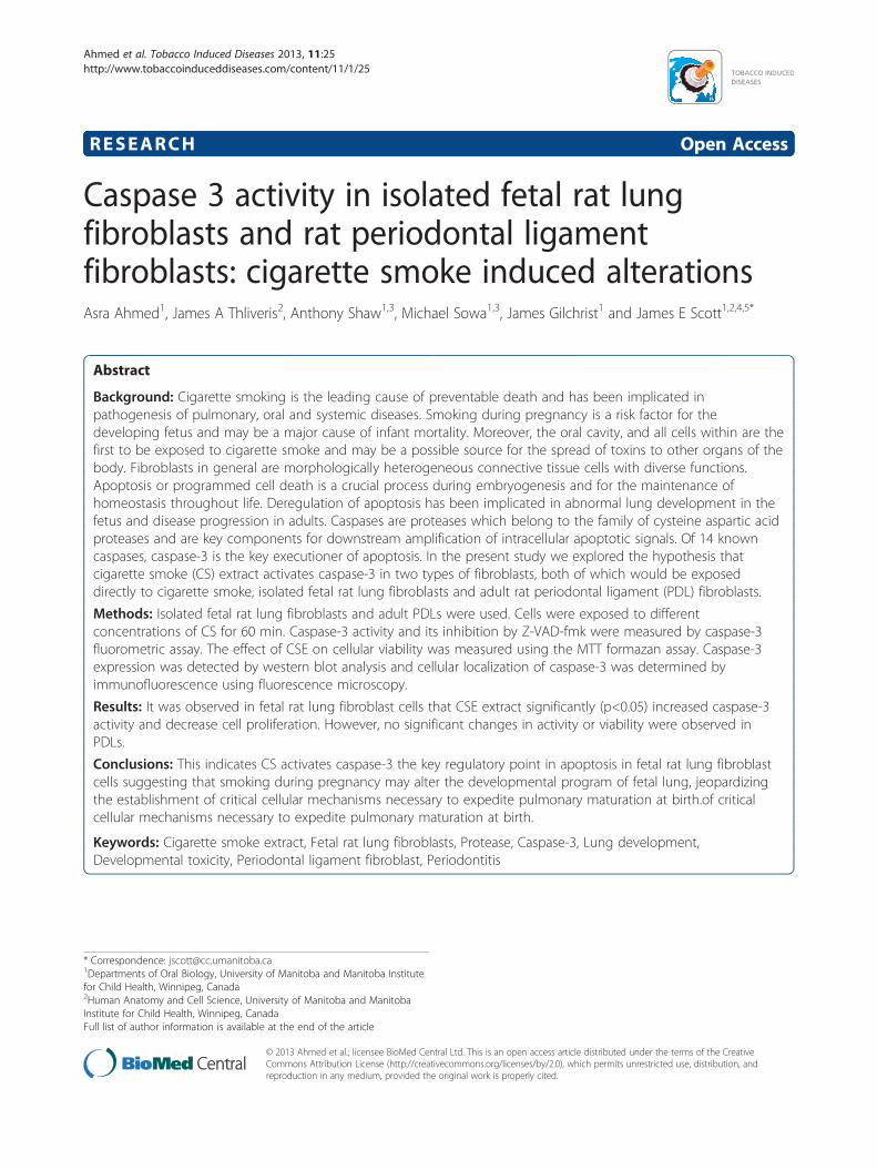
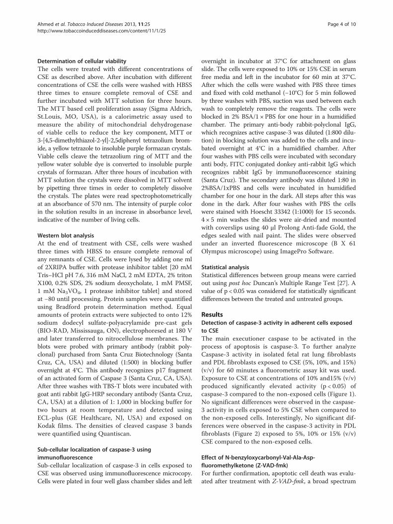
ResultsDetection of caspase-3 activity in adherent cells exposedto CSEThe main executioner caspase to be activated in theprocess of apoptosis is caspase-3. To further analyzeCaspase-3 activity in isolated fetal rat lung fibroblastsand PDL fibroblasts exposed to CSE (5%, 10%, and 15%)(v/v) for 60 minutes a fluorometric assay kit was used.Exposure to CSE at concentrations of 10% and15% (v/v)produced significantly elevated activity (p < 0.05) ofcaspase-3 compared to the non-exposed cells (Figure 1).No significant differences were observed in the caspase-3 activity in cells exposed to 5% CSE when compared tothe non-exposed cells. Interestingly, No significant dif-ferences were observed in the caspase-3 activity in PDLfibroblasts (Figure 2) exposed to 5%, 10% or 15% (v/v)CSE compared to the non-exposed cells.
Effect of N-benzyloxycarbonyl-Val-Ala-Asp-fluoromethylketone (Z-VAD-fmk)For further confirmation, apoptotic cell death was evalu-ated after treatment with Z-VAD-fmk, a broad spectrum
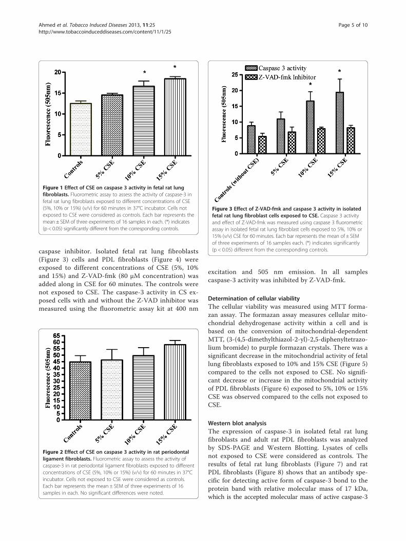
Figure 1 Effect of CSE on caspase 3 activity in fetal rat lungfibroblasts. Fluorometric assay to assess the activity of caspase-3 infetal rat lung fibroblasts exposed to different concentrations of CSE(5%, 10% or 15%) (v/v) for 60 minutes in 37°C incubator. Cells notexposed to CSE were considered as controls. Each bar represents themean ± SEM of three experiments of 16 samples in each. (*) indicates(p < 0.05) significantly different from the corresponding controls.
Figure 3 Effect of Z-VAD-fmk and caspase 3 activity in isolatedfetal rat lung fibroblast cells exposed to CSE. Caspase 3 activityand effect of Z-VAD-fmk was measured using caspase 3 fluorometricassay in isolated fetal rat lung fibroblast cells exposed to 5%, 10% or15% (v/v) CSE for 60 minutes. Each bar represents the mean of ± SEMof three experiments of 16 samples each. (*) indicates significantly(p < 0.05) different from the corresponding controls.
Ahmed et al. Tobacco Induced Diseases 2013, 11:25 Page 5 of 10http://www.tobaccoinduceddiseases.com/content/11/1/25
caspase inhibitor. Isolated fetal rat lung fibroblasts(Figure 3) cells and PDL fibroblasts (Figure 4) wereexposed to different concentrations of CSE (5%, 10%and 15%) and Z-VAD-fmk (80 μM concentration) wasadded along in CSE for 60 minutes. The controls werenot exposed to CSE. The caspase-3 activity in CS ex-posed cells with and without the Z-VAD inhibitor wasmeasured using the fluorometric assay kit at 400 nm
Figure 2 Effect of CSE on caspase 3 activity in rat periodontalligament fibroblasts. Fluorometric assay to assess the activity ofcaspase-3 in rat periodontal ligament fibroblasts exposed to differentconcentrations of CSE (5%, 10% or 15%) (v/v) for 60 minutes in 37°Cincubator. Cells not exposed to CSE were considered as controls.Each bar represents the mean ± SEM of three experiments of 16samples in each. No significant differences were noted.
excitation and 505 nm emission. In all samplescaspase-3 activity was inhibited by Z-VAD-fmk.
Determination of cellular viabilityThe cellular viability was measured using MTT forma-zan assay. The formazan assay measures cellular mito-chondrial dehydrogenase activity within a cell and isbased on the conversion of mitochondrial-dependentMTT, (3-(4,5-dimethylthiazol-2-yl)-2,5-diphenyltetrazo-lium bromide) to purple formazan crystals. There was asignificant decrease in the mitochondrial activity of fetallung fibroblasts exposed to 10% and 15% CSE (Figure 5)compared to the cells not exposed to CSE. No signifi-cant decrease or increase in the mitochondrial activityof PDL fibroblasts (Figure 6) exposed to 5%, 10% or 15%CSE was observed compared to the cells not exposed toCSE.
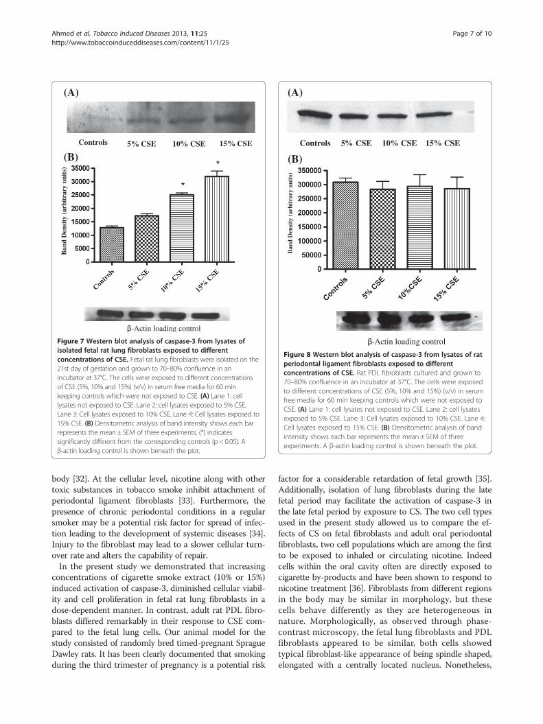
Western blot analysisThe expression of caspase-3 in isolated fetal rat lungfibroblasts and adult rat PDL fibroblasts was analyzedby SDS-PAGE and Western Blotting. Lysates of cellsnot exposed to CSE were considered as controls. Theresults of fetal rat lung fibroblasts (Figure 7) and ratPDL fibroblasts (Figure 8) shows that an antibody spe-cific for detecting active form of caspase-3 bond to theprotein band with relative molecular mass of 17 kDa,which is the accepted molecular mass of active caspase-3
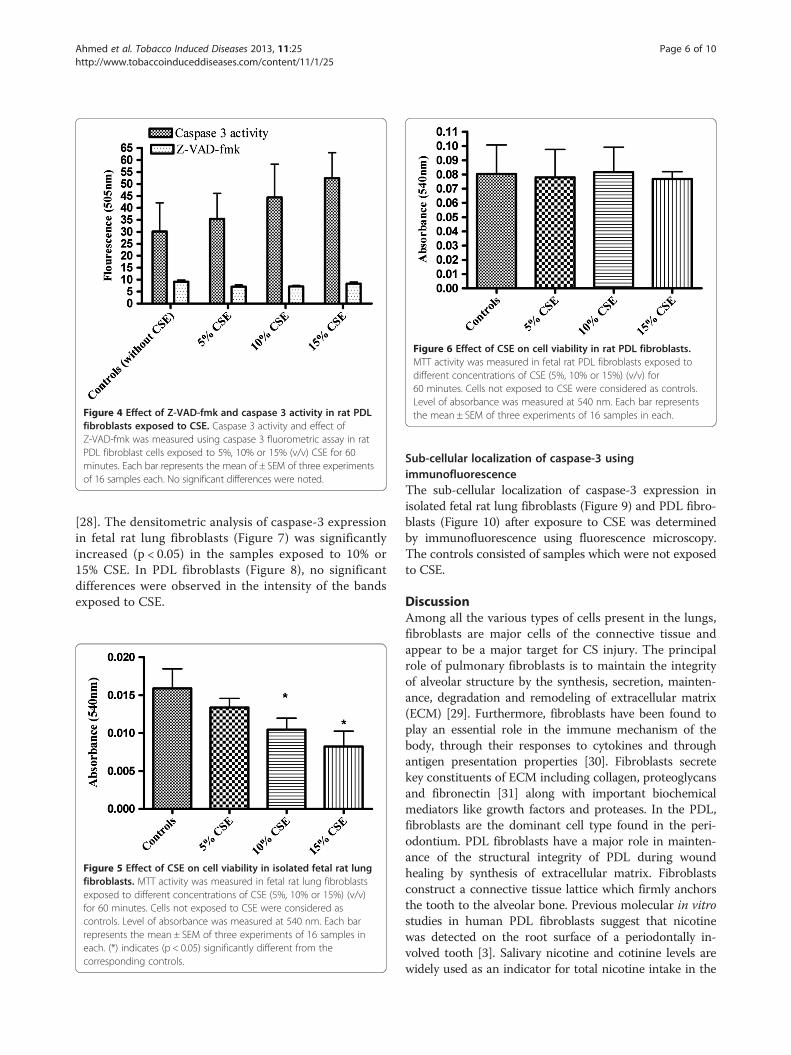
Figure 6 Effect of CSE on cell viability in rat PDL fibroblasts.MTT activity was measured in fetal rat PDL fibroblasts exposed todifferent concentrations of CSE (5%, 10% or 15%) (v/v) for60 minutes. Cells not exposed to CSE were considered as controls.Level of absorbance was measured at 540 nm. Each bar representsthe mean ± SEM of three experiments of 16 samples in each.Figure 4 Effect of Z-VAD-fmk and caspase 3 activity in rat PDL
fibroblasts exposed to CSE. Caspase 3 activity and effect ofZ-VAD-fmk was measured using caspase 3 fluorometric assay in ratPDL fibroblast cells exposed to 5%, 10% or 15% (v/v) CSE for 60minutes. Each bar represents the mean of ± SEM of three experimentsof 16 samples each. No significant differences were noted.
Ahmed et al. Tobacco Induced Diseases 2013, 11:25 Page 6 of 10http://www.tobaccoinduceddiseases.com/content/11/1/25
[28]. The densitometric analysis of caspase-3 expressionin fetal rat lung fibroblasts (Figure 7) was significantlyincreased (p < 0.05) in the samples exposed to 10% or15% CSE. In PDL fibroblasts (Figure 8), no significantdifferences were observed in the intensity of the bandsexposed to CSE.
Figure 5 Effect of CSE on cell viability in isolated fetal rat lungfibroblasts. MTT activity was measured in fetal rat lung fibroblastsexposed to different concentrations of CSE (5%, 10% or 15%) (v/v)for 60 minutes. Cells not exposed to CSE were considered ascontrols. Level of absorbance was measured at 540 nm. Each barrepresents the mean ± SEM of three experiments of 16 samples ineach. (*) indicates (p < 0.05) significantly different from thecorresponding controls.
Sub-cellular localization of caspase-3 usingimmunofluorescenceThe sub-cellular localization of caspase-3 expression inisolated fetal rat lung fibroblasts (Figure 9) and PDL fibro-blasts (Figure 10) after exposure to CSE was determinedby immunofluorescence using fluorescence microscopy.The controls consisted of samples which were not exposedto CSE.
DiscussionAmong all the various types of cells present in the lungs,fibroblasts are major cells of the connective tissue andappear to be a major target for CS injury. The principalrole of pulmonary fibroblasts is to maintain the integrityof alveolar structure by the synthesis, secretion, mainten-ance, degradation and remodeling of extracellular matrix(ECM) [29]. Furthermore, fibroblasts have been found toplay an essential role in the immune mechanism of thebody, through their responses to cytokines and throughantigen presentation properties [30]. Fibroblasts secretekey constituents of ECM including collagen, proteoglycansand fibronectin [31] along with important biochemicalmediators like growth factors and proteases. In the PDL,fibroblasts are the dominant cell type found in the peri-odontium. PDL fibroblasts have a major role in mainten-ance of the structural integrity of PDL during woundhealing by synthesis of extracellular matrix. Fibroblastsconstruct a connective tissue lattice which firmly anchorsthe tooth to the alveolar bone. Previous molecular in vitrostudies in human PDL fibroblasts suggest that nicotinewas detected on the root surface of a periodontally in-volved tooth [3]. Salivary nicotine and cotinine levels arewidely used as an indicator for total nicotine intake in the
(A)
-Actin loading control
Controls 5 CSE 10 CSE 15 CSE
(B)
Ban
d D
ensi
ty (
arbi
trar
y un
its)
Figure 7 Western blot analysis of caspase-3 from lysates ofisolated fetal rat lung fibroblasts exposed to differentconcentrations of CSE. Fetal rat lung fibroblasts were isolated on the21st day of gestation and grown to 70–80% confluence in anincubator at 37°C. The cells were exposed to different concentrationsof CSE (5%, 10% and 15%) (v/v) in serum free media for 60 minkeeping controls which were not exposed to CSE. (A) Lane 1: celllysates not exposed to CSE. Lane 2: cell lysates exposed to 5% CSE.Lane 3: Cell lysates exposed to 10% CSE. Lane 4: Cell lysates exposed to15% CSE. (B) Densitometric analysis of band intensity shows each barrepresents the mean ± SEM of three experiments. (*) indicatessignificantly different from the corresponding controls (p < 0.05). Aβ-actin loading control is shown beneath the plot.
(A)
-Actin loading control
Ban
d D
ensi
ty (
arbi
trar
y un
its)
(B)
Controls 5 CSE 10 CSE 15 CSE
Figure 8 Western blot analysis of caspase-3 from lysates of ratperiodontal ligament fibroblasts exposed to differentconcentrations of CSE. Rat PDL fibroblasts cultured and grown to70–80% confluence in an incubator at 37°C. The cells were exposedto different concentrations of CSE (5%, 10% and 15%) (v/v) in serumfree media for 60 min keeping controls which were not exposed toCSE. (A) Lane 1: cell lysates not exposed to CSE. Lane 2: cell lysatesexposed to 5% CSE. Lane 3: Cell lysates exposed to 10% CSE. Lane 4:Cell lysates exposed to 15% CSE. (B) Densitometric analysis of bandintensity shows each bar represents the mean ± SEM of threeexperiments. A β-actin loading control is shown beneath the plot.
Ahmed et al. Tobacco Induced Diseases 2013, 11:25 Page 7 of 10http://www.tobaccoinduceddiseases.com/content/11/1/25
body [32]. At the cellular level, nicotine along with othertoxic substances in tobacco smoke inhibit attachment ofperiodontal ligament fibroblasts [33]. Furthermore, thepresence of chronic periodontal conditions in a regularsmoker may be a potential risk factor for spread of infec-tion leading to the development of systemic diseases [34].Injury to the fibroblast may lead to a slower cellular turn-over rate and alters the capability of repair.In the present study we demonstrated that increasing
concentrations of cigarette smoke extract (10% or 15%)induced activation of caspase-3, diminished cellular viabil-ity and cell proliferation in fetal rat lung fibroblasts in adose-dependent manner. In contrast, adult rat PDL fibro-blasts differed remarkably in their response to CSE com-pared to the fetal lung cells. Our animal model for thestudy consisted of randomly bred timed-pregnant SpragueDawley rats. It has been clearly documented that smokingduring the third trimester of pregnancy is a potential risk
factor for a considerable retardation of fetal growth [35].Additionally, isolation of lung fibroblasts during the latefetal period may facilitate the activation of caspase-3 inthe late fetal period by exposure to CS. The two cell typesused in the present study allowed us to compare the ef-fects of CS on fetal fibroblasts and adult oral periodontalfibroblasts, two cell populations which are among the firstto be exposed to inhaled or circulating nicotine. Indeedcells within the oral cavity often are directly exposed tocigarette by-products and have been shown to respond tonicotine treatment [36]. Fibroblasts from different regionsin the body may be similar in morphology, but thesecells behave differently as they are heterogeneous innature. Morphologically, as observed through phase-contrast microscopy, the fetal lung fibroblasts and PDLfibroblasts appeared to be similar, both cells showedtypical fibroblast-like appearance of being spindle shaped,elongated with a centrally located nucleus. Nonetheless,

(B)
FITC Merge
FITC
DAPI
DAPI Merge
(C)
(A)
Figure 9 Immunofluorescence staining for detection of caspase 3 expression in isolated fetal rat lung fibroblasts. Isolated fetal rat lungfibroblasts were exposed to CSE (15% v/v) for three hours. Immunofluorescence was performed using caspase 3 rabbit polyclonal IGg antibody.Caspase 3 was visualized using donkey anti-rabbit IGg-FITC (green fluorescence) and counter stained with Hoescht 33342 (nuclear staining - blue).Image (A) shows controls not exposed to CSE. Image (B) shows expression of caspase 3 (green) primarily localized in the cytoplasm. Image (C) showsan enlarged image of a single cell.
Ahmed et al. Tobacco Induced Diseases 2013, 11:25 Page 8 of 10http://www.tobaccoinduceddiseases.com/content/11/1/25
the proliferation rates of these cells differed, the fetal lungfibroblasts having a faster rate of proliferation comparedto the adult PDL fibroblasts. The observations of thepresent study suggest that the two cell types used in thisstudy differ from each other in vitro. Furthermore, studieshave shown that fibroblasts of the periodontium showphenotypic differences from the gingival fibroblasts [37]. Itis speculated that the difference in the periodontal andgingival fibroblasts could be due the difference in thedoubling times of these two cells [37] and the difference inthe origin of the cells [38]. Similarly, we speculated thatthe difference in behavior of fetal lung fibroblasts andPDL fibroblasts when treated with CSE in vitro couldbe due to the difference in the origin and doublingtime of these two cells. PDL fibroblasts originate fromthe ecto-mesenchyme of the investing layer of thedental follicle and the dental papilla [39], whereas lungfibroblasts are mesenchymal in origin. Indeed the
differences observed herein may relate to susceptibilityto the nicotine concentrations in the CSE as Alparet al. [40] observed that periodontal fibroblasts wereonly influenced by nicotine concentrations above7.8 mM. Such a high nicotine level would most likelybe toxic as determination of the levels used in thepresent study place undiluted CSE at 0.20 μM.Apoptosis or programmed cell death is a normal and
essential biological process for the development, main-tenance of homeostasis and host defense [41]. Thehighly regulated process of apoptosis can be triggeredby extra-cellular or intra-cellular signals resulting in celldeath with the absence of inflammation. Moreover,apoptosis plays an important role during lung develop-ment and during postnatal adaptation of lung after birthfor proper gas exchange [42]. Considerable research inthe past supports a role of apoptosis in remodeling oflung tissue after lung injury [43]. The deregulation of

(A) FITC DAPI Merge
(B) FITC DAPI Merge
(C)
Figure 10 Immunofluorescence staining for detection of caspase 3 expression in rat PDL cells. Rat PDL cells were exposed to CSE (15% v/v)for three hours. Immunofluorescence was performed using caspase 3 rabbit polyclonal IGg antibody. Caspase 3 was visualized using donkey anti-rabbitIGg-FITC (green fluorescence) and counter stained with Hoescht 33342 (nuclear staining - blue). Image (A) shows controls not exposed to CSE. Image(B) shows expression of caspase 3 (green) primarily localized in the cytoplasm. Image (C) shows enlarged image of a single cell.
Ahmed et al. Tobacco Induced Diseases 2013, 11:25 Page 9 of 10http://www.tobaccoinduceddiseases.com/content/11/1/25
apoptosis may lead to development of lung disease. In-creased apoptosis in the epithelial lung cells may resultin inadequate re-epithelialisation [44]. PDL fibroblasts incontrast require an ongoing process of apoptosis which isimportant for the maintenance of periodontal health. Inresponse to apoptotic stimuli the inactive groups of intra-cellular caspases become activated and the process of celldeath is carried out through proteolytic cleavage. Al-though caspase-3 is the central caspase in the caspase cas-cade that mediates the execution of apoptotic process ofcell death [45], little is known about the ability of CSE toactivate caspase-3 in fetal lung cells and PDL fibroblasts.Our present in vitro study suggests that exposure of
fetal rat lung fibroblasts to CS may trigger activation ofcaspase-3 leading to cell death which consequently altercellular viability and proliferation in a dose dependentmanner. We subsequently found that exposure of ratPDL fibroblasts to CS did not show a significant changein the activation of caspase-3. The activation of this en-zyme in particular may result in changes in extracellularmatrix secretion, remodeling and repair of injured tissues.Although the present in vitro study gives an insight of the
changes in cellular functions, clearly further studies are re-quired to elucidate the initiating factors inducing caspase-3 activation in smoke exposed tissues. Furthermore thecomplex nature of whole cigarette smoke makes it difficultto attribute the present observations to any particularcomponent. Indeed the nonuniform nature of cigarettesmoking as well as the variability of dose exposure andinterpretion of this in terms of the more controlled envir-onment of an in vitro study remain problematic. The iden-tification of the critical role of caspases has been beneficialin understanding the ways by which this enzyme contrib-utes during tissue injury and repair. Our results suggestthat modulation of caspase-3 may have therapeuticpotential in the prevention of lung related diseases dueto cigarette smoke exposure.
Competing interestThe authors declare that they have no competing interests.
Authors' contributionAA carried out the experimental work and drafted the manuscript. JAT, JGand JES edited and revised the manuscript and supervised the work of AA.All authors read and approved the final manuscript.
Ahmed et al. Tobacco Induced Diseases 2013, 11:25 Page 10 of 10http://www.tobaccoinduceddiseases.com/content/11/1/25
AcknowledgementsThis work was supported by the Natural Science and Engineering ResearchCouncil of Canada (NSERC) and the Manitoba Institute for Child Health.
Author details1Departments of Oral Biology, University of Manitoba and Manitoba Institutefor Child Health, Winnipeg, Canada. 2Human Anatomy and Cell Science,University of Manitoba and Manitoba Institute for Child Health, Winnipeg,Canada. 3The National Research Council Biodiagnostics Institute, University ofManitoba and Manitoba Institute for Child Health, Winnipeg, Canada. 4TheBiology of Breathing Group, University of Manitoba and Manitoba Institutefor Child Health, Winnipeg, Canada. 5Department of Oral Biology, Faculty ofDentistry, University of Manitoba, Winnipeg, Manitoba, Canada.
Received: 18 October 2012 Accepted: 1 December 2013Published: 6 December 2013
References1. Roquer JM, Figueras J, Botet F, Jimenez R: Influence on fetal growth of exposure
to tobacco smoke during pregnancy. Acta Paediatr 1995, 84(2):118–121.2. DiFranza JR, Aligne CA, Weitzman M: Prenatal and postnatal
environmental tobacco smoke exposure and children’s health. Pediatrics2004, 113(4 Suppl):1007–1015.
3. Chang YC, Huang FM, Tai KW, Yang LC, Chou MY: Mechanisms ofcytotoxicity of nicotine in human periodontal ligament fibroblastcultures in vitro. J Periodontal Res 2002, 37(4):279–285.
4. Spooner BS, Wessells NK: Mammalian lung development: interactions inprimordium formation and bronchial morphogenesis. J Exp Zool 1970,175(4):445–454.
5. Nishino H, Nemoto N, Lu W, Sakurai I: Significance of apoptosis inmorphogenesis of human lung development: light microscopicobservation using in situ DNA end-labeling and ultrastructural study.Med Electron Microsc 1999, 32(1):57–61.
6. Haanen C, Vermes I: Apoptosis: programmed cell death in fetaldevelopment. Eur J Obstet Gynecol Reprod Biol 1996, 64(1):129–133.
7. Naik AS, Kallapur SG, SG Bachurski CJ, Jobe AH, AH Michna J, Kramer BW,Ikegami M: Effects of ventilation with different positive end-expiratorypressures on cytokine expression in the preterm lamb lung. Am J RespirCrit Care Med 2001, 164(3):494–498.
8. Wright JR: Pulmonary surfactant: a front line of lung host defense.J Clin Invest 2003, 111(10):1453–1455.
9. Joshi S, Kotecha S: Lung growth and development. Early Hum Dev 2007,83(12):789–794.
10. Habek D, Habek JC, Ivanisevic M, Djelmis J: Fetal tobacco syndrome andperinatal outcome. Fetal Diagn Ther 2002, 17(6):367–371.
11. Habek D, Habek JC, Jugovic D, Salihagic A: Intrauterine hypoxia andsudden infant death syndrome. Acta Med Croatica 2002, 56(3):109–118.
12. Ishii T, Matsuse T, Igarashi H, Masuda M, Teramoto S, Ouchi Y: Tobaccosmoke reduces viability in human lung fibroblasts: protective effect ofglutathione S-transferase P1. Am J Physiol Lung Cell Mol Physiol 2001,280(6):L1189–L1195.
13. Kosmider B, Messier EM, Chu HW, Mason RJ: Human alveolar epithelial cellinjury induced by cigarette smoke. PLoS One 2011, 6(12):e26059.
14. Krall EA, EA Garvey AJ, Garcia RI: Alveolar bone loss and tooth loss in malecigar and pipe smokers. J Am Dent Assoc 1999, 130(1):57–64.
15. Nicholson DW, Thornberry NA: Caspases: killer proteases. Trends BiochemSci 1997, 22(8):299–306.
16. Fadeel B, Orrenius S, Zhivotovsky B: The most unkindest cut of all: on themultiple roles of mammalian caspases. Leukemia 2000, 14(8):1514–1525.
17. Degterev A, Boyce M, Yuan J: A decade of caspases. Oncogene 2003,22(53):8543–8567.
18. Green D, Kroemer G: The central executioners of apoptosis: caspases ormitochondria? Trends Cell Biol 1998, 8(7):267–271.
19. Thornberry NA, Lazebnik Y: Caspases: enemies within. Science 1998,281(5381):1312–1316.
20. Tyas L, Brophy VA, Pope A, Rivett AJ, Tavare JM: Rapid caspase-3 activationduring apoptosis revealed using fluorescence-resonance energy transfer.EMBO Rep 2000, 1(3):266–270.
21. Giansanti V, Torriglia A, Scovassi AI: Conversation between apoptosis andautophagy: “Is it your turn or mine?”. Apoptosis 2011, 16:321–333.
22. Janoff A, Carp H: Possible mechanisms of emphysema in smokers:cigarette smoke condensate suppresses protease inhibition in vitro.Am Rev Respir Dis 1977, 116(1):65–72.
23. Tabassian AR, Nylen ES, Giron AE, Snider RH, Cassidy MM, Becker KL:Evidence for cigarette smoke-induced calcitonin secretion from lungs ofman and hamster. Life Sci 1988, 42(23):2323–2329.
24. Hastings C, Rand T, Bergen HT, Thliveris JA, Shaw AR, Lombaert GA, MantschHH, Giles BL, Dakshinamurti S, Scott JE: Stachybotrys chartarum alterssurfactant-related phospholipid synthesis and CTP:cholinephosphatecytidylyltransferase activity in isolated fetal rat type II cells. Toxicol Sci 2005,84(1):186–194.
25. Lekic PC, Nayak BN, Al-Sanea R, Tenenbaum H, Ganss B, McCulloch C: Celltransplantation in wounded mixed connective tissues. Anat Rec A DiscovMol Cell Evol Biol 2005, 287(2):1256–1263.
26. Harrington HA, Ho KL, Ghosh S, Tung KC: Construction and analysis of amodular model of caspase activation in apoptosis. Theor Biol Med Model2008, 5:26.
27. Ott L: An Introduction to Statistical Methods and Data Analysis. NorthScituate: Duxbury Press; 1977.
28. Kilic M, Schafer R, Hoppe J, Kagerhuber U: Formation of noncanonical highmolecular weight caspase-3 and −6 complexes and activation ofcaspase-12 during serum starvation induced apoptosis in AKR-2B mousefibroblasts. Cell Death Differ 2002, 9(2):125–137.
29. McGowan SE, Torday JS: The pulmonary lipofibroblast (lipid interstitialcell) and its contributions to alveolar development. Annu Rev Physiol1997, 59:43–62.
30. Fries KM, Blieden T, Looney RJ, Sempowski GD, Silvera MR, Willis RA, Phipps RP:Evidence of fibroblast heterogeneity and the role of fibroblastsubpopulations in fibrosis. Clin Immunol Immunopathol 1994, 72(3):283–292.
31. Bradley KH, Kawanami O, Ferrans VJ, Crystal RG: The fibroblast of humanlung alveolar structures: a differentiated cell with a major role in lungstructure and function. Methods Cell Biol 1980, 21A:37–64.
32. Rose JE, Levin ED, Benowitz N: Saliva nicotine as an index of plasma levelsin nicotine skin patch users. Ther Drug Monit 1993, 15(5):431–435.
33. James JA, Sayers NM, Drucker DB, Hull PS: Effects of tobacco products onthe attachment and growth of periodontal ligament fibroblasts.J Periodontol 1999, 70(5):518–525.
34. Vellappally S, Fiala Z, Smejkalova J, Jacob V, Somanathan R: Smoking relatedsystemic and oral diseases. Acta Med (Hradec Kralove) 2007, 50(3):161–166.
35. Lieberman E, Gremy I, Lang JM, Cohen AP: Low birthweight at term andthe timing of fetal exposure to maternal smoking. Am J Public Health1994, 84(7):1127–1131.
36. Yanagita M, Kojima Y, Kawahara T, Kajikawa T, Oohara H, Takedachi M, YamadaS, Murakami S: Suppressive effects of nicotine on the cytodifferentiation ofmurine periodontal ligament cells. Oral Dis 2010, 16(8):812–817.
37. Somerman MJ, Archer SY, Imm GR, Foster RA: A comparative study ofhuman periodontal ligament cells and gingival fibroblasts in vitro.J Dent Res 1988, 67(1):66–70.
38. Lekic PC, Pender N, McCulloch CA: Is fibroblast heterogeneity relevant tothe health, diseases, and treatments of periodontal tissues? Crit Rev OralBiol Med 1997, 8(3):253–268.
39. McCulloch CA, Lekic P, McKee MD: Role of physical forces in regulating theform and function of the periodontal ligament. Periodontol 2000 2000, 24:56–72.
40. Alpar B, Leyhausen G, Sapotnick A, Gunay H, Geurtsen W: Nicotine-inducedalterations in human primary periodontal ligament and gingivafibroblast cultures. Clin Oral Investig 1998, 2(1):40–46.
41. Chang HY, Yang X: Proteases for cell suicide: functions and regulation ofcaspases. Microbiol Mol Biol Rev 2000, 64(4):821–846.
42. Kresch MJ, Christian C, Wu F, Hussain N: Ontogeny of apoptosis duringlung development. Pediatr Res 1998, 43(3):426–431.
43. Martin TR, Hagimoto N, Nakamura M, Matute-Bello G: Apoptosis andepithelial injury in the lungs. Proc Am Thorac Soc 2005, 2(3):214–220.
44. Drakopanagiotakis F, Xifteri A, Polychronopoulos V, Bouros D: Apoptosis inlung injury and fibrosis. Eur Respir J 2008, 32(6):1631–1638.
45. Kumar S: Caspase function in programmed cell death. Cell Death Differ2007, 14(1):32–43.
doi:10.1186/1617-9625-11-25Cite this article as: Ahmed et al.: Caspase 3 activity in isolated fetal ratlung fibroblasts and rat periodontal ligament fibroblasts: cigarettesmoke induced alterations. Tobacco Induced Diseases 2013 11:25.