respiratory syncytial virus attachment glycoprotein contribution to
TRANSCRIPT

Respiratory Syncytial Virus Attachment Glycoprotein Contribution toInfection Depends on the Specific Fusion Protein
Jia Meng,a,b Anne L. Hotard,a,b Michael G. Currier,a,b Sujin Lee,a,b Christopher C. Stobart,a,b Martin L. Moorea,b
Department of Pediatrics, Emory University of School of Medicine, Atlanta, Georgia, USAa; Children’s Healthcare of Atlanta, Atlanta, Georgia, USAb
ABSTRACT
Human respiratory syncytial virus (RSV) is an important pathogen causing acute lower respiratory tract disease in children. TheRSV attachment glycoprotein (G) is not required for infection, as G-null RSV replicates efficiently in several cell lines. Our labo-ratory previously reported that the viral fusion (F) protein is a determinant of strain-dependent pathogenesis. Here, we hypothe-sized that virus dependence on G is determined by the strain specificity of F. We generated recombinant viruses expressing Gand F, or null for G, from the laboratory A2 strain (Katushka RSV-A2GA2F [kRSV-A2GA2F] and kRSV-GstopA2F) or the clinicalisolate A2001/2-20 (kRSV-2-20G2-20F and kRSV-Gstop2-20F). We quantified the virus cell binding, entry kinetics, infectivity,and growth kinetics of these four recombinant viruses in vitro. RSV expressing the 2-20 G protein exhibited the greatest bindingactivity. Compared to the parental viruses expressing G and F, removal of 2-20 G had more deleterious effects on binding, entry,infectivity, and growth than removal of A2 G. Overall, RSV expressing 2-20 F had a high dependence on G for binding, entry, andinfection.
IMPORTANCE
RSV is the leading cause of childhood acute respiratory disease requiring hospitalization. As with other paramyxoviruses, twomajor RSV surface viral glycoproteins, the G attachment protein and the F fusion protein, mediate virus binding and subsequentmembrane fusion, respectively. Previous work on the RSV A2 prototypical strain demonstrated that the G protein is functionallydispensable for in vitro replication. This is in contrast to other paramyxoviruses that require attachment protein function as aprerequisite for fusion. We reevaluated this requirement for RSV using G and F proteins from clinical isolate 2-20. Compared tothe laboratory A2 strain, the G protein from 2-20 had greater contributions to virus binding, entry, infectivity, and in vitrogrowth kinetics. Thus, the clinical isolate 2-20 F protein function depended more on its G protein, suggesting that RSV has ahigher dependence on G than previously thought.
Human respiratory syncytial virus (hRSV or RSV) causes anannual global 3.4 million estimated severe acute lower respi-
ratory tract infections (ALRI) in children younger than 5 years ofage (1). In the United States, about 132,000 to 172,000 childrenyounger than 5 years of age are hospitalized due to RSV every year(2). Thus far, there are no licensed vaccines, although there aremultiple vaccine candidates undergoing clinical trials (3). Devel-opment of antivirals against RSV is also an active field of researchand clinical development (4–6).
RSV is a member of the Paramyxoviridae family, Pneumovirusgenus. Members of the paramyxovirus family encode two majorglycoproteins important early during infection for attachment tothe host cell and the subsequent entry process. Paramyxovirusfusion mediated by the viral fusion (F) protein is generally initi-ated by interaction with the homologous attachment proteinupon receptor engagement (reviewed in references 7 and 8). Sev-eral studies on RSV subgroup A and B strains indicated that G isnot functionally required for efficient in vitro replication in certaincell lines but is needed for optimal growth in vivo (9–11). Al-though not required for in vitro replication, G was shown to en-hance passage of a RSV minigenome (12), and in a later study,viruses lacking G required more passages in cell culture to reachtiters similar to those of viruses expressing G (10). RSV G was alsoshown to enhance cell-to-cell fusion, in an apparently strain-specific manner (10, 13). Similarly, human metapneumovirus(HMPV), another pneumovirus, does not require its G protein forinfection (reviewed in reference 14). For both HMPV and RSV,
the attachment function of G can be substituted by the F protein(15, 16). The RSV G protein has long been thought to mediate themajority of virus binding to host cells via interaction with glycos-aminoglycans (GAGs) (17–19), while F is reported to bind a pro-tein receptor (20). Considering that previous studies regarding therequirement for G during RSV infection were done with proto-typical strains of this virus, we set out to reevaluate the functionsof this major attachment protein using protein from a clinicalisolate strain (A2001/2-20) compared to the prototypical A2strain. We generated recombinant RSV strains harboring differentcombinations of the G and F proteins (GF viruses; Katushka RSV-A2GA2F [kRSV-A2GA2F] and kRSV-2-20G2-20F), along with vi-ruses that do not express the G gene but maintain an almost iden-tical genomic sequence composition in the G gene region (Gstopviruses; kRSV-GstopA2F and kRSV-Gstop2-20F). By comparingthe G functions of each GF and Gstop virus pair, we found that
Received 24 August 2015 Accepted 4 October 2015
Accepted manuscript posted online 14 October 2015
Citation Meng J, Hotard AL, Currier MG, Lee S, Stobart CC, Moore ML. 2016.Respiratory syncytial virus attachment glycoprotein contribution to infectiondepends on the specific fusion protein. J Virol 90:245–253.doi:10.1128/JVI.02140-15.
Editor: D. S. Lyles
Address correspondence to Martin L. Moore, [email protected].
Copyright © 2015, American Society for Microbiology. All Rights Reserved.
crossmark
January 2016 Volume 90 Number 1 jvi.asm.org 245Journal of Virology
on February 24, 2018 by guest
http://jvi.asm.org/
Dow
nloaded from

there are greater contributions of 2-20 G than of A2 G to aspects ofthe RSV life cycle, including enhanced binding to the cell, viralentry, infectivity, and overall in vitro growth rate. Our study re-sults show that the F protein from a clinical RSV strain has agreater dependence on its homologous G protein than the F pro-tein of the prototypical A2 strain.
MATERIALS AND METHODSCell lines. HEp-2 (ATCC CCL-23) and BEAS-2B cells were maintained asdescribed previously (21). BSR T7/5 cells (a gift from Ursula Buchholz,National Institutes of Health, Bethesda, MD) were cultured in Glasgow’sminimal essential medium (GMEM) containing 10% fetal bovine serum(FBS) and 1 �g/ml porcine serum albumin (PSA), and during everyother passage, these cells were selected with Geneticin at 1 mg/ml.Chinese hamster ovary (CHO-K1) (ATCC, CCL-61) cells and a hepa-rin sulfate-deficient derivative of that cell line, pgsD-677 (ATCC, CRL-2244), were cultured in Kaighn’s modified F-12K medium (plus L-glu-tamine) supplemented with 10% FBS and 1 �g/ml PSA, according toATCC instructions.
Generation of recombinant RSV strains. The F glycoprotein genes ofRSV A2 (GenBank accession number FJ614814) and A2001/2-20 (RSV2-20 G [GenBank accession number JF279545] and RSV 2-20 F [GenBankaccession number JF279544]) (21) were synthesized by GeneArt gene syn-thesis (Life Technologies) and cloned into a bacterial artificial chromo-some (BAC) containing the antigenomic cDNA of RSV A2-K-line19F,described previously (22). All recombinant RSV strains generated in thisstudy express a far-red fluorescent gene (monomeric Katushka-2[mKate2]) in the first gene position— hence the kRSV designationthroughout. To generate the recombinant RSV strains without G proteinexpression (Gstop viruses), both of the Met codons (Met1 and Met48) inthe G open reading frame (ORF) were changed to Ile, with the first Met orIle followed by a stop codon (11). Recombinant viruses were recovered bycotransfection of the RSV antigenomic BAC and four human codon bias-optimized RSV helper plasmids (N, P, L, and M2-1) into BSR T7/5 cells asdescribed previously (22). The viruses were propagated in HEp-2 cells andthe sequences of the glycoprotein ORFs confirmed. Viruses used in thisstudy were prepared by harvesting infected HEp-2 cells followed by son-ication, as described previously (23). For binding assays, virus stocks werepurified by sucrose gradient centrifugation to remove the majority ofcellular proteins from the virus fraction (24). Briefly, the infected HEp-2cells were frozen at �80°C and later thawed at 37°C. Cells were scrapeddown and were transferred along with the medium to 50-ml conical tubes.After centrifugation at 2,000 rpm for 10 min at 4°C, supernatants werepooled and layered onto 20% sucrose-containing MEM for subsequentultracentrifugation at 16,000 � g for 3 h at 4°C (SW32 rotor; BeckmanCoulter). The resulting pellets were resuspended in MEM, and aliquotswere frozen in liquid nitrogen before being stored at �80°C until use.These sucrose-purified virus stocks had infectious titers comparable tothose measured for the starting material, accounting for volume change.
RSV binding assay and Western blotting. BEAS-2B cells were seededthe prior day to be subconfluent for the experiment. Input volumes ofsucrose-purified virus stocks were determined by first loading equal PFUsin SDS-PAGE gels and blotting for N and then normalizing stock dilutions(no more than 2-fold to 3-fold) to N levels. The cells were washed withcold phosphate-buffered saline (PBS), placed on ice, and inoculated withvirus for 2 h. The inocula were removed by three ice-cold PBS washes, andcells were lysed in radioimmunoprecipitation assay (RIPA) buffer (Sig-ma-Aldrich, St. Louis, MO; catalog no. R0278) supplemented with 1�protease inhibitor cocktail (Thermo Scientific, Rockford, IL; catalog no.78430). The lysates were cleared by centrifugation at 13,200 rpm for 10min at 4°C, and the supernatants were used for Western blotting.
Protein samples were mixed 1:1 with Laemmli sample buffer (Sigma-Aldrich) and heated at 95°C for 10 min. Samples were separated by 10%SDS-PAGE, transferred onto polyvinylidene difluoride (PVDF) mem-branes, and blocked with 5% nonfat dry milk–Tris-buffered saline con-
taining 0.1% Tween 20 (TBST). Blots were probed with a mouse mono-clonal antibody (clone D14; generously provided by Edward Walsh,University of Rochester, Rochester, NY) against RSV N protein followedby a horseradish peroxidase-conjugated secondary antibody. For GAPDH(glyceraldehyde-3-phosphate dehydrogenase) blots, mouse anti-GAPDH(6C5; GeneTex, Irvine, CA) was used. Chemiluminescent signal was de-tected with WesternBright Quantum substrate (Advansta, Menlo Park,CA). Images of Western blots were analyzed using ImageLab (v3.0.11).Relevant bands were defined manually for each group, and the total dark-pixel volume for each band was taken. The bands for the glycoproteinswere then normalized by dividing their values by the respective RSV Nband volumes.
FFU assay. HEp-2 cells were seeded in 96-well plates the day before theexperiment to reach 70% confluence for the assay. Fifty microliters ofvirus samples (serially diluted 10-fold) was inoculated onto the cells andincubated 1.5 h at room temperature with gentle rocking. After virusadsorption, 150 �l 0.75% methylcellulose (EMD, Gibbstown, NJ) in com-plete media was added to each well and then cells were incubated at 37°C5% CO2 for 2 days. Wells containing 1 to 50 fluorescent-focus units (FFU)were counted and used for calculation of the virus titer in the samples. Thelimit of detection of this assay is 1 FFU per well, corresponding to 20FFU/ml.
Infectivity and virus growth kinetics. Cells were seeded into 6-wellplates to be 70% confluent for infection at a multiplicity of infection(MOI) of 1.0. Cells were washed with PBS once before infection in a totalvolume of 500 �l per well at room temperature for 1 h with gentle rocking.The cells were then washed twice with PBS to remove the remaining in-oculum. For infectivity, cells were harvested with trypsin 24 h postinfec-tion and quantified using an LSR II flow cytometer (Becton Dickinson,Franklin Lakes, NJ) by detecting the mKate2 signal with a 532-nm-wave-length laser with a 610/20 filter. For growth kinetics, triplicate wells ofinfected cells were scraped in medium and resuspended at the indicatedtimes, and aliquots were frozen at �80°C until titration by the FFU assaydescribed above.
RSV entry assay. This assay was performed as described previouslywith some modifications (25). BEAS-2B cells (70% confluent in 12-wellplates) were placed on ice for 5 min and washed once with ice-cold PBSbefore addition of virus at an MOI of 1.0. Binding of virus to cells pro-ceeded for 2 h on ice with gentle shaking until the inocula were removed,and cells were washed twice with ice-cold PBS. Five hundred microliters ofice-cold RPMI 1640 medium was added to each well. Plates were thenwarmed at 37°C for the indicated times (30 s or 1, 2, 3, 4, or 5 min). At theend of each time period, medium was removed followed by addition of500 �l citrate buffer (400 mM sodium citrate, 10 mM potassium chloride,135 mM sodium chloride, pH 3.0) for 2 min to inactivate any remainingextracellular virus. Cells were then washed in PBS at room temperatureonce before addition of complete medium, and incubation was continuedat 37°C and 5% CO2 for 20 h. Infected mKate2-positive (mKate2�) cellswere counted on an LSR II cytometer and analyzed using FlowJo software(Tree Star, Ashland, OR) as a quantification of the amount of virus thatentered during the short warming period.
Culture and infection of primary cells. Normal human bronchial(NHBE) cells at the air-liquid interface (NHBE/ALI cells) were obtainedfrom Lonza (Allendale, NJ) and cultured according to the recommendedprotocols. For differentiation, cells were seeded onto 24-well, collagen-coated transwell supports (BD Bioscience, Bedford, MA). Cells weremaintained and differentiated as described previously (26).
For binding assays, differentiated cells were cooled at 4°C and washedonce with cold PBS prior to addition of virus. Equivalent amounts of virus(determined by Western blotting; see above) were used to infect the apicalsurface of NHBE/ALI cells for 2 h at 4°C. Following infection, the inocu-lum was removed and the apical surface of cells was washed three timeswith cold PBS. RIPA buffer supplemented with protease inhibitor cocktailwas used for cell lysis. Lysates were centrifuged at 14,000 � g for 10 min forclarification prior to Western blotting.
Meng et al.
246 jvi.asm.org January 2016 Volume 90 Number 1Journal of Virology
on February 24, 2018 by guest
http://jvi.asm.org/
Dow
nloaded from

To determine virus growth kinetics in these cells, differentiatedNHBE/ALI cells were washed with PBS and apically infected at an MOI of1.0 for 2 h at 37°C. Following infection, inocula were removed by threeapical PBS washes. Virus was collected from the apical surface of cells dailyby adding differential medium to the apical chamber, incubating cells inmedia for 10 min at 37°C, and removing media for later use in virustitration. Virus collection was performed twice per well, for a total volumeof 300 �l per well for each time point. All samples were snap-frozen inliquid nitrogen until titration by FFU assay.
Statistical analysis. Statistical analyses were performed using Graph-Pad Prism software version 6.0 (San Diego, CA). Data are represented asmeans with standard errors of the means (SEM). One-way and two-wayanalysis of variance (ANOVA) with Tukey’s post hoc test with a P value of0.05 was used.
RESULTSGeneration of recombinant viruses. In order to assess the de-pendence on G protein of specific F proteins in the context ofRSV infection, we used a chimeric virus approach. We gener-ated recombinant RSV containing both G and F from either theprototypical A2 strain or the low-passage-number clinical iso-late A2001/2-20 strain. We also generated G-null mutants ex-pressing either the F protein of A2 or the F protein of 2-20.Thus, only the presence or absence of F differed between theG-null viruses. As the RSV G protein is produced both as amembrane-bound form and as a secreted form due to the pres-ence of an alternative translation initiation site, we mutatedboth initiation methionines to isoleucine, as previously de-scribed (11), and we changed the second codon in the G open
reading frame to a premature stop codon to abolish the expres-sion of this protein without perturbing the gene order (Fig.1A). We did not change the codon following the second methi-onine to abolish the secreted G expression because previouswork showed that changing the second methionine to isoleu-cine is sufficient to abolish secreted G (11). As expected, West-ern blot analyses of sucrose-purified virus stocks showed thatneither of the Gstop viruses (kRSV-GstopA2F and kRSV-Gstop2-20F) expressed detectable G protein (Fig. 1B). Therewas no significant difference in the levels of mature G forkRSV-A2GA2F and kRSV-2-20G2-20F (Fig. 1B). There wasalso no statistically significant difference in the levels of F pro-tein abundance for kRSV-A2GA2F and kRSV-GstopA2F andfor kRSV-2-20G2-20F and kRSV-Gstop2-20F (Fig. 1B), consis-tent with previously published data showing that the absence ofG did not alter the F protein level in the virions (10). The Flevels of kRSV-2-20G2-20F were 28% higher than those ofkRSV-A2GA2F, normalized to N, which was not statisticallysignificant by one-way ANOVA. Similar results were found inanalyzing N, G, and F levels in HEp-2 cell lysate virus stocksthat were not sucrose purified (data not shown).
Differential contributions of G proteins to virus attachmentto host cells. The F protein of the prototypical A2 strain binds tohost cells, possibly through interactions with heparan sulfate (16,19, 27). We asked whether the relative contributions of G and F tocell binding differ between the A2 and 2-20 glycoproteins. To testthis, we used sucrose-purified virus stocks and normalized the
FIG 1 Schematic design of the recombinant viruses and quantification of surface glycoproteins in purified virions. (A) RSV genome with G gene open readingframe (amino acids 1 to 298) enlarged to illustrate the mutations made to generate the Gstop virus. The two methionines were changed to isoleucine, and thesecond codon (serine) was changed to a stop codon. (B) Western blot showing the F, G, and N protein expression levels (left) and densitometry combined fromthe results of four independent experiments (right). Data are represented as means � SEM.
RSV Strain-Specific Functionality of G
January 2016 Volume 90 Number 1 jvi.asm.org 247Journal of Virology
on February 24, 2018 by guest
http://jvi.asm.org/
Dow
nloaded from

relative amounts of virions used as inputs for this binding assaybased on the N protein expression levels determined by Westernblotting. The N protein level was previously reported to correlatewith radiolabeled activity in virus preparations (10). The amountof kRSV-2-20G2-20F bound to BEAS-2B cells (a human bronchialepithelial cell line) was approximately 5-fold higher than theamount of kRSV-A2GA2F (Fig. 2). Removal of the A2 G proteinfrom the virus resulted in loss of approximately half of cell binding(comparing kRSV-GstopA2F to kRSV-A2GA2F in Fig. 2), consis-tent with previously published data (19). However, removal of2-20 G protein from the virus resulted in loss of 90% of cell bind-ing (comparing kRSV-Gstop2-20F to kRSV-2-20G2-20F in Fig.2). We found similar virus attachment results using these viruses
that were not sucrose purified (data not shown). Thus, 2-20 Gdisplayed a greater contribution to virus binding to BEAS-2B cellsthan A2 G.
Greater contribution of 2-20 G than A2 G to virus entry ki-netics. To further explore the functional differences between 2-20G and A2 G, we tested whether virus entry into host cells would bedifferentially affected by the removal of A2 or 2-20 G protein. Wequantified entry kinetics of all four viruses in BEAS-2B cells by theuse of a citric acid wash entry assay (25, 28). kRSV-A2GA2F hadthe fastest entry kinetics, followed by kRSV-GstopA2F (Fig. 3A).Removal of A2 G resulted in 3-fold-lower entry efficiency of thevirus (Fig. 3B). Removal of 2-20 G from kRSV-2-20G2-20F re-sulted in approximately 8-fold-lower entry efficiency (Fig. 3C).
A
kRS
V-A
2GA
2F
kRS
V-2-
20G
2-20
F
kRS
V-G
stop
A2F
kRS
V-G
stop
2-20
F
Bound RSV N
GAPDH
Input RSV N
B
0
1
2
3
4
5
Rat
io o
f RSV
N to
GAP
DH
kRSV-A2GA2FkRSV-2-20G2-20FkRSV-GstopA2FkRSV-Gstop2-20F
**
0
20
40
60
80
100
boun
d vi
rus
(%)
rela
tive
to G
+F v
irus
kRSV-A2GA2F
kRSV-GstopA2FkRSV-2-20G2-20F
kRSV-Gstop2-20F
FIG 2 Greater contribution of 2-20 G than A2 G to binding BEAS-2B cells. (A)Western blots showing results of a representative binding assay. Input virusinocula were normalized based on the N protein expression levels as shown onthe bottom blot. (B) Densitometry analysis of bound virus. Virus bindingactivity (top panel) was calculated by normalizing the N level (bound RSV Nfrom the top blot) to the GAPDH level of the sample. This value for the Gstopviruses was also normalized to that of its parental virus containing both F andG and is represented as the percentage of bound virus compared to the per-centage of G�F virus (bottom panel). *, P � 0.05 (comparing the bracketedgroups using one-way ANOVA). Data are shown as means � SEM and repre-sent the combined results of three replicate experiments.
FIG 3 Entry kinetics in BEAS-2B cells. (A) Entry kinetics of kRSV-A2GA2F,kRSV-2-20G2-20F, kRSV-GstopA2F, and kRSV-Gstop2-20F in BEAS-2B cellsusing an acid wash protocol as described in Materials and Methods. (B) Con-tribution of A2 G protein to the entry kinetics of kRSV-A2GA2F compared tokRSV-GstopA2F. (C) Contribution of 2-20 G protein to the entry kinetics ofkRSV-2-20G2-20F compared to kRSV-Gstop2-20F. *, P � 0.05 (comparingGstop virus to GF virus at the same time point using one-way ANOVA). Dataare represented as means � SEM. The graphs in panels B and C were con-structed using the data in panel A. The data represent the combined resultsfrom three independent experiments.
Meng et al.
248 jvi.asm.org January 2016 Volume 90 Number 1Journal of Virology
on February 24, 2018 by guest
http://jvi.asm.org/
Dow
nloaded from

These data demonstrate that although kRSV-2-20G2-20F hadlower entry efficiency than kRSV-A2GA2F, 2-20 G played a rela-tively greater role in cell entry than A2 G. Additionally, the A2 Fprotein was significantly more efficient than the 2-20 F protein atmediating entry in BEAS-2B cells.
In vitro infectivity. As 2-20 G contributed relatively morethan A2 G to virus binding to host cells and entry kinetics, wecompared the roles of A2 G and 2-20 G in infectivity in vitro.Infectivity of kRSV-A2GA2F, kRSV-2-20G2-20F, kRSV-GstopA2F,and kRSV-Gstop2-20F was assayed in three cell lines, BEAS-2B,CHO-K1, and pgsD-677. CHO-K1 and pgsD-677 were used forcomparing the effects of the presence and absence of GAGs(pgsD-677 is a heparan sulfate-deficient CHO-K1 derivative).In BEAS-2B and CHO-K1 cells, kRSV-A2GA2F and kRSV-2-20G2-20F exhibited greater infectivity than kRSV-GstopA2F andkRSV-Gstop2-20F, respectively (Fig. 4). In pgsD-677 cells,kRSV-A2GA2F and kRSV-GstopA2F exhibited similar levels ofinfectivity, while kRSV-2-20G2-20F displayed greater infectivitythan kRSV-Gstop2-20F (Fig. 4). To compare the relative contri-
butions of the G proteins to virus infectivity, we normalized thepercentage of cells infected by the Gstop viruses to the percentageof cells infected by the viruses containing both G and F proteins(kRSV-GstopA2F normalized to kRSV-A2GA2F and kRSV-Gstop2-20F normalized to kRSV-2-20G2-20F; Fig. 4). In BEAS-2Band CHO-K1 cells (both expressing heparan sulfate), A2 G con-tributed to 30% to 40% infectivity whereas 2-20 G contributed tomore than 80% (Fig. 4A and B). Moreover, the contribution of2-20 G protein was evident in the heparan sulfate-deficient pgsD-677 cell line whereas A2 G exhibited no significant contribution toinfection in these cells (Fig. 4C). These data are consistent with theestablished role of A2 G binding to GAGs (10, 19) and suggest thatthe 2-20 G protein utilizes an additional host factor(s) in vitro.
In vitro growth kinetics in HEp-2 and BEAS-2B cell lines. Wecompared the growth kinetics of kRSV-A2GA2F, kRSV-2-20G2-20F, kRSV-GstopA2F, and kRSV-Gstop2-20F in theHEp-2 and BEAS-2B cell lines using an MOI of 0.01. As re-ported previously, the absence of G protein from A2 reducedthe infectious yield of the virus in HEp-2 cells (Fig. 5A) (11).
A
B
C
0
20
40
60
80
BEAS-2B
% k
atus
hka+
cel
ls kRSV-A2GA2FkRSV-2-20G2-20FkRSV-GstopA2FkRSV-Gstop2-20F
0
5
10
15
20
25
CHO-K1
% k
atus
hka+
cel
ls kRSV-A2GA2FkRSV-2-20G2-20FkRSV-GstopA2FkRSV-Gstop2-20F
0.0
0.2
0.4
0.6
0.8
1.0
1.2
CHO-K1
Rel
ativ
e to
G+F
viru
s
*
*
0.0
0.2
0.4
0.6
0.8
1.0
1.2
BEAS-2B
Rel
ativ
e to
G+F
viru
s
*
*
0.00.20.40.60.81.01.21.41.6
pgsD-677
% k
atus
hka+
cel
ls kRSV-A2GA2FkRSV-2-20G2-20FkRSV-GstopA2FkRSV-Gstop2-20F
0.00.20.40.60.81.01.21.41.6
pgsD-677
Rel
ativ
e to
G+F
viru
s
*
FIG 4 Infectivity in BEAS-2B, CHO-K1, and pgsD-677 cell lines. BEAS-2B (A), CHO-K1 (B), and pgsD-677 (C) cells were infected with kRSV-A2GA2F,kRSV-2-20G2-20F, kRSV-GstopA2F, or kRSV-Gstop2-20F at an MOI of 1. Twenty-four hours later, infected cells were quantified using flow cytometry. The leftpanels show the percentages of RSV-infected cells. Brackets represent a P value of �0.05 for the indicated group comparison. The right panels show the levels ofinfectivity of the virus without G protein normalized to that of the virus containing both G and F proteins of the same strain (kRSV-GstopA2F normalized tokRSV-A2GA2F and kRSV-Gstop2-20F normalized to kRSV-2-20G2-20F) expressed as values relative to the levels seen with G�F virus on the y axis. *, P � 0.05(comparing each Gstop virus to the corresponding parental GF virus using one-way ANOVA). Data are represented as means � SEM. Each graph depicts datafrom the combined results of three independent experiments.
RSV Strain-Specific Functionality of G
January 2016 Volume 90 Number 1 jvi.asm.org 249Journal of Virology
on February 24, 2018 by guest
http://jvi.asm.org/
Dow
nloaded from

The origin of the G and F glycoprotein did not affect the growthof the virus, as kRSV-A2GA2F and kRSV-2-20G2-20F grew tosimilar levels in both cell lines (Fig. 5A). However, the absenceof 2-20 G from the virus resulted in reduced infectious yield inHEp-2 cells and, to a greater degree, in BEAS-2B cells (Fig. 5B).Compared to kRSV-2-20G2-20F, kRSV-Gstop2-20F consis-tently had a titer in the BEAS-2B cell line that was more than 2log10 lower. This growth deficiency was greater than that ofkRSV-GstopA2F relative to kRSV-A2GA2F (�1 log10 differ-ence), suggesting that the 2-20 G protein contributes more to invitro growth than the A2 G protein.
Contribution of G proteins to binding and growth kinetics inprimary airway epithelial cells. Because RSV primarily infectsciliated airway epithelial cells, we investigated the effects of theabsence of G on virus binding and growth in normal human bron-chial epithelial (NHBE) cells cultured at the air-liquid interface(ALI). In a binding assay using well-differentiated NHBE/ALIcells, we found that kRSV-2-20G2-20F bound more efficientlythan kRSV-A2GA2F (Fig. 6A and B). Additionally, when we nor-malized binding of the Gstop viruses with binding of the parentalviruses to NHBE/ALI cells, we demonstrated that A2 G contrib-uted very little to binding by kRSV-A2GA2F (Fig. 6B). This was incontrast to removal of 2-20 G from kRSV-2-20G2-20F, whichresulted in loss of approximately 70% of binding (Fig. 6B). Theseresults imply that most of the binding difference between kRSV-A2GA2F and kRSV-2-20G2-20F in these cells is due to the G pro-tein and that 2-20 G serves a greater role than A2 G in bindingprimary airway epithelial cells.
We also monitored the ability of kRSV-A2GA2F, kRSV-2-20G2-20F, kRSV-Gstop-A2F, and kRSV-Gstop-2-20F to infectNHBE/ALI cells. By days 5 and 6 postinfection, kRSV-2-20G2-20Fexhibited greater infectious yield from NHBE/ALI cells thankRSV-A2GA2F (Fig. 6C). Neither Gstop virus was able to replicateto detectable levels in NHBE/ALI cells. While we did not detect adifference in the G-specific contributions of these viruses to infec-tivity in NHBE/ALI cells, our data indicate that, together, the 2-20
G and F proteins provide an advantage to kRSV-2-20G2-20F rel-ative to kRSV-A2GA2F in primary human bronchial epithelialcells.
DISCUSSION
Previous studies analyzing functions of the RSV G protein usedprototypical strains such as A2 (10, 11) or a subgroup B strain (9).In the present studies, deletion of the attachment protein from thevirus attenuated virus replication in HEp-2 but not Vero cell lines.We reevaluated the requirement for G in the context of a recentclinical isolate strain of RSV and expanded our study to include ahuman bronchial epithelial cell line (BEAS-2B) as well as primarynormal human bronchial epithelial cells cultured at the air-liquidinterface (NHBE/ALI cells). We generated recombinant virusescontaining G and F glycoproteins of either A2 or the 2-20 clinicalisolate and compared the functional contributions of G from theA2 and 2-20 strains.
Congruent with previously published reports, removal of Gfrom the A2 strain resulted in a decrease in virus-to-cell bindingand infectivity compared to parental A2 results (10, 11). In ourstudy, Gstop-A2F was approximately one-third less efficient atbinding and one-third less infectious in BEAS-2B cells than kRSV-A2GA2F. We found that the 2-20 G protein showed greater con-tributions to virus attachment to host cells, virus entry, infectivity,and overall growth kinetics than the A2 G protein. In contrast tothe A2 G protein, the 2-20 G protein played a role in infection ofheparan sulfate-deficient cells, suggesting that the ability of 2-20 Gto bind to a receptor(s) other than GAGs may contribute to thehigh level of attachment. The level of entry of BEAS-2B cells byvirus expressing 2-20 G and 2-20 F was reduced approximately10-fold compared to that seen with virus expressing A2 G and A2F. It is likely that the lower entry efficiency of the virus expressing2-20 G and 2-20 F is due to the 2-20 F protein, because the entry ofthe G-null virus expressing 2-20 F was approximately 40-foldlower than the entry of the G-null virus expressing A2F.
Earlier reports determined that A2 G is involved in binding of
FIG 5 Contribution of G protein to virus in vitro growth kinetics. HEp-2 cells (A) and BEAS-2B cells (B) were infected with kRSV-A2GA2F, kRSV-2-20G2-20F,kRSV-GstopA2F, or kRSV-Gstop2-20F using an MOI of 0.01. Time zero represents the calculated input titer. Virus titers at each time point thereafter wereassayed by FFU assay. *, P � 0.05 (comparing the Gstop virus and parental GF virus at the same time point using one-way ANOVA). Data are represented asmeans � SEM. LOD, limit of detection. FFU, fluorescent-focus unit. Each graph depicts data from the combined results of four independent experiments in eachcell line.
Meng et al.
250 jvi.asm.org January 2016 Volume 90 Number 1Journal of Virology
on February 24, 2018 by guest
http://jvi.asm.org/
Dow
nloaded from
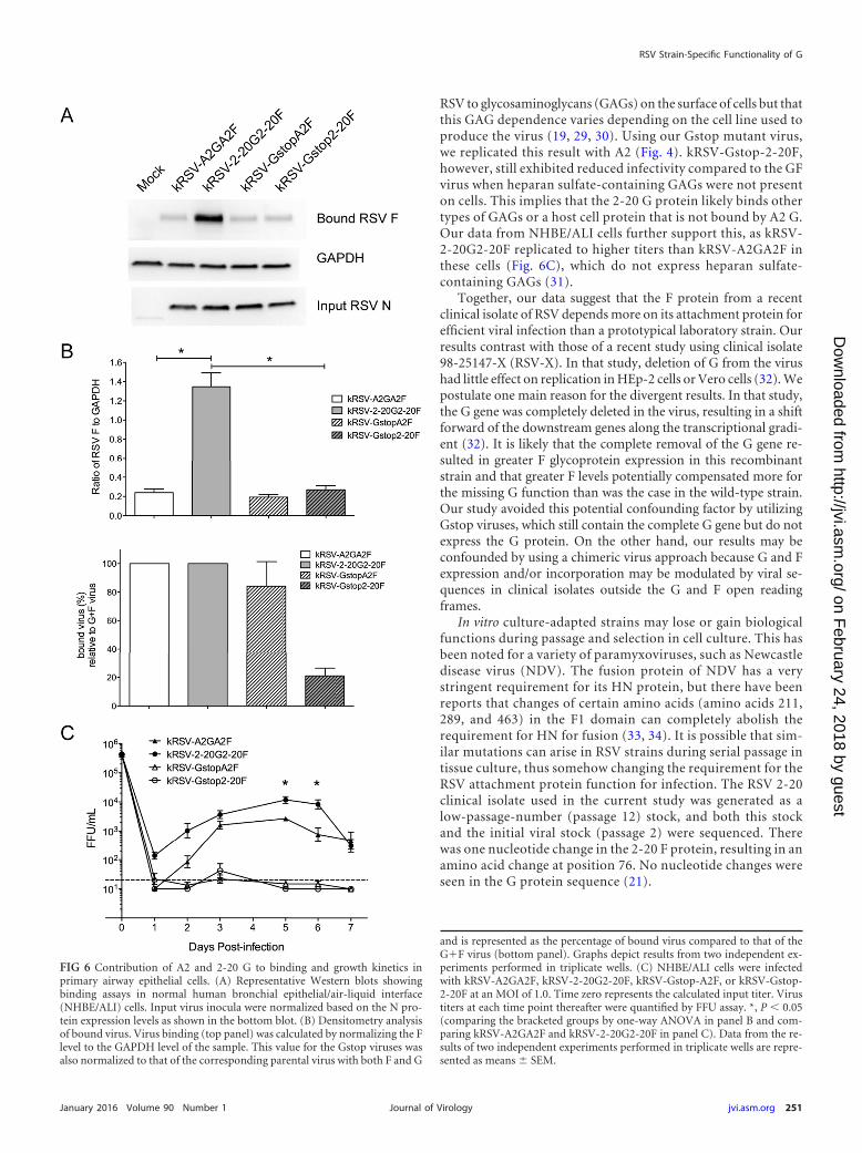
RSV to glycosaminoglycans (GAGs) on the surface of cells but thatthis GAG dependence varies depending on the cell line used toproduce the virus (19, 29, 30). Using our Gstop mutant virus,we replicated this result with A2 (Fig. 4). kRSV-Gstop-2-20F,however, still exhibited reduced infectivity compared to the GFvirus when heparan sulfate-containing GAGs were not presenton cells. This implies that the 2-20 G protein likely binds othertypes of GAGs or a host cell protein that is not bound by A2 G.Our data from NHBE/ALI cells further support this, as kRSV-2-20G2-20F replicated to higher titers than kRSV-A2GA2F inthese cells (Fig. 6C), which do not express heparan sulfate-containing GAGs (31).
Together, our data suggest that the F protein from a recentclinical isolate of RSV depends more on its attachment protein forefficient viral infection than a prototypical laboratory strain. Ourresults contrast with those of a recent study using clinical isolate98-25147-X (RSV-X). In that study, deletion of G from the virushad little effect on replication in HEp-2 cells or Vero cells (32). Wepostulate one main reason for the divergent results. In that study,the G gene was completely deleted in the virus, resulting in a shiftforward of the downstream genes along the transcriptional gradi-ent (32). It is likely that the complete removal of the G gene re-sulted in greater F glycoprotein expression in this recombinantstrain and that greater F levels potentially compensated more forthe missing G function than was the case in the wild-type strain.Our study avoided this potential confounding factor by utilizingGstop viruses, which still contain the complete G gene but do notexpress the G protein. On the other hand, our results may beconfounded by using a chimeric virus approach because G and Fexpression and/or incorporation may be modulated by viral se-quences in clinical isolates outside the G and F open readingframes.
In vitro culture-adapted strains may lose or gain biologicalfunctions during passage and selection in cell culture. This hasbeen noted for a variety of paramyxoviruses, such as Newcastledisease virus (NDV). The fusion protein of NDV has a verystringent requirement for its HN protein, but there have beenreports that changes of certain amino acids (amino acids 211,289, and 463) in the F1 domain can completely abolish therequirement for HN for fusion (33, 34). It is possible that sim-ilar mutations can arise in RSV strains during serial passage intissue culture, thus somehow changing the requirement for theRSV attachment protein function for infection. The RSV 2-20clinical isolate used in the current study was generated as alow-passage-number (passage 12) stock, and both this stockand the initial viral stock (passage 2) were sequenced. Therewas one nucleotide change in the 2-20 F protein, resulting in anamino acid change at position 76. No nucleotide changes wereseen in the G protein sequence (21).
FIG 6 Contribution of A2 and 2-20 G to binding and growth kinetics inprimary airway epithelial cells. (A) Representative Western blots showingbinding assays in normal human bronchial epithelial/air-liquid interface(NHBE/ALI) cells. Input virus inocula were normalized based on the N pro-tein expression levels as shown in the bottom blot. (B) Densitometry analysisof bound virus. Virus binding (top panel) was calculated by normalizing the Flevel to the GAPDH level of the sample. This value for the Gstop viruses wasalso normalized to that of the corresponding parental virus with both F and G
and is represented as the percentage of bound virus compared to that of theG�F virus (bottom panel). Graphs depict results from two independent ex-periments performed in triplicate wells. (C) NHBE/ALI cells were infectedwith kRSV-A2GA2F, kRSV-2-20G2-20F, kRSV-Gstop-A2F, or kRSV-Gstop-2-20F at an MOI of 1.0. Time zero represents the calculated input titer. Virustiters at each time point thereafter were quantified by FFU assay. *, P � 0.05(comparing the bracketed groups by one-way ANOVA in panel B and com-paring kRSV-A2GA2F and kRSV-2-20G2-20F in panel C). Data from the re-sults of two independent experiments performed in triplicate wells are repre-sented as means � SEM.
RSV Strain-Specific Functionality of G
January 2016 Volume 90 Number 1 jvi.asm.org 251Journal of Virology
on February 24, 2018 by guest
http://jvi.asm.org/
Dow
nloaded from

There is some evidence suggesting that RSV strain differ-ences contribute to different pathogenesis outcomes (21, 35–37). Several studies using different clinical isolates of RSV com-pared to the prototypical A2 strain demonstrated that somestrains are more virulent in well-differentiated cell culture sys-tems or in animal models (21, 35, 37). Thus, reevaluating thegene functions for clinical isolates of RSV may contribute toour understanding of the pathogenesis of RSV. The strain usedin this study, 2-20, has been shown to induce disease that ismore severe than that seen with RSV A2 in the BALB/c mousemodel (21, 38, 39). The contribution of the 2-20 G protein maybe linked to its pathogenesis, and it would be interesting toexplore whether the relatively greater contribution of G can beextended to other RSV clinical isolates. The results from thesefurther studies could potentially guide vaccine design and an-tiviral drug development.
Although many members of the Paramyxoviridae family, suchas NDV and measles virus, have a strict requirement for theattachment protein (HN or H) to trigger fusion, there are someknown exceptions in this virus family. The fusion protein ofparainfluenza virus 5 (PIV5) has been shown to induce mem-brane fusion independently of the abundance of its HN pro-tein, which is thought to provide only the binding activity nec-essary for the optimal distance between the fusion protein andthe cellular target(s) (40). In addition to RSV, other membersof the Pneumovirus genus, namely, human metapneumovirusand bovine RSV, harbor attachment glycoproteins which aredispensable for virus growth in vitro (41, 42). Previous workdetermined that the 2-20 G protein enhanced fusion of the 2-20F protein (13). The G protein of RSV has been thought tosimply facilitate fusion enhancement by bridging the twomembranes in close proximity (7). At this point, we cannot ruleout this scenario for RSV 2-20 G as it did enhance the bindingactivity of the virus to the host cells, as shown in this study.Future studies may determine the role of specific domains in2-20 G as important for boosting 2-20 F fusion activity. As thevirus containing only A2 F still retained more than half of thebinding activity seen with the virus with only 2-20 F (Fig. 2),differences in the F binding activity could be the determiningfactor for their different levels of dependence on the G proteinfor infection. Further studies are needed to completely dissectthe interaction of RSV G and F proteins in regard to fusionactivity, and, as a whole, more focus on clinically relevant vi-ruses will aid our knowledge of this evolving pathogen.
ACKNOWLEDGMENTS
We thank the Emory Children’s Pediatric Research Center flow cytometrycore supported by Children’s Healthcare of Atlanta (CHOA).
We thank Edward Walsh for the monoclonal antibody to the RSV Nprotein and Ursula Buchholz and Karl-Klaus Conzelmann for the BSR-T7/5 cell line. We also thank Nancy Ulbrandt (MedImmune) for provid-ing motavizumab antibody.
M.L.M. and Emory University are entitled to licensing fees derivedfrom various agreements that Emory has entered into related to productsused in this research described in this paper. This study could affect thepersonal financial status of M.L.M. The terms of this agreement have beenreviewed and approved by Emory University in accordance with its con-flict-of-interest policies.
FUNDING INFORMATIONHHS | NIH | National Institute of Allergy and Infectious Diseases (NIAID)provided funding to Michael M. Moore under grant numbers1R01AI087798 and 1U19AI095227.
This study was also supported by funds from Emory University andCHOA. Additional support was provided by the NIH through EmoryVaccinology Training Grant T32AI074492 (Christopher C. Stobart), byEmory and CHOA under joint grant number 33515 (Michael G. Currier,Christopher C. Stobart, Sujin Lee, and Martin L. Moore), and by Emorythrough Nelson Memorial Fund R6336510 (Anne L. Hotard and MartinL. Moore).
REFERENCES1. Nair H, Nokes DJ, Gessner BD, Dherani M, Madhi SA, Singleton RJ,
O’Brien KL, Roca A, Wright PF, Bruce N, Chandran A, Theodoratou E,Sutanto A, Sedyaningsih ER, Ngama M, Munywoki PK, KartasasmitaC, Simoes EA, Rudan I, Weber MW, Campbell H. 2010. Global burdenof acute lower respiratory infections due to respiratory syncytial virus inyoung children: a systematic review and meta-analysis. Lancet 375:1545–1555. http://dx.doi.org/10.1016/S0140-6736(10)60206-1.
2. Stockman LJ, Curns AT, Anderson LJ, Fischer-Langley G. 2012. Respi-ratory syncytial virus-associated hospitalizations among infants andyoung children in the United States, 1997–2006. Pediatr Infect Dis J 31:5–9. http://dx.doi.org/10.1097/INF.0b013e31822e68e6.
3. Guvenel AK, Chiu C, Openshaw PJ. 2014. Current concepts and progressin RSV vaccine development. Expert Rev Vaccines 13:333–344. http://dx.doi.org/10.1586/14760584.2014.878653.
4. DeVincenzo JP, Whitley RJ, Mackman RL, Scaglioni-Weinlich C,Harrison L, Farrell E, McBride S, Lambkin-Williams R, Jordan R,Xin Y, Ramanathan S, O’Riordan T, Lewis SA, Li X, Toback SL, LinSL, Chien JW. 2014. Oral GS-5806 activity in a respiratory syncytialvirus challenge study. N Engl J Med 371:711–722. http://dx.doi.org/10.1056/NEJMoa1401184.
5. Mackman RL, Sangi M, Sperandio D, Parrish JP, Eisenberg E, PerronM, Hui H, Zhang L, Siegel D, Yang H, Saunders O, Boojamra C, Lee G,Samuel D, Babaoglu K, Carey A, Gilbert BE, Piedra PA, Strickley R,Iwata Q, Hayes J, Stray K, Kinkade A, Theodore D, Jordan R, Desai M,Cihlar T. 2015. Discovery of an oral respiratory syncytial virus (RSV)fusion inhibitor (GS-5806) and clinical proof of concept in a human RSVchallenge study. J Med Chem 58:1630 –1643. http://dx.doi.org/10.1021/jm5017768.
6. Wang G, Deval J, Hong J, Dyatkina N, Prhavc M, Taylor J, Fung A, JinZ, Stevens SK, Serebryany V, Liu J, Zhang Q, Tam Y, Chanda SM,Smith DB, Symons JA, Blatt LM, Beigelman L. 2015. Discovery of4=-chloromethyl-2=-deoxy-3=,5=-di-O-isobutyryl-2=-fluorocytidine(ALS-8176), a first-in-class RSV polymerase inhibitor for treatment ofhuman respiratory syncytial virus infection. J Med Chem 58:1862–1878.http://dx.doi.org/10.1021/jm5017279.
7. Chang A, Dutch RE. 2012. Paramyxovirus fusion and entry: multiplepaths to a common end. Viruses 4:613– 636.
8. Plattet P, Plemper RK. 2013. Envelope protein dynamics in paramyxovirusentry. mBio 4:e00413-13. http://dx.doi.org/10.1128/.00413-13.
9. Karron RA, Buonagurio DA, Georgiu AF, Whitehead SS, Adamus JE,Clements-Mann ML, Harris DO, Randolph VB, Udem SA, Murphy BR,Sidhu MS. 1997. Respiratory syncytial virus (RSV) SH and G proteins arenot essential for viral replication in vitro: clinical evaluation and molecu-lar characterization of a cold-passaged, attenuated RSV subgroup B mu-tant. Proc Natl Acad Sci U S A 94:13961–13966. http://dx.doi.org/10.1073/pnas.94.25.13961.
10. Techaarpornkul S, Barretto N, Peeples ME. 2001. Functional analysis ofrecombinant respiratory syncytial virus deletion mutants lacking the smallhydrophobic and/or attachment glycoprotein gene. J Virol 75:6825– 6834.
11. Teng MN, Whitehead SS, Collins PL. 2001. Contribution of therespiratory syncytial virus G glycoprotein and its secreted and mem-brane-bound forms to virus replication in vitro and in vivo. Virology289:283–296.
12. Teng MN, Collins PL. 1998. Identification of the respiratory syncytialvirus proteins required for formation and passage of helper-dependentinfectious particles. J Virol 72:5707–5716.
13. Stokes KL, Currier MG, Sakamoto K, Lee S, Collins PL, Plemper RK,Moore ML. 2013. The respiratory syncytial virus fusion protein and neu-
Meng et al.
252 jvi.asm.org January 2016 Volume 90 Number 1Journal of Virology
on February 24, 2018 by guest
http://jvi.asm.org/
Dow
nloaded from

trophils mediate the airway mucin response to pathogenic respiratorysyncytial virus infection. J Virol 87:10070 –10082. http://dx.doi.org/10.1128/JVI.01347-13.
14. Cox RG, Williams JV. 2013. Breaking in: human metapneumovirus fu-sion and entry. Viruses 5:192–210. http://dx.doi.org/10.3390/v5010192.
15. Cox RG, Livesay SB, Johnson M, Ohi MD, Williams JV. 2012. Thehuman metapneumovirus fusion protein mediates entry via an interac-tion with RGD-binding integrins. J Virol 86:12148 –12160. http://dx.doi.org/10.1128/JVI.01133-12.
16. Feldman SA, Audet S, Beeler JA. 2000. The fusion glycoprotein of humanrespiratory syncytial virus facilitates virus attachment and infectivity viaan interaction with cellular heparan sulfate. J Virol 74:6442– 6447. http://dx.doi.org/10.1128/JVI.74.14.6442-6447.2000.
17. Feldman SA, Hendry RM, Beeler JA. 1999. Identification of a linearheparin binding domain for human respiratory syncytial virus attachmentglycoprotein G. J Virol 73:6610 – 6617.
18. Levine S, Klaiber-Franco R, Paradiso PR. 1987. Demonstration thatglycoprotein G is the attachment protein of respiratory syncytial virus. JGen Virol 68(Pt 9):2521–2524. http://dx.doi.org/10.1099/0022-1317-68-9-2521.
19. Techaarpornkul S, Collins PL, Peeples ME. 2002. Respiratory syncytialvirus with the fusion protein as its only viral glycoprotein is less dependenton cellular glycosaminoglycans for attachment than complete virus. Vi-rology 294:296 –304. http://dx.doi.org/10.1006/viro.2001.1340.
20. Tayyari F, Marchant D, Moraes TJ, Duan W, Mastrangelo P, HegeleRG. 2011. Identification of nucleolin as a cellular receptor for humanrespiratory syncytial virus. Nat Med 17:1132–1135.
21. Stokes KL, Chi MH, Sakamoto K, Newcomb DC, Currier MG, Hucka-bee MM, Lee S, Goleniewska K, Pretto C, Williams JV, Hotard A,Sherrill TP, Peebles RS, Jr, Moore ML. 2011. Differential pathogenesis ofrespiratory syncytial virus clinical isolates in BALB/c mice. J Virol 85:5782–5793.
22. Hotard AL, Shaikh FY, Lee S, Yan D, Teng MN, Plemper RK, Crowe JE,Jr, Moore ML. 2012. A stabilized respiratory syncytial virus reverse genet-ics system amenable to recombination-mediated mutagenesis. Virology434:129 –136. http://dx.doi.org/10.1016/j.virol.2012.09.022.
23. Graham BS, Perkins MD, Wright PF, Karzon DT. 1988. Primary respi-ratory syncytial virus infection in mice. J Med Virol 26:153–162. http://dx.doi.org/10.1002/jmv.1890260207.
24. Boyoglu-Barnum S, Gaston KA, Todd SO, Boyoglu C, Chirkova T,Barnum TR, Jorquera P, Haynes LM, Tripp RA, Moore ML, AndersonLJ. 2013. A respiratory syncytial virus (RSV) anti-G protein F(ab=)2monoclonal antibody suppresses mucous production and breathing effortin RSV rA2-line19F-infected BALB/c mice. J Virol 87:10955–10967.
25. White LK, Yoon JJ, Lee JK, Sun A, Du Y, Fu H, Snyder JP, Plemper RK.2007. Nonnucleoside inhibitor of measles virus RNA-dependent RNApolymerase complex activity. Antimicrob Agents Chemother 51:2293–2303. http://dx.doi.org/10.1128/AAC.00289-07.
26. Meng J, Lee S, Hotard AL, Moore ML. 2014. Refining the balance ofattenuation and immunogenicity of respiratory syncytial virus by targetedcodon deoptimization of virulence genes. mBio 5:e01704-14. http://dx.doi.org/10.1128/mBio.01704-14.
27. Crim RL, Audet SA, Feldman SA, Mostowski HS, Beeler JA. 2007.Identification of linear heparin-binding peptides derived from humanrespiratory syncytial virus fusion glycoprotein that inhibit infectivity. JVirol 81:261–271. http://dx.doi.org/10.1128/JVI.01226-06.
28. Yan D, Lee S, Thakkar VD, Luo M, Moore ML, Plemper RK. 2014.Cross-resistance mechanism of respiratory syncytial virus against struc-turally diverse entry inhibitors. Proc Natl Acad Sci U S A 111:E3441–E3449. http://dx.doi.org/10.1073/pnas.1405198111.
29. Hallak LK, Spillmann D, Collins PL, Peeples ME. 2000. Glycosamino-glycan sulfation requirements for respiratory syncytial virus infection. JVirol 74:10508 –10513. http://dx.doi.org/10.1128/JVI.74.22.10508-10513.2000.
30. Kwilas S, Liesman RM, Zhang L, Walsh E, Pickles RJ, Peeples ME.2009. Respiratory syncytial virus grown in Vero cells contains a trun-cated attachment protein that alters its infectivity and dependence onglycosaminoglycans. J Virol 83:10710 –10718. http://dx.doi.org/10.1128/JVI.00986-09.
31. Monzon ME, Casalino-Matsuda SM, Forteza RM. 2006. Identification ofglycosaminoglycans in human airway secretions. Am J Respir Cell MolBiol 34:135–141.
32. Widjojoatmodjo MN, Boes J, van Bers M, van Remmerden Y, RohollPJ, Luytjes W. 2010. A highly attenuated recombinant human respiratorysyncytial virus lacking the G protein induces long-lasting protection incotton rats. Virol J 7:114. http://dx.doi.org/10.1186/1743-422X-7-114.
33. Ayllón J, Villar E, Muñoz-Barroso I. 2010. Mutations in the ectodomainof Newcastle disease virus fusion protein confer a hemagglutinin-neuraminidase-independent phenotype. J Virol 84:1066 –1075. http://dx.doi.org/10.1128/JVI.01473-09.
34. Sergel TA, McGinnes LW, Morrison TG. 2000. A single amino acidchange in the Newcastle disease virus fusion protein alters the requirementfor HN protein in fusion. J Virol 74:5101–5107. http://dx.doi.org/10.1128/JVI.74.11.5101-5107.2000.
35. Villenave R, Thavagnanam S, Sarlang S, Parker J, Douglas I, SkibinskiG, Heaney LG, McKaigue JP, Coyle PV, Shields MD, Power UF. 2012.In vitro modeling of respiratory syncytial virus infection of pediatric bron-chial epithelium, the primary target of infection in vivo. Proc Natl Acad SciU S A 109:5040 –5045. http://dx.doi.org/10.1073/pnas.1110203109.
36. Melero JA, Moore ML. 2013. Influence of respiratory syncytial virusstrain differences on pathogenesis and immunity. Curr Top MicrobiolImmunol 372:59 – 82.
37. Derscheid RJ, van Geelen A, Gallup JM, Kienzle T, Shelly DA, Cihlar T,King RR, Ackermann MR. 2014. Human respiratory syncytial virusMemphis 37 causes acute respiratory disease in perinatal lamb lung. BioresOpen Access 3:60 – 69. http://dx.doi.org/10.1089/biores.2013.0044.
38. de Almeida Nagata DE, Demoor T, Ptaschinski C, Ting HA, Jang S,Reed M, Mukherjee S, Lukacs NW. 2014. IL-27R-mediated regulation ofIL-17 controls the development of respiratory syncytial virus-associatedpathogenesis. Am J Pathol 184:1807–1818. http://dx.doi.org/10.1016/j.ajpath.2014.02.004.
39. Petersen BC, Dolgachev V, Rasky A, Lukacs NW. 2014. IL-17E (IL-25)and IL-17RB promote respiratory syncytial virus-induced pulmonary dis-ease. J Leukoc Biol 95:809 – 815. http://dx.doi.org/10.1189/jlb.0913482.
40. Dutch RE, Joshi SB, Lamb RA. 1998. Membrane fusion promoted byincreasing surface densities of the paramyxovirus F and HN proteins:comparison of fusion reactions mediated by simian virus 5 F, humanparainfluenza virus type 3 F, and influenza virus HA. J Virol 72:7745–7753.
41. Biacchesi S, Skiadopoulos MH, Yang L, Lamirande EW, Tran KC,Murphy BR, Collins PL, Buchholz UJ. 2004. Recombinant human Meta-pneumovirus lacking the small hydrophobic SH and/or attachment Gglycoprotein: deletion of G yields a promising vaccine candidate. J Virol78:12877–12887. http://dx.doi.org/10.1128/JVI.78.23.12877-12887.2004.
42. Karger A, Schmidt U, Buchholz UJ. 2001. Recombinant bovine respira-tory syncytial virus with deletions of the G or SH genes: G and F proteinsbind heparin. J Gen Virol 82:631– 640. http://dx.doi.org/10.1099/0022-1317-82-3-631.
RSV Strain-Specific Functionality of G
January 2016 Volume 90 Number 1 jvi.asm.org 253Journal of Virology
on February 24, 2018 by guest
http://jvi.asm.org/
Dow
nloaded from