respiratory system diagnostic procedure
DESCRIPTION
RESPIRATORY SYSTEM DIAGNOSTIC PROCEDURETRANSCRIPT

RESPIRATORY SYSTEM
CHEST X-RAYA chest X-ray is a radiology test that involves exposing the chest briefly to radiation to produce an image of the chest and the internal organs of the chest. An X-ray film is positioned against the body opposite the camera, which sends out a very small dose of a radiation beam. As the radiation penetrates the body, it is absorbed in varying amounts by different body tissues depending on the tissue's composition of air, water, blood, bone, or muscle.
Procedure
Patients obtaining a chest X-ray will often be requested to use an X-ray gown, and extra metallic objects such as jewelry are removed from the chest and/or neck areas. These objects can block X-ray penetration, making the result less accurate. Patients may be asked to take a deep breath and hold it during the chest X-ray in order to inflate the lungs to their maximum, which increases the visibility of different tissues within the chest.
The chest X-ray procedure often involves a view from the back to the front of the body as well as a view from the side. The view from the side is called a lateral chest X-ray. Occasionally, different angles are added in order for the radiologist to interpret certain specific areas of the chest.
The radiology technologist or technician is a trained, certified assistant to the radiologist who will help the patient during the X-ray and actually perform the X-ray test procedure. After the chest X-ray is taken and recorded on the X-ray film, the film is placed into a developing machine, and this picture (which is essentially a photographic negative) is examined and interpreted by the radiologist.

COMPUTER TOMOGRAPHY
Computed Tomography (CT) is a powerful nondestructive evaluation (NDE) technique for producing 2-D and 3-D cross-sectional images of an object from flat X-ray images. Characteristics of the internal structure of an object such as dimensions, shape, internal defects, and density are readily available from CT images.
Procedure During the test, you will lie on a table that is attached to the CT scanner, which is
a large doughnut-shaped machine. The CT scanner sends X-rays through the body area being studied. Each rotation of the scanner takes less than a second and provides a picture of a thin slice of the organ or area. All of the pictures are saved as a group on a computer. They also can be printed.
LUNG SCANA lung scan is a nuclear scanning test that is most commonly used to detect a blood clot that is preventing normal blood flow to part of a lung (pulmonary embolism).
Two types of lung scans are usually done together:
Ventilation scan. During a ventilation scan, a radioactive tracer gas or mist is inhaled into the lungs. Pictures from this scan can show areas of the lungs that are not receiving enough air or that retain too much air. Areas of the lung that retain too much air show up as bright or "hot" spots on the pictures. Areas that are not receiving enough air show up as dark or "cold" spots.

Perfusion scan. During a perfusion scan, a radioactive tracer substance is injected into a vein in the arm. It travels through the bloodstream and into the lungs. Pictures from this scan can show areas of the lungs that are not receiving enough blood. The tracer is absorbed evenly in areas of the lung where the blood flow is normal. These areas show up with the tracer distributed evenly. Areas that are not receiving enough blood show up as cold spots.
Prepare
Before your lung scan, tell your doctor if:
You are or might be pregnant. You are breast-feeding. Use formula (discard your breast milk) for 1 to 2 days
after the scan until the radioactive tracer has been eliminated from your body.
Within the past 4 days, you have had an X-ray test using barium contrast material (such as a barium enema) or have taken a medicine (such as Pepto-Bismol) that contains bismuth. Barium and bismuth can interfere with test results.
POSITRON EMISSION TOMOGRAPHY
Positron emission tomography (PET) is a test that uses a special type of camera and a tracer (radioactive chemical) to look at organs in the body.During the test, the tracer liquid is put into a vein (intravenous, or IV) in your arm. The tracer moves through your body, where much of it collects in the specific organ or tissue. The tracer gives off tiny positively charged particles (positrons). The camera records the positrons and turns the recording into pictures on a computer.
Prepare
Tell your doctor if you have diabetes. If you take medicine to control diabetes, you may need to take less than your normal dose. Talk with your doctor about how much medicine you should take.

Tell your doctor about any medicines and herbal remedies you take. You may need to stop taking some medicines or change your dose before this test.Do not smoke or drink caffeine or alcohol for 24 hours before this test.
It Is Done To: Study the brain's blood flow and metabolic activity. A PET scan can help a
doctor find nervous system problems, such as Alzheimer's disease, Parkinson's disease, multiple sclerosis, transient ischemic attack (TIA), amyotrophic lateral sclerosis (ALS), Huntington's disease, stroke, and schizophrenia.
Find changes in the brain that may cause epilepsy. Evaluate some cancers, especially lymphoma or cancers of the head and neck,
brain, lung, colon, or prostate. In its early stages, cancer may show up more clearly on a PET scan than on a CT scan or an MRI.
FLUOROSCOPYFluoroscopy is the method that provides real-time X ray imaging that is especially
useful for guiding a variety of diagnostic and interventional procedures. The ability of fluoroscopy to display motion is provided by a continuous series of images produced at a maximum rate of 25-30 complete images per second. This is similar to the way conventional television or video transmits images.

Procedure
Fluoroscopy may be performed on an outpatient basis or as part of your stay in a hospital. Procedures may vary depending on your condition and your physician's practices.
Generally, fluoroscopy follows this process:
1. You will be asked to remove any clothing or jewelry that may interfere with the exposure of the body area to be examined.
2. If you are asked to remove clothing, you will be given a gown to wear.
3. A contrast substance may be given, depending upon the type of procedure that is being performed, via swallowing, enema, or an intravenous (IV) line in your hand or arm.
4. You will be positioned on the x-ray table. Depending upon the type of procedure, you may be asked to assume different positions, move a specific body part, or hold your breath at intervals while the fluoroscopy is being performed.
5. For procedures that require catheter insertion, such as cardiac catheterization or catheter placement into a joint or other body part, an additional line insertion site may be used in the groin, elbow, or other site.
6. A special x-ray scanner will be used to produce the fluoroscopic images of the body structure being examined or treated.
7. A dye or contrast substance may be injected into the IV line in order to better visualize the organs or structures being studied.
8. In the case of arthrography (visualization of a joint), any fluid in the joint may be aspirated (withdrawn with a needle) prior to the injection of the contrast substance. After the contrast is injected, you may be asked to move the joint for a few minutes in order to evenly distribute the contrast substance throughout the joint.
9. The type of procedure being performed and the body part being examined and/or treated will determine the length of the procedure.
10. After the procedure has been completed, the IV line will be removed.

Endoscopic Studies
BRONCHOSCOPY
Bronchoscopy is a procedure in which a thin tube is threaded through the nose or mouth into the windpipe and lungs. This allows the clinician to look inside a patient's airway for abnormalities like blockages, bleeding, tumors, or inflammation. The clinician often takes samples from inside the lungs: biopsies, fluid (bronchoalveolar lavage), or endobronchial brushing. The clinician may use either a rigid or a flexible bronchoscope. Bronchoscopy was developed in the late nineteenth century as a way for physicians to access the airways through the mouth. Since then, it has developed into an important way to diagnose and treat diseases and conditions of the lungs and trachea.
LARYNGOSCOPY
Examination of the larynx (voice box) with a mirror (indirect laryngoscopy) or with a laryngoscope (direct laryngoscopy).

THORACOSCOPY
Thoracoscopy uses an endoscope to visually examine the pleura, lungs, and mediastinum and to obtain tissue for testing purposes. An endoscope is an illuminated optic instrument that is inserted through an incision.
Procedure The anesthesiologist gives you general anesthesia, which lets you sleep and
keeps you free from pain during surgery. Once you're asleep, you're positioned
comfortably on your side.
Several small incisions are made in your side.
The surgeon inserts a thin, tubelike instrument containing a tiny camera through
one of the incisions. This camera allows the surgeon to view your lungs on a
video monitor. Surgical instruments are inserted through the other incisions.
When the procedure is finished, one or more tubes may be temporarily placed
in the chest to drain fluid and air. The incisions are then closed with sutures or
staples.

Invasive Procedures
THORACENTESIS
Thoracentesis is a procedure to remove fluid from the space between the lining
of the outside of the lungs (pleura) and the wall of the chest.
Procedure
A small area of skin on your chest or back is washed with a sterilizing liquid. Some numbing medicine (local anesthetic) is injected in this area.
A needle is placed through the skin of the chest wall into the space around the lungs, called the pleural space. Fluid is collected and may be sent to a laboratory for testing (pleural fluid analysis).
Normally, very little fluid is in the pleural space. A buildup of too much fluid between the layers of the pleura is called a pleural effusion.
The test is performed to determine the cause of the extra fluid, or to relieve symptoms from the fluid buildup.

LUNG BIOPSY
A lung needle biopsy is a method to remove a piece of lung tissue for
examination.
Procedure
A chest x-ray or chest CT scan may be used to find the exact spot for the biopsy. If the biopsy is done using a CT scan, you may be lying down during the exam. A needle biopsy of the lung may also be performed during bronchoscopy or mediastinoscopy. You sit with your arms resting forward on a table. You should try to keep still and not cough during the biopsy. The doctor will ask you to hold your breath. The skin is scrubbed and a local pain-killing medicine (anesthetic) is injected.
The physician will make a small (about 1/8-inch) cut in the skin, and will insert the biopsy needle into the abnormal tissue, tumor, or lung tissue. A small piece of tissue is removed with the needle and sent to a laboratory for examination. When the biopsy is done, pressure is placed over the site. Once bleeding has stopped, a bandage is applied. A chest x-ray is taken immediately after the biopsy. The procedure usually takes 30 - 60 minutes. Laboratory analysis usually takes a few days.

THORACOTOMYIn a thoracotomy (THOR-uh-KAH-tuh-mee), the surgeon makes a long incision
between two ribs, from front to back, on one side of your chest. Once the chest wall has been opened in this way, the surgeon can remove all or part of a diseased or damaged lung. A thoracotomy is typically performed to treat a lung abscess, lung cancer, or the blebs (balloon-like sacs pressing on the lung) that sometimes form in emphysema. The operation requires 6 to 8 days of hospitalization.
Preaparation
The Week Before Surgery: You'll probably need to stop taking aspirin and ibuprofen; the doctor will tell you
when. If you're taking aspirin for your heart, don't stop without asking the doctor first. Also ask whether you can take any over-the-counter medicines.
Your doctor will tell you whether you need to have blood drawn.
The Night Before Surgery: Your physician may suggest you take a sleeping pill. Just before surgery, you should not eat or drink anything (even water). Your doctor
will tell you when to begin fasting.
Call Your Doctor If... You have a cold or flu or are running a high temperature. The operation may need to
be postponed. The problems for which you are having the operation get any worse.
When You Arrive Check with your doctor before taking insulin, diabetes pills, blood pressure
medicine, heart pills, or any other medication on the day of surgery. Do not wear contact lenses to the hospital.
Non-invasive Procedures
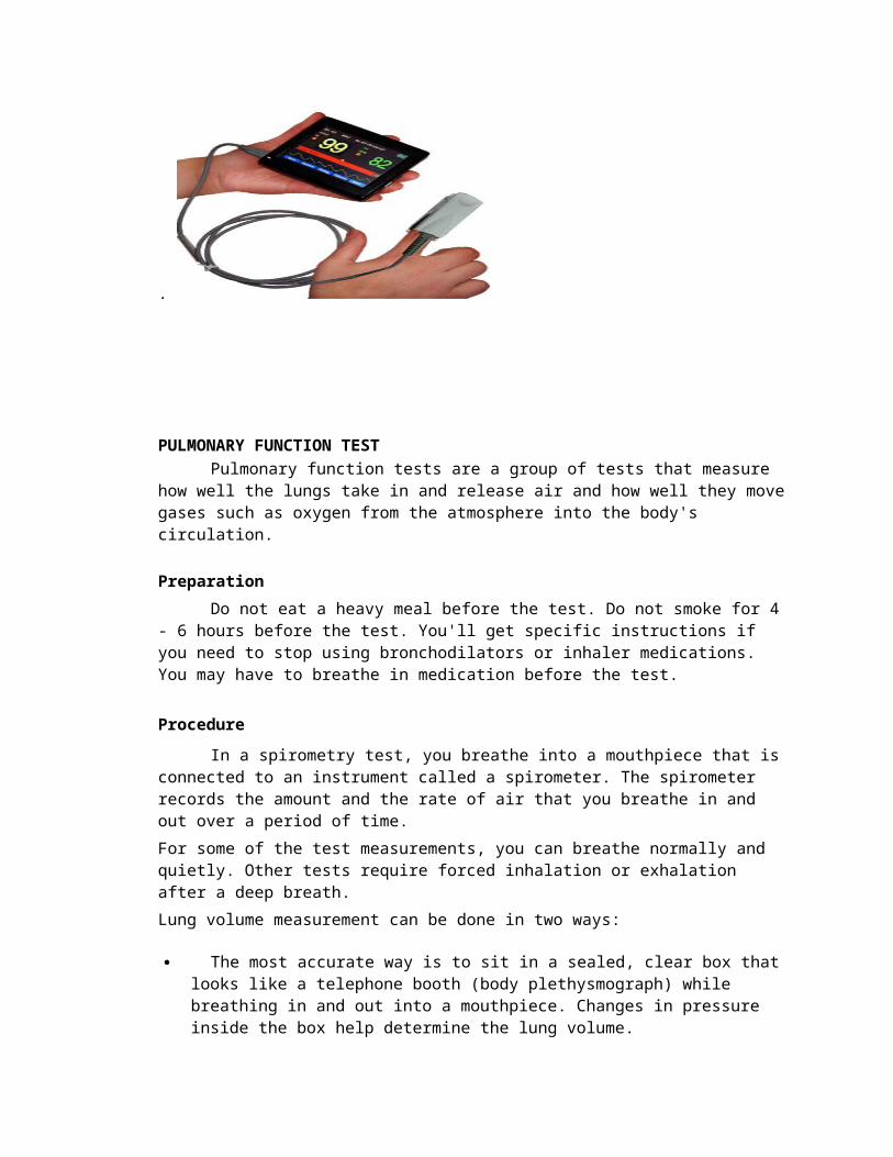
PULSE OXIMETRY

A pulse oximeter is a medical device that indirectly measures the oxygen saturation of a patient's blood (as opposed to measuring oxygen saturation directly through a blood sample) and changes in blood volume in the skin, producing a photoplethysmograph. It is often attached to a medical monitor so staff can see a patient's oxygenation at all times. Most monitors also display the heart rate. Portable, battery-operated pulse oximeters are also available for home blood-oxygen monitoring. The original oximeter was made by Milliken in the 1940s.[1] The precursor to today's modern pulse oximeter was developed in 1972, by Aoyagi at Nihon Kohden using the ratio of red to infrared light absorption of pulsating components at the measuring site. It was commercialized by Biox in 1981. The device did not see wide adoption in the United States until the late 1980s.
.
PULMONARY FUNCTION TESTPulmonary function tests are a group of tests that measure how well the lungs
take in and release air and how well they move gases such as oxygen from the atmosphere into the body's circulation.
Preparation
Do not eat a heavy meal before the test. Do not smoke for 4 - 6 hours before the test. You'll get specific instructions if you need to stop using bronchodilators or inhaler medications. You may have to breathe in medication before the test.
Procedure
In a spirometry test, you breathe into a mouthpiece that is connected to an instrument called a spirometer. The spirometer records the amount and the rate of air that you breathe in and out over a period of time.

For some of the test measurements, you can breathe normally and quietly. Other tests require forced inhalation or exhalation after a deep breath.
Lung volume measurement can be done in two ways:
The most accurate way is to sit in a sealed, clear box that looks like a telephone booth (body plethysmograph) while breathing in and out into a mouthpiece. Changes in pressure inside the box help determine the lung volume.
Lung volume can also be measured when you breathe nitrogen or helium gas through a tube for a certain period of time. The concentration of the gas in a chamber attached to the tube is measured to estimate the lung volume.
To measure diffusion capacity, you breathe a harmless gas for a very short time, often one breath. The concentration of the gas in the air you breathe out is measured. The difference in the amount of gas inhaled and exhaled measures how effectively gas travels from the lungs into the blood.
Laboratory Tests
SPUTUM CULTURE
Sputum is material coughed up from the lungs and expectorated (spit out) through the mouth. A sputum culture is done to find and identify the microorganism causing an infection of the lower respiratory tract such as pneumonia (an infection of the lung). If a microorganism is found, more testing is done to determine which antibiotics will be effective in treating the infection.
Based on the clinical condition of the patient, the physician determines what group of microorganism is likely to be causing the infection, and then orders one or more specific types of cultures: bacterial, viral, or fungal (for yeast and molds). For all culture types, the sputum must be collected into a sterile container. The sputum specimen must be collected carefully, so that bacteria that normally live in the mouth and saliva don't contaminate the sputum and complicate the process of identifying the cause of the infectious agent. Once in the laboratory, each culture type is handled differently.

ARTERIAL BLOOD GAS
An arterial blood gas (ABG) test measures the acidity (pH) and the levels of oxygen and carbon dioxide in the blood from an artery. This test is used to check how well your lungs are able to move oxygen into the blood and remove carbon dioxide from the blood.As blood passes through your lungs, oxygen moves into the blood while carbon dioxide moves out of the blood into the lungs. An ABG test uses blood drawn from an artery, where the oxygen and carbon dioxide levels can be measured before they enter body tissues. An ABG measures:
Partial pressure of oxygen (PaO2). This measures the pressure of oxygen dissolved in the blood and how well oxygen is able to move from the airspace of the lungs into the blood.
Partial pressure of carbon dioxide (PaCO2). This measures how much carbon dioxide is dissolved in the blood and how well carbon dioxide is able to move out of the body.
pH. The pH measures hydrogen ions (H+) in blood. The pH of blood is usually between 7.35 and 7.45. A pH of less than 7.0 is called acid and a pH greater than 7.0 is called basic (alkaline). So blood is slightly basic.
Bicarbonate (HCO3). Bicarbonate is a chemical (buffer) that keeps the pH of blood from becoming too acidic or too basic.
Oxygen content (O2CT) and oxygen saturation (O2Sat) values. O2 content measures the amount of oxygen in the blood. Oxygen saturation measures how much of the hemoglobin in the red blood cells is carrying oxygen (O2).
Blood for an ABG test is taken from an artery. Most other blood tests are done on a sample of blood taken from a vein, after the blood has already passed through the body's tissues where the oxygen is used up and carbon dioxide is produced.
Preparation
Tell your doctor if you:
Have had bleeding problems or take blood thinners, such as aspirin or warfarin(Coumadin).
Are taking any medicines. Are allergic to any medicines, such as those used to numb the skin(anesthetics).
If you are on oxygen therapy, the oxygen may be turned off for 20 minutes before the blood test. This is called a "room air" test. If you cannot breathe without the oxygen, the oxygen will not be turned off.Talk to your doctor about any concerns you have regarding the need for the test, its risks, how it will be done, or what the results may mean. To help you understand the importance of this test, fill out the medical test information form (What is aPDF document?).
MANTOUX TEST
A Mantoux Test is a skin test which is used to test someone for signs of exposure to Mycobacterium tuberculosis, thebacterium which causes tuberculosis infection. This type oftest may be ordered in a wide variety of circumstances, from a routine physical to a medical examination performed as part of the requirements for a job. Because tuberculosis is highly infections, people who seek employment in schools and government agencies are often asked to get a Mantoux Testas a term of employme
Procedure
In this test, a medical professional injects a small amount of protein from the tuberculosis bacterium under the skin of the arm. This substance is known as tuberculin or purified protein derivative (PPD). It cannot cause tuberculosis, but it will spur the immune system to react, causing an area of raised skin to appear at the injection site if someone has been exposed to or infected with tuberculosis.
After the injection, the patient is sent home for two to three days. He or she must return to a doctor's office to have thetest read. In a negative test, little to no swelling has

occurred. Positive tests cause the appearance of a raised disc which can be between five and 15 millimeters in diameter. If someone has a compromised immune system, a five millimeter reading is considered positive. In individuals with risk factors for tuberculosis exposure, the raised skin should measure at least 10 millimeters for a positive reading, while individuals who are at low risk would have positive readings if the area was 15 millimeters or more in diameter.
It is possible to get a false negative on a Mantoux Test, in the case of someone who was exposed lightly, or infected and treated more than 15 years prior to the test. People who have been infected and treated may experience false positives, in which the test makes it look like the patient is actively infected, but he or she cannot pass the bacterium on to others because it is not present. In these cases, additional diagnostic tests can be used to confirm that the patient is free of the bacterium.
If a Mantoux Test is positive, the patient will be advised to seek medical advice from a doctor who can perform additional diagnostic tests and offer treatment recommendations. Generally these tests will include x-rays to check on lung function. If the patient does indeed have an active tuberculosis infection, antibiotic medications can be provided to kill the bacteria in the body. A person with a positive Mantoux Test may be barred from employment in certain types of jobs until he or she can provide documentation indicating freedom from tuberculosis.
Pulmonary/ Therapies/ Interventions
INCENTIVE SPIROMETRYAn incentive spirometer is a medical device commonly used after surgery or with
certain lung conditions such as COPD or asthma.

As it measures how well your lungs fill up with each breath, an incentive spirometer helps exercise your lungs to help keep your alveoli (air sacs where oxygen and carbon dioxide are exchanged) inflated.ProcedureDifficulty: AverageTime Required: 10 times every hour while awakeHere's How:1. Sit or lie upright in a comfortable position.2. Hold the incentive spirometer upright, with both hands.3. Slide the indicator (located in the left-hand column when you are facing the
spirometer) to the desired level. For example, start at 1250 milliliters and slowly increase as your treatment progresses.
4. Place the mouthpiece into your mouth and tightly seal your lips around it.5. With your lips tightly sealed around the mouthpiece, breathe in slowly and as
deeply as possible. The piston that is resting below the indicator should now rise toward the top of the column.
6. Hold your breath for at least 3 seconds and allow the piston to fall back to the bottom of the column.
7. After each set of deep breathing, cough to help clear your airways of mucus.8. Rest for a few seconds and repeat steps two through eight, 10 times each hour
while you are awake.
CHEST TUBESA chest tube insertion involves the surgical placement of a hollow, flexible
drainage tube into the chest.Procedure
Chest tubes are inserted to drain blood, fluid, or air and to allow the lungs to fully expand. The tube is placed between the ribs and into the space between the inner lining and the outer lining of the lung (pleural space).
The area where the tube will be inserted is numbed (local anesthesia). Sometimes sedation (medication to make you relaxed and sleepy) is also used. The chest tube is inserted through an incision between the ribs into the chest and is connected to a bottle or canister that contains sterile water. Suction is attached to the system for drainage. A stitch (suture) and adhesive tape keep the tube in place.
The chest tube usually stays in place until x-rays show that all the blood, fluid, or air has drained from the chest and the lung has fully re-expanded. When the chest tube is no longer needed, it can be easily removed. Most people don't need medications to sedate or numb them while the chest tube is removed. Antibiotics may be used to prevent or treat infection.

In certain people, the chest tube may be inserted using a minimally invasive technique guided by x-ray. Sometimes chest tubes are placed during major lung or heart surgery while the person is under general anesthesia.
MECHANICAL VENTILATION
A mechanical ventilator is a machine that generates a controlled flow of gas into a patient’s airways. Oxygen and air are received from cylinders or wall outlets, the gas is pressure reduced and blended according to the prescribed inspired oxygen tension (FiO2), accumulated in a receptacle within the machine, and delivered to the patient using one of many available modes of ventilation.
The central premise of positive pressure ventilation is that gas flows along a pressure gradient between the upper airway and the alveoli. The magnitude, rate and duration of flow are determined by the operator. Flow is either volume targeted and pressure variable, or pressure limited and volume variable. The pattern of flow may be either sinusoidal (which is normal), decelerating or constant. Flow is controlled by an array of sensors and microprocessors. Conventionally, inspiration is active and expiration is passive (although modern ventilators have active exhalation valves).
There are two phases in the respiratory cycle, high lung volume and lower lung volume (inhalation and exhalation). Gas exchange occurs in both phases. Inhalation serves to replenish alveolar gas. Prolonging the duration of the higher volume cycle enhances oxygen uptake, while increasing intrathoracic pressure and reducing time available for CO2 removal.

CHEST PHYSIOTHERAPY
Chest Physiotherapy is the removal of excess secretions (also called mucus, phlegm, sputum) from inside the lungs, by physical means. It is used to assist a cough, re-educate breathing muscles and to try to improve ventilation of the lungs.
Procedure
The first way of trying to remove the secretions is by postural drainage. This uses gravity and correct positioning to bring the secretions into the throat where it is easier to remove them. The lungs are divided into segments called lobes and at times, certain lobes can be more affected than others.If the bottom lobes have more secretions, then the child/adult will be tipped head down. If one lung is more affected than the other, then they will be positioned on the opposite side.Many children with SMA do not like lying on their front because they find it difficult to move and breathe. It is important for your physiotherapist to be aware of this and to adapt the positions accordingly. Some also do not like to be on their back for the same reason. When tipping the child over pillows to get them 'head down', the pillows should be placed under the pelvis, NOT under the chest.In babies, it may be more usual for the upper lobes to be affected and then the baby will be propped in sitting position to try and clear some of the secretions.
Another technique is percussion. This involves a form of 'patting' the chest to vibrate the lungs and help the secretions move. It is not hitting! 'Vibrations' and 'patting' do what they say, to try and clear the airways.Assisted coughing is a very important adjunct to chest physiotherapy and when done well is effective and comfortable. It assists the work of the diaphragm to increase the cough pressure and try and force the secretions out.Some physiotherapists prefer a technique called active cycle of breathing which involves taking deep breaths and trying to 'Huff' the air out. Huffing is that funny thing we all do if we feel we have something in the back of our throat. The problem with this sort of treatment in SMA is that often the children cannot take a big enough breath for this to be effective.Manual hyperinflation or bagging: this is a technique most often used in intensive care but some physiotherapists do use this in a ward or home situation. It involves the use of a facemask attached to a special rubber or plastic 'bag'. By

pressing the bag, air can be pushed into the chest to help it expand. This is not as easy as it sounds. There are machines that can do this, the 'Bird' or 'Cough Machine'
POSTURAL DRAINAGEPostural drainage is an important way to treat bronchiolitis (swelling and too
much mucus in the airways of the lungs). When you do postural drainage, you get into a position that helps drain fluid out of your lungs. Your doctor may recommend postural drainage for you. It may help:
Treat or prevent an infection Make breathing easier
Prevent more problems with your lungs
A respiratory therapist, nurse, or doctor should show you the best position for you.
Procedure
The best time to do postural drainage is either before a meal or 1 ½ hours after a meal, when your stomach is emptiest.
Use one of the following positions:
Sitting Lying on your back, stomach, or side
Sitting or lying with your head, flat, up, or down
Stay in the position as long as your doctor has told you to (usually at least 5 minutes). Wear comfortable clothes, and use pillows to get as comfortable as possible. Repeat the position as often as your doctor has told you to.
Breathe in slowly through your nose, and then out through your mouth. Breathing out should take about twice as long as breathing in.

Percussion or Vibration
Your doctor may also recommend percussion or vibration to you.
Percussion can help break up thick fluids in your lungs. Either you or someone else claps a hand on your ribs while you are lying down. You can do this with or without clothing on your chest:
Form a cup shape with your hand and wrist. Clap your hand and wrist against your chest (or have someone clap your back, if
your doctor tells you to).
You should hear a hollow or popping sound, NOT a slapping sound.
Do not clap so hard that it hurts.
Vibration is like percussion, but with a flat hand that gently shakes your ribs.
Take a deep breath and then blow out hard. With a flat hand, gently shake your ribs.
Ask your doctor, nurse, or respiratory therapist how to do this the right way.
Do percussion or vibration for 5 to 7 minutes in each area of the chest. Do this on all of the areas of your chest or back that your doctor tells you to. When you finish, take a deep breath and cough.
INTERMITTENT POSITIVE PRESSURE BRAETHING
The active inflation of the lungs during inhalation under positive pressure from a cycling valve.

SUCTIONING
The upper airway warms, cleans and moistens the air we breathe. The trach tube bypasses these mechanisms, so that the air moving through the tube is cooler, dryer and not as clean. In response to these changes, the body produces more mucus. Suctioning clears mucus from thetracheostomy tube and is essential for proper breathing. Also, secretions left in the tube could become contaminated and a chest infection could develop. Avoid suctioning too frequently as this could lead to more secretion buildup.
Removing mucus from trach tube without suctioning
1. Bend forward and cough. Catch the mucus from the tube, not from the nose and mouth.
2. Squirt sterile normal saline solutions (approximately 5cc) into the trach tube to help clear the mucus and cough again.
3. Remove the inner tube (cannula).
4. Suction.
5. Call 911 if breathing is still not normal after doing all of the above steps.
6. Remove the entire trach tube and try to place the spare tube.
7. Continue trying to cough, instill saline, and suction until breathing is normal or help arrives
Procedure
1. Wash your hands.

2. Turn on the suction machine and connect the suction connection tubing to the machine.
3. Use a clean suction catheter when suctioning the patient. Whenever the suction catheter is to be reused, place the catheter in a container of distilled/sterile water and apply suction for approximately 30 seconds to clear secretions from the inside. Next, rinse the catheter with running water for a few minutes then soak in a solution of one part vinegar and one part distilled/sterile water for 15 minutes. Stir the solution frequently. Rinse the catheters in cool water and air-dry. Allow the catheters to dry in a clear container. Do not reuse catheters if they become stiff or cracked.
4. Connect the catheter to the suction connection tubing.
5. Lay the patient flat on his/her back with a small towel/blanket rolled under the shoulders. Some patients may prefer a sitting position which can also be tried.
6. Wet the catheter with sterile/distilled water for lubrication and to test the suction machine and circuit.
7. Remove the inner cannula from the tracheostomy tube (if applicable). The patient may not have an inner cannula. If that is the case, skip this step and go to number 8.
a. There are different types of inner cannulas, so caregivers will need to learn the specific manner to remove their patient's. Usually rotating the inner cannula in a specific direction will remove it.
b. Be careful not to accidentally remove the entire tracheostomy tube while removing the inner cannula. Often by securing one hand on the tracheostomy tube?s flange (neck plate) one can/ will prevent?accidental removal.
c. Place the inner cannula in a jar for soaking (if it is disposable, then throw it out).
8. Carefully insert the catheter into the tracheostomy tube. Allow the catheter to follow the natural curvature of the tracheostomy tube. The distance to the location of catheter becomes easier to determine with experience. The least traumatic technique is to pre-measure the length of the tracheostomy tube then introduce the catheter only to that length. For example if the patient?s tracheostomy tube is 4 cm long, place the catheter 4 cm into the tracheostomy tube. Often, there will be instances when this technique of suctioning (called tip suctioning) will not clear the patient?s secretions. For those situations, the catheter may need to be inserted several mm beyond the end of the tracheostomy tube (called deep suctioning). With experience, caregivers will be able to judge the distance to insert the tracheostomy tube without measuring.

9. Place your thumb over the suction vent (side of the catheter) intermittently while you remove the catheter. Do not leave the catheter in the tracheostomy tube for more than 5-10 seconds since the patient will not be able to breathe well with the catheter in place.
10. Allow the patient to recover from the suctioning and to catch his/her breath. Wait for at least 10 seconds.
11. Suction a small amount of distilled/sterile water with the suction catheter to clear any residual debris/secretions.
12. Insert the inner cannula from extra tracheostomy tube (if applicable).
13. Turn off suction machine and discard catheter (clean according to step 3 if to be reused).
14. Clean inner cannula (if applicable).
OXYGEN THERAPYYour body needs oxygen to get or make energy from the food you eat. Your
lungs deliver oxygen to your blood. If your lungs can’t get enough oxygen to your blood, you can feel short of breath and very uncomfortable. Oxygen therapy can help.The oxygen comes through nasal prongs, a mask, or a breathing tube. If you havechronic obstructive pulmonary disease or other types of lung diseases, you may have a portable oxygen tank or a machine in your home.A different kind of oxygen therapy is called hyperbaric oxygen therapy. It uses oxygen at high pressure to treat wounds and serious infections.

THORACIC SURGERIES AND PROCEDURESThoracic surgery is any surgery performed in the chest (thorax).The purpose of
thoracic surgery is to treat diseased or injured organs in the thorax, including the esophagus (muscular tube that passes food to the stomach), trachea (windpipe that branches to form the right bronchus and the left bronchus), pleura (membranes that cover and protect the lung), mediastinum (area separating the left and right lungs), chest wall, diaphragm, heart, and lungs.
General thoracic surgery is a field that specializes in diseases of the lungs and
esophagus. The field also encompasses accidents and injuries to the chest, esophageal
disorders (esophageal cancer or esophagitis), lung cancer, lung transplantation , and
surgery for emphysema.
PNEUMONECTOMY
The removal of an entire lung is performed chiefly for cancer when the lesion cannot be removed by a less extensive procedure.
Procedure
A postural or anterolateral thoracotomy incision is made, sometimes with resection of a rib. The pulmonary artery and the pulmonary veins are ligated and severed. The main bronchus is divided and the lung removed. The bronchial stump is stapled, and the empty hemithorax prevents shift.
LOBECTOMY
When the pathology is limited to one rea of a lung, a lobectomy is performed (removal of a lobe). Lobectomy, which is more common than pneumonectomy, may be

carried out for bronchogenic carcinoma, giant emphysematous blebs or bullae, benign tumors, metastatic malignant tumor, bronchiectasis, and fungus infections.
Procedure
The surgeon make a thoracotomy incision: its exact location depends on the lobe to be resected .when the pleural space is entered, the involved lung collapses and the lobar vessels and the bronchus are ligated and divided. After the lobe is removed, the remaining lobes of the lung are reexpanded. Usually, two chest catheters are inserted for drainage. Sometimes, only one catheter is needed. The chest tube is connected to a chest drainage apparatus for several days.
SEGMENTECTOMY (Segmental resection)
Some lesions are located in only one segment of the lung. Bronchopolmunary segments are subdivisions of the lung that function as individual units. They are held together by delicate connective tissue.
WEDGE RESECTION
Wedge resection of a small, well-circumscribed lesion may be performed without regard for the location of the intersegmental planes.the pleural cavity usually is drained because of the possibility of an air or blood leak.
BRONCHOPLASTIC RESECTION
It is a procedure in which only one lobar bronchus, together with a part of the right and left bronchus, is excised.the distal bronchus is reanastomed to the proximal bronchus or trachea.
LUNG VOLUME REDUCTION
It is a surgical procedure involving the removal of 20% to 30% of a patient’s lung through a midsternal incision or video thoracoscopy.
VIDEO T HORACOSCOPY
It is an endoscopic procedure that allows the surgeon to look into the thorax without making a large incision.
