respiratory system-parts ii&iii
TRANSCRIPT
RESPIRATORY SYSTEM-Parts II&III
Riddhi JaiswalDisclaimer: All photographs and facts have been taken from reliable academic sources and this presentation is entirely for teaching-learning
purposes
5/5/2020

OBJECTIVES (after revising previous part)
1. Pleural effusion/Pneumothorax
2. SARS-COV2
3. Obstructive (Emphysema/Chronic bronchitis/Asthma/Bronchiectasis)
4. Tumors of lung and pleura
5. Restrictive diseases (Fibrosis/Granulomatous/Surfactant dysfunction)
5/5/2020
5/5/2020
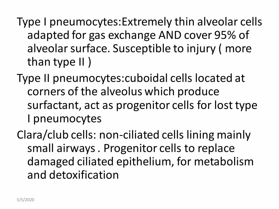
Type I pneumocytes:Extremely thin alveolar cells adapted for gas exchange AND cover 95% of alveolar surface. Susceptible to injury ( more than type II )
Type II pneumocytes:cuboidal cells located at corners of the alveolus which produce surfactant, act as progenitor cells for lost type I pneumocytes
Clara/club cells: non-ciliated cells lining mainly small airways . Progenitor cells to replace damaged ciliated epithelium, for metabolism and detoxification
5/5/2020
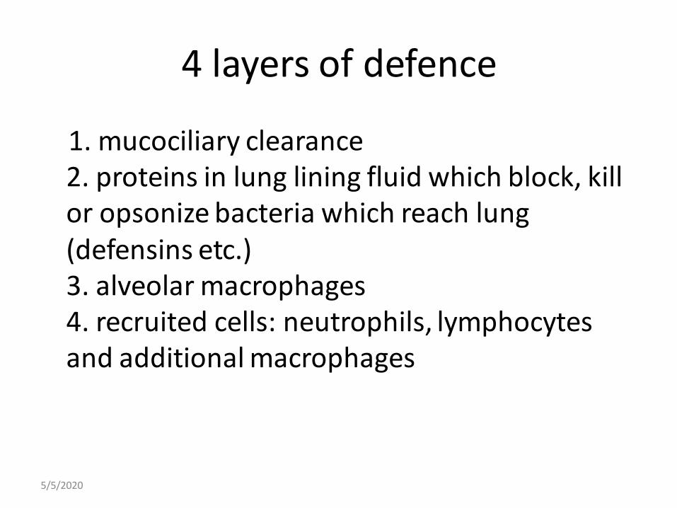
4 layers of defence
1. mucociliary clearance2. proteins in lung lining fluid which block, kill or opsonize bacteria which reach lung (defensins etc.)3. alveolar macrophages4. recruited cells: neutrophils, lymphocytes and additional macrophages
5/5/2020
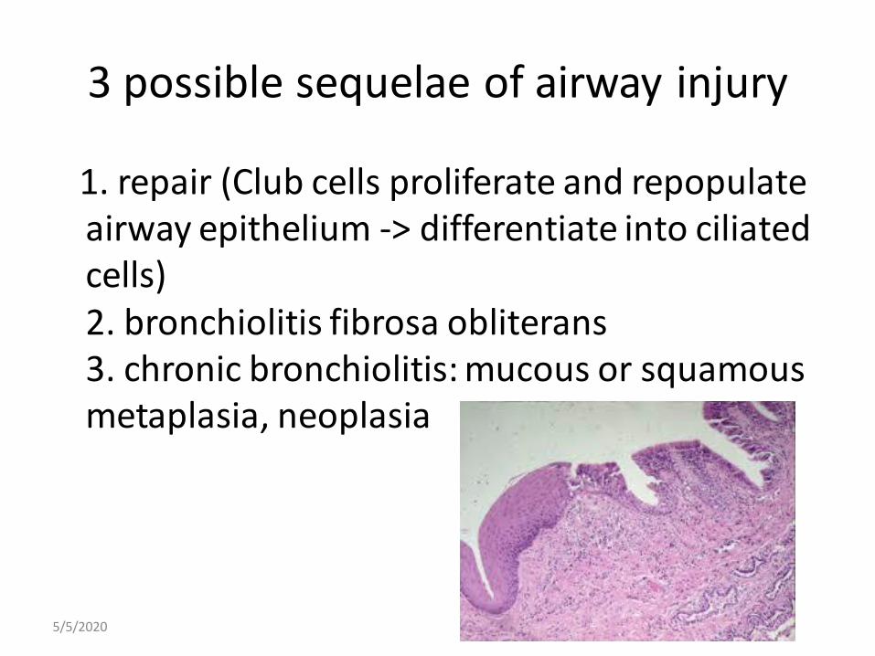
3 possible sequelae of airway injury
1. repair (Club cells proliferate and repopulate airway epithelium -> differentiate into ciliated cells)2. bronchiolitis fibrosa obliterans3. chronic bronchiolitis: mucous or squamousmetaplasia, neoplasia
5/5/2020

Specimens/Samples received in Pathology
• Sputum
• Bronchoalveolar lavage
• Pleural fluid
• FNAC lymph nodes, lung mass
• Needle/ core biopsies
• Lobectomy
• Post mortem viscera
5/5/2020

Chest X rays
5/5/2020
1PLEURAL EFFUSION/PNEUMOTHORAX
• The potential space between layers of visceral and parietal pleura, containing a capillary layer of serous pleural fluid
• Normally, 10 to 20 mL of pleural fluid, similar in composition to plasma but lower in protein (< 1.5 g/dL), facilitating movement between the lungs and chest wall
• X ray, fluid cytology, fluid biochemical analysis
5/5/2020

5/5/2020
ACUTE LUNG INJURY: ALI( non cardiogenic pulmonary edema )
• abrupt onset of severe hypoxemia without evidence of heart failure, profound dyspnea
• many cases have lesions of interstitial lung disease and bilateral pulmonary infiltrates
• ARDS is a manifestation of severe ALI
5/5/2020
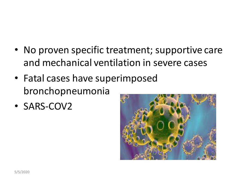
• No proven specific treatment; supportive care and mechanical ventilation in severe cases
• Fatal cases have superimposed bronchopneumonia
• SARS-COV2
5/5/2020

Pathogenesis
• Injury to pneumocytes
• Endothelial activation+surfactant abnormality
• TNF, Cytokines, Adhesion molecules etc
• Extravasation of neutrophils, macrophages
• Accumulation of intra-alveolar & interstitial fluid with hyaline membrane formation
• Diffuse alveolar damage+Necrosis
• Resolution may occur resulting in fibrosis
5/5/2020

2OBSTRUCTIVE DISEASES
1. mucous, neutrophils fill airway lumen2. inflammation within bronchiole wall -> predisposes to bronchoconstriction and reactivity3. inflammation -> edema -> thickening of airway wall -> decreased luminal diameter -> increased airway resistance
FEV1/FVC < 0.7 indicates obstruction
5/5/2020

• Emphysema
smoking+ similar features: COPD (irreversible)
• Chronic Bronchitis
• Asthma (reversible)
• Bronchiectasis
5/5/2020

EMPHYSEMA
5/5/2020

Pathogenesis
• Inflammatory mediators, leukocytes, protease-antiprotease imbalance, oxidants in smoke lead to NRF2 gene inactivation & infection
• Symptoms don’t appear until at least one-third of functioning pulmonary parenchyma is damaged
5/5/2020
CHRONIC BRONCHITIS
• Clinically persistent cough with sputum production for at least three months in at least two consecutive years in absence of any other identifiable cause
• IL13, marked increase in goblet cells, loss of cilia leading to plug formation
• Reid index is increased ( normal is 0.4)
5/5/2020
ASTHMA
• It is a chronic disorder of conducting airways, caused by immunological reaction leading to episodic bronchoconstriction and mucus production due to allergenssmokemoldsanimal dander etc
• Extrinsic/Intrinsic OR Trigger based
5/5/2020
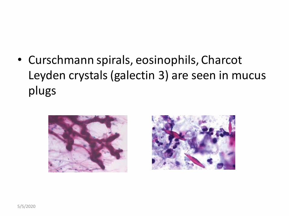
• Curschmann spirals, eosinophils, Charcot Leyden crystals (galectin 3) are seen in mucus plugs
5/5/2020
BRONCHIECTASIS
• Destruction of smooth muscle and elastic tissue by chronic necrotising infections leading to permanent dilatation of bronchi and bronchioles
• Cystic fibrosis: sodium chloride high in sweat
pancreatic insufficiency - non absorption of fats by the body causing malnutrition andchronic recurrent pulmonary infections and secretions that block airways
productive cough with purulent sputum• Primary ciliary dyskinesia (Kartagener syndrome with situs
inversus)
5/5/2020
RESTRICTIVE DISEASES
• disorders caused by pulmonary ( pleuritis, pneumoconiosis ) or extrapulmonaryrestriction ( obesity, poliomyelitis, kyphoscoliosis) that produce impairment in lung volume expansion and abnormal reduction in pulmonary ventilation
• flows are decreased, exhaled air comes out more slowly (FEV1/FVC remains normal)
5/5/2020
• Idiopathic pulmonary fibrosis
• Non specific interstitial pneumonia
• Cryptogenic organising pneumonia (BOOP)
• Autoimmune diseases
• Radiation induced
• Surfactant dysregulation (protein C, S)
• Hypersensitivity pneumonitis (farmer’s lung etc)• Sarcoidosis (non necrotising granuloma; Schaumann & Asteroid
bodies)
• Pneumoconiosis (coal, silica, asbestos)
5/5/2020
Pathogenesis
Acute: edema & hyaline membrane formation
Subacute to Chronic: proliferation of type II pneumocytes and interstitial fibrosis
- interstitial fibrosis is a permanent impediment to lung function-honeycomb lung
5/5/2020
5/5/2020

5/5/2020

3MEDIASTINAL MASSES
• In adults, thymomas and lymphomas (both Hodgkin and non-Hodgkin) are the most common anterior lesions, lymph node enlargement and vascular masses are the most common middle lesions, and neurogenictumors and esophageal abnormalities are the most common posterior lesions
• CT with IV contrast is the most valuable imaging technique.
5/5/2020
• Fine-needle aspiration techniques usually suffice for carcinomatous lesions, but a cutting-needle biopsy should be done whenever lymphoma, thymoma, or a neural mass is suspected
• If ectopic thyroid tissue is considered, thyroid-stimulating hormone is measured
5/5/2020

The following are symptoms of lung cancer
• Persistent cough/ worsening over time
• Hemoptysis (blood in cough)
• Chest pain
• Dyspnoea (shortness of breath)
• Paraneoplastic ( ADH,ACTH etc ) & systemic (Horner’s etc) syndromes
• Cervical/mediastinal lymphadenopathy
5/5/2020
Molecular aspects of lung tumors
• Many procarcinogens are activated via p450 polymorphism
• Squamous cell and small cell carcinoma frequently show p53 mutation & loss of tumor suppressor gene Rb activation (smoking)
• Adenocarcinoma showgain of function mutation in EGFR, ALK,ROS,MET pathways
5/5/2020
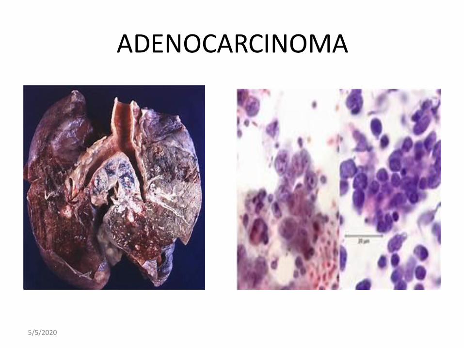
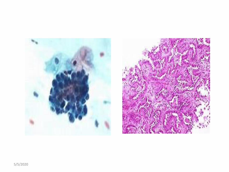
ADENOCARCINOMA
5/5/2020
5/5/2020
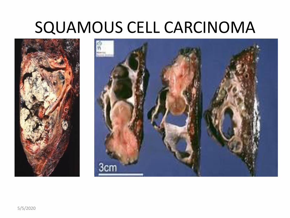
SQUAMOUS CELL CARCINOMA
5/5/2020

5/5/2020
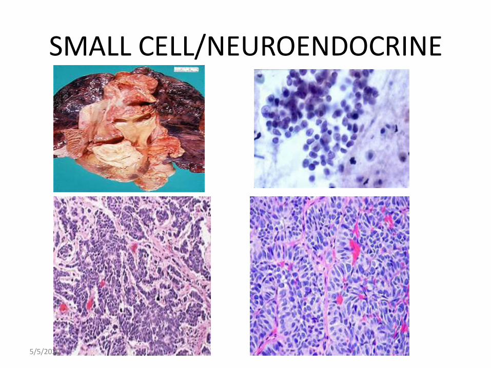
SMALL CELL/NEUROENDOCRINE
5/5/2020

MESOTHELIOMA (pleural)
5/5/2020

• 80% are associated with deletion of tumor suppressor gene CDKN2A/INK4a
• FISH is the molecular technique used to demonstrate 9p chromosome involvement
• Solitary Fibrous Tumors are associated with NAB2 & STAT 6 genes
5/5/2020

MUST KNOW
• Functional unit of lung/ structural division• Stages of pneumonia• Tuberculosis vs sarcoidosis• COPD• Asbestos and lung cancer• Paraneoplastic syndromes• Hypersensitivity• Mediators of inflammation• Molecular aspects & target therapy in lung cancer• COVID 19
5/5/2020

Thank you
5/5/2020