retromandibular transparotid approach for subcondylar
TRANSCRIPT

Retromandibular transparotid approach for subcondylar mandibular fractures: A retrospective studyGökhan Göçmen, DDS, PhD, Altan Varol, DDS, PhD, Onur Atalı, DDS, PhD,Sertaç Aktop, DDS, PhD, Selçuk Basa, DDS, PhD
Department of Oral and Maxillofacial Surgery, Marmara University Faculty of Dentistry, İstanbul-Turkey
ABSTRACT
BACKGROUND: The aim was to evaluate the effectiveness and complications of retromandibular transparotid approach performed for the reduction of dislocated subcondylar fractures.
METHODS: Fourteen patients with subcondylar mandibular fractures were evaluated (8 male, 6 female, age range 19–43 years). The primary predictor variable in the present study was time (preoperative vs postoperative). The primary outcome variables were inflammatory complication, facial nerve deficit (House and Brackmann classification), and presence of parotid fistula. The secondary outcome variables were occlusal disturbances, maximal interincisal opening (MIO), and temporomandibular joint (TMJ) pain (VAS).
RESULTS: Excellent occlusion and function was observed postoperatively. One salivary fistula occurred after surgery but was healed after 3 weeks. No inflammatory complication was observed. Three patients had grade III and one patient had grade II facial nerve deficit, all recovered in 6 weeks. All patients were free of pain and no malocclusion was observed. MIO was ranging from 34 to 58 mm (mean 44.4 mm) after 6 months.
CONCLUSION: The retromandibular transparotid approach is feasible and safe. It facilitates reduction and fixation of subcondylar fractures with functional outcomes and rare complications.
Keywords: Internal fixation; open reduction; retromandibular; subcondylar; transparotid.
region.[1,3–5] However, each approach has its own limits to ex-pose directly the region of a subcondylar fracture. The only approach that allows direct access and ease of manipulation of bony stumps in such fracture pattern is the retromandibu-lar or transparotid approach.
The aim of this study was to perform retrospective evalu-ation of subcondylar fractures treated between 2012–2015 years using transparotid approach.
MATERIALS AND METHODS
Study Design and SampleThis retrospective cohort study enrolled 14 patients (8 male, 6 female). Ethics committee approval was obtained from the appropriate institution (Approval No: 154/6122011). The study sample was derived from the population of patients who received ORIF for mandibular subcondylar fractures be-tween 2012 and 2015. The mean patient age was 26 (range 19–43) years. Inclusion criteria was severely dislocated or displaced collum fractures requiring surgical reduction. Exclu-sion criteria were Type VI or I fractures according to Spiessl and Schroll Classification,[6] and using additional surgical ac-cess other than the transparotid approach.
O R I G I N A L A R T I C L E
Ulus Travma Acil Cerrahi Derg, May 2017, Vol. 23, No. 3 251
Address for correspondence: Gökhan Göçmen, M.D.
Marmara Üniversitesi Diş Hekimliği Fakültesi, Ağız Diş ve Çene
Cerrahisi Anabilim Dalı, İstanbul, Turkey
Tel: +90 216 - 421 16 21 E-mail: [email protected]
Ulus Travma Acil Cerrahi Derg2017;23(3):251–257doi: 10.5505/tjtes.2016.43669
Copyright 2017TJTES
INTRODUCTION
Management of subcondylar fractures is a controversial topic in maxillofacial traumatology as similar functional results have been reported with non-surgical (conservative) and open re-duction and internal fixation (ORIF).[1] However, follow-up studies have confirmed better functional outcomes with prop-er occlusion, temporomandibular joint function/ mobility, and faster return to preoperative status when ORIF was used.[2]
Preauricular, submandibular, face-lift, bicoronal, retroman-dibular, and endoscopic assisted intraoral approaches can be used alone or with combination to expose the subcondylar

Göçmen et al. Retromandibular transparotid approach for subcondylar mandibular fractures
Study VariablesThe primary predictor variable in the present study was time (preoperative vs postoperative). The primary outcome vari-ables were inflammatory complication, facial nerve deficit (I- VI House-Brackmann classification),[7] presence of parotid fistula. The secondary outcome variables were occlusal dis-turbances, maximal interincisal opening (MIO), and TMJ pain (Visual Analog Scale (VAS) from no pain =0 to worst pain imaginable =10).
Patient Selection and TreatmentThe etiology was fall, traffic accidents, interpersonal violence, and gunshot injury. Twelve patients had unilateral subcondy-lar fractures, two patients had bilateral subcondylar fractures and five patients had medially dislocated fracture pattern, and 5 patients had lateral overriding dislocated subcondylar frac-tures (Table 1). Five patients had accompanying parasymphi-sial/ symphisial mandibular fractures.
The unilateral fracture pattern had pathognomic skeletal de-formity characterized by malocclusion with significant chin deviation to fractured site and non-occlusion on contralat-eral site. The bilateral subcondylar fracture presented with anterior open-bite, malocclusion and absence of mandibular protrusion. Additionally, clockwise rotation of the distal seg-ment and shortening of the posterior facial height occurred in bilateral cases.
All patients were operated using the transparotid retro-mandibular approach under general anesthesia. Arch bars or intermaxillary fixation (IMF) screws were placed. IMF was achieved using orthodontic elastics with 5/16 size during pro-
cedure and removed following rigid fixation. The parasymphi-sial fractures were reduced first using 2 straight adaptation miniplates (2.0 mm profile system, Trimed, Elektron Medikal, Turkey) or KLS Martin (2.0 mm CMF Trauma Module) cranio maxillofacial fracture (CMF) system.
Local hemostasis during dissection was achieved by subcuta-neous injection of 2 ml epinephrine diluted in 1000 cc saline. Local anesthetic was avoided to facilitate facial nerve moni-toring during transparotid dissection. A skin incision start-ing from 5 mm under the ear lobe was extended down to the retro-angular skin (Fig. 1) until the parotid capsule was reached. The capsule was incised and blunt dissection was continued with a hemostatic clamp until the posterior border of ramus was reached. Nerve stimulator was used to identify any branches of the facial nerve running in the dissection field. None of the patients required ligation of the retromandibular vein. The Senn-Miller retractors were used to pull the skin flap from both caudal and cranial flap poles and the pterygo-masseteric sling was incised sharply with no: 15 blade. The periosteum was reflected and fractures site was exposed. The ramus was retracted inferiorly to create a room for reduc-tion of dislocated fragment with the help of a bone reduction forceps. In few cases, this traction was done using a stainless steel wire introduced with a Reverdin’s needle from the sub-mandibular region. The wire was anchored to a head of 2.0 mm profile titanium screw (mostly 12 mm in length) that was placed perpendicularly to the ramus through the retroman-dibular approach and caudal distraction of the distal segment was done until reduction of the fractures was completed.
Two 2.0 mm profile adaptation miniplates were placed at the
Ulus Travma Acil Cerrahi Derg, May 2017, Vol. 23, No. 3252
Table 1. Description of the mandibular condylar neck fractures in 14 patients
Patient Gender Age Spiessl Etiology Dislocation pattern Accompanying Pre-op classification fractures MIO
F.A. Female 19 Cl2 Fall Right, lateral over-ride No 23
H.S Male 23 Cl2 Personal violence Left, lateral, over-ride No 23
A.A. Male 38 Cl4 Fall Right, anteromedial No 25
A.K. Female 19 Cl4 Fall Left, medial No 22
S.S. Male 19 Cl2 Personal violence Right, lateral over-ride Unilateral parasymphis 20
S.K. Male 23 Cl2 Traffic accident Left, lateral override No 28
E.T. Female 43 Cl2 Personal violence Left, lateral, over-ride Contralateral parasymphis 20
E.Ç Male 18 Cl4 Traffic accident Bilateral, medial Left parasymphis 25
S.K.V Female 38 Cl4 Traffic accident Right, lateral No 28
E.M.C. Male 23 Cl2 Traffic accident Left, medial No 30
E.L. Male 15 CL2 Gun shot Nondislocated Maxillary fracture 24
G.K. Female 34 Cl4 Fall Bilateral medial Symphisial 18
M.K. Female 25 cl 4 Personal violence Medial No 29
K.I. Male 27 Cl3 Fall Lateral No 21

Göçmen et al. Retromandibular transparotid approach for subcondylar mandibular fractures
posterior and anterior region of the subcondylar fracture. The plates were placed divergent caudally to resist and over-come the pull of lateral pterygoid muscle in medially dislo-cated fractures owing to unfavorable fracture pattern. The
anterior plate was placed parallel to mandibular notch and the posterior plate was placed parallel to border of the sub-condylar region (Fig. 1). In lateral over-riding fracture patterns (Fig. 2), one miniplate was placed and an additional plate was
Ulus Travma Acil Cerrahi Derg, May 2017, Vol. 23, No. 3 253
(a)
(d)
(h) (j) (k)(i)
(e) (g)
(f)
(b) (c)
Figure 1. (a) Pre-op 3D CT view. (b) Note medially dislocated bilateral subcondylar fragment and left parasymphis fracture. (c) Shortening of the ramus height can be observed in panaromic X-Ray. (d) Extraoral view of anterior open bite. (e, f) Right and left intraoral view of open bite due to posterior premature dental contact. (g, h) Post-op coronal CT showing miniplates in position. (i) Post-op 6 months panaromic X-Ray. (j, k) Mouth opening and complete inter-occlusal relationship 1 month after surgery.
(a) (b) (c) (d)
Figure 2. (a) Pre-op 3D CT view of lateral over-riding fracture of left condylar head. (b) Intra-op view of one miniplate placed parallel to border of the subcondylar region. (c) Post-op 3D CT showing miniplate in position. (d) Post-op 6 months’ skull PA X-review.

used to provide rigid ORIF when minimal mobility of condylar stump was observed (Fig. 3, 4). Condylar function and centric occlusion were double checked to ensure anatomic reduction after removing IMF before wound closure.
The wound was closed in layers after copious saline irrigation. We confirmed that watertight closure of the parotid capsule was achieved to avoid any salivary fistula. The skin was closed with 6-0 nylon sutures. Regime type I diet was recommended for postoperative period of 3 weeks. None of the patients were maintained under IMF following procedure. Antibiotic and non-steroidal anti-inflammatory drugs were prescribed for 5 days. The patients were encouraged to undergo physical therapy after a week.
Surgical field was checked for signs of infection (increased pain, swelling, redness, drainage of pus from the area, and fe-ver). Occlusal disturbance was controlled observing maxillo-mandibular relationship in centric relation of TMJ. The pain at TMJ was evaluated using VAS and MIO for 6 months after surgery.
RESULTS
There were 8 male (57.2%), 6 female (42.8%) patients with
age ranging between 19–43 years. The fractures were as-sessed as low-neck type (n=13) (92.86%) and high neck type (n=1) (7.14%) of the condyle/subcondylar unit. There was no patient drop out.
Six patients (42.85%) had fracture dislocations with the con-dylar head out of the glenoid fossa (Spiessl & Schroll Class IV) and 8 patients (57.15%) had displaced fractures (Spiessl & Schroll Class II). Isolated fractures of the mandibular condyle were seen in 9 patients; the other 5 had additional fractures of the mandible symphisial, and 1 had maxillary fracture. The mechanisms of injury were fall in 5 patients, personal violence in 4 patients, traffic accidents in 4 patients, and 1 gunshot injury (Table 1).
Salivary fistula occurred in one patient immediately after sur-gery which was closed spontaneously after applying pressure with dressing for 4 weeks. Three patients had Grade III facial nerve paralyzes according to House & Brackman classifica-tion. The facial nerve paralyses resolved completely within 3 weeks in 2 patients and the other one after 6 weeks, 1 patient had nerve weakness in Grade II that recovered in 4 weeks.
In all patients, pre-injury occlusion and mouth opening was
Ulus Travma Acil Cerrahi Derg, May 2017, Vol. 23, No. 3254
Göçmen et al. Retromandibular transparotid approach for subcondylar mandibular fractures
(a)
(d) (e)
(b) (c)
Figure 3. (a, b) Pre-op 3D CT showing right over-riding subcondylar fragment and left parasymphis fracture. (c) Intra-op view of fixation with two 2.0 miniplates, were placed at the posterior and anterior region of the subcondylar fracture. Secondary plate was used to provide minimal mobility of condylar stump. (d) 2 straight adaptation miniplates were placed for anatomic reduction of parasymphis fracture. (e) post-op 6 months panaromic X-Ray.

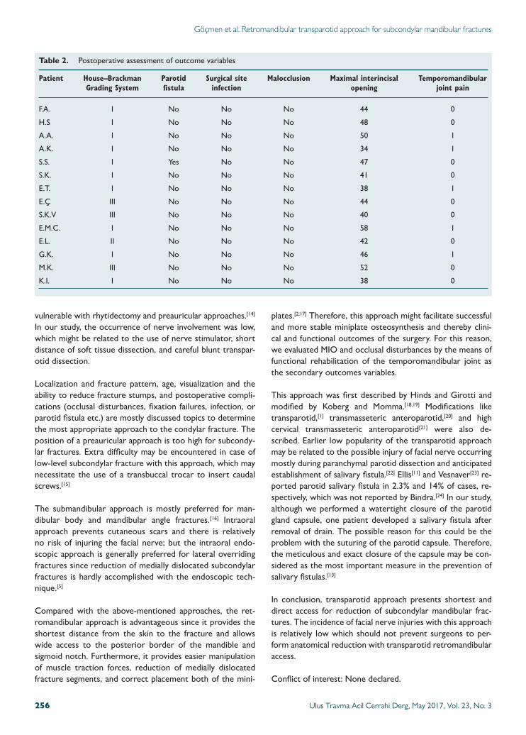
achieved. This was assessed by clinical examination of the in-terdental relationship during TMJ in centric relation as well as subjectively by the patients. MIO was 34–58 mm (mean 44.4 mm). All operated 16 joints were pain free after 6 months. There were 4 cases (29%) with facial nerve injury, affecting the buccal or marginal mandibular branches or both (Grade II or III / House & Brackmann classification). Facial nerve inju-ries in those cases resolved spontaneously and completely af-ter 3–6 weeks. No plate fractures were observed in our cases serial. Postoperative salivary fistulae developed in 1 patient (7%), and lasted for 4 weeks. No incident of greater auricular nerve anesthesia/ paresthesia, postoperative hematoma and wound infection was observed (Table 2).
DISCUSSIONThe treatment approach to the mandibular condyle frac-tures is one of the most widely debated subject in maxil-lofacial traumatology.[8] Conservative treatment may lead to malocclusion, anterior open-bite, facial asymmetry, internal derangement, chronic joint pain, and reduced mobility.[9] The miniplate osteosynthesis of the subcondylar process using
an extraoral approach is currently the most popular method followed by the miniplate osteosynthesis using a transoral approach.[10] The purpose of this study was to conduct ret-rospective evaluation of subcondylar fractures treated using retromandibular transparotid approach.
Although ORIF provides better function and anatomical re-duction, possible risk of injuring to the facial nerve is the major complication that discourages many surgeons to per-form ORIF with any transcutaneous open approach.[3] The traditional retromandibular approach may lead to facial nerve injury in 30% cases.[11,12] Preauricular and submandibular ap-proaches are other options that might be used for open re-duction, as they facilitate better exposure of the operating field from caudal and cranial side, and simplify fracture repo-sitioning compared to the cosmetically more favorable tran-soral approach or its endoscopically assisted modifications.[2,4]
However, a more forceful retraction of the soft tissues might be necessary because of the longer working distance, imply-ing a higher risk of facial nerve disturbances.[13] Additionally, temporal, and zygomatic branches of the facial nerve might be
Ulus Travma Acil Cerrahi Derg, May 2017, Vol. 23, No. 3 255
Göçmen et al. Retromandibular transparotid approach for subcondylar mandibular fractures
(a)
(c)
(b)
Figure 4. (a) Pre-op 3D CT view of lateral over-riding fracture of left condylar fragment and right parasymphis fracture. (b) Intra-op view of two miniplates placed after the ramus was distracted inferiorly with a stainless steel wire. (c) Post-op 6 months panaromic X-Ray.
vulnerable with rhytidectomy and preauricular approaches.[14] In our study, the occurrence of nerve involvement was low, which might be related to the use of nerve stimulator, short distance of soft tissue dissection, and careful blunt transpar-otid dissection.
Localization and fracture pattern, age, visualization and the ability to reduce fracture stumps, and postoperative compli-cations (occlusal disturbances, fixation failures, infection, or parotid fistula etc.) are mostly discussed topics to determine the most appropriate approach to the condylar fracture. The position of a preauricular approach is too high for subcondy-lar fractures. Extra difficulty may be encountered in case of low-level subcondylar fracture with this approach, which may necessitate the use of a transbuccal trocar to insert caudal screws.[15]
The submandibular approach is mostly preferred for man-dibular body and mandibular angle fractures.[16] Intraoral approach prevents cutaneous scars and there is relatively no risk of injuring the facial nerve; but the intraoral endo-scopic approach is generally preferred for lateral overriding fractures since reduction of medially dislocated subcondylar fractures is hardly accomplished with the endoscopic tech-nique.[5]
Compared with the above-mentioned approaches, the ret-romandibular approach is advantageous since it provides the shortest distance from the skin to the fracture and allows wide access to the posterior border of the mandible and sigmoid notch. Furthermore, it provides easier manipulation of muscle traction forces, reduction of medially dislocated fracture segments, and correct placement both of the mini-
plates.[2,17] Therefore, this approach might facilitate successful and more stable miniplate osteosynthesis and thereby clini-cal and functional outcomes of the surgery. For this reason, we evaluated MIO and occlusal disturbances by the means of functional rehabilitation of the temporomandibular joint as the secondary outcomes variables.
This approach was first described by Hinds and Girotti and modified by Koberg and Momma.[18,19] Modifications like transparotid,[1] transmasseteric anteroparotid,[20] and high cervical transmasseteric anteroparotid[21] were also de-scribed. Earlier low popularity of the transparotid approach may be related to the possible injury of facial nerve occurring mostly during paranchymal parotid dissection and anticipated establishment of salivary fistula.[22] Ellis[11] and Vesnaver[23] re-ported parotid salivary fistula in 2.3% and 14% of cases, re-spectively, which was not reported by Bindra.[24] In our study, although we performed a watertight closure of the parotid gland capsule, one patient developed a salivary fistula after removal of drain. The possible reason for this could be the problem with the suturing of the parotid capsule. Therefore, the meticulous and exact closure of the capsule may be con-sidered as the most important measure in the prevention of salivary fistulas.[13]
In conclusion, transparotid approach presents shortest and direct access for reduction of subcondylar mandibular frac-tures. The incidence of facial nerve injuries with this approach is relatively low which should not prevent surgeons to per-form anatomical reduction with transparotid retromandibular access.
Conflict of interest: None declared.
Ulus Travma Acil Cerrahi Derg, May 2017, Vol. 23, No. 3256
Göçmen et al. Retromandibular transparotid approach for subcondylar mandibular fractures
Table 2. Postoperative assessment of outcome variables
Patient House–Brackman Parotid Surgical site Malocclusion Maximal interincisal Temporomandibular Grading System fistula infection opening joint pain
F.A. I No No No 44 0
H.S I No No No 48 0
A.A. I No No No 50 1
A.K. I No No No 34 1
S.S. I Yes No No 47 0
S.K. I No No No 41 0
E.T. I No No No 38 1
E.Ç III No No No 44 0
S.K.V III No No No 40 0
E.M.C. I No No No 58 1
E.L. II No No No 42 0
G.K. I No No No 46 1
M.K. III No No No 52 0
K.I. I No No No 38 0

REFERENCES
1. Handschel J, Rüggeberg T, Depprich R, Schwarz F, Meyer U, Kübler NR, et al. Comparison of various approaches for the treatment of fractures of the mandibular condylar process. J Craniomaxillofac Surg 2012;40:397–401. [CrossRef ]
2. Ellis E 3rd, Dean J. Rigid fixation of mandibular condyle fractures. Oral Surg Oral Med Oral Pathol 1993;76:6–15. [CrossRef ]
3. Kyzas PA, Saeed A, Tabbenor O. The treatment of mandibular condyle fractures: a meta-analysis. J Craniomaxillofac Surg 2012;40:438–52.
4. Lachner J, Clanton JT, Waite PD. Open reduction and internal rigid fixa-tion of subcondylar fractures via an intraoral approach. Oral Surg Oral Med Oral Pathol 1991;71:257–61. [CrossRef ]
5. Martin M, Lee C. Endoscopic mandibular condyle fracture repair. Atlas Oral Maxillofac Surg Clin North Am 2003;11:169–78. [CrossRef ]
6. Spiessl B, Schroll K. Spezielle Frakturenund Luxationslehre. Ein kurzes Handbuch in fünf Bänden. Band I/1 Gesichtsschädel 1972:317.
7. House JW, Brackmann DE. Facial nerve grading system. Otolaryngol Head Neck Surg 1985;93:146–7. [CrossRef ]
8. Saponaro A, Stecco A, Brucoli M, Armienti F, Stellin L, Favano F, et al. Magnetic resonance imaging in the postsurgical evaluation of patients with mandibular condyle fractures treated using the transparotid approach: our experience. J Oral Maxillofac Surg 2009;67:1815–20. [CrossRef ]
9. Brandt MT, Haug RH. Open versus closed reduction of adult mandibular condyle fractures: a review of the literature regarding the evolution of cur-rent thoughts on management. J Oral Maxillofac Surg 2003;61:1324–32.
10. Klatt J, Pohlenz P, Blessmann M, Blake F, Eichhorn W, Schmelzle R, et al. Clinical follow-up examination of surgically treated fractures of the condylar process using the transparotid approach. J Oral Maxillofac Surg 2010;68:611–7. [CrossRef ]
11. Ellis E 3rd, McFadden D, Simon P, Throckmorton G. Surgical compli-cations with open treatment of mandibular condylar process fractures. J Oral Maxillofac Surg 2000;58:950–8. [CrossRef ]
12. Tang W, Gao C, Long J, Lin Y, Wang H, Liu L, et al. Application of modi-fied retromandibular approach indirectly from the anterior edge of the parotid gland in the surgical treatment of condylar fracture. J Oral Maxil-lofac Surg 2009;67:552–8. [CrossRef ]
13. Dalla Torre D, Burtscher D, Widmann G, Pichler A, Rasse M, Puelacher
W. Surgical treatment of mandibular condyle fractures using the retro-mandibular anterior transparotid approach and a triangular-positioned double miniplate osteosynthesis technique: A clinical and radiological evaluation of 124 fractures. J Craniomaxillofac Surg 2015;43:944–9.
14. Yang L, Patil PM. The retromandibular transparotid approach to man-dibular subcondylar fractures. Int J Oral Maxillofac Surg 2012;41:494–9.
15. MacArthur CJ, Donald PJ, Knowles J, Moore HC. Open reduction-fix-ation of mandibular subcondylar fractures. A review. Arch Otolaryngol Head Neck Surg 1993;119:403–6. [CrossRef ]
16. Widmark G, Bågenholm T, Kahnberg KE, Lindahl L. Open reduction of subcondylar fractures. A study of functional rehabilitation. Int J Oral Maxillofac Surg 1996;25:107–11. [CrossRef ]
17. Kanno T, Sukegawa S, Tatsumi H, Nariai Y, Ishibashi H, Furuki Y, et al. The retromandibular transparotid approach for reduction and rigid in-ternal fixation using two locking miniplates in mandibular condylar neck fractures. Int J Oral Maxillofac Surg 2014;43:177–84. [CrossRef ]
18. Hinds EC, Girotti WJ. Vertical subcondylar osteotomy: a reappraisal. Oral Surg Oral Med Oral Pathol 1967;24:164–70. [CrossRef ]
19. Koberg WR, Momma WG. Treatment of fractures of the articular pro-cess by functional stable osteosynthesis using miniaturized dynamic com-pression plates. Int J Oral Surg 1978;7:256–62. [CrossRef ]
20. Wilson AW, Ethunandan M, Brennan PA. Transmasseteric antero-pa-rotid approach for open reduction and internal fixation of condylar frac-tures. Br J Oral Maxillofac Surg 2005;43:57–60. [CrossRef ]
21. Trost O, Abu El-Naaj I, Trouilloud P, Danino A, Malka G. High cervical transmasseteric anteroparotid approach for open reduction and internal fixation of condylar fracture. J Oral Maxillofac Surg 2008;66:201–4.
22. Chossegros C, Cheynet F, Blanc JL, Bourezak Z. Short retroman-dibular approach of subcondylar fractures: clinical and radiologic long-term evaluation. Oral Surg Oral Med Oral Pathol Oral Radiol Endod 1996;82:248–52. [CrossRef ]
23. Vesnaver A, Gorjanc M, Eberlinc A, Dovsak DA, Kansky AA. The peri-auricular transparotid approach for open reduction and internal fixation of condylar fractures. J Craniomaxillofac Surg 2005;33:169–79. [CrossRef ]
24. Bindra S, Choudhary K, Sharma P, Sheorain A, Sharma CB. Manage-ment of mandibular sub condylar and condylar fractures using retroman-dibular approach and assessment of associated surgical complications. J Maxillofac Oral Surg 2010;9:355–62. [CrossRef ]
Ulus Travma Acil Cerrahi Derg, May 2017, Vol. 23, No. 3 257
Göçmen et al. Retromandibular transparotid approach for subcondylar mandibular fractures
OLGU SUNUMU
Subkondiler mandibula kırıklarına retromandibular transparotid yaklaşım:Geriye dönük bir çalışmaDr. Gökhan Göçmen, Dr. Altan Varol, Dr. Onur Atalı, Dr. Sertaç Aktop, Dr. Selçuk BasaMarmara Üniversitesi Diş Hekimliği Fakültesi, Ağız Diş ve Çene Cerrahisi Anabilim Dalı, İstanbul
AMAÇ: Bu çalışmanın amacı dislokasyon olmuş subkondiler kırıkların redüksiyonunda uygulanan retromandibular transparotid yaklaşımın etkinliği-nin ve komplikasyonlarının değerlendirilmesidir.GEREÇ VE YÖNTEM: Subkondiler mandibular kırık görülen 14 hasta değerlendirildi (8 erkek, 6 kadın, yaş aralığı: 21–56). Bu çalışmada ameliyat öncesi ve sonrasında elde edilen sonuçlar karşılaştırıldı. Birincil olarak değerlendirilen sonuçlar iltihabi komplikasyon gelişimi, fasiyal sinir hasarı (Ho-use ve Brackmann sınıflandırmasına göre) ve parotis fistül oluşumu idi. İkincil olarak değerlendirilen sonuçlar oklüzyon bozuklukları, maksimum ağız açıklığı ve temporomandibular eklemde görülen ağrıydı. (Görsel Analog Skalası’na göre).BULGULAR: Ameliyat sonrası dönemde hatasız bir oklüzyon ve fonksiyon gözlemlendi. Bir hastada parotis fistül oluştu fakat üç hafta içerisinde kapandı. Hiçbir hastada iltihabi komplikasyon gelişmedi. Üç hastada 3. seviye, bir hastada 2. seviye fasiyal sinir hasarı gelişti, fakat bütün fasiyal sinir hasarlarının altı hafta içerisinde iyileşti. Hiçbir hasta da ağrı veya oklüzyon bozukluğu olmadı. Altı ay sonraki maksimum ağız açıklığı 34–58 mm (or-talama 44.4 mm) olarak ölçüldü.TARTIŞMA: Retromandibular transparotid yaklaşım kullanışlı ve güvenli bir yaklaşımdır. Subkondiler kırıkların redüksiyonunu ve fiksasyonunu kolay-laşlaştıran bu yaklaşımda komplikasyon oranı az olup fonksiyonel sonuçlar elde edilir.Anahtar sözcükler: Açık redüksiyon; internal fiksasyon; retromandibular; subkondiler; transparotid.
Ulus Travma Acil Cerrahi Derg 2017;23(3):251–257 doi: 10.5505/tjtes.2016.43669
ORİJİNAL ÇALIŞMA - ÖZET