a comparative study of intraoral versus retromandibular
TRANSCRIPT

RESEARCH ARTICLE Open Access
A comparative study of intraoral versusretromandibular approach in themanagement of subcondylar fractureSeung Min Nam1, Yong Bae Kim1* , Sun Jae Lee1, Eun Soo Park1 and Jang Hyun Lee2
Abstract
Background: The purpose of this study was to compare the outcomes and effectiveness between intraoralapproach and retromandibular approach for treatment of subcondylar fracture of mandible.
Methods: Between March 2011 and October 2013, 24 patients with subcondylar fractures of the mandible weretreated by a single surgeon with an intraoral approach using an angulated screwdriver (n = 14) or by anothersurgeon using a retromandibular approach (n = 10). The interincisal distance was measured 1 week (T0), 6 weeks(T1), 3 months (T2), and 6 months (T3) postoperatively. We also compare the average operation time and the costof operation between the two groups.
Results: At 6 months postoperatively, all 24 patients achieved satisfactory ranges of temporomandibular jointmovement, with an interincisal distance > 40 mm without deviation and with stable centric occlusion. The intraoralgroup had the median interincisal distance of 14 mm at T0, 38 mm at T1, 42.5 mm at T2, and 43 mm at T3, whilethe retromandibular group had that of 15, 29, 35, and 42.5 mm respectively. There was no statistically significantdifference between the intraoral and the retromandibular group at T0 and T4. However, significant differences werenoted T1 and T2 (p < 0.01). The differences of average operation time between the intraoral (81 min) andretromandibular group (45 min) were statistically significant (p < 0.01). The cost of an operation was 369.96 ± 8.14(United States dollar [USD]) in intraoral group and was 345.48 ± 0.0 (USD) in retromandibular group. The differencesbetween the two groups were statistically significant (p < 0.01).
Conclusion: In open reduction of a subcondylar fracture of the mandible, a intraoral approach using an angulatedscrewdriver is superior to the retromandibular approach in terms of interincisal distance, although the operationtime is longer.
Keywords: Fracture fixation internal, Mandible, Mandibular condyle, Mandibular fractures, Oral surgical procedures
BackgroundMandibular fractures, including fractures of the subcondy-lar and condylar regions, are common facial fractures [1].Subcondylar fractures account for 20–62% of all mandibu-lar fractures [2–4] but their management remains contro-versial [5–7]. Although closed reduction is the most usefulmethod, it can be difficult to achieve anatomical reductionwith this technique compared with open reduction and in-ternal fixation (ORIF) [6–8]. The most surgeons agreed
the consensus that the proper surgical indications forORIF are the displaced bilateral or unilateral fractures ofthe mandibular condylar neck or subcondyle [9–11].Among the numerous surgical methods that can be
used in the treatment of subcondylar fracture [11–16],extraoral rather than intraoral approaches are generallypreferred because they can be provided a sufficient surgi-cal vision. However, compared with intraoral approaches,extraoral approaches commonly have a high rate of post-operative complications, such as salivary fistula formation,visible scarring, and facial nerve injury [6, 7, 16–18].We previously reported the clinical outcomes of pa-
tients with subcondylar fractures of the mandible treated
* Correspondence: [email protected] of Plastic and Reconstructive Surgery, College of Medicine,Soonchunhyang University, 170 Jomaru-ro, Bucheon 14584, Republic ofKoreaFull list of author information is available at the end of the article
© The Author(s). 2019 Open Access This article is distributed under the terms of the Creative Commons Attribution 4.0International License (http://creativecommons.org/licenses/by/4.0/), which permits unrestricted use, distribution, andreproduction in any medium, provided you give appropriate credit to the original author(s) and the source, provide a link tothe Creative Commons license, and indicate if changes were made. The Creative Commons Public Domain Dedication waiver(http://creativecommons.org/publicdomain/zero/1.0/) applies to the data made available in this article, unless otherwise stated.
Nam et al. BMC Surgery (2019) 19:28 https://doi.org/10.1186/s12893-019-0487-7

with ORIF through a modified intraoral approach withan angulated screwdriver [19]. However, the effectivenessof that technique in the present setting is unclear.Therefore, in this study we compared the clinical resultsachieved with our intraoral approach using an angulatedscrewdriver versus a retromandibular approach in pa-tients with subcondylar fractures of the mandible, whowere treated by ORIF.
MethodsBetween March 2011 and October 2013, 24 patients withsubcondylar fractures of the mandible were treated eitherby one surgeon (S.M. Nam) using an intraoral approachwith an angulated screwdriver (n = 14) or by another sur-geon (E.S. Park) using a retromandibular approach (n =10). The inclusion criteria were an age older than 15 yearsand presentation with a displaced subcondyle and occlu-sion disturbances. Patients with contralateral condylar orsubcondylar fractures or condylar neck fractures were ex-cluded from this study. The subcondylar fractures of man-dible are defined the fracture line is positioned below thelevel of the most inferior part on the sigmoid notch. [7]The study conformed to the principles dictated by theDeclaration of Helsinki, and written consent was obtainedfrom each patient for both the surgery and the publicationof photographs of the results. The study was reviewed andapproved by the Institutional Review Board for HumanSubject Research (SCHBC 2017–01–002-002) and in-formed consent was obtained from each patient.Before the miniplate osteosynthetic fixation of subcon-
dylar fracture, under local anesthesia, the madibulomaxil-lary fixation (MMF) was performed to achieve the centricocclusion using skeletal fixation with bone-anchoring skel-etal self-drilling screws (Dual-Top Anchor System; JeilMedical, Seoul, Korea) and the rubber bands in all pa-tients. After the ranged from 3 to 7 days, the rigid and in-ternal fixation of subcondylar fracture wad performed. Inpreoperative clinical examination, the dental occlusionwas evaluated with clinical examination and a review ofphotograph was taken using self-cheek retractor. The dir-ection and type of fracture fragments were evaluated pre-operatively with computed tomography.Patients were taken a liquid diet until the postoperative
7 days and educated to maintain a soft diet for the next 4weeks. The MMF was maintained until postoperative 7days and then, after the rubber bands were removed,mouth-opening physiotherapy was performed. The pa-tients can be coming back to daily life at postoperative 8–9 days. The absorbable sutures in the intraoral approachwere stitched out at postoperative 7–9 days and thenon-absorbable Nylon sutures in the retromandibular ap-proach were stitched out 5 days postoperatively. Thebone-anchoring skeletal self-drilling screws were removedat postoperative 6 weeks in outpatient clinics.
Surgical procedureThe retromandibular approachThe surgical technique was similar to that described by El-lis and Dean [16], Ebenezer et al. [20]. Briefly, a 3–3.5-cmincision was made 0.5 cm below the earlobe, not extend-ing below the angle of the mandible. A blunt metzenbaumscissors was used to achieve blunt dissection through thesubcutaneous tissue to the parotid capsule, which wasthen incised to allow dissection through the parotid gland,parallel to the branches of the facial nerve. When the lat-ter were encountered, they were first carefully dissectedand then retracted to minimize tension. The periosteumwas incised at the posterior border of the mandibularramus. After subperiosteal dissection of the ramus andsubcondylar areas, reduction and fixation of the fracturesegments were achieved. The mandibular ramus wasretracted inferiorly using manual pressure, which resultedin sufficient working space for reduction of the fracturesegments using one or two 2.0-mm miniplates. Special at-tention was paid to complete closure of the parotid cap-sule. After the occlusion was confirmed and the woundwas copiously irrigated, the wound was repaired.
The intraoral approach with an angulated screwdriversystemOur modified intraoral approach with an angulated screw-driver was described in a previous report [19]. Briefly, anintraoral incision was made in the mucosa overlying theexternal oblique ridge. The subperiosteal dissection wasperformed under the masseter muscle from the lateral tothe posterior border of the ascending mandibular ramusand the insertion of the temporalis muscle was detachedfrom the coronoid process. Through this procedure, itcould be provided a sufficient operative space and surgicalvision for the ORIF of subcondylar fracture. Using anObwegeser channel retractor, it could be visualized fromthe sigmoid notch to the mandibular angle of the entiresurface of the ramus. The fracture site and dislocated con-dylar segment were also identified.An 11-mm titanium screw was placed at the mandibu-
lar angle to hang the wire used the inferior ramus re-tractor. A stab incision was made using an 18-G needlein the 1 cm inferior to the mandible to allow insertion ofa 27-gauge wire. The wire was hung on the titaniumscrew to be used the inferior retractor of the ramus.During the manual inferior traction of ramus, the dis-placed subcondylar fracture segment was reduced usinga periosteal elevator. When the fracture segment waswell positioned within the mandibular fossa, we slowlyreleased the manual inferior traction of the ramus tomaintain reduction status (Fig. 1).After fracture reduction was confirmed using an angu-
lated mirror, MMF was applied to maintain the centric oc-clusion. The fracture fragment can be maintained through
Nam et al. BMC Surgery (2019) 19:28 Page 2 of 8

the Obwegeser channel retractor was positioned under theinferolateral border of the condyle during fixation of theplate and screw. The four-hole 2.0-mm titanium miniplatewas placed at the perpendicular position to the fracture siteand was fixed with the 6mm screw at the proximal frac-ture segment using an angulated screwdriver. Then, withthe plate pulled to eliminate the bone gap of fracture site,it was fixed with the screw onto the mandibular ramus andthen additionally fixed with screw in remained 2 holes.After the wound was irrigated and bleeding control con-ducted, we confirmed the centric occlusion and the stabil-ity of internal fixation at the fracture site.
Clinical examinationThe interincisal distance was evaluated at 1 week(T0), 6weeks(T1), 3 months(T2), and 6months(T3) after sur-gery and the occlusion was evaluated (Fig. 2). The aver-age operation times of the intraoral and retromandibulargroups were determined from the respective anesthesiarecords. The cost of an operation analysis betweenintraoral and retromandibular groups was performedwith direct payment data.Clinical and radiological evaluations were performed
during the postoperative follow-up period. Clinical out-comes of both approaches were evaluated with the oc-clusion status, range of mouth opening (and deviation),wound infection, nonunion, and plate and screw loosen-ing or exposure.
Statistical analysisStatistical analyses were performed using SPSS version20.0 (SPSS Inc., Chicago, IL, USA). The Mann-WhitneyU test was used to compare the interincisal distances,the average operation time and the operation cost ofeach approach. A p value < 0.05 was considered to indi-cate statistical significance.
ResultsFourteen patients with subcondylar fractures of themandible were treated via a intraoral approach using anangulated screwdriver. In this group, three patients had iso-lated subcondylar fractures of the mandible and 9 patientshad other mandibular fractures. Among the 10 patientswith subcondylar fractures treated via a retromandibularapproach, 7 had isolated subcondylar fractures and 3 pa-tients had other mandibular fractures (Table 1). During thefollow-up period (range, 6–10months), no major postoper-ative complication was encountered. There was no surgicalsite infection, bleeding, plate exposure or fracture, bonyabsorption or condylar necrosis, or salivary fistula. Onepatient in the retromandibular group had immediatepostoperative facial expression weakness but full recoveryoccurred within 3months postoperatively.Radiological follow-up was performed using computed
tomography, Towne’s and mandibular radiographic series,and orthopantomography, which reduction and fixation ofall fractures in both groups (Figs. 3 and 4). Towne’s and
Fig. 1 Illustration of the operative procedure for treatment of a subcondylar fracture using intraoral approach with an angulated screwdriver.Reprinted from “Transoral open reduction for subcondylar fractures of the mandible using an angulated screwdriver system” by Nam SM, Kim YB,Cha HG, Wee SY, Choi CY, Annals of Plastic Surgery, 2015, 75(3), 295–301 [19]. Copyright by Wolters Kluwer Health
Nam et al. BMC Surgery (2019) 19:28 Page 3 of 8

mandibular radiographic series were performed at 1 week,6 weeks, 3months and 6months after surgery and the com-puted tomography was performed at 3months after sur-gery. The orthopantomography was performed at 1 daypostoperatively. Clinical examination revealed ipsilateral de-viation on mouth opening in one patient in the transoral
group and one in the retromandibular group for about 1month postoperatively. These patients were treated withmouth-opening physiotherapy at an outpatient clinic andgradually improved. At 6months postoperatively, all pa-tients had satisfactory ranges of TMJ movement, interinci-sal distances of > 40mm without deviation, and stablecentric occlusion. There were no clinically problematicsymptoms related to TMJ movement in all patients.
Fig. 2 The interincisal distances were evaluated after surgery. In thispatient, the interincisal distance was 38 mm 6weeks after surgery (a)and the patient had neutron-occlusion postoperatively (b)
Table 1 Demographic characteristics of patients
Variable Intraoralapproach
Retromandibularapproach
p-valuea
Sample size (n = 24) 14 (58.3) 10 (41.7)
Sex (male) 13 (92.9) 7 (70.0) 0.139
Associated mandibular fracture
None 3 (21.4) 7 (70.0)
Symphysis 6 (42.9) 1 (10.0)
Parasymphysis 5 (35.7) 2 (20.0) 0.062
Injury side (right) 5 (35.7) 5 (50.0) 0.484
Age (years) 36.71 ± 4.16 29.30 ± 3.34 0.151
Data are presented as number (%) or mean ± standard deviationap-values were computed by Chi-square test or Fisher’s exact test forcategorical variables and Mann-Whitney U test for continuous variables
Fig. 3 A 40 year-old male with a left subcondylar fracture of themandible was treated using the retromandibular approach. ModifiedTowne’s radiographic images show the medially deviatedsubcondylar fracture (a) and the postoperative reduction state of thefracture following rigid internal fixation (b)
Nam et al. BMC Surgery (2019) 19:28 Page 4 of 8
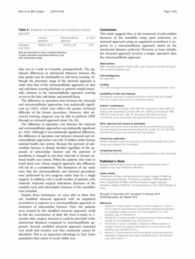
At 1 week postoperatively, the median interincisal dis-tances were 14mm (13–15 mm) in the intraoral groupand 15 mm (15–16mm) in the retromandibular group.At postoperative 6 weeks, the median interincisal dis-tances were 38 mm (37–39 mm) and 29mm (29–30mm), respectively. After 3 months, the median interinci-sal distances were 42.5 mm (41–44mm) in the intraoralgroup and 35mm (34–35mm) in the retromandibulargroup. After 6 months, the corresponding values were43mm (42–45 mm) and 42.5 mm (41–44mm). The dif-ferences between the two groups were statistically sig-nificant at 6 weeks and 3months (p < 0.01) but not at 1week or 6 months postoperatively (Fig. 5 and Table 2).Based on the anesthesia records, the average operation
time in the retromandibular group was significantlyshorter than in the intraoral group (p < 0.01). The aver-age operation time in retromandibular group was 45 minand in intraoral group was 81min (Fig. 6 and Table 3).The cost of an operation in intraoral group was 369.96
± 8.14 (USD) and in retromandibular group was 345.48 ±0.0 (USD). The differences between the two groups werestatistically significant (p < 0.01) (Fig. 7 and Table 4).
DiscussionNondisplaced subcondylar fractures of the mandible areusually treated with a closed procedure and MMF. Insuch cases, short-term MMF stimulates the callus for-mation, which prevents mobility of fracture segments.After the MMF is removed, mouth-opening physiother-apy is initiated [21, 22]. The advantages of closed
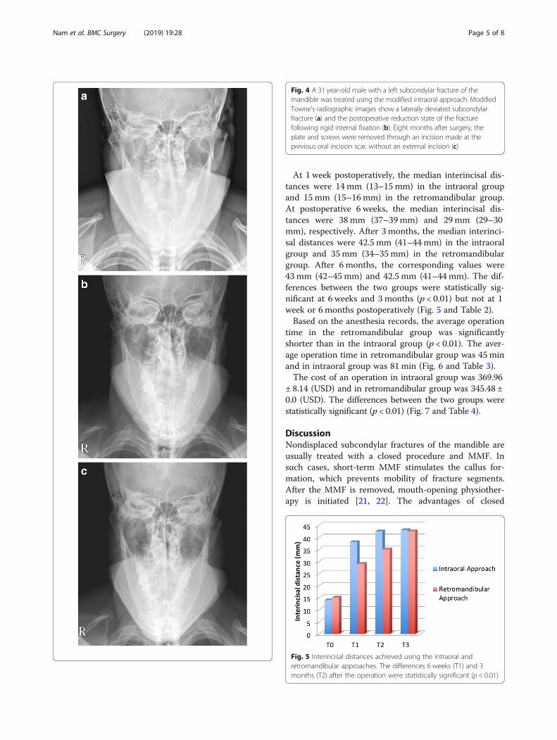
Fig. 4 A 31 year-old male with a left subcondylar fracture of themandible was treated using the modified intraoral approach. ModifiedTowne’s radiographic images show a laterally deviated subcondylarfracture (a) and the postoperative reduction state of the fracturefollowing rigid internal fixation (b). Eight months after surgery, theplate and screws were removed through an incision made at theprevious oral incision scar, without an external incision (c)
Fig. 5 Interincisal distances achieved using the intraoral andretromandibular approaches. The differences 6 weeks (T1) and 3months (T2) after the operation were statistically significant (p < 0.01)
Nam et al. BMC Surgery (2019) 19:28 Page 5 of 8

treatment were the uneventful healing and a minimalrisk of long-term disability or pain [21–24]. However, itcan lead to the open-bite deformity or malocclusion indisplaced subcondylar fractures due to the shortening ofthe ascending ramus [25–28]. In such cases, open reduc-tion is therefore recommended [10, 16, 28–31]. None-theless, the indications for surgical treatment ofdisplaced fractures are still debatable due to the postop-erative scar and the possibility of facial nerve injury dur-ing external approaches [6, 16, 32–34]. However,surgical treatment of lower neck and subcondylar frac-tures may be better than closed treatment with respectto postoperative mouth opening [35].In this study, only patients with subcondylar fractures
of the mandible were included. Patients with fracturesabove the condylar neck were excluded because, in thesefractures, a intraoral approach does not provide suffi-cient space to allow fixation of the plate. Moreover, sur-gical treatment of fractures above the condylar neck canresult in limited TMJ movement and prevent the correctevaluation of interincisal distances.The retromandibular approach has several advantages.
First, it provides an excellent operative field and directvisual alignment of the fracture fragments [36, 37], withexposure of the entire fracture from the posterior borderto the condylar process. Second, it allows for a reductionin the distance from the skin incision to the fracture site
and for fixation of the plate without the need for a trans-cutaneous trocar [38]. Third, it avoids damage to the fa-cial nerve [39]. Although facial expression weaknesssometimes occurs in patients treated via a retromandib-ular approach, this may be caused by facial muscle weak-ness rather than nerve damage [36]. Immediateweakness of the facial nerve has been described in someretromandibular treated patients, but full recovery oc-curred within 3 months [34, 40]. In other studies, nosimilarly treated patient had facial nerve weakness [39–41]. Salivary fistula is a potential complication of the ret-romandibular approach [34, 42, 43]. In our study, water-tight closure of the parotid capsule prevented theformation of salivary fistulas.The surgical indications for an intraoral approach are
considered to be limited to moderately dislocated subcon-dylar or mandibular ramus fractures, because, otherwise,there is insufficient surgical space to allow the use of anangulated screwdriver [35]. However, the inferior tractionof the ramus using can be provided the sufficient surgicalto allow performance of a modified intraoral approach withan angulated screwdriver, as shown in this and our previousstudy [19]. In addition to the adequate surgical space, animportant advantage of our modified intraoral approachusing an angulated screwdriver is that it eliminates the riskof external scarring, including scarring inflicted during re-moval of the palate and screw, and avoids both facial nervedamage and salivary fistula formation.A comparison of interincisal distances achieved using the
intraoral and retromandibular approaches showed signifi-cant differences only at 6 weeks and 3months (p < 0.01),
Table 2 Comparison of interincisal distance according tosurgical approach
Interincisal distance (mm) p-valuea
Intraoral approach Retromandibular approach
1 week (T0) 14 (13–15) 15 (15–16) 0.07
6 weeks (T1) 38 (37–39) 29 (29–30) < 0.01
3 months (T2) 42.5 (41–44) 35 (34–35) < 0.01
6 months (T3) 43 (42–45) 42.5 (41–44) 0.403ap-value calculated using Mann-Whitney U test
Fig. 6 Average operation times of the intraoral and retromandibularapproaches. The difference between the two procedures wassignificant (p < 0.01)
Table 3 Comparison of operation time according to surgicalapproach
Intraoral approach Retromandibular approach p-valuea
Operationtime (min)
81 (65–100) 45 (40–45) < 0.01
ap-value calculated using Mann-Whitney U test
Fig. 7 Comparison of operation cost between the intraoral andretromandibular approaches. The difference between the twoprocedures was significant (p < 0.01). USD United States dollar
Nam et al. BMC Surgery (2019) 19:28 Page 6 of 8
thus not at 1 week or 6months, postoperatively. The sig-nificant differences in interincisal distances between thetime points may be attributable to soft-tissue scarring. Al-though the dissection range of the intraoral approach iswider than that of the retromandibular approach, no skinand soft-tissue scarring develops in patients treated intrao-rally, whereas, in the retromandibular approach, scarringoccurs in the skin, soft tissue, and parotid fascia.The difference in operation time between the intraoral
and retromandibular approaches was statistically signifi-cant (p < 0.01), which may reflect the greater technicaldifficulty of the former procedure. However, after ad-vanced training, surgeons may be able to perform ORIFthrough an intraoral approach alone [16, 44].The difference in operation cost between the intraoral
and retromandibular approaches was statistically significant(p < 0.01). Although it was statistically significant difference,the difference of operation cost between intraoral and ret-romandibular approaches was only 25 dollars under Koreannational health care system. Because the payment of sub-condylar fracture is already decided regardless of the ap-proach of subcondylar fracture and the payment ofanesthesia is charged in one-hour intervals in Korean na-tional health care system. When the patients who want toavoid facial scar choose surgical approach, this differencewill not be a consideration. The limitations of our studywere that the retromandibular and intraoral procedureswere performed by two surgeons rather than by a singlesurgeon. In addition, only a small number of patients, withrelatively restricted surgical indications (fractures of thecondylar neck and subcondylar fractures of the mandible)were included.Despite these limitations, we were able to show that
our modified intraoral approach with an angulatedscrewdriver is superior to a retromandibular approach intreatment of subcondylar fracture. First, the patientswere treated by the modified intraoral approach couldbe felt the convenience of daily life from 6 weeks to 3months after surgery, because it could be provided widerinterincisal distances compared to retromandibular ap-proach. Second, modified intraoral approach remainedvery small stab incision scar that commonly cannot beidentified. This is an important advantage in East Asianpopulation that wants to avoid visible scar.
ConclusionThis study suggests that, in the treatment of subcondylarfractures of the mandible using open reduction, anintraoral approach using an angulated screwdriver is su-perior to a retromandibular approach, based on theinterincisal distance achieved. However, at least initially,the intraoral approach involves a longer operation thanthe retromandibular approach.
AbbreviationsMMF: mandibulomaxillary fixation; ORIF: open reduction and internal fixation;TMJ: temporomandibular joint
AcknowledgmentsNot applicable.
FundingThis work was supported by the Soonchunhyang University Research Fund.
Availability of data and materialsThe datasets used and/or analysed during the current study are availablefrom the corresponding author on reasonable request.
Authors’ contributionsStudy conception and design: SMN, YBK, ESP. Acquisition of data: SMN, SJL.Analysis and interpretation of data: SMN, JHL. Drafting of manuscript: SMN,YBK, ESP. Critical revision: YBK, JHL. All authors discussed the results,commented on the manuscript, and approved the final manuscript.
Ethics approval and consent to participateThe study with human tissue was approved by Soonchunhyang UniversityBucheon Hospital Institutional Review Board (No. SCHBC 2017–01–002-002)and informed consent was obtained from each patient.
Consent for publicationWritten informed consent for publication of their clinical details and clinicalimages was obtained from the patient.
Competing interestsThe authors declare that there are no competing interests in this manuscript.
Publisher’s NoteSpringer Nature remains neutral with regard to jurisdictional claims inpublished maps and institutional affiliations.
Author details1Department of Plastic and Reconstructive Surgery, College of Medicine,Soonchunhyang University, 170 Jomaru-ro, Bucheon 14584, Republic ofKorea. 2Department of Plastic and Reconstructive Surgery, HanyangUniversity, College of Medicine, 153, Gyeongchun-ro, Guri 11923, Republic ofKorea.
Received: 27 November 2017 Accepted: 14 February 2019
References1. Salgarelli AC, Anesi A, Bellini P, Pollastri G, Tanza D, Barberini S, Chiarini L.
How to improve retromandibular transmasseteric anteroparotid approachfor mandibular condylar fractures: our clinical experience. Int J OralMaxillofac Surg. 2013;42(4):464–9.
2. Mitchell DA. A multicentre audit of unilateral fractures of the mandibularcondyle. Br J Oral Maxillofac Surg. 1997;35(4):230–6.
3. Silvennoinen U, Iizuka T, Lindqvist C, Oikarinen K. Different patterns ofcondylar fractures: an analysis of 382 patients in a 3-year period. J OralMaxillofac Surg. 1992;50(10):1032–7.
4. Boutros SG. Closed reduction and fluoroscopically assisted percutaneousstabilization of displaced subcondylar mandible fractures. Plast ReconstrSurg. 2005;116(4):971–7.
Table 4 Comparison of operation cost according to surgicalapproach
Intraoralapproach
Retromandibularapproach
p-valuea
Operationcost (USD)
369.96 ± 8.14 345.48 ± 0.0 < 0.01
Data are presented as mean ± standard deviationap-value calculated using Mann-Whitney U testUSD United States dollar
Nam et al. BMC Surgery (2019) 19:28 Page 7 of 8
5. Ellis E, 3rd, Moos KF, el-Attar A: Ten years of mandibular fractures: ananalysis of 2,137 cases. Oral Surg Oral Med Oral Pathol 1985, 59(2):120–129.
6. Schmelzeisen R, Cienfuegos-Monroy R, Schon R, Chen CT, Cunningham L, Jr.,Goldhahn S: Patient benefit from endoscopically assisted fixation of condylar neckfractures--a randomized controlled trial. J Oral Maxillofac Surg 2009, 67(1):147–158.
7. Ellis E, 3rd: Method to determine when open treatment of condylar processfractures is not necessary. J Oral Maxillofac Surg 2009, 67(8):1685–1690.
8. Takenoshita Y, Ishibashi H, Oka M. Comparison of functional recovery afternonsurgical and surgical treatment of condylar fractures. J Oral MaxillofacSurg. 1990;48(11):1191–5.
9. Zide MF. Open reduction of mandibular condyle fractures. Indications andtechnique Clin Plast Surg. 1989;16(1):69–76.
10. Zide MF, Kent JN. Indications for open reduction of mandibular condylefractures. J Oral Maxillofac Surg. 1983;41(2):89–98.
11. Meyer C, Zink S, Chatelain B, Wilk A. Clinical experience with osteosynthesisof subcondylar fractures of the mandible using TCP plates. JCraniomaxillofac Surg. 2008;36(5):260–8.
12. Baek RM, Min KH, Heo CY, Eun SC. The perilobule approach to subcondylarfractures. Ann Plast Surg. 2011;66(3):253–6.
13. Kheradpir AR, Chien AT, Julian RS, 3rd: Transoral osteosynthesis ofsubcondylar fractures of the mandible using a fenestrated Levassier-Merrillretractor. J Oral Maxillofac Surg 2011, 69(7):2006–2011.
14. Nicolai G, Lore B, De Marinis L, Calabrese L. Combined surgical approachretromandibular and intraoral to subcondylar mandibular fractures. JCraniofac Surg. 2011;22(4):1354–7.
15. Trost O, Trouilloud P, Malka G. Open reduction and internal fixation of lowsubcondylar fractures of mandible through high cervical transmassetericanteroparotid approach. J Oral Maxillofac Surg. 2009;67(11):2446–51.
16. Ellis E, 3rd, Dean J: Rigid fixation of mandibular condyle fractures. Oral SurgOral Med Oral Pathol 1993, 76(1):6–15.
17. Bhavsar D, Barkdull G, Berger J, Tenenhaus M. A novel surgical approach tosubcondylar fractures of mandible. J Craniofac Surg. 2008;19(2):496–9.
18. Schon R, Gutwald R, Schramm A, Gellrich NC, Schmelzeisen R. Endoscopy-assisted open treatment of condylar fractures of the mandible: extraoral vsintraoral approach. Int J Oral Maxillofac Surg. 2002;31(3):237–43.
19. Nam SM, Kim YB, Cha HG, Wee SY, Choi CY. Transoral open reduction forsubcondylar fractures of the mandible using an angulated screwdriversystem. Ann Plast Surg. 2015;75(3):295–301.
20. Ebenezer V, Ramalingam B. Comparison of approaches for the rigid fixationof sub-condylar fractures. J maxillofacial and oral surgery. 2011;10(1):38–44.
21. Haug RH, Assael LA. Outcomes of open versus closed treatment ofmandibular subcondylar fractures. J Oral Maxillofac Surg. 2001;59(4):370–5discussion 375-376.
22. Smets LM, Van Damme PA, Stoelinga PJ. Non-surgical treatment of condylarfractures in adults: a retrospective analysis. J Craniomaxillofac Surg. 2003;31(3):162–7.
23. Villarreal PM, Monje F, Junquera LM, Mateo J, Morillo AJ, Gonzalez C.Mandibular condyle fractures: determinants of treatment and outcome. JOral Maxillofac Surg. 2004;62(2):155–63.
24. Yang WG, Chen CT, Tsay PK, Chen YR. Functional results of unilateralmandibular condylar process fractures after open and closed treatment. JTrauma. 2002;52(3):498–503.
25. Schon R, Gellrich NC, Schmelzeisen R. Frontiers in maxillofacial endoscopicsurgery. Atlas Oral Maxillofac Surg Clin North Am. 2003;11(2):209–38.
26. Schon R, Schmelzeisen R. Endoscopic fracture treatment. Ann R AustralasColl Dent Surg. 2002;16:40–5.
27. Dahlstrom L, Kahnberg KE, Lindahl L. 15 years follow-up on condylarfractures. Int J Oral Maxillofac Surg. 1989;18(1):18–23.
28. Ellis E, 3rd, Simon P, Throckmorton GS: Occlusal results after open or closedtreatment of fractures of the mandibular condylar process. J Oral MaxillofacSurg 2000, 58(3):260–268.
29. Hall MB. Condylar fractures: surgical management. J Oral Maxillofac Surg.1994;52(11):1189–92.
30. Umstadt HE, Ellers M, Muller HH, Austermann KH. Functional reconstructionof the TM joint in cases of severely displaced fractures and fracturedislocation. J Craniomaxillofac Surg. 2000;28(2):97–105.
31. Widmark G, Bagenholm T, Kahnberg KE, Lindahl L. Open reduction ofsubcondylar fractures. A study of functional rehabilitation Int J OralMaxillofac Surg. 1996;25(2):107–11.
32. Kanno T, Sukegawa S, Fujioka M, Takabatake K, Furuki Y. Transoral openreduction with rigid internal fixation for subcondylar fractures of the
mandible using a small angulated screwdriver system: is endoscopicassistance necessary? J Oral Maxillofac Surg. 2011;69(11):e372–84.
33. Kitagawa Y, Sano K, Nakamura M, Miyauchi K. Transoral osteosynthesis at themandibular ramus and subcondyle using angular screwing instrument andbiodegradable miniplate system. J Oral Maxillofac Surg. 2004;62(8):1041–3.
34. Ellis E, 3rd, McFadden D, Simon P, Throckmorton G: Surgical complicationswith open treatment of mandibular condylar process fractures. J OralMaxillofac Surg 2000, 58(9):950–958.
35. Nogami S, Takahashi T, Yamauchi K, Miyamoto I, Kaneuji T, Yamamoto N,Yoshiga D, Yamashita Y. Clinical comparison between the retromandibularapproach for reduction and fixation and endoscope-assisted openreduction and internal fixation for mandibular condyle fractures. J CraniofacSurg. 2012;23(6):1815–8.
36. Bindra S, Choudhary K, Sharma P, Sheorain A, Sharma CB. Management ofmandibular sub condylar and condylar fractures using retromandibularapproach and assessment of associated surgical complications. Jmaxillofacial and oral surgery. 2010;9(4):355–62.
37. Chossegros C, Cheynet F, Blanc JL, Bourezak Z. Short retromandibularapproach of subcondylar fractures. clinical and radiologic long-term evaluationOral Surg Oral Med Oral Pathol Oral Radiol Endod. 1996;82(3):248–52.
38. Ellis E 3rd, Throckmorton GS, Palmieri C. Open treatment of condylar processfractures: assessment of adequacy of repositioning and maintenance ofstability. J Oral Maxillofac Surg. 2000;58(1):27–34 discussion 35.
39. Hyde N, Manisali M, Aghabeigi B, Sneddon K, Newman L. The role of openreduction and internal fixation in unilateral fractures of the mandibularcondyle: a prospective study. Br J Oral Maxillofac Surg. 2002;40(1):19–22.
40. Devlin MF, Hislop WS, Carton AT. Open reduction and internal fixation offractured mandibular condyles by a retromandibular approach: surgicalmorbidity and informed consent. Br J Oral Maxillofac Surg. 2002;40(1):23–5.
41. Klatt J, Pohlenz P, Blessmann M, Blake F, Eichhorn W, Schmelzle R, HeilandM. Clinical follow-up examination of surgically treated fractures of thecondylar process using the transparotid approach. J Oral Maxillofac Surg.2010;68(3):611–7.
42. Vesnaver A, Gorjanc M, Eberlinc A, Dovsak DA, Kansky AA. The periauriculartransparotid approach for open reduction and internal fixation of condylarfractures. J Craniomaxillofac Surg. 2005;33(3):169–79.
43. Vogt A, Roser M, Weingart D. Transparotidean approach to surgicalmanagement of condylar neck fractures. A prospective study. Mund KieferGesichtschir. 2005;9(4):246–50.
44. Undt G, Kermer C, Rasse M, Sinko K, Ewers R. Transoral miniplateosteosynthesis of condylar neck fractures. Oral Surg Oral Med Oral PatholOral Radiol Endod. 1999;88(5):534–43.
Nam et al. BMC Surgery (2019) 19:28 Page 8 of 8