review article blackwell publishing, ltd. the discovery of
TRANSCRIPT

© 2005 The Royal Microscopical Society
Journal of Microscopy, Vol. 217, Pt 1 January 2005, pp. 3–15
Blackwell Publishing, Ltd.
R E V I E W A RT I C L E
The discovery of aequorin and green fluorescent protein
O. S H I M O M U R A
The Photoprotein Laboratory, 324 Sippewissett Road, Falmouth, MA 02540, U.S.A.
We discovered aequorin and green fluorescent protein (GFP)in 1961 from the same species of jellyfish (Shimomura
et al
.,1962). Our target was a luminescent substance, aequorin,and GFP was isolated as a by-product of aequorin owing to itsbright conspicuous fluorescence. Both are unusual proteinsbut they had no particular importance when we first reportedthem. Their importance became apparent in the course ofstudies, and now, 40 years after their discovery, they are wellknown and widely used, aequorin as a calcium probe and GFPas a marker protein.
In the characterization of these proteins, information obtainedfrom the bioluminescence of the ostracod
Cypridina
playedan extremely important role. Without the information gainedon
Cypridina
luminescence, the characterization of aequorinwould not have been possible. Because I had studied the biolu-minescence of
Cypridina
before I worked on aequorin, I wouldlike to begin my story with my encounter with
Cypridina
.It was the spring of 1955. I was working as a teaching
assistant at the Pharmacy School of Nagasaki University, andit was my fourth year in the role. My supervisor, ProfessorShungo Yasunaga, was interested in my education and wantedme to broaden my knowledge. He kindly gave me a leave ofabsence for one year, and arranged for me to work at thelaboratory of Professor Yoshimasa Hirata, at Nagoya Univer-sity, as a visiting researcher. Professor Hirata was an expert inthe chemistry of natural products. At my first meeting withProfessor Hirata, he showed me dried
Cypridina
stored in alarge vacuum desiccator and asked me to purify the luciferinand crystallize it.
Cypridina
is a tiny egg-shaped crustacean ostracod only 2–3 mm in length (illustrated here in Fig. 1), and is abundant inshallow seas around Japan and south-east Asia. At night, theostracod swims along and, upon encountering a predator,ejects luciferin and luciferase into the water, producing acloud of blue luminescence. The animal then swims away intothe darkness of the surrounding water. The luminescence isemitted by an enzyme reaction, commonly known as theluciferin–luciferase reaction, in which the reaction of luciferinwith oxygen to produce light and oxyluciferin is catalysed bythe enzyme luciferase. When
Cypridina
is dried, the specimen
keeps its luminescence property almost permanently, and itwill emit light again by simply wetting it with water. Becauseof this property, the Japanese military collected a large quantityof this organism during the World War II for intended use as alow-intensity light source.
Professor Hirata’s objective was the structural determinationof
Cypridina
luciferin. For that purpose, pure luciferin was essential,and crystallization was the only practical means to confirmpurity at that time. The luciferin of
Cypridina
had been studiedat Newton Harvey’s laboratory at Princeton University for almost30 years at the time, but no information on the chemicalnature of the luciferin had been gained. Professor Hirata wasfully aware that
Cypridina
luciferin is an extremely unstablesubstance, and it is rapidly oxidized in air. He mentioned to methat he could not give this project to any of his students becauseit was too risky as a degree subject owing to the extraordinaryinstability of luciferin, and therefore the difficulty of the projectand its slim chances of success.
I made several small-scale tests of luciferin extraction, todetermine how unstable the compound was and also to esti-mate the amount of luciferin in dried
Cypridina
. The luciferincontent was indeed very low, probably about 10 p.p.m., andin solution it was highly sensitive to air, being oxidized anddestroyed even with a trace of contaminating oxygen. Therefore,extraction had to be carried out in an atmosphere in whichoxygen was completely absent. Nitrogen gas and argon containtrace amounts of oxygen that are difficult to remove, andtherefore they are not suitable. Despite its risk of explosion, theonly method was to use hydrogen gas that had been purified bypassing it though a red-heated copper catalyst to remove oxygen.I planned to extract 500 g of dried
Cypridina
with methanol inthe complete absence of air, at a temperature lower than40
°
C, to obtain 2 or 3 mg of purified luciferin for crystalli-zation. The 500 g of
Cypridina
contained about half a millionorganisms, and it was 10 times the amount used at PrincetonUniversity. Professor Hirata agreed to my plan, and his glassblower constructed an over-sized soxhlet apparatus for me.
I extracted luciferin in hydrogen under reduced pressure usingthis apparatus (Fig. 2), purified the extract in an atmosphereof nitrogen and then tried to crystallize the purified luciferin. Itried all combinations of solvents and salts I could think of,but all my efforts ended with the creation of amorphous
Correspondence to: O. Shimomura. E-mail: [email protected]

4
O. S H I M O M U R A
© 2005 The Royal Microscopical Society,
Journal of Microscopy
,
217
, 3–15
Fig. 1. The ostracod Cypridina. Photo by Toshio Goto.
Fig. 2. Our apparatus for extracting luciferin inhydrogen under reduced pressure.

A E Q U O R I N A N D G R E E N F L U O R E S C E N T P RO T E I N
5
© 2005 The Royal Microscopical Society,
Journal of Microscopy
,
217
, 3–15
precipitates, and any leftover luciferin became useless byoxidation by the next day. I therefore had to extract and purifyfresh batches of luciferin for my further efforts of crystalliza-tion. The whole experimental process involved day-and-nightwork for 7 days. I wasted six or seven batches of purified luci-ferin in unsuccessful efforts until finally I saw the first crystalsof
Cypridina
luciferin on one cold morning of February 1956;however, this occurred in an unexpected way.
On the previous night, I had some leftover purified luciferinafter my crystallization attempts. Because I could not think ofany further ideas to improve the crystallization, I decided touse the material for amino acid analysis. Thus, I added anequal amount of concentrated hydrochloric acid to the luci-ferin solution. The colour of the solution instantly changed
from yellow to dark red. Because it was late at night, I wenthome without heating the sample. The next morning, I sawthat the solution had discoloured and become a light orangecolour, with a small amount of dark precipitate at the bottomof the test tube. Under the microscope, the precipitate appearedas fine red needle-like crystals, as illustrated in Fig. 3. Theresult indicated that the luciferin – unusually – could becrystallized in a high concentration of hydrochloric acid. Iachieved this unexpected result 10 months after I had begunthe crystallization experiments.
This successful crystallization (Shimomura
et al
., 1957) madeit possible to investigate the chemical structure of
Cypridina
luciferin and the mechanism of its luminescence reaction. Thecomplete structures of
Cypridina
luciferin and its oxidation
Fig. 3. Crystallized luciferin.

6
O. S H I M O M U R A
© 2005 The Royal Microscopical Society,
Journal of Microscopy
,
217
, 3–15
process were elucidated 10 years later (Kishi
et al
., 1966;McCapra & Chang, 1967; Stone, 1968).
The luminescence reaction of
Cypridina
luciferin is illustratedin Fig. 4. Luciferin (Fig. 4, upper left) contains an imidazo-pyrazinone skeleton. In the presence of luciferase and oxygen,it forms an intermediate peroxide at position 2, followed by itscyclization into a dioxetanone ring. The dioxetanone instantlydecomposes into oxyluciferin and CO
2
accompanied by theemission of light. Oxyluciferin contains a 2-aminopyrazineskeleton in its acylated form, and this compound is slowlyhydrolysed into etioluciferin containing a 2-aminopyrazineskeleton. It was these structures that made it possible for us todetermine the structure of the aequorin chromophore in theearly 1970s.
Sadly, in 1959 my boss Dr Yasunaga died of cancer. In thesame year, I received an invitation from Dr Frank Johnson ofPrinceton University to join his laboratory as a research asso-ciate. He offered me travel expenses, but I chose to apply for aFulbright Travel Grant, because in that way I would be betterprepared to go to America. Fulbright grants at that time werevery well managed by the U.S. State Department and weremeticulously planned. I was given a 4-week course in basicEnglish conversation and a 3-day orientation prior to my
departure. In August 1960, I was among the Fulbrightrecipients onboard
Hikawa-maru
leaving Yokohama forSeattle. At the departure, a thousand well-wishers jammed thepier to see us off with coloured tapes. After 13 days of comfortablevoyage to Seattle and then 3 nights of travel by rail across thecontinent, I arrived at Princeton with great excitement.
Shortly after my arrival, Dr Johnson asked if I would beinterested in studying the bioluminescence of the jellyfish
Aequorea
. He showed me a small vial containing white powder,and said that it was a freeze-dried extract of the jellyfish thatwould emit light if moistened with water. We went into adarkroom and tested it. The powder, however, did not emitany light. Despite this unsuccessful demonstration, I was quiteimpressed by Dr Johnson’s description of the brilliant lumines-cence of the live jellyfish and the great abundance of specimensat Friday Harbor in the State of Washington. I agreed to studythe jellyfish.
In the early summer of 1961, we travelled from Princeton toFriday Harbor. Dr Johnson purchased a new station wagonfor the trip and Dr Johnson’s assistant Yo Saiga and my wifeaccompanied us. The car was fully loaded with equipment,chemicals and luggage for four. At the time, the interstatehighway was not yet complete and we drove though Chicago
Fig. 4. The luminescence reaction of Cypridina luciferin.
A E Q U O R I N A N D G R E E N F L U O R E S C E N T P RO T E I N
7
© 2005 The Royal Microscopical Society,
Journal of Microscopy
,
217
, 3–15
and Minneapolis and then took U.S. route 2 all the way to thewest coast. Dr Johnson was the only driver and he drove for12 h every day. After 7 days of driving, we arrived at Anacortesand then took the ferry to Friday Harbor. The director of FridayHarbor Laboratory at the time was Dr Robert Fernald. Heassigned us to Laboratory 1, and we shared a room with threeother scientists, one of whom was Dixy Lee Ray, who laterbecame governor of the State of Washington. She was alwaysaccompanied by a dog, her well-known trademark, and althoughthe lab was in a sanctuary barred to dogs, she circumventedthis by declaring that the animal was her assistant.
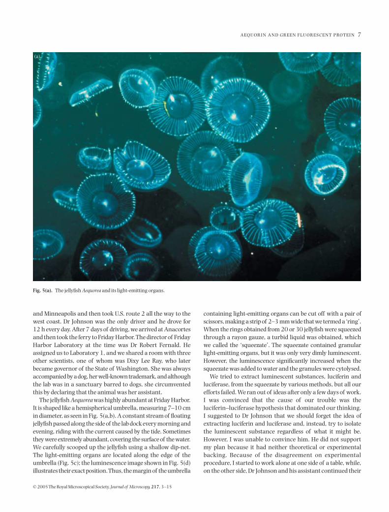
The jellyfish
Aequorea
was highly abundant at Friday Harbor.It is shaped like a hemispherical umbrella, measuring 7–10 cmin diameter, as seen in Fig. 5(a,b). A constant stream of floatingjellyfish passed along the side of the lab dock every morning andevening, riding with the current caused by the tide. Sometimesthey were extremely abundant, covering the surface of the water.We carefully scooped up the jellyfish using a shallow dip-net.The light-emitting organs are located along the edge of theumbrella (Fig. 5c); the luminescence image shown in Fig. 5(d)illustrates their exact position. Thus, the margin of the umbrella
containing light-emitting organs can be cut off with a pair ofscissors, making a strip of 2–3 mm wide that we termed a ‘ring’.When the rings obtained from 20 or 30 jellyfish were squeezedthrough a rayon gauze, a turbid liquid was obtained, whichwe called the ‘squeezate’. The squeezate contained granularlight-emitting organs, but it was only very dimly luminescent.However, the luminescence significantly increased when thesqueezate was added to water and the granules were cytolysed.
We tried to extract luminescent substances, luciferin andluciferase, from the squeezate by various methods, but all ourefforts failed. We ran out of ideas after only a few days of work.I was convinced that the cause of our trouble was theluciferin–luciferase hypothesis that dominated our thinking.I suggested to Dr Johnson that we should forget the idea ofextracting luciferin and luciferase and, instead, try to isolatethe luminescent substance regardless of what it might be.However, I was unable to convince him. He did not supportmy plan because it had neither theoretical or experimentalbacking. Because of the disagreement on experimentalprocedure, I started to work alone at one side of a table, while,on the other side, Dr Johnson and his assistant continued their
Fig. 5(a). The jellyfish Aequorea and its light-emitting organs.

8
O. S H I M O M U R A
© 2005 The Royal Microscopical Society,
Journal of Microscopy
,
217
, 3–15
efforts to extract luciferin by grinding luminous tissues withsand. It was an awkward situation.
The principle in the isolation of bioluminescent substancesis simple, but it is not always easy in practice. To avoid theloss of luminescence activity, the luminescent substance isextracted from the tissue under conditions that reversiblyinhibit luminescence, or that cause a selective inactivation orremoval of a co-factor necessary for light emission. For exam-ple, in the case of a luciferin–luciferase system, the luciferin isusually extracted with methanol or boiling water, whichinhibits luminescence by inactivating the luciferase. The luci-ferase itself can then be obtained from cold aqueous extractafter the luciferin has been exhausted by spontaneous lumi-nescence reactions. If a co-factor is involved in light emission,its exhaustion causes luminescence to stop, as in the case ofthe co-factor ATP in firefly bioluminescence.
In the case of
Aequorea
, however, I could not find any condi-tion that would reversibly inhibit luminescence. I tried knownenzymatic co-factors, such as ATP, FMN and NADH, but noneshowed any activation effect, indicating that they were not
involved. I tried various enzyme inhibitors and they either hadno effect or completely destroyed the ability of the material toluminesce. I became conceptually exhausted and could notcome up with one single further idea.
I spent the next several days soul-searching, trying to imag-ine the reaction that might occur in luminescing jellyfish andsearching for a way to extract the luminescent principle. Ioften meditated on the problem on a drifting rowing boatunder clear summer skies. Friday Harbor in summer at thattime was quiet and peaceful, quite different from the present-day, when it is saturated with busy pleasure boats and noisyseaplanes. Meditation afloat was safe but if I fell asleep the boatwas carried away by the tide so that I had to row for a long timeto get back to the laboratory.
One afternoon, a thought suddenly struck me, a thoughtso simple that I should have had it much sooner: even if aluciferin–luciferase system is not involved in the jellyfishluminescence, an enzyme or protein is probably involved inthe luminescence reaction. If so, the activity of this enzyme orprotein can probably be altered by a pH change. There might
Fig. 5(b). Continued.

A E Q U O R I N A N D G R E E N F L U O R E S C E N T P RO T E I N
9
© 2005 The Royal Microscopical Society,
Journal of Microscopy
,
217
, 3–15
be a certain degree of acidity at which an enzyme or protein isreversibly inactivated. I immediately went back to the lab,made a squeezate, and tested a small portion of it with acetatebuffer solutions of various acidities. I found that the squeezatewas luminous at pH 6 and pH 5, but not at pH 4. I filtered therest of squeezate, and mixed the solid part containing granularlight-emitting organs with pH 4 buffer. After 2–3 min, whenthe cells were cytolysed, I filtered the mixture. The filtrate, nowfree of cells and debris, was almost without luminescence, butit regained its luminescence when neutralized with a smallamount of sodium bicarbonate. The experiment showed thatthe luminescence substance was reversibly inhibited at pH 4,and it was extracted into the solution, indicating that I hadsucceeded in extracting the bioluminescence substance, atleast in principle.
A surprise came very shortly afterwards. I threw the solu-tion I had just tested into a sink, and the inside of the sink lit upwith a bright blue flash. As the overflow from an aquarium
was flowing into the sink, I thought that it was the presence ofthe seawater that had caused the luminescence. So I mixed asmall amount of seawater with the extract: it gave explosivelystrong luminescence. As the composition of seawater isknown, I easily worked out that the activator was Ca
2+
. I calledDr Johnson and repeated the experiment for him. The discov-ery of Ca
2+
as the activator in turn suggested that EDTA shouldserve as a better inhibitor of luminescence than acidification.It was the first important breakthrough in the study of aequorin.
We devised a simple procedure to extract the luminescencesubstance from the jellyfish using EDTA (Fig. 6). Our next taskwas to catch and process as many jellyfish as possible. Wecollected jellyfish from 6 a.m. to 8 a.m., then after a quickbreakfast we would cut rings from the jellyfish until noon. Wedevoted all afternoon to the extraction. After dinner, we againcollected jellyfish from 7 p.m. to 9 p.m., and the catch was keptin an aquarium for the next day. We soon found that thebottleneck in the operation was the step of cutting rings with
Fig. 5(c). Continued.

10
O. S H I M O M U R A
© 2005 The Royal Microscopical Society,
Journal of Microscopy
,
217
, 3–15
scissors, which is a delicate and very slow process. Even afterconsiderable practice, it took more than 1 min to cut one jellyfishring, and 3 h work by four people could not produce morethan 500 rings. To increase productivity, we hired severalhighschool students, trained them and paid them 2 cents foreach ring they cut. We also decided to buy jellyfish from localchildren, paying a penny per jellyfish. However, just when ouroperation was in full swing, the jellyfish suddenly vanishedfrom the area. We therefore extracted only about 10 000 speci-mens of
Aequorea
in the summer of 1961.We returned to Princeton with the jellyfish extract packed
in dry-ice, and then began purification of the light-emittingprinciple by numerous repetitions of column chromatography.The purification was completed in early 1962, with a total yieldof 5 mg of protein, of which only 1 mg was highly purified. Duringthe purification of aequorin, another protein with a brightgreen fluorescence was separated and also purified. It was laternamed GFP. The luminescent substance was a protein with amolecular weight of about 20 000, and it emitted blue lightwhen a trace of Ca
2+
was added, even in the absence of oxygen.We named the protein ‘aequorin’ (Shimomura
et al
., 1962).
Fig. 5(d). Continued.
Fig. 6. Our method to extract the luminescence substance from Aequoreausing EDTA.

A E Q U O R I N A N D G R E E N F L U O R E S C E N T P RO T E I N
11
© 2005 The Royal Microscopical Society,
Journal of Microscopy
,
217
, 3–15
Aequorin is an extraordinary protein that stores a largeamount of energy and releases the energy when calcium isadded, resembling a charged battery that releases its chargedenergy by a short circuit. We were greatly interested instudying the mechanism of its luminescence reaction. How-ever, every attempt to extract the chromophore of aequorinresulted in the self-destruction of the chromophore by anintramolecular reaction. We therefore decided to postponefurther study on aequorin.
Five years later, Ridgway & Ashley (1967) reported the firstbiological application of aequorin. They observed transientCa
2+
signals in single muscle fibres of barnacle with microin-jected aequorin. The importance of aequorin became obviousand we decided to resume our study. However, there was no wayto extract the native chromophore. Therefore, we aimed at afragment of the chromophore that is formed when aequorin isdenatured with urea in the presence of 2-mercaptethanol. Thiscompound was blue-fluorescent and named AF-350 based on itsabsorption maximum at 350 nm (Shimomura & Johnson, 1969).
To obtain 1 mg of AF-350, 100–200 mg of pure aequorinwas required, which would require about 2.5 tons of jellyfishor 50 000 animals. To process 50 000 jellyfish in one summer,
we would need to collect, cut and extract 3000 jellyfish eachday, allowing for days of bad weather and poor fishing. It was aworkload that could not be accomplished by collecting jelly-fish at the lab dock and cutting rings with scissors at a rate ofone per minute. To solve the problem, Dr Johnson constructedtwo jellyfish cutting machines, as illustrated in Fig. 7. Themachine had a rotating meat slicer blade; jellyfish were placedon the cutting platform and, using the jellyfish turner, the jel-lyfish was rotated against the slicer blade by hand. The ringwas cleanly cut and dropped into a receiving jar. With thesemachines we could cut 1200 rings in an hour. We alsoexploited new fishing grounds at the town dock to obatin morejellyfish and buckets filled with jellyfish were transported tothe lab usually by car and sometimes by Boston whaler.
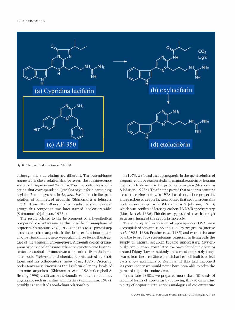
After five years of hard work and with hundreds of thous-ands of jellyfish, the chemical structure of AF-350 was finallydetermined (Shimomura & Johnson, 1972). The result wassurprising: AF-350 and
Cypridina
etioluciferin have the same2-aminopyrazine skeleton, as illustrated in Fig. 8. Thestructure of AF-350 (Fig. 8c) contained the skeleton of a 2-aminopyrazine, the skeleton identical with that which existsin
Cypridina
oxyluciferin (Fig. 8b) and etioluciferin (Fig. 8d),
Fig. 7. Our apparatus for cutting the light-emitting rings from Aequorea. F. H. Johnson, Naval Research Reviews, February 1970, 16–23 (Office of NavalResearch, Dept. of Navy, Washington, DC.
12
O. S H I M O M U R A
© 2005 The Royal Microscopical Society,
Journal of Microscopy
,
217
, 3–15
although the side chains are different. The resemblancesuggested a close relationship between the luminescencesystems of
Aequorea
and
Cypridina
. Thus, we looked for a com-pound that corresponds to
Cypridina
oxyluciferin containingacylated 2-aminopyrazine in
Aequorea
. We found it in the spentsolution of luminesced aequorin (Shimomura & Johnson,1973). It was AF-350 acylated with
p
-hydroxyphenylacetylgroup; this compound was later named ‘coelenteramide’(Shimomura & Johnson, 1975a).
The result pointed to the involvement of a hypotheticalcompound coelenterazine as the possible chromophore ofaequorin (Shimomura
et al
., 1974) and this was a pivotal stepin our research on aequorin. In the absence of the informationon
Cypridina
luminescence, we could not have found the struc-ture of the aequorin chromophore. Although coelenterazinewas a hypothetical substance when the structure was first pre-sented, the actual substance was soon isolated from the lumi-nous squid
Watasenia
and chemically synthesized by ShojiInoue and his collaborators (Inoue
et al.
, 1975). Presently,coelenterazine is known as the luciferin of many kinds ofluminous organisms (Shimomura
et al
., 1980; Campbell &Herring, 1990), and it can be also found in various non-luminousorganisms, such as sardine and herring (Shimomura, 1987),possibly as a result of a food-chain relationship.
In 1975, we found that apoaequorin in the spent solution ofaequorin could be regenerated into original aequorin by treatingit with coelenterazine in the presence of oxygen (Shimomura& Johnson, 1975b). This finding proved that aequorin containsa coelenterazine moiety. In 1978, based on various propertiesand reactions of aequorin, we proposed that aequorin containscoelenterazine-2-peroxide (Shimomura & Johnson, 1978),which was confirmed later by carbon-13 NMR spectrometry(Musicki
et al
., 1986). This discovery provided us with a roughstructural image of the aequorin molecule.
The cloning and expression of apoaequorin cDNA wereaccomplished between 1985 and 1987 by two groups (Inouye
et al
., 1985, 1986; Prasher
et al
., 1985) and when it becamepossible to produce recombinant aequorin in living cells thesupply of natural aequorin became unnecessary. Mysteri-ously, two or three years later, the once abundant
Aequorea
around Friday Harbor suddenly and almost completely disap-peared from the area. Since then, it has been difficult to collecteven a few specimens of
Aequorea
. If this had happened20 years sooner we would never have been able to solve thepuzzle of aequorin luminescence.
In the late 1980s, we prepared more than 30 kinds ofmodified forms of aequorins by replacing the coelenterazinemoiety of aequorin with various analogues of coelenterazine
Fig. 8. The chemical structure of AF-350.

A E Q U O R I N A N D G R E E N F L U O R E S C E N T P RO T E I N
13
© 2005 The Royal Microscopical Society,
Journal of Microscopy
,
217
, 3–15
(Shimomura
et al
., 1988, 1989, 1993). Those aequorins, termedsemisynthetic aequorins, have different calcium sensitivities.During the same period, I learned that attempts at X-raycrystallography of aequorin were in progress at three differentlaboratories. I waited for their results, but none appeared andso in 1997 we decided to take up the X-ray project ourselvesand organized a team. We first developed a new technique toproduce high-purity recombinant aequorin (Shimomura &Inouye, 1999) and then the X-ray structure of aequorin wasfinally determined in 2000 (Head
et al
., 2000). The X-raystructure revealed that aequorin is a globular protein. Thecoelenterazine moiety is shielded in the central cavity of theprotein and the outside molecules have no access to the coe-lenterazine moiety. The peroxide group of the coelenterazinemoiety is stabilized by hydrogen bonds to the protein residuesthat include Tyrosine 184. Figure 9 illustrates the overallaequorin reactions. Binding of two Ca
2+
ions to aequorin causesconformational changes of the protein part of aequorin,resulting in the opening of the protein and the decompositionof coelenterazine peroxide into coelenteramide and CO
2
,
accompanied by emission of light. Apoaequorin can beregenerated into aequorin with coelenterazine and oxygen.
With respect to GFP, Davenport and Nicol first describedgreen fluorescence of the light organs of
Aequorea
in 1955(Davenport & Nicol, 1955). In 1961, we found during thepurification of aequorin that the green fluorescent substancein
Aequorea
is a protein (Shimomura
et al
., 1962). We calledthe protein ‘green protein’ because its precipitated form isgreen under room light. Hastings & Morin (1969), however,called it ‘green fluorescent protein’, and I thought the namewas more appropriate than ‘green protein’. Morin & Hastings(1971) suggested that the emission of green luminescencefrom
Aequorea
involves a Förster-type energy transfer fromaequorin to GFP. In 1974, we purified GFP completely andcrystallized it (Morize
et al
., 1974) (Fig. 10).In 1979, we elucidated the structure of the GFP chromo-
phore (Shimomura, 1979). In this work, we first denaturedGFP by heat then digested the denatured GFP with papain.From the digest, we isolated and purified a fragment containingthe chromophore. At the moment I saw the absorption
Fig. 9. The reactions involving aequorin.

14
O. S H I M O M U R A
© 2005 The Royal Microscopical Society,
Journal of Microscopy
,
217
, 3–15
spectrum of the purified chromophore, I recognized itsresemblance to the spectrum of an imidazolone that I hadsynthesized during my study of
Cypridina
luciferin in the late1950s (Shimomura & Eguchi, 1960). Acid hydrolysis of thechromophore produced
p
-hydroxybenzaldehyde, and severalamino acids suggested the presence of
p
-hydroxybenzalgroup. Therefore, I synthesized a second compound, thespectral characteristics of which satisfactorily matched thoseof the GFP chromophore. From this, I deduced the structure ofthe GFP chromophore. In the same year, I learned that Dr BillWard was studying GFP and I decided to discontinue my workon GFP to concentrate on my studies of bioluminescence.
Since then, the structure of GFP chromophore has beenverified by Cody
et al
. (1993). The X-ray structure of GFP wassolved in 1996 by two different groups (Ormo
et al
., 1996;Yang
et al
., 1996). cDNA of
Aequorea
GFP was cloned byPrasher
et al.
(1992) and expressed in living organisms byChalfie
et al
. (1994) and also by Inouye & Tsuji (1994). The
successful expression of GFP has established the basis of usingGFP as a marker protein for gene expression, resulting in thepresent widespread popularity of GFP. The usefulness of GFPhas been further enhanced by the discovery of red-fluorescentGFP-like proteins in anthozoan corals (Matz
et al
., 1999) andalso by various improvements and developments. I am, as anoriginal co-discoverer of this protein, very happy to see thegrowing usefulness of GFP.
In retrospect, my work on
Aequorea
was helped by manypeople in various ways and I was certainly fortunate. My threementors played decisive roles in my life and research. In 1955,Dr Yasunaga sent me to Professor Hirata’s lab in Nagoya, givingme a career as a scientist instead of a pharmacist. ProfessorHirata gave me the subject of
Cypridina
luciferin to study. Myfirst research work, the crystallization of
Cypridina
luciferin,was very difficult, and I succeeded only after 10 months ofvery hard effort. Even if my success was accidental, it gave meself-confidence, and a feeling of ‘if it is not impossible, then I
Fig. 10. Crystallized GFP. Photo by Shinya Inouye.

A E Q U O R I N A N D G R E E N F L U O R E S C E N T P RO T E I N
15
© 2005 The Royal Microscopical Society,
Journal of Microscopy
,
217
, 3–15
can do it’. Then in 1961, Dr Frank Johnson gave me the sub-ject of
Aequorea
to study, and in this bioluminescence system,the information on
Cypridina
luminescence was essential indetermining the chromophore structure of aequorin. It was alucky coincidence that the
Cypridina
project had come first. Ibelieve, however, that my self-belief supported me in solvingthe difficult problems involved in the study of aequorin. Basedon my experience, I think it is important not to give up when adifficult problem is encountered in research. Overcoming adifficulty may reward us with a self-belief that could be morevaluable than the solution to the problem.
References
Campbell, A.K. & Herring, P.J. (1990) Imidazopyrazine bioluminescencein copepods and other marine organisms.
Mar. Biol
.
104
, 219–225.Chalfie, M., Tu, Y., Euskirchen, G., Ward, W.W. & Prasher, D.C. (1994)
Green fluorescent protein as a marker for gene expression.
Science
,
263
,802–805.
Cody, C.W., Prasher, D.C., Westler, W.M., Prendergast, F.G. & Ward, W.W.(1993) Chemical structure of the hexapeptide chromophore of the
Aequorea
green-fluorescent protein.
Biochemistry
,
32
, 1212–1218.Davenport, D. & Nicol, J.A.C. (1955) Luminescence of hydromedusae.
Proc. Roy. Soc. B
,
144
, 399–411.Hastings, J.W. & Morin, J.G. (1969) Comparative biochemistry of calcium-
activated photoproteins from the ctenophore,
Mnemiopsis
and thecoelenterates
Aequorea
,
Obelia
,
Pelagia
and
Renilla
.
Biol. Bull
.
137
,402.
Head, J.F., Inouye, S., Teranishi, K. & Shimomura, O. (2000) The crystalstructure of the photoprotein aequorin at 2.3A resolution.
Nature
, 405,372–376.
Inoue, S., Sugiura, S., Kakoi, H. & Hashizume, K. (1975) Squid biolumi-nescence II. Isolation from Watasenia scintillans and synthesis of 2-(p-hydroxybenzyl)-6-( p-hydroxyphenyl)-3,7-dihydroimidazo[1,2-a]pyrazin-3-one. Chem. Lett. 141–144.
Inouye, S., Noguchi, M., Sakaki, Y., Takagi, Y., Miyata, T., Iwanaga, S.,Miyata, T. & Tsuji, F.I. (1985) Cloning and sequence analysis of cDNAfor the luminescent protein aequorin. Proc. Natl Acad. Sci. U.S.A. 82,3154–3158.
Inouye, S., Sakaki, Y., Goto, T. & Tsuji, F.I. (1986) Expression of apoaequorincomplementary DNA in Escherichia coli. Biochemistry, 25, 8425–8429.
Inouye, S. & Tsuji, F.I. (1994) Aequorea green fluorescence protein. Expres-sion of the gene and fluorescence characteristics of the recombinantprotein. FEBS Lett. 341, 277–280.
Kishi, Y., Goto, T., Hirata, Y., Shimomura, O. & Johnson, F.H. (1966)Cypridina bioluminescence I: structure of Cypridina luciferin. Tetrahe-dron Lett. 3427–3436.
Matz, M.V., Fradkov, A.F., Labas, Y.A., Savitsky, A.P., Zaraisky, A.G.,Markelov, M.L. & Lukyanov, S.A. (1999) Fluorescent proteins fromnonbioluminescent anthozoa species. Nature Biotechnol. 17, 969–973.
McCapra, F. & Chang, Y.C. (1967) The chemiluminescence of a Cypridinaluciferin analogue. Chem. Commun. 1011–1012.
Morin, J.G. & Hastings, J.W. (1971) Energy transfer in a bioluminescentsystem. J. Cell. Physiol. 77, 313–318.
Morise, H., Shimomura, O., Johnson, F.H. & Winant, J. (1974) Intermole-cular energy transfer in the bioluminescent system of Aequorea. Bio-chemistry, 13, 2656–2662.
Musicki, B., Kishi, Y. & Shimomura, O. (1986) Structure of the functionalpart of photoprotein aequorin. Chem. Commun. 1986, 1566–1568.
Ormo, M., Cubitt, A.B., Kallio, K., Gross, L.A., Tsien, R.Y. & Remington, S.J.(1996) Crystal structure of the Aequorea victoria green fluorescentprotein. Science, 273, 1392–1395.
Prasher, D.C., Eckenrode, V.K., Ward, W.W., Prendergast, F.G. &Cormier, M.J. (1992) Primary structure of the Aequorea victoria greenfluorescent protein. Gene, 111, 229–233.
Prasher, D., McCann, R.O. & Cormier, M.J. (1985) Cloning and expressionof the cDNA coding for aequorin, a bioluminescent calcium-bindingprotein. Biochem. Biophys. Res. Commun. 126, 1259–1268.
Ridgway, E.B. & Ashley, C.C. (1967) Calcium transients in single musclefibers. Biochem. Biophys. Res. Commun. 29, 229–234.
Shimomura, O. (1979) Structure of the chromophore of Aequorea greenfluorescrent protein. FEBS Lett. 104, 220–222.
Shimomura, O. (1987) Presence of coelenterazine in non-bioluminescentmarine organisms. Comp. Biochem. Physiol. 86B, 361–363.
Shimomura, O. & Eguchi, S. (1960) Studies on 5-imidazolone. I–II. NipponKagaku Zasshi, 81, 1434–1439.
Shimomura, O., Goto, T. & Hirata, Y. (1957) Crystalline Cypridina luci-ferin. Bull. Chem. Soc. Japan, 30, 929–933.
Shimomura, O., Inoue, S., Johnson, F.H. & Haneda, Y. (1980) Widespreadoccurrence of coelenterazine in marine bioluminescence. Comp. Bio-chem. Physiol. 65B, 435–437.
Shimomura, O. & Inouye, S. (1999) The in situ regeneration and extractionof recombinant aequorin from Escherichia coli cells and the purificationof extracted aequorin. Protein Expression Purification, 16, 91–95.
Shimomura, O. & Johnson, F.H. (1969) Properties of the bioluminescentprotein aequorin. Biochemistry, 8, 3991–3997.
Shimomura, O. & Johnson, F.H. (1972) Structure of the light-emittingmoiety of aequorin. Biochemistry, 11, 1602–1608.
Shimomura, O. & Johnson, F.H. (1973) Chemical nature of light emitter inbioluminescence of aequorin. Tetrahedron Lett. 2963–2966.
Shimomura, O. & Johnson, F.H. (1975a) Chemical Nature of bioluminescencesystems in coelenterates. Proc. Natl Acad. Sci. U.S.A. 72, 1546–1549.
Shimomura, O. & Johnson, F.H. (1975b) Regeneration of the photoproteinaequorin. Nature, 256, 236–238.
Shimomura, O. & Johnson, F.H. (1978) Peroxidized coelenterazine, theactive group in the photoprotein aequorin. Proc. Natl Acad. Sci. U.S.A.75, 2611–2615.
Shimomura, O., Johnson, F.H. & Morise, H. (1974) Mechanism of theluminescent intramolecular reaction of aequorin. Biochemistry, 13,3278–3286.
Shimomura, O., Johnson, F.H. & Saiga, Y. (1962) Extraction, purificationand properties of aequorin, a bioluminescent protein from the lumi-nous hydromedusan, Aequorea. J. Cell. Comp. Physiol. 59, 223–239.
Shimomura, O., Musicki, B. & Kishi, Y. (1988) Semi-synthetic aequorin:an improved tool for the measurement of calcium ion concentration.Biochem. J. 251, 405–410.
Shimomura, O., Musicki, B. & Kishi, Y. (1989) Semi-synthetic aequorinswith improved sensitivity to Ca2+ ions. Biochem. J. 261, 913–920.
Shimomura, O., Musicki, B., Kishi, Y. & Inouye, S. (1993) Light-emittingproperties of recombinant semi-synthetic aequorins and recombinantfluorescein-conjugated aequorin for measuring cellular calcium. CellCalcium, 14, 373–378.
Stone, H. (1968) The enzyme catalyzed oxidation of Cypridina luciferin.Biochem. Biophys. Res. Commun. 31, 386–391.
Yang, F., Moss, L.G. & Phillips, G.N.J. (1996) The molecular structure ofgreen fluorescent protein. Nature Biotechnol. 14, 1246–1251.