review article cell membrane for · tures. furthermore, the cell surface is intimately...
TRANSCRIPT

Review articleJournal of Medical Genetics (1975). 12, 174.
Cell membrane receptors for serological reagentsG. W. G. BIRD
West Midlands Regional Blood Transfusion Service, Vincent Drive, Edgbaston, Birmingham B15 2SG
Many useful genetic markers are cell surface struc-tures. Furthermore, the cell surface is intimatelyinvolved in cellular immunity, and often in humoralimmunity. It is therefore important, both in gene-tics and immunogenetics, to understand cell surfacestructure.Although the cell membrane has been actively
studied for many years, progress in this field isunderstandably slow. It is generally agreed thatthe cell membrane is fundamentally a phospholipidbilayer with inner and outer hydrophilic areas and acentral hydrophobic region. Besides lipids, themembrane contains proteins and carbohydrates; thecarbohydrates occur as glycolipids or glycoproteins.Whereas the protein and lipid portions of the cell
membrane occupy the plane of the membrane, thecarbohydrate chains of the glycolipid and glycopro-tein components of the cell membrane project out-wards, so that their N-terminal ends are exposedabove the membrane surface. Recent work,chiefly by Singer and Nicolson (1972) and Nicolson(1973), on the structural organization ofmembranes,in terms of the thermodynamics of macromolecularsystems, has led to the conclusion that the cell mem-brane is a mosaic of alternating globular proteins andphospholipid units, in which the phospholipid unitsrepresent the bricks and the globular protein themortar (Fig. 1). The mosaic structure is dynamicrather than static, with considerable lateral move-ment of the phospholipid and protein components.Different membrane components move laterally inthe plane of the membrane at different rates, de-pending on restraints applied from outside or insidethe cell. The movement may account for certainapparent changes on the cell surface, such as theappearance of 'new' HL-A specificities on culturedcells (Mackintosh, Hardy, and Aviet, 1971), or forchanges observed after treatment of intact cells by
Received 26 June 1974.
enzymes. Indeed, it has been suggested (Singerand Nicolson, 1972) that after proteolysis or malig-nant transformation, agglutinin-binding sites diffuseand aggregate in clusters (Fig. 2). This mightexplain, for example, the very strong agglutinationof papain-treated A2 cells by the virtually A1-specific agglutinin obtained from the seeds of Doli-chos biflorus. Although this lateral movement maysometimes affect the 'display' of surface compo-nents, molecular orientation is generally maintained.
Celi surface carbohydratesWe know more about the structure of erythro-
cytes than about any other cells, and more aboutthose antigens which are carbohydrate than thosewhich are lipid, protein, or lipoprotein.There are only seven monosaccharide components
of the human red cell membrane (Winzler, 1969):galactose, mannose, fucose, glucose, acetylgalactos-amine, acetylglucosamine and N-acetylneuraminic(sialic) acid. All human erythrocyte membranecarbohydrate chains are therefore combinations ofthese seven sugars, and differ from one another inthe serial arrangement of the sugar components, andthe nature of the chemical linkages between them.There are therefore structural similarities in recep-tor sites for various serological reagents, so that it isnot surprising that they crossreact; what is surpris-ing therefore is that many reagents are very highlyspecific.
The ABH and Lewis systemsThe ABH and Lewis blood group antigenic de-
terminants are the carbohydrate moieties of glyco-proteins or glycolipids. Part of the work which ledto the elucidation of ABH and Lewis structure wasdone, not on red cells, but on blood group specificsubstances present in soluble form in certain bodyfluids, eg, saliva of secretors, pseudomucinous
174
on January 29, 2020 by guest. Protected by copyright.
http://jmg.bm
j.com/
J Med G
enet: first published as 10.1136/jmg.12.2.174 on 1 June 1975. D
ownloaded from

Cell membrane receptors for serological reagents
FIG. 1. The fluid mosaic model of the cell membrane; schematic three-dimensional and cross-sectional views, showing the phospholipidbilayer and the globular integral protein (solid bodies with stippled surfaces). (According to Fig. 3 in Singer, S. J. and Nicolson, G. L.,'The fluid mosaic model of the structure of cell membranes', Science, 175, 18 February 1972. Copyright 1972 by the American Associationfor the Advancement of Science. Reproduced with the permission of authors and the editor, Science.)
ovarian cyst fluids (Kabat, 1956; Morgan and Wat-kins, 1959; 1969).The earliest indication that a single component
sugar (the 'immunodominant' sugar) is more closelyinvolved in serological specificity than any other wasobtained by means of agglutination-inhibition testswith simple sugars (Morgan and Watkins, 1953).This approach was based on the observations ofLandsteiner (1920) that low molecular weight sub-stances with structures identical with, or closelysimilar to, the immunologically determinant groupof a complex antigen combine with the agglutinatingreagent, and completely inhibit its reaction with thecorresponding antigenic determinant. The agglu-tinating reagents first used for this purpose byMorgan and Watkins (1953) were those specific forsmall configurations, such as the anti-H agglutininspresent in the sera of certain eels, or in Lotus tetra-gonolobus seeds, and which are therefore inhibited bysimple sugars.The capacity of L-fucose, a component sugar of
H-substance, to inhibit the anti-H agglutinins of eelserum or Lotus tetragonolobus shows that this sugaris an important determinant of H-specificity. Simi-lar studies show that N-acetyl-D-galactosamine isan important structural determinant of A- and D-galactose of B-specificity. These observations havebeen confirmed by other methods.
Biosynthesis of ABH and LewisThe primary product of a blood group gene is an
enzyme (glycosyltransferase) which attaches thecharacteristic end sugar to a preformed substrate(Morgan and Watkins, 1969), presumably created bythe sequential action of other transferases.The precursor substrate for the ABH and Lewis
substances consists of two carbohydrate chains(Table IA). The product of the H gene, a fucosyl-transferase, adds L-fucose to both chains (Table IB).The product of the A gene, an N-acetyl-D-galactos-aminyltransferase, adds N-acetyl-D-galactosamine
175
on January 29, 2020 by guest. Protected by copyright.
http://jmg.bm
j.com/
J Med G
enet: first published as 10.1136/jmg.12.2.174 on 1 June 1975. D
ownloaded from

G. W. G. Bird
A
B
FIG. 2. Two mechanisms to explain the enhanced action of lectinson malignantly transformed or protease-treated erythrocytes: (A) re-
moval of steric hindrance; (B) clustering of receptor sites. (Accordingto Fig. 7 in Singer, S. J. and Nicolson, G. L., 'The fluid mosaic modelof the structure of cell membranes', Science, 175, 18 February 1972.Copyright 1972 by the American Association for the Advancement ofScience. Reproduced with the permission of authors and the editor,Science.)
to the H-chains (Table IC). Similarly the productof the B gene, a D-galactosyltransferase, adds D-galactose to the H-chain (Table ID).The product, Lea, of the L (Lewis) gene, which is
a different fucosyltransferase to the product of theH gene, attaches L-fucose to the penultimate sugaronly of chain 1 (Table IE). The ABH and Lewisgenes, which are carried on different chromosomes,act on the same substrate, and therefore interact intheir phenotype expression. In the presence ofboth H and L genes, the precursor substance is con-verted as shown in Table IF. It will be seen thatthe Leb character, which at one time was thought tobe the product of a gene allelic to the Lea (L) gene, isreally an interaction product of the H and L genes.Indeed, in the presence ofHand L genes, one of theprecursor chains is converted into Leb and theother into H. It is not surprising therefore thatcrossreactivity between H and Leb is frequentlyobserved.
GlycosyltransferasesThe glycosyltransferases involved in ABH and
Lewis biosynthesis are present in various tissues andbody fluids (Race and Watkins, 1972). They arepresent in soluble form in serum. The origin andfunction ofglycosyltransferases are unknown. Theycan be shown to act in vitro by incubating 0 cells in amixture containing a-N-acetyl-D-galactosamyl-transferase and nucleotide-bound N-acetyl-D-
galactosamine. The 0 cells then develop the Acharacter. Similar results are obtained with the B-enzyme and the B-sugar. That transferases do notact on erythrocytes in vivo is evident from observa-tions made on blood group chimieras in which, forexample, a mixture of group 0 and A1 erythrocytespersists throughout life. Race and Watkins (1972)state that the co-existence in chimaeras of two inde-pendent cell populations indicates that the final stepin the formation of A- or B-active sites on the ery-throcyte surface is not mediated in the external en-vironment of the cells.The biosynthetic pathways established by the
work of Morgan and Watkins (1959; 1969) and ofKabat (1956) have firm experimental support andare generally accepted. Some alternative hypo-theses have been proposed, notably by Weiner,Socha, and Gordon (1972), who propose that H isnot a precursor ofA and B, and that the H, A, and Bgenes compete with one another for a common pre-cursor substrate.
Red cellsSince information derived from the study of
blood group specific substances in body fluids doesnot necessarily apply to red blood cells, the cellsthemselves are now being actively studied. It hasbeen shown that whereas in body fluids, the ABHblood group specific substances are glycoproteins,they occur in the red cells as glycolipids and glyco-proteins. They occur as glycolipids in the red cellsof all persons and also as glycoproteins on the redcells of secretors of blood group specific substances(Gardas and Koscielak, 1971). One of the mostvaluable and perhaps the least harsh of the methodsof investigating the fine structure of the red cellsurface is to study the reactions of plant seed lectinsof known chemical specificity before and aftertreatment of red cells with various enzymes.
LectinsIndeed, the current 'explosion' in the applica-
tion of lectins to the study of cell surface structurehas greatly exceeded the expectations of earlierworkers in this field. The subject has been re-viewed by Bird (1959) and Boyd (1963). Lectinsare now being extensively and usefully applied tostructural studies not only of red blood cells but alsoof white blood cells, tumour cells, bacteria andviruses. A recent number (vol. 234) of the Annalsof the New York Academy of Sciences is devotedto a symposium held on this subject. Lectinsare particularly useful in studies of cell membranestructure because they combine with single sugar
176
on January 29, 2020 by guest. Protected by copyright.
http://jmg.bm
j.com/
J Med G
enet: first published as 10.1136/jmg.12.2.174 on 1 June 1975. D
ownloaded from

Cell membrane receptors for serological reagents
TABLE IBIOSYNTHESIS OF ABH-LEWIS AND (?) I SUBSTANCES
Designation Gene Structure of Gene Product Specificityof Chain
1 -k31 P-Gal - j-GNAc- >
A1I- 4
2 P-Gal > P-GNAc > May be related toI-specificity
]~~~~~~~~~~~~~131 P-Gal > P-GNAc > H
2 1 -2--2a-Fuc
B H1 434
2 a lo-Gal >, l-GNAc > Ht 1 --*2a-Fuc
1-*3 1-31 za-GalNAc > P-Gal B P-GNAc > A
t 1 -*21a-FucC H and A
1 -*3 1 42 a-GalNAC > P-Gal > P-GNAc > A
t 1 -*2a-Fuc
1-..3 1-*3-31 a-Gal > P-Gal > P-GNAc -> B
t 1 --*2a a-Fuc
D HandBI-*3 1-44
2 c-Gal > P-Gal > P-GNAc - Bt 1 -*2a-Fuc
c=- 31 P9-Gal -> P-GNAc-*La&
t 1 --*4B L a-Fuc
2 P-Gal -> P-GNAc1 -*3
1 P-Gal -* P-GNAc -> LabftI-*..2 tlI -*4
F ~~~~~~Hand Le ax-Fuc at-Fuc2 ~~~~~P-Gal -> P-GNAc >~ H
t 1 -*).2a-Fuc
Fuc =fucose; Gal =galactose; GNAc =N-acetyl-D-glucosam-ine; GalNAc =N-acetylgalactosamine.
units, usually in terminal position in the carbohy-drate chains. The specificity of some lectins isgiven in Table II. The mere presence of the reac-
tive sugar is not the sole determinant of lectin speci-ficity. Other factors which influence lectin activityinclude the nature of the next-to-terminal structure,the type of chemical linkage to this structure, theamount of steric hindrance provided by neighbour-ing structures, and the number and distribution ofreceptor sites.
The 'Bombay' blood group
In the rare 'Bombay' blood group (H-negative,genotype hh), the H-forming, a-fucosyltransferase is
absent, so that the precursor substrate is not con-verted into H. Therefore, even ifA or B genes are
present, they cannot act, because their normal sub-strate, H-substance, is absent. 'Bombay' bloodtherefore has no ABH antigens.
The MN systemMuch work on the genetics of the MN blood
group system has been based on the assumption thatthe M and N characters are the products of allelicMand N genes. Recent studies of the biosynthesis ofantigens of the MN system show that this assum-tion is wrong. It now seems certain (Springer andHuprikar, 1972) that N is a basal substance, analo-
177
on January 29, 2020 by guest. Protected by copyright.
http://jmg.bm
j.com/
J Med G
enet: first published as 10.1136/jmg.12.2.174 on 1 June 1975. D
ownloaded from
G. W. G. Bird
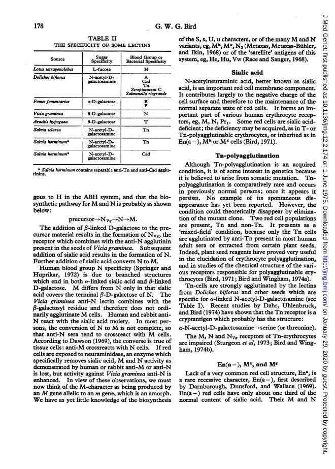
TABLE IITHE SPECIFICITY OF SOME LECTINS
Source Sugar Blood Group orource_________ Specificity Bacterial Specificity
Lotus tetragonolobus L-fucose H
Dolichos biflorus N-acetyl-D- Agalactosamine Cad
TnStreptococcus C
Salmonella riograndePomes fomentarius a-D-galactose B
P
Vicia graminca |-D-galactose N
Arachis hypogaea 1-D-galactose T
Salvia sclarea N-acetyl-D- Tngalactosamine
Salvia horminum* N-acetyl-D- Tngalactosaniine
Salvia horminum* N-acetyl-D- Cadgalactosamine
* Salvia horminwn contains separable anti-Tn and anti-Cad agglu-tirins.
gous to H in the ABH system, and that the bio-synthetic pathway forM and N is probably as shownbelow:
precursor--NNvg--*N--*M.The addition of ,8-linked D-galactose to the pre-
cursor material results in the formation of Nvg, thereceptor which combines with the anti-N agglutininpresent in the seeds of Vicia graminea. Subsequentaddition of sialic acid results in the formation of N.Further addition of sialic acid converts N to M.Human blood group N specificity (Springer and
Huprikar, 1972) is due to branched structureswhich end in both a-linked sialic acid and ,8-linkedD-galactose. M differs from N only in that sialicacid covers the terminal P-D-galactose of N. TheVicia graminea anti-N lectin combines with the,B-galactosyl residue and therefore does not ordi-narily agglutinateM cells. Human and rabbit anti-N react with the sialic acid moiety. In most per-sons, the conversion ofN to M is not complete, sothat anti-N sera tend to crossreact with M cells.According to Dawson (1969), the converse is true oftissue cells: anti-M crossreacts with N cells. If redcells are exposed to neuraminidase, an enzyme whichspecifically removes sialic acid, M and N activity asdemonstrated by human or rabbit anti-M or anti-Nis lost, but activity against Vicia graminea anti-N isenhanced. In view of these observations, we mustnow think of the M-character as being produced byanM gene allelic to an m gene, which is an amorph.We have as yet little knowledge of the biosynthesis
ofthe S, s, U, u characters, or of the manyM andNvariants, eg, Mk, M', N2 (Metaxas, Metaxas-Buhler,and Ikin, 1968) or of the 'satellite' antigens of thissystem, eg, He, Hu, Vw (Race and Sanger, 1968).
Sialic acidN-acetylneuraminic acid, better known as sialic
acid, is an important red cell membrane component.It contributes largely to the negative charge of thecell surface and therefore to the maintenance of thenormal separate state of red cells. It forms an im-portant part of various human erythrocyte recep-tors, eg, M, N, Pr,. Some red cells are sialic acid-deficient; the deficiency may be acquired, as in T- orTn-polyagglutinable erythrocytes, or inherited as inEn(a -), Mk or Mg cells (Bird, 1971).
Tn-polyagglutinationAlthough Tn-polyagglutination is an acquired
condition, it is of some interest in genetics becauseit is believed to arise from somatic mutation. Tn-polyagglutination is comparatively rare and occursin previously normal persons; once it appears itpersists. No example of its spontaneous dis-appearance has yet been reported. However, thecondition could theoretically disappear by elimina-tion of the mutant clone. Two red cell populationsare present, Tn and non-Tn. It presents as a'mixed-field' condition, because only the Tn cellsare agglutinated by anti-Tn present in most humanadult sera or extracted from certain plant seeds.Indeed, plant seed reagents have proved very usefulin the elucidation of erythrocyte polyagglutination,and in studies of the chemical structure of the vari-ous receptors responsible for polyagglutinable ery-throcytes (Bird, 1971; Bird and Wingham, 1974a).
Tn-cells are strongly agglutinated by the lectinsfrom Dolichos biflorus and other seeds which arespecific for a-linked N-acetyl-D-galactosamine (seeTable I). Recent studies by Dahr, Uhlenbruck,and Bird (1974) have shown that the Tn receptor is acryptantigen which probably has the structure:
a-N-acetyl-D-galactosamine--serine (or threonine).The M, N and Nvg receptors of Tn-erythrocytes
are impaired (Sturgeon et al, 1973; Bird and Wing-ham, 1974b).
En(a -), Mk, and MlLack of a very common red cell structure, Ena, is
a rare recessive character, En(a -), first describedby Darnborough, Dunsford, and Wallace (1969).En(a -) red cells have only about one third of thenormal content of sialic acid. Their M and N
178
on January 29, 2020 by guest. Protected by copyright.
http://jmg.bm
j.com/
J Med G
enet: first published as 10.1136/jmg.12.2.174 on 1 June 1975. D
ownloaded from

Cell membrane receptors for serological reagents
TABLE IIICLASSIFICATION OF CAD/Sd- GROUPS*
Reactions with Mixed-Dolichos biflorus field Polyagglutination
CAD 1 + + + No Yes
CAD 2 + + + Yes No
CAD 3 + + Yes No
Strong Sd + Yes No
Sd5L No
* There may be some overlap between the various categories.
antigens are weak, as are their receptors for theMaclura aurantiaca lectin, and the cells are agglu-tinated when suspended in physiological saline solu-tion by 'incomplete' antibody (Darnborough et al,1969; Furuhjelm et al, 1969; Bird and Wingham,1973). En(a -) persons can produce immune anti-Ena antibodies.
Furuhjelm et al (1969) suspect that the conditionis caused by the lack of an enzyme needed for thebuilding of a normal cell surface (the term Enstands for envelope) and that the En(a -) conditioncan be thought of as 'an inborn error of metabolism'.Darnborough et al (1969) postulated the involve-
ment of two codominant genes, Ena and Enb. Theterm En is generally preferred to Enb. There aretherefore three genotypes:
Ena EnaEna EnEn En.
The cells of heterozygotes react in a similar man-ner to those of homozygotes, but to a lesser extent.The Mk gene gives rise to none of the antigens of
the MN or Ss series; the presence of its product ismanifest in the heterozygous state by single dosereactions with anti-M or anti-N, and by indicationsof abnormality of the red cell surface, ie, sialic aciddeficiency, which causes the cells to be agglutinatedby various 'incomplete' antibodies. There is pro-bably no anti_Mk antibody as such. The Mk geneis probably an amorph, analogous to the 0 gene ofthe ABO system, or the d gene, if any, of the Rhesussystem. It might be a 'built-in' inhibitor gene atthe MNSs locus. The subject has been extensivelytreated by Metaxas, Metaxas-Buhler, and Romanski(1971). The reactions of Mk cells are similar tothose ofEna En heterozygotes.Mg cells react similarly to Mk (Nordling et al,
1969). Anti-Mg is a relatively common antibody.En(a -), Mk, and Mg are however different to oneanother (Nordling et al, 1969).
En(a -), Mk, and M' cells have so far shown noevidence of shortened survival.
pPl-substance is present in hydatid cyst fluid.
Morgan, Watkins, and Cory (1972) have isolated aPl-active glycoprotein from this material. Theimmunodominant sugar in Pl-specificity is an a-D-galactosyl residue.
Studies by Feizi et al (1971a and b) on solubleI-substance obtained from milk showed that I-specificity is related to the ABH and Lewis glyco-proteins. These workers concluded that the syn-thesis of I-substance precedes that of ABH andLewis. Earlier studies with enzymes which de-stroy I-substance had suggested that /3-N-acetyl-D-glucosamine and ,B-D-galactose residues are in-volved in I-specificity.Work on the Ii system is greatly bedevilled by
heterogeneity of both antigens and antibodies.Feizi et al (1971b) have shown that the specificity ofone anti-I serum is probably directed towards thestructure 3-D-Gal-(>4)--A->3-D-GNAc-s. Thisstructure is present in Chain 2 of the ABH andLewis precursor substance (see Table IA).
Cad/SdaCad is a rare inherited blood group character first
reported by Cazal et al (1968). Cad cells are agglu-tinated, irrespective of ABO blood group, by Doli-chos biflorus seed extract. Cad is inherited inde-pendently of ABH. Subsequently, various gradesof Cad-positivity were described (Cazal, Monis, andBizot, 1971).
Sanger et al (1971) made the interesting observa-tion that Cad is probably a strong form of the Sid orSda antigen. An attempted classification of thevarious Cad/Sda grades is shown in Table III.This classification is arbitrary; there may be someoverlap between one category and the next.The capacity of the Dolichos biflorus agglutinin
strongly to agglutinate Cad cells shows that ct-N-acetyl-D-galactosamine is also the chief structuraldeterminant of Cad specificity. Thus, there is aresemblance between the A, Tn, and Cad receptors.It is therefore remarkable that there are some plantseed reagents which can clearly distinguish thesethree receptors (Bird and Wingham, 1974a).
RhesusComparatively little is known of the structure of
the Rhesus antigens. Studies by Green (1968a and
179
on January 29, 2020 by guest. Protected by copyright.
http://jmg.bm
j.com/
J Med G
enet: first published as 10.1136/jmg.12.2.174 on 1 June 1975. D
ownloaded from
G. W. G. Bird
b) suggest that they are probably lipoproteins. Ex-traction of the red cell membrane lipid with L-butanol causes loss of Rhesus activity which can berestored by subsequent exposure of the membraneto the extract. Inactivation of Rh determinants byheat or treatment with some sulphydryl compoundsindicates that the protein moiety is an essential partof Rh antigen structure.An interesting genetic condition is the Rhn
state, in which the red cells are devoid of the pro-ducts of any of the Rhesus genes. The conditionarises either when a person is homozygous for a rarerecessive gene X°r, allelic to a common regulatorgene Xlr, or when a person is homozygous for a raresilent or amorphic gene R,ull, within the Rhesuslocus. The regulator type can be recognized whena parent or child has normal Rhesus antigens. Inthe amorphic type, family studies may show, forexample, an apparently CDe/CDe parent with anapparently cDE/cDE child. The explanation, ofcourse, is that the parent is CDe/ --- and thechild --- /cDE.The absence ofspecific lipoprotein sites apparently
has an adverse effect on the red cell membrane, sothat in the Rh,ull condition there is a mild haemo-lytic anaemia (Schmidt and Holland, 1971), and thesodium pump has to work twice as hard as in normalpersons (Levine, 1974).The haemolytic anaemia is associated with
shortened red cell survival, stomatocytosis, in-creased red cell fragility, mild spherocytosis, and ahigh reticulocyte count. Schmidt and Holland(1971) gave the name 'Rhn,ul disease' to this con-dition; the name is not satisfactory because a similarcondition occurs in Rhmod, in which Rh antigens arepresent but are weaker than normal (Chown et al,1972).
In the 'Bombay' blood group, however, red cellsurvival is normal. Levine et al (1973) attribute thedifference to the fact that the defect in the Rhnui1condition involves sites in the plane of the mem-brane, whereas that in the 'Bombay' blood groupinvolves oligosaccharide chains, which projectabove the plane of the cell surface.
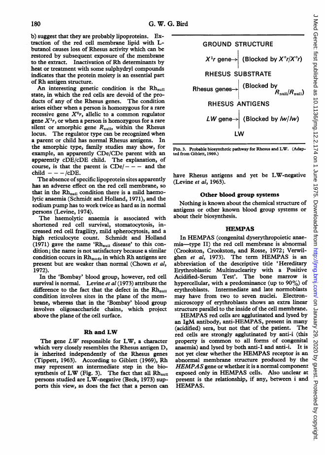
Rh and LWThe gene LW responsible for LW, a character
which very closely resembles the Rhesus antigen D,is inherited independently of the Rhesus genes(Tippett, 1963). According to Giblett (1969), Rhmay represent an intermediate step in the bio-synthesis ofLW (Fig. 3). The fact that all Rhnul1persons studied are LW-negative (Beck, 1973) sup-ports this view, as does the fact that a person can
GROUND STRUCTURE
Xlr gene-* (Blocked by X°r/X°r)
RHESUS SUBSTRATE
Rhesus genes-+ (Blocked byRnull/Rnull)
RHESUS ANTIGENS
LW gene-* (Blocked by 1w/lw)
LW
FIG. 3. Probable biosynthetic pathway for Rhesus and LW. (Adap-ted from Giblett, 1969.)
have Rhesus antigens and yet be LW-negative(Levine et al, 1963).
Other blood group systemsNothing is known about the chemical structure of
antigens or other known blood group systems orabout their biosynthesis.
HEMPASIn HEMPAS (congenital dyserythropoietic anae-
mia-type II) the red cell membrane is abnormal(Crookston, Crookston, and Rosse, 1972; Verwil-ghen et al, 1973). The term HEMPAS is anabbreviation of the descriptive title 'HereditaryErythroblastic Multinuclearity with a PositiveAcidified-Serum Test'. The bone marrow ishypercellular, with a predominance (up to 90%) oferythroblasts. Intermediate and late normoblastsmay have from two to seven nuclei. Electron-microscopy of erythroblasts shows an extra linearstructure parallel to the inside of the cell membrane.HEMPAS red cells are agglutinated and lysed by
an IgM antibody, anti-HEMPAS, present in many(acidified) sera, but not that of the patient. Thered cells are strongly agglutinated by anti-i (thisproperty is common to all forms of congenitalanaemia) and lysed by both anti-I and anti-i. It isnot yet clear whether the HEMPAS receptor is anabnormal membrane structure produced by theHEMPAS gene or whether it is a normal componentexposed only in HEMPAS cells. Also unclear atpresent is the relationship, if any, between i andHEMPAS.
180
on January 29, 2020 by guest. Protected by copyright.
http://jmg.bm
j.com/
J Med G
enet: first published as 10.1136/jmg.12.2.174 on 1 June 1975. D
ownloaded from

Cell membrane receptors for serological reagents
Leucocyte antigensABH antigens occur in both red and white blood
cells (Race and Sanger, 1968), and I and i can easilybe detected in lymphocytes (Shumak et al, 1971).The HL-A system is a complex immunogenic
system controlled by genes at two or more loci.The ABH and HL-A systems are of major im-portance in human transplantation because theirantigens are not confined to blood cells but arepresent in most tissue cells.The red cell antigens Bga, Bgb and Bgc are pro-
bably identical with, or very similar to, the HL-Aantigens 7, W17 and W28, respectively (Morton,Pickles, and Sutton, 1969; Morton et al, 1971).Recent work by Nordhagen and 0rjaswter (1974),who used an AutoAnalyzer technique, have shownthat other HL-A antigens may be present, probablyin modified form, on red cells; apparently such anti-gens can be better demonstrated on reticulocytesthan on mature cells.Most clinical studies ofHL-A antigens have been
made on material obtained from cells by means ofdetergents, enzymic digestion or ultrasonic wavebombardment. These methods are harsh and mayproduce a heterogenous population of proteins sothat purification is difficult. Since the HL-A anti-gens present in serum are the same as those on cells,Billing and Terasaki (1974) have studied the HL-Aantigens of serum and have found them to be glyco-proteins with a molecular weight of 130 000. Noevidence is available as to whether the active siteon the molecule is carbohydrate or protein. Theo-retical and experimental evidence (Reisfeldand Kahan, 1972), however, suggest that histo-compatibility loci code for polypeptide chains ratherthan carbohydrate moieties. This would accountfor the number of variants which characterize theHL-A system and for the broad specificity of someHL-A antibodies.
Platelet antigensABH and HL-A antigens are present in platelets.
There are also blood group systems exclusive toplatelets: Ko, PIA, and PlE (Svejgaard, 1969).Nothing is known of their chemistry or biosynthesis.
Relationships of blood group systemsto one another
Since the lipid, protein, and carbohydrate buildingstones of the red cell surface are limited in number,it is not really surprising that structural relationshipsbetween various blood group systems have beendemonstrated.The relationship between ABH and Lewis, which
has been mentioned above, is perhaps the easiest tounderstand because it involves the competitiveaction of independent genes on the same substrate.The relationship of I and i to ABH is not so clear.Indeed, Ii is an unsatisfactory blood group systembecause its genetics is obscure. Serological inter-action betweenABH and Ii has been known for sometime because of the occurrence of antibodies withthe specificity anti-HI, anti-A,I, and anti-BI (Raceand Sanger, 1968). The relationship has beenascribed to steric interaction(Chessin and McGinnis,1968; Bird and Wingham, 1970b). As mentionedabove, Feizi et al (1971a) believe that the I-sub-stance may be part of the ABH-Lewis precursorsubstance.A relationship between H, Lewis, and I was
demonstrated by the findings of an antibody withthe specificity anti-ILebH (Tegoli et al, 1971), and arelationship between P and I by the demonstrationof anti-IP, (Issitt et al, 1968). The discovery of theLuke blood group character (Tippett et al, 1965)revealed an association between the ABH and Pblood groups. A further indication of an ABH-Prelationship is given by the reactions of the agglu-tinins from Fomes fomentarius or certain fish ovawhich were originally thought to be anti-B butwhich were later found also to be anti-P and there-fore to react poorly or not at all with group B cells ofthe genotype pp (Anstee, 1972). All these reagentsare specific for cz-D-galactose which is the immuno-dominant sugar in B and P specificity. There isalso a relationship between the Lutheran, Auberger,P1, and i antigens (Crawford, Tippett, and Sanger,1974).A relationship between ABO and MN is sug-
gested by the action of agglutinins from Moluccellalaevis seeds (Bird and Wingham, 1970a). Thisagglutinin has the specificity anti-A+ N. It is asingle agglutinin reactive with both A, MM and 0,NN cells, and which can be completely absorbedby either. No clear relationship betweenMN and Ihas yet been established; an MN-active glycopro-tein was, however, reported by Dzierzkowa-Borodej, Lisowska, and Seyfriedowa (1970) to inhibit anti-I.A curious association of ABH and Rh was mani-
fest by the remarkable anti-D serum described byIkin, Mourant, and Pugh (1953), which, whentested with red cells suspended in physiologicalsaline solution, reacted only with D-positive cells ofgroup A.An MN-Rh relationship has been revealed by the
fact that in the regulator type of Rhn,1l cells, the U,and to some extent s, antigens are impaired (Schmidtand Vos, 1967). Perhaps a common substrate is
181
on January 29, 2020 by guest. Protected by copyright.
http://jmg.bm
j.com/
J Med G
enet: first published as 10.1136/jmg.12.2.174 on 1 June 1975. D
ownloaded from

Auberger
Lutheran
/\/'\
Cad(Sda) , ABH Lewis
//MN Rhesus Duffy
En LW
FIG. 4. Diagrammatic representation of known genetic or struc-
tural relationships between various blood group systems,
established; --------: not yet substantiated.
used by the genes of these two systems. En(a -)cells are sialic acid-deficient and therefore their Mand N antigens are depressed. The apparentlyantithetical relationship between Rh and En(a -) isprobably due to the enhanced agglutinability ofEn(a -) cells.When it was discovered (Ruddle et al, 1972) that
the Rhesus and Duffy gene loci were situated on thesame chromosome (No. 1), many blood workers hadto revise their fundamental concepts of the laws ofinheritance. Since most family studies showRhesus and Duffy to segregate independently withbut occasional 'disturbance' (Mohr, 1953/1954),it is now quite obvious that genes carried on thesame chromosome, but which are sufficiently farapart, appear to segregate independently. Genesthat are close together on the same chromosome andtherefore transmitted together are said to be linked,but those that are beyond measurable linkage dis-tance are termed syntenic. The Rhesus and Duffygenes are syntenic.The genetic association of Rhesus and Duffy ex-
plains a puzzling blood group mosaic reported byJenkins and Marsh (1965). A healthy blood donorhad two red cell populations: one Rlr, Fy(a + b +),and the other rr, Fy(a-b + ). The rr, Fy(a-)component could not have arisen by the conventionalmode of inheritance; some change must have occur-
red in chromosome No. 1. Re-investigation, how-
ever, revealed no visible chromosomal abnormalityin peripheral lymphocytes (Marsh and Chaganti,1973).Another indication of an Rh-Duffy relationship is
provided by the reactions of the antibody anti-Fy5(Colledge, Pezzulich, and Marsh, 1973). Thisantibody reacts with cells which are either Fy(a +)or Fy(b +) but fails to react with Rhnuii cells evenwhen they are Fy(a +) or Fy(b +). These observa-tions are not yet satisfactorily explained; it is pos-sible that the Rh and Duffy genes act on the samesubstrate.The relationship between Rh and LW has been
mentioned in a previous section.The A-Cad/Sda relationship is dependent on the
fact that a-N-acetyl-D-galactosamine is the immu-nodominant sugar in both the A and Cad receptors.The probable identity of red cell Bg and other
groups with certain HL-A antigens has been men-tioned above.Some of these various relationship are entirely
due to structural similarities, eg, A and Cad, or tostructural defects, eg, impaired M and N in En(a-)cells. Other relationships, although primarilygenetic, are also clearly structural in the sense thatthe genes compete for a common substrate, eg, ABHand Lewis.
ConclusionMuch knowledge has been obtained on the
molecular organization of cell surface structures,many of which are of importance in genetics and inclinical medicine. Plant seed agglutinins (lectins)have made a very useful contribution to our under-standing of cell surface topography.
REFMNCWSAnstee, D. J. (1972). Immunochemistry of Cell Surface Galactosyl
Antigens. PhD Thesis, Bristol.Beck, M. L. (1973). The LW system: a review and current con-
cepts. In A Seminar on Recent Advances in Immunohaematology.American Association of Blood Banks, 26th Annual Meeting.
Billing, R. J. and Terasaki, P. I. (1974). Purification ofHL-A anti-gens from normal serum. Journal of Immunology, 112, 1124-1130.
Bird, G. W. G. (1959). Haemagglutinins in seeds. British MedicalBulletin, 15, 165-168.
Bird, G. W. G. (1971). Erythrocyte polyagglutination. NouvelleRevue Fran;aise d'Hematologie, 11, 885-896.
Bird, G. W. G. and Wingham, J. (1970a). Agglutinins for antigensof two different human blood group systems in the seeds ofMoluccella laevis. Vox Sanguinis, 18, 235-239.
Bird, G. W. G. and Wingham, J. (1970b). Anti-H from Cerastiumtomentosum seeds. A comparison with other seed anti-H agglu-tinins. Vox Sanguinis, 19, 132-139.
Bird, G. W. G. and Wingham, J. (1973). The action of seed andother reagents on En(a-) erythrocytes. Vox Sanguinis, 24,48-57.
Bird, G. W. G. and Wingham, J. (1974a). Haemagglutinins fromSaitfia. Vox Sanguinis, 26, 163-166.
Bird, G. W. G. and Wingham, J. (1974b). The M, N and Nvgreceptors of Tn-erythrocytes. Vox Sanguinis, 26, 171-175.
182 G. W. G. Bird
on January 29, 2020 by guest. Protected by copyright.
http://jmg.bm
j.com/
J Med G
enet: first published as 10.1136/jmg.12.2.174 on 1 June 1975. D
ownloaded from

Cell membrane receptors
Boyd, W. C. (1963). Lectins: their present status. Vox Sanguinis,8, 1-32.
Cazal, P., Monis, M., and Bizot, M. (1971). Les antigenes Cad etleurs rapports avec les antigens A. Revue Franfaise de Trans-fusion, 14, 321-334.
Cazal, P., Monis, M., Caubel, J., and Brives, J. (1968). Polyagglu-tinabilite hereditaire dominante: antigene prive (Cad) correspon-dent a un anticorps public et a une lectine de Dolichos biflorus.Revue Fran;aise de Transfusion, 11, 209-221.
Chessin, L. N. and McGinnis, M. H. (1968). Further evidence forthe serological association of the O(H) and I blood groups. VoxSanguinis, 14, 194-201.
Chown, B., Lewis, M., Kaita, H., and Lowen, B. (1972). An un-linked modifier of Rh blood groups: effects when heterozygousand when homozygous. American J7ournal of Human Genetics, 24,623-637.
Colledge, K. I., Pezzulich, M., and Marsh, W. L. (1973). Anti-Fy5,an antibody disclosing a probable association between the Rhesusand Duffy blood group genes. Vox Sanguinis, 24, 193-199.
Crawford, M. N., Tippett, P., and Sanger, R. (1974). AntigensAua, i and P1 of cells of the dominant type of Lu(a - b -). VoxSanguinis, 26, 283-287.
Crookston, J. H., Crookston, M. C., and Rosse, W. F. (1972). Red-cell abnormalities in HEMPAS (hereditary erythroblastic multi-nuclearity with a positive acidified-serum test). British Journalof Haematology, 23 (Suppl.) 83-91.
Dahr, W., Uhlenbruck, G., and Bird, G. W. G. (1974). CrypticA-like receptor sites in human erythrocyte glycoproteins: proposednature of Tn-antigens. Vox Sanguinis, 27, 29-42.
Darnborough, J., Dunsford, I., and Wallace, J. A. (1969). The Enaantigen and antibody. A genetical modification of human redcells affecting their blood grouping reactions. Vox Sanguinis, 17,241-255.
Dawson, A. (1969). Antigenic markers on cultured human cells.III. The MN antigens. Vox Sanguinis, 17, 393-405.
Dzierzkowa-Borodej, W., Lisowska, E., and Seyfriedowa, H. (1970).The activity of glycoproteins from erythrocytes and protein frac-tions of human colostrum towards anti-I antibodies. LifeSciences, 9, 111-120.
Feizi, T., Kabat, E. A., Vicari, G., Anderson, B., and Marsh, W. L.(1971a). Immunochemical studies on blood groups. XLVII.The I antigen complex: precursors in the A, B, H, Lea and Lebblood group system-haemagglutination-inhibition studies.Journal of Experimental Medicine, 133, 39-52.
Feizi, T., Kabat, E. A., Vicari, G., Anderson, B., and Marsh, W. L.(1971b). Immunochemical studies on blood groups. XLIX.The I antigen complex: specificity differences among anti-I serarevealed by quantitative precipitin studies; partial structure of theI determinant specific for one anti-I serum. journal of Immuno-logy, 106, 1578-1592.
Furuhjelm, U., Myllylla, G., Nevalinna, H. R., Nordling, S.,Pirkola, A., Gavin, J., Gooch, A., Sanger, R., and Tippett, P. (1969).The red cell phenotype En(a -) and anti-Ena: serological andphysiochemical aspects. Vox Sanguinis, 17, 256-278.
Gardas, A. and Ko§cielak, J. (1971). A, B and H blood groupspecificities in glycoprotein and glycolipid fractions of humanerythrocyte membrane. Absence of blood group active glycopro-teins in the membranes of non-secretors. Vox Sanguinis, 20,137-149.
Giblett, E. (1969). Genetic Markers in Human Blood, p. 309.Blackwell, Oxford.
Green, F. A. (1968a). Rh antigenicity. An essential componentsoluble in butanol. Nature, 219, 86-87.
Green, F. A. (1968b). Phospholipid requirement for Rh antigenicactivity. journal of Biological Chemistry, 243, 5519-5521.
Ikin, E. W., Mourant, A. E., and Pugh, V. W. (1953). An anti-Rhserum acting differently with 0 and A red cells. Vox Sanguinis, 3,74-78.
Issitt, P. D., Tegoli, J., Jackson, V., Sanders, C. W., and Allen, F. H.(1968). Anti-IP1: antibodies that show an association between theI and P blood group systems. Vox Sanguinis, 14, 1-8.
Jenkins, W. J. and Marsh, W. L. (1965). Somatic mutation affect-ing the Rhesus and Duffy blood group systems. Transfusion(USA), 5, 6-10.
Kabat, E. A. (1956). Blood Group Substances: Their Chemistry andImmunochemistry. Academic Press, New York.
Landsteiner, K. (1920). Spezifische Serumrealtionen mit einfachzusammengesetzten Substanzen von bekannter Konstitution
for serological reagents 183
(organischen Sauren). XIV. Mitteilung uber Antigene undserologische Spezifitat. Biochemische Zeitschrift, 104, 280-299.
Levine, P. (1974). Discussion on paper by G. W. G. Bird on Plantand other agglutinins in the study of some human erythrocytemembrane anomalies. In Conference on biomedical perspectivesof agglutinins of invertebrate and plant origins; 21-23 May 1973.Annals of the New York Academy of Sciences 234, 137
Levine, P., Celano, M. J., Wallace, J., and Sanger, R. (1963). Ahuman 'D-like' antibody. Nature, 198,596-597.
Levine, P., Tripodi, D., Struck, J., Zmijewski, C. M., and Pollack,W. (1973). Hemolytic anaemia associated with Rhnull but notwith Bombay blood. Vox Sanguinis, 24, 417-424.
Mackintosh, P., Hardy, D. A., and Aviet, T. (1971). Lymphocyte-typing changes after short-term culture. Lancet, 1, 1019.
Marsh, W. L. and Chaganti, R. S. K. (1973). Blood group mosaic-ism involving the Rhesus and Duffy blood groups. Transfusion(USA), 13, 314-315.
Metaxas, M. N., Metaxas-Biihler, M., and Ikin, E. W. (1968). Com-plexities of the MN locus. Vox Sanguinis, 15, 102-117.
Metaxas, M. N., Metaxas-Buhler, M., and Romanski, Y. (1971).The inheritance of the blood group gene Mk and some considera-tions of its possible nature. Vox Sanguinis, 20, 509-518.
Mohr, J. (1953/1954). Note on the inheritance of the Duffy blood-group system and its possible interaction with the Rhesus groups.Annals of Eugenics, 18, 318-324.
Morgan, W. T. J. and Watkins, W. M. (1953). The inhibition of thehaemagglutinins in plant seeds by human blood group substancesand simple sugars. British Journal of Experimental Pathology, 34,94-103.
Morgan, W. T. J. and Watkins, W. M. (1959). Some aspects of thebiochemistry of the human blood-group substances. BritishMedical Bulletin, 15, 109-113.
Morgan, W. T. J. and Watkins, W. M. (1969). Genetic and bio-chemical aspects of human blood group A-, B-, H-, Lea- andLeb-specificity. British Medical Bulletin, 25, 30-34.
Morgan, W. T. J., Watkins, W. M., and Cory, H. T. (1972). Ablood group P,-active glycoprotein. In Abstracts of the XIIIthCongress of the International Society for Blood Transfusion, Wash-ington, p. 24.
Morton, J. A., Pickles, M. M., and Sutton, L., (1969). The correla-tion of the Bga blood group with the HL-A7 leucocyte group.Demonstration of antigenetic sites on red cells and leucocytes.Vox Sanguinis, 17, 536-547.
Morton, J. A., Pickles, M. M., Sutton, L., and Skov, F. (1971).Identification of further antigens on red cells and lymphocytes.Vox Sanguinis, 21, 141-153.
Nicolson, G. L. (1973). The relationship of a fluid membranestructure to cell agglutination and surface topography. SeriesHaematologica, 6, 275-291.
Nordhagen, R. and 0rjasster, H. (1974). Association betweenHL-A and red cell antigens. An autoanalyzer study. VoxSanguinis, 26, 97-106.
Nordling, S., Sanger, R., Gavin, J., Furuhjelm, U., Myllyla, G., andMetaxas, M. N. (1969). Mk and Mg: Some serological andphysicochemical observations. Vox Sanguinis, 17, 300-302.
Race, R. R. and Sanger, R. (1968). Blood Groups in Man, 5thedition. Blackwell, Oxford.
Race, C. and Watkins, W. M. (1972). The action of the bloodgroup B gene-specified-a-galactosyltransferase from human serumand stomach mucosal extracts on group 0 and 'Bombay' Oherythrocytes. Vox Sanguinis, 23, 385-401.
Reisfield, R. A. and Kahan, B. D. (1972). The molecular nature ofHL-A antigens. In Transplantation Antigens. Markers ofBiological Individuality, ed. by B. D. Kahan and R. A. Reisfeld,pp. 489-507. Academic Press, New York.
Ruddle, F., Riciutti, F., McMorris, F. A., Tischfield, J., Creagan, R.,Darlington, G., and Chen, T. (1972). Somatic cell geneticassignment of Peptidase C and the Rh linkage group to chromo-some A1 in man. Science, 176, 1429-1431.
Sanger, R., Gavin, J., Tippett, P., Teesdale, P., and Eldon, K. (1971).Plant agglutinin for another human blood-group. Lancet, 1,1130.
Schmidt, P. J. and Holland, P. V. (1971). Rhnull disease. Biblio-theca Haematologica, 38, 230-233.
Schmidt, P. J. and Vos, G. H. (1967). Multiple phenotype abnor-malities associated with Rhnull ( . ). Vox Sanguinis,13, 18-20.
on January 29, 2020 by guest. Protected by copyright.
http://jmg.bm
j.com/
J Med G
enet: first published as 10.1136/jmg.12.2.174 on 1 June 1975. D
ownloaded from

G. W. G. BirdShumak, K. H., Rachewich, R. A., Crookston, M. C., and Crookston,
J. H. (1971). Antigens of the Ii system on lymphocytes. NatureNew Biology, 231, 148-149.
Singer, S. J. and Nicolson, G. L. (1972). The fluid mosaic model ofthe structure of cell membranes. Science, 175, 720-731.
Springer, G. F. and Huprikar, S. V. (1972). On the biochemicaland genetic basis of the human blood-group MN specificities.Haematologica, 6, 81-92.
Sturgeon, P., McQuiston, D. T., Taswell, H. F., and Allan, C. J.(1973). Permanent mixed-field polyagglutinability (PMFP).I. Serological observations. Vox Sanguinis, 25, 481-497.
Svejgaard, A. (1969). Iso-antigenic systems of human blood plate-lets. A survey. Series Haematologica, 2, No. 3.
Tegoli, J., Cortez, M., Jensen, L., and Marsh, W. L. (1971). Anew antibody anti-ILebH, specific for a determinant formed by thecombined action of the I, Le, Se and H gene products. VoxSanguinis, 21, 397-404.
Tippett, P. (1963). Scrological Study of the Inheritance of UnusualRh and Other Blood Group Phenotypes. PhD Thesis, London.
Tippett, P., Sanger, R., Race, R. R., Swanson, J., and Busch, S.(1965). An agglutinin associated with the P and ABO bloodgroup system. Vox Sanguinis, 10, 269-280.
Verwilghen, R. L., Lewis, S. M., Dacie, J. V., Crookston, J. H., andcrookston, M. C. (1973). HEMPAS: congenital dyserythro-poietic anaemia (type II). Quarterly,Journal of Medicine, 42, 257-278.
Weiner, A. S., Socha, W. W., and Gordon, E. B. (1972). The re-lationship of the H specificity to the ABO blood groups. II. Ob-servations on whites, negroes and Chinese. Vox Sanguinis, 22,97-106.
Winzler, R. J. (1969). A glycoprotein in human erythrocyte mem-branes. In Red Cell Membrane. Structure and Function, ed. byG. A. Jamieson and T. J. Greenwalt, pp. 157-171. Lippincott,Philadelphia.
184
on January 29, 2020 by guest. Protected by copyright.
http://jmg.bm
j.com/
J Med G
enet: first published as 10.1136/jmg.12.2.174 on 1 June 1975. D
ownloaded from