review bacteriorhodopsin: a high-resolution structural ...€¦ · review bacteriorhodopsin: a...
TRANSCRIPT

Review
Bacteriorhodopsin: a high-resolution structural view of
vectorial proton transport
Richard Neutzea, Eva Pebay-Peyroulab, Karl Edmana, Antoine Royantb,c,Javier Navarrod, Ehud M. Landaud,*
aDepartment of Molecular Biotechnology, Chalmers University of Technology, Box 462, S-40530 Goteborg, Swedenb Institut de Biologie Structurale, UMR5075, CEA-CNRS, Universite Joseph Fourier, 41 rue Jules Horowitz, F-38027 Grenoble Cedex 1, France
cEuropean Synchrotron Radiation Facility, 6 rue Jules Horowitz, BP 220, F-38043 Grenoble Cedex, FrancedMembrane Protein Laboratory, Department of Physiology and Biophysics, Sealy Centers for Structural Biology and Molecular Science,
The University of Texas Medical Branch, 301 University Boulevard, Galveston, TX 77555-0437, USA
Received 16 January 2002; accepted 30 July 2002
Abstract
Recent 3-D structures of several intermediates in the photocycle of bacteriorhodopsin (bR) provide a detailed structural picture of this
molecular proton pump in action. In this review, we describe the sequence of conformational changes of bR following the photoisomerization
of its all-trans retinal chromophore, which is covalently bound via a protonated Schiff base to Lys216 in helix G, to a 13-cis configuration.
The initial changes are localized near the protein’s active site and a key water molecule is disordered. This water molecule serves as a
keystone for the ground state of bR since, within the framework of the complex counter ion, it is important both for stabilizing the structure of
the extracellular half of the protein, and for maintaining the high pKa of the Schiff base (the primary proton donor) and the low pKa of Asp85
(the primary proton acceptor). Subsequent structural rearrangements propagate out from the active site towards the extracellular half of the
protein, with a local flex of helix C exaggerating an early movement of Asp85 towards the Schiff base, thereby facilitating proton transfer
between these two groups. Other coupled rearrangements indicate the mechanism of proton release to the extracellular medium. On the
cytoplasmic half of the protein, a local unwinding of helix G near the backbone of Lys216 provides sites for water molecules to order and
define a pathway for the reprotonation of the Schiff base from Asp96 later in the photocycle. A steric clash of the photoisomerized retinal
with Trp182 in helix F drives an outward tilt of the cytoplasmic half of this helix, opening the proton transport channel and enabling a proton
to be taken up from the cytoplasm. Although bR is the first integral membrane protein to have its catalytic mechanism structurally
characterized in detail, several key results were anticipated in advance of the structural model and the general framework for vectorial proton
transport has, by and large, been preserved.
D 2002 Elsevier Science B.V. All rights reserved.
Keywords: Bacteriorhodopsin; Energy transduction; Kinetic crystallography; Proton pumping; Structural mechanism
1. Introduction
Vectorial ion transport across a cell membrane is a
beautiful and ubiquitous function in biology. Proton pump-
ing lies at the heart of Mitchell’s theory [1] of chemiosmotic
coupling, whereby light [2] or electrochemical energy [3,4]
is first converted into a proton-motive potential by a chain
of integral membrane proton pumps [5], and the energy thus
stored within this potential is harvested by ATP synthase [6]
so as to regenerate ATP from ADP and Pi. The continuous
regeneration of a proton-motive potential thus displaces the
ATP X ADP+Pi equilibrium approximately 10 orders of
magnitude from equilibrium and the excess of ATP acts as
the basic energy currency of the cell. X-ray structures of the
calcium pump Ca2 +-ATPase [7] and the light-driven chlor-
ide pump halorhodopsin (hR) [8] provide a structural basis
for understanding how cation and anion concentrations are
controlled within the cell using very different sources of
energy.
0005-2736/02/$ - see front matter D 2002 Elsevier Science B.V. All rights reserved.
PII: S0005 -2736 (02 )00566 -7
Abbreviations: bR, bacteriorhodopsin; hR, halorhodopsin; SR, sensory
rhodopsin; O.D., optical density; FTIR, Fourier transform infrared; LT, Low
temperature; r, root mean square electron density
* Corresponding author. Tel.: +1-409-772-8635; fax: +1-409-772-
1301.
E-mail address: [email protected] (E.M. Landau).
www.bba-direct.com
Biochimica et Biophysica Acta 1565 (2002) 144–167
Bacteriorhodopsin (bR) is the simplest known light-
driven proton pump. This small integral membrane protein
belongs to the family of archaeal rhodopsins, all of which
exhibit a heptahelical transmembrane architecture, and con-
tain a buried retinal chromophore covalently bound to a
conserved lysine residue in helix G via a protonated Schiff
base. Although first discovered within halophilic archaebac-
teria [9,10], related rhodopsins were later identified in
eubacteria [11] and unicellular eukaryotes [12]. In addition
to bR, the family of archaeal rhodopsins include hR, which
harvests light so as to pump chloride ions from the extrac-
ellular medium into the cytoplasm [13], and two sensory
rhodopsins (SRI and SRII) [14] which mediate positive or
negative phototaxis in the host archaea in response to
changing environmental conditions. All archaeal rhodopsins
undergo a series of light-induced reactions, called a photo-
cycle, the primary event of which is the photoisomerization
of the all-trans retinal chromophore to the 13-cis config-
uration. Despite their functional diversity, it has often been
suggested that a common mechanism of action is manifest
within the family of archaeal rhodopsins [13,14]. This idea
is further emphasized by the observation that under certain
conditions, ion selectivity and vectoriality may be reversed
[13]. Nevertheless, at a detailed molecular level, subtle yet
significant differences are to be expected.
Due to bR’s simplicity and stability, and to the fact that
bR is highly enriched in the native membrane, it has long
served as the prototype for elucidating general features of
vectorial ion transport across a cell membrane. An enor-
mous body of biophysical knowledge deriving from a
plethora of biochemical, genetic and spectroscopic inves-
tigations has been built up over the three decades since bR
was first identified as a proton pump [10]. Three-dimen-
sional structures of bR at increasing resolutions [15–18]
reveal the positions of a number of charged residues and
water molecules arranged to form the proton translocation
channel that extends across the membrane through the
center of the heptahelical bundle. Light-induced structural
changes cause these residues and water molecules to act as
if members of a relay team, passing a proton from one group
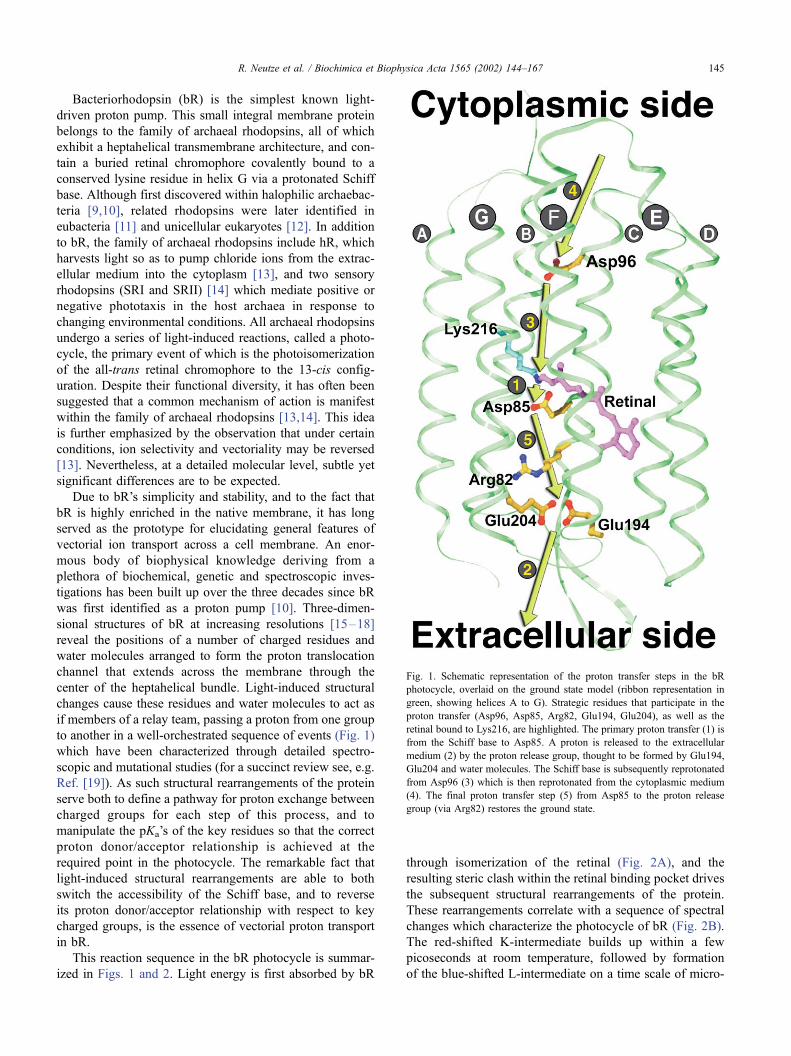
to another in a well-orchestrated sequence of events (Fig. 1)
which have been characterized through detailed spectro-
scopic and mutational studies (for a succinct review see, e.g.
Ref. [19]). As such structural rearrangements of the protein
serve both to define a pathway for proton exchange between
charged groups for each step of this process, and to
manipulate the pKa’s of the key residues so that the correct
proton donor/acceptor relationship is achieved at the
required point in the photocycle. The remarkable fact that
light-induced structural rearrangements are able to both
switch the accessibility of the Schiff base, and to reverse
its proton donor/acceptor relationship with respect to key
charged groups, is the essence of vectorial proton transport
in bR.
This reaction sequence in the bR photocycle is summar-
ized in Figs. 1 and 2. Light energy is first absorbed by bR
through isomerization of the retinal (Fig. 2A), and the
resulting steric clash within the retinal binding pocket drives
the subsequent structural rearrangements of the protein.
These rearrangements correlate with a sequence of spectral
changes which characterize the photocycle of bR (Fig. 2B).
The red-shifted K-intermediate builds up within a few
picoseconds at room temperature, followed by formation
of the blue-shifted L-intermediate on a time scale of micro-
Fig. 1. Schematic representation of the proton transfer steps in the bR
photocycle, overlaid on the ground state model (ribbon representation in
green, showing helices A to G). Strategic residues that participate in the
proton transfer (Asp96, Asp85, Arg82, Glu194, Glu204), as well as the
retinal bound to Lys216, are highlighted. The primary proton transfer (1) is
from the Schiff base to Asp85. A proton is released to the extracellular
medium (2) by the proton release group, thought to be formed by Glu194,
Glu204 and water molecules. The Schiff base is subsequently reprotonated
from Asp96 (3) which is then reprotonated from the cytoplasmic medium
(4). The final proton transfer step (5) from Asp85 to the proton release
group (via Arg82) restores the ground state.
R. Neutze et al. / Biochimica et Biophysica Acta 1565 (2002) 144–167 145

seconds. The primary proton transfer event from the Schiff
base to Asp85 (step 1 in Fig. 1) defines the L-to-M spectral
transition, which induces a large blue shift in the absorption
maximum. In wild-type bR under physiological conditions,
a proton is released to the extracellular medium (step 2 in
Fig. 1) on a similar time scale [20]. The identity of the
proton release group is somewhat ambiguous, but is
believed to be formed by Glu194, Glu204 and structural
waters [21,22]. It is accepted that, on the sub-millisecond
time scale, there is a spectrally silent transition [23] from an
early M (labeled M1) to a late M (labeled M2), which is
associated with a large structural rearrangement on the
cytoplasmic side of the protein [24]. These changes ensure
a switch in accessibility of the retinal, so that the Schiff base
can subsequently be reprotonated from Asp96 (step 3 in Fig.
1) on the cytoplasmic side [25], corresponding spectrally to
the M2 to N transition. Asp96, in turn, is reprotonated from
the cytoplasmic medium (step 4 in Fig. 1), and the absorp-
tion peak is further red-shifted as the retinal thermally
reisomerizes to recover the all-trans configuration, associ-
ated with the N to O transition. Finally, the ground state is
recovered when a proton is transferred from Asp85 to the
putative release group on the extracellular side via Arg82
(step 5 in Fig. 1).
In this review, we discuss the new insights into the
molecular mechanism of light-induced proton pumping
which have emerged from several recent high-resolution
structures. These were obtained by developing intermediate
trapping protocols, and applying them to crystals of wild-
type bR [26–29] and of bR mutants [30–32], as well as
analysis of ground state structures of mutants of bR that may
serve as structural analogues for the late intermediates in the
photocycle [33,34]. While the basic picture outlined above
is preserved, several important new details are revealed
which shed light on the key steps of vectorial proton
transport. In Section 2, we briefly sketch the most important
results from the highest resolution structures of the ground
state of bR which laid the framework for the intermediate
trapping studies in 3-D crystals. In Section 3, we describe
the different methodologies employed to structurally char-
acterize in 3-D the intermediates of the bR photocycle.
Section 4 presents the major structural results in progres-
sion, describing the early intermediate immediately follow-
ing photoisomerization of the retinal chromophore; the
sequence of events which set the stage for the primary
proton transfer event from the protonated Schiff base to the
primary acceptor Asp85; the coupled structural rearrange-
ments which assist proton release to the extracellular
medium; and the large-scale movements which facilitate
the Schiff base reprotonation from Asp96 on the cytoplas-
mic side of the protein, as well as proton uptake from the
cytoplasmic medium. Evidently, no single experiment yields
the full story in its own right. Nevertheless, by combining
the observed structural rearrangements and drawing com-
parisons with the wealth of biochemical, genetic and spec-
troscopic evidence, a coherent structural mechanism for
light-induced proton pumping by bR emerges. Section 5
reviews an ongoing debate concerning the specific nature of
the primary proton transport event. In closing, we summa-
rize the overall structural mechanism and speculate to the
extent that the central themes may be extended to aid the
understanding of other ion translocation processes.
2. Structure of the ground state of bacteriorhodopsin
Discovered in 1967 [9] and having its functional role
identified shortly afterward [10], the first electron crystal-
lography structural model for bR [35], recovered from tilted
2-D samples, appeared as early as 1975. This remarkable
pace of discovery, which resulted in bR being the first
integral membrane protein from which any structural infor-
mation was gleaned, was due to bR’s natural propensity to
form well-ordered two-dimensional crystals within the bac-
terial membrane of Halobacterium salinarum, combined
with the development of electron crystallography as an
important tool in structural biology. Nevertheless, it took a
further 15 years before a high-resolution 3-D structure for
Fig. 2. Schematic representation of light-induced retinal isomerization and
the bR photocycle. (A) The retinal chromophore is covalently bound to
Lys216 via a protonated Schiff base. Following absorption of a photon the
all-trans retinal is isomerized to the 13-cis configuration. (B) Retinal
isomerization is the first event in the photocycle. The spectral intermediates,
their absorption maxima, and their lifetimes at room temperature are shown.
R. Neutze et al. / Biochimica et Biophysica Acta 1565 (2002) 144–167146

bR was published by Henderson et al. [15], and this stands
out as a seminal contribution towards understanding the
structural mechanism of bR. From this basis, a series of
improved and refined electron crystallography structures
followed [36–38], eventually achieving a resolution below
3 A.
Three-dimensional crystallization experiments on bR
were extensively performed, and appeared quite promising
early on using detergent-solubilized bR [39]. Nevertheless,
crystals were disordered and it was not until 1996 that the
inception of a novel concept for the crystallization of
membrane proteins utilizing the properties of lipidic cubic
phases [40] resulted in the first well diffracting 3-D crystals
of bR [41]. The reasoning underlying this approach was that
membrane proteins might crystallize more readily in a lipid
bilayer environment, provided that they could be incorpo-
rated into an appropriate matrix, retain their native proper-
ties, and diffuse in three dimensions. Bicontinuous cubic
phases are highly viscous and transparent materials com-
posed of lipids and water, and provide a stable, structured
matrix in which diffusion of both water-soluble and mem-
brane proteins takes place. It was suggested that labile
membrane proteins, incorporated into continuous lipid
bilayers, could be stabilized, diffusing rather freely along
the bilayer, similar to the lateral diffusion of lipids. Upon
nucleation, this ‘‘feeding’’ mechanism would eventually
lead to well-ordered crystals. More details as to this crys-
tallization technique and its general applicability may be
found in recent references [42–46]. Other well-diffracting
3-D crystals of bR have since been grown from detergent-
solubilized bR by epitaxy [16] and vesicle fusion [47].
The rapid pace of elucidation of the structural mechanism
of bR which followed the development of lipidic cubic
phase crystallization was unanticipated. In the intervening 5
years, X-ray structures of bR from lipidic cubic phase-
grown crystals have been reported at 1.9 A resolution [17]
and 1.55 A resolution [18]. Since the 1.9 A structure was
free from merohedral twinning, both structures are of similar
quality and that at lower resolution is slightly more com-
plete. These results were followed by the X-ray structures of
the low-temperature K [26] (2.1 A), L [27] (2.1 A), and M
(2.25 [28] and 2.0 A [29]) intermediates of wild-type bR. In
addition, M-state structures have been published for the
D96N (2.0 A) [30] and E204Q [31] (1.8 A) bR mutants, as
well as the ground state structure of the D85S bR mutant,
claimed to model the O state (2.2 A) [34]. All these studies
used lipidic cubic phase grown crystals. Furthermore, high-
resolution X-ray structures of hR [8] (1.8 A) and SRII from
Natronobacterium pharaonis (2.1 A [48] and 2.4 A [49])
have recently been described using the same crystallization
approach. As such, the concept and implementation of
lipidic cubic phase membrane protein crystallization [40]
represents a significant contribution to understanding the
structural mechanisms of the archaeal rhodopsin family.
Since diffraction quality crystals of two bacterial reaction
centers have also been reported using this technique [44], it
is not unreasonable to expect that X-ray structures of other
membrane proteins may eventually be obtained using the
lipidic cubic phase crystallization methodology.
Several important features have emerged from the X-ray
structures of the ground state of bR at better than 2 A
resolution [17,18]. Most strikingly, the detailed atomic
nature of the Schiff base’s complex counter ion, which
stabilizes the high pKa of 13.5 [50] of the Schiff base and
the low pKa of 2.2 [51] of the Asp85 was revealed, and is
illustrated in Fig. 3. This counter ion is stabilized by a
network of water-mediated hydrogen bonds which extends
from the Schiff base, the primary proton donor, to the
extracellular surface. A key water molecule, Wat402, forms
H-bonds to the Schiff base nitrogen and the two negatively
Fig. 3. Refined structure and electron density of the extracellular half of bR
in the ground state. The 2Fobs–Fcalc refined electron density map (blue,
contoured at 1.2 r) shows the structure of the Schiff base’s complex counter
ion formed by Asp85, Asp212, Wat400, Wat401, Wat402 and Arg82. The
high pKa of the Schiff base (13.5) is stabilized by a H-bond to Wat402
within the framework of this complex counter ion. The low pKa of Asp85
(2.2) is stabilized by H-bonds to Thr89, Wat401 and Wat402. The close
proximity of the positively charged guanidinium group of Arg82 also helps
to stabilize the protonation states of both the Schiff base and Asp85. The
proton release group is thought to be formed by Glu194, Glu204 and water
molecules. The ground state model and crystallographic observations are
available from entries 1qhj and r1qhjsf of the protein data bank. Details are
found in Ref. [17]. This figure was drawn using a modified version of
Bobscript [130] and rendered using Raster3D [131].
R. Neutze et al. / Biochimica et Biophysica Acta 1565 (2002) 144–167 147

charged aspartates (Asp85, the primary proton acceptor, and
Asp212). As was anticipated by Gat and Sheves [52] through
studies on model retinal compounds, within the framework
of this complex counter ion, this water molecule stabilizes
the unusually high pKa of the Schiff base. Two further water
molecules (Wat400 and Wat401) combine to form a penta-
gon, and link the two negatively charged aspartates to the
positively charged guanidinium group of Arg82, which is
located further away towards the extracellular surface, but in
the ground state structure is orientated towards the active
site. The presence of three H-bonds to Asp85 (Od2 receives aH-bond from Thr89 and Od1 receives H-bonds from Wat401
and Wat402) as well as the position of the positive charge of
Arg82 stabilize the negative charge on the carboxylate group
to the extent that the pKa of Asp85 is 2.2 [51]. Arg82 in turn
is connected through a series of H-bonds (Fig. 3) to the
putative proton release group, formed by Glu194, Glu204
and water molecules [21,22].
Due to the 11 pKa units difference between the primary
proton donor and acceptor, even though these two groups
share H-bonds to the same water molecule in the resting
state, it is 1011 times more probable that a proton resides on
the Schiff base rather than Asp85 under physiological
conditions. These somewhat extreme pKa values illustrate
how the active site of bR has been optimized to ensure that
Asp85 is not protonated by the nearby Schiff base, or by the
extracellular medium prior to photoactivation. Similar argu-
ments show that the Schiff base cannot be deprotonated by
Asp85, any other nearby group, or by the cytoplasmic
medium, guaranteeing the optimal protonation state in the
bR resting state. It should be emphasized that this network
of water-mediated H-bonds on the extracellular half of the
protein (Fig. 3) also plays a significant structural role,
making the extracellular half of the protein more rigid and
brittle than the cytoplasmic half. An asymmetry in the
distribution of flexibility has been observed in neutron
diffraction studies [53] and is also reflected in the distribu-
tion of crystallographic temperature factors [16–18], which
are systematically lower on the extracellular half of the
protein.
Whereas several well-defined water molecules appear
along the proton translocation channel on the extracellular
side of the protein (Fig. 3), on the cytoplasmic side there are
relatively few. One conserved water (Wat501) appears
between Trp182 and Ala215 [17,18], and another crystallo-
graphic water (Wat502) was identified near the carbonyl
oxygen of Lys216 [18]. An H-bond from Ala215 to Wat501
creates a local distortion of the H-bond pattern of helix G in
this region, and this was recently assigned as a k-bulge [18],providing a more detailed description of the bend in helix G
which was seen as early as 1990 in the electron crystallog-
raphy structure [15]. With the exception of Asp96 and
Thr46, which share a H-bond, the cytoplasmic half of the
proton translocation channel consists predominantly of
hydrophobic residues. These pack tightly against each other
and create a hydrophobic barrier preventing the leakage of
protons back across the membrane, which would otherwise
dissipate the energy stored within the proton-motive poten-
tial. Nevertheless, it is necessary that a proton enters the
proton translocation channel from the cytoplasm during the
latter half of the photocycle. To assist this, the four aspar-
tates at the cytoplasmic surface of bR (Asp36, Asp38,
Asp102 and Asp104) serve to attract protons from the
cytoplasm. In addition, a number of 2-D studies of photo-
activated bR in projection [24,54–61] have firmly estab-
lished that a large-scale movement of helices on the
cytoplasmic half of the protein occurs following the depro-
tonation of the Schiff base, which opens up this hydro-
phobic barrier to the cytoplasmic medium.
Several other details regarding the interactions between
the bR molecules within the trimer, or interactions with lipid
molecules [16–18], have also emerged from the higher
resolution structure and are reviewed elsewhere [45,62].
3. Intermediate trapping and mutation protocols
Structural characterization of any reaction intermediate
within crystals requires that the protein is functionally active
in the crystalline state and that conditions can be found
under which a sufficient population of the desired inter-
mediate builds up [63]. In the case of lipidic cubic phase
grown 3-D crystals of bR, the protein packs in purple
membrane-like layers that are stacked along the direction
perpendicular to the plane of the membrane. Fourier trans-
form infrared (FTIR) studies have established that bR’s
photocycle in 3-D crystals closely resembles that of bR in
the native purple membrane [64]. A common strategy for
trapping structural intermediates of light-driven macromo-
lecules has been to illuminate 3-D crystals at low temper-
ature [65–68], although reaction initiation at room
temperature [69], as well as pump-probe experiments using
a broad spectrum X-ray probe [70,71], have also been
reported. Other strategies are to select a mutant for which
the decay of the desired intermediate is slowed, and thereby
recover a relatively high population of this intermediate.
Alternatively, one may determine the structure of a mutant
(or mutants) for which there are reasons to believe that its
ground state structure models a specific photo-intermediate.
In the last 3 years, structural results for the intermediates of
the bR photocycle have been reported using all of these
approaches, with the exception of room temperature pump-
probe studies which appear unlikely to succeed due to rapid
disordering of the crystal lattice.
Studies of the dynamics of bR at low temperature have
established two phase transitions that occur as a function of
temperature, corresponding to distinct changes in the nature
of the allowed motions. As shown in Fig. 4, the first phase
transition is at 150 K, where a change in the linear depend-
ence of the mean square amplitude of motion with temper-
ature sets in [53], and was observed by Zaccai et al. using
neutron scattering. A second phase transition occurs at 240
R. Neutze et al. / Biochimica et Biophysica Acta 1565 (2002) 144–167148

K, and was first identified using FTIR spectroscopy [72]. In
that work, Ormos established that the retinal, when depro-
tonated, becomes reprotonated from the extracellular side of
the protein below 240 K, but from the cytoplasmic side
above 240 K. Evidence for a phase transition at 240 K is also
suggested in the neutron scattering data [53] (Fig. 4). As is
illustrated schematically in Fig. 4, there exists a correlation
between the temperature at which these phase transitions
occur, and the temperature at which specific photo-inter-
mediates become populated when bR is illuminated.
A generally accepted protocol for trapping the K-inter-
mediate at low temperature (KLT) is to illuminate bR with
green light near (or below) 110 K [73–75]. Similarly, most
low temperature studies on the L-intermediate have either
illuminated bR with red light at 170 K [76–82], or have first
illuminated bR with green light at lower temperature and
then warmed the sample up to 170 K [83]. It is necessary to
raise the temperature to at least 150 K in order to ensure that
the protein has sufficient thermal energy to cross the energy
barrier associated with the K to L transition, and this
correlates with a distinct change in the nature of the allowed
motions above 150 K [53] (Fig. 4). Similarly, 2-D structural
studies in projection on the M-intermediates of bR have
shown that while no large-scale structural rearrangements
have been reproducibly observed below 240 K [75,81],
studies performed at 240 K or above have repeatedly
observed significant movements near the cytoplasmic sides
of helices E, F and G [24,54–61]. These movements, which
are characteristic of the spectrally silent M1 to M2 transition
(Fig. 2B), correlate with the phase transition observed at 240
K, in which the accessibility of the Schiff base switches
from the extracellular to the cytoplasmic side [72]. As such,
any protocol for trapping the M2 (or a later) intermediate of
the bR photocycle must ensure that the temperature is raised
Fig. 4. Low-temperature dynamics of bR. Hydrogen mean square
amplitudes ( y-axis) measured by neutron scattering are plotted as a function
of temperature (x-axis). Experimental and theoretical details are given in
Ref. [53]. A phase transition is visible at 150 K and (possibly) at 240 K,
representing a change in the nature of the allowed motions. The phase
transition at 240 K switches the accessibility of the Schiff base from the
extracellular to the cytoplasmic side [72]. A correlation with low-temper-
ature studies of the bR photocycle intermediates is represented schemati-
cally. Illumination of bR at 110 K is commonly used to generate and trap the
K-intermediate; likewise, illumination of bR at 170 K is commonly used to
trap the L-intermediate; at 230 K to trap the early M-intermediate; and
illumination above 240 K is used to trap the late M, as well as the later
intermediates. The K, L, M1 and M2 photo-intermediates and their
accessibility are shown schematically. For K, L and M1, the cytoplasmic
(upper) half of the proton translocation channel is closed, but becomes open
in M2. Photoisomerization breaks the accessibility to the extracellular side
below 150 (denoted schematically as two white circles), but the Schiff base
becomes accessible to the extracellular side (lower circle red) above 150 K,
and switches its accessibility to the cytoplasmic side (upper circle red) above
240 K [72]. Experimental data from Ref. [53] is reproduced with permission
from G. Zaccai. The schematic representation of the intermediates and their
accessibility are modified from those of Ref. [24].
Fig. 5. Difference spectra recorded from single 3-D bR crystals at low
temperature. (A) Following 30 s illumination with green (k= 532 nm) light
at 170 K, the difference spectrum (green) shows a mixture of K170 K (red)
and M170 K (orange) in the ratio of 4:1. (B) After a further delay of 40 s in
the dark, this evolved to the difference spectrum (green) yielding a mixture
of 1:3:1 for the K170 K (red): L170 K (blue): M170 K (orange) spectral
intermediates, with L170 K becoming the dominant species. The purple lines
give the spectral decomposition. (C) Following 30 s illumination with red
(k= 635 nm) light at 150 K plus a further delay of 40 s in the dark, the
difference spectrum (red) is characteristic of an almost pure L150 K. The
changes in electron density which resulted when following the trapping
protocols B and C are show in Fig. 6C and B, respectively. Details of the
spectral decomposition are given in Ref. [85]. Data shown in A and B are
reproduced with permission from Photochemistry and Photobiology.
R. Neutze et al. / Biochimica et Biophysica Acta 1565 (2002) 144–167 149

above 240 K for a sufficiently long period such that large-
scale rearrangements can occur.
In our work on the low-temperature K-intermediate of bR
[26], we used green light to illuminate 3-D crystals cooled to
110 K in a stream of nitrogen gas. Green light illumination
established a photostationary state containing a substantial
population of K110 K [73,74], which was characterized
spectrally using single crystal microspectrophotometry
[84]. The long-distance view of the difference Fourier
map [26] showed the structural rearrangements to be clus-
tered in the immediate vicinity of the active site, as was also
the case in studies on an early intermediate of photoactive
yellow protein [67]. Crystallographic refinement with partial
occupancy established that 35% K110 K was trapped in 3-D
crystals under these conditions.
In characterizing the structural changes associated with
the low-temperature L-intermediate [27] we worked at 170
K [76–82]. We used green light illumination for 30 s
followed by a further 40 s delay in the dark, at which point
we froze crystals in liquid nitrogen and later mounted them
in the X-ray beam. Fig. 5A shows the difference spectrum of
the photostationary state after 30 s illumination with green
light, which indicates a mixture of 48% K170 K and 12%
M170 K [85]. After a further 40 s delay in the dark, during
which most of the K170 K evolved to L170 K (Fig. 5B), the
crystals exhibit a mixture of 12% K170 K, 36% L170 K and
12% M170 K [85]. The long-distance view of the difference
Fourier map [27] showed that the structural rearrangements
evolved along the proton translocation channel towards the
extracellular medium. Since the illumination conditions
were similar to those used for trapping K110 K, the structural
evolution we observed from K110 K to L170 K (Fig. 6) arose
from the extra thermal energy at 170 K, which correlates
with the phase transition at 150 K [53] (Fig. 4). Crystallo-
graphic refinement with partial occupancy converged from
above and from below to a value of 70% for the trapped
photo-intermediate species [27,85], and the well-ordered
structure (Fig. 8 shows the 2Fobs–Fcalc electron density
map) was labeled LLT in recognition of the fact that this
was the predominant intermediate trapped, although the
presence of a small component of MLT was acknowledged
in the publication [27]. An apparent discrepancy between
the population estimates deriving from the spectroscopic
(60%) and crystallographic (70%) analyses stems from the
limitations of both techniques [85], since the white light
probe used to record a difference spectrum unavoidably
shoots a fraction of the trapped species back to the ground
state, whereas partial occupancy refinement with the ground
state model held fixed tends to overestimate slightly the
population of the intermediate species.
Since the publication of this structural result [27], it has
been suggested that the total trapped population, based on
spectroscopy alone, was approximately 30%, and hence the
X-ray crystallographic result contained a dominant (rather
than a minor) contribution from the M-intermediate [86–
88]. These spectral assignments, which account for less than
50% of the total population of the trapped intermediate
[27,85], were based upon the assumption that spectra re-
corded from 3-D crystals of bR at 170 K are identical in all
respects to those recorded from purple membrane samples in
suspension at other temperatures and pH. It is well known
that absorption spectroscopy of proteins can depend on the
medium in which they reside, the pH and the temperature.
Specifically, spectra recorded from 3-D crystals often differ
from those recorded from samples in suspension [89–91]
and this is also true for 3-D crystals of bR grown in a lipidic
cubic phase. In keeping with this, the maximum extinction
of the M-intermediate at 410 nm in 3-D crystals of the
D96N bR mutant [62] was equal to that of the ground state
peak at 570 nm, although scatter at short wavelengths
somewhat affected the spectral observations. That the spec-
tral fingerprint for the M-intermediate at 410 nm in 3-D
crystals of wild-type bR cooled to 170 K [85] is strong
relative to its fingerprint in samples in suspension at higher
temperatures was central to this issue, and has been elabo-
rated upon through a more detailed spectral analysis [85].
Furthermore, illumination of 3-D crystals of bR with red
light at 150 K results in a significant population of L150 K,
Fig. 6. Evolution of structural changes resulting from photoactivation of bR at low temperature, overlaid (in A, B and C) on the ground state bR model. Positive
and negative difference electron densities are shown in blue and yellow, respectively. (A) Stereo view of the difference Fourier map resulting from illumination
of a single 3-D crystal of wild-type bR with green (k= 532 nm) light at 110 K contoured at 4 r (r= r.m.s. electron density for the unit cell). These changes
characterize the KLT intermediate. A negative difference electron density peak indicates the disordering of Wat402, as is a weaker peak visible on Wat401.
Paired negative and positive difference electron density peaks indicate a small movement of Asp85 towards the Schiff base as well as movements of the side
chain and backbone of Lys216. The circle between Asp85, Wat400 and Wat402 highlights positive difference electron density which may correlate with the
reordering of one disordered water molecule. (B) Stereo view of the difference Fourier map resulting from 30 s illumination of a 3-D bR crystal with red light at
150 K, followed by a delay of 40 s prior to quenching in liquid nitrogen. This map is contoured at 2.8 r. (C) Stereo view of the difference Fourier map resulting
from 30 s illumination of a 3-D bR crystal with green light at 170 K, followed by a delay of 40 s prior to quenching in liquid nitrogen. This map is contoured at
3.4 r. B and C show similar structural rearrangements, which characterize the LLT.intermediate. Negative electron density peaks arise on Wat400, Wat401 and
Wat402, and (in C) a positive peak is visible at the center of this triad. Both maps show paired negative and positive difference density peaks corresponding to a
reorientation of the guanidinium group of Arg82 towards the extracellular medium. Extended paired negative and positive electron density peaks stretch along
the backbone of helix C, indicating a local flex of this helix towards the proton translocation channel. Electron density peaks indicating a movement of the side
chain of Trp182 towards the cytoplasm are also visible. (D) Structural models for KLT (blue, a refinement with fewer constraints, crystallographic observations
are available from entries 1QKO and 1QKP of the protein data bank) and LLT (red, model and crystallographic observations are available from entry 1EPO of
the protein data bank) overlaid on the ground state (green backbone and colored residues, model and crystallographic observations are available from entries
1qhj and r1qhjsf of the protein data bank). All the major changes described above are reproduced through crystallographic refinement. This figure was drawn
using the Swiss PDB Viewer [132].
R. Neutze et al. / Biochimica et Biophysica Acta 1565 (2002) 144–167150

R. Neutze et al. / Biochimica et Biophysica Acta 1565 (2002) 144–167 151

whereas those of K150 K and M150 K are negligible (Fig. 5C).
It was therefore decisive that the analysis of the X-ray
diffraction data to 2.3 A recorded following this trapping
protocol (Fig. 6B and C show the difference Fourier maps
recovered using the red and green light trapping protocols,
respectively) reproduced the earlier structural result [27].
Four studies have recently reported X-ray structures of
the M-intermediate from wild-type bR [28,29], and from the
D96N [30] and E204Q [31] bR mutants, all using 3-D
crystals grown in a lipidic cubic phase. The trapping
protocols for three of these studies [28,30,31] were similar,
but they differed markedly from protocols successfully used
to observe large-scale light-induced movements in bR
[24,54–61]. bR crystals were first frozen in liquid nitrogen
and then mounted in a cryo-cooled stream of nitrogen gas.
The cold nitrogen stream was subsequently blocked for a
short period of time, during which crystals thawed and were
simultaneously illuminated with either yellow [30,31] or
green [28] light. After either 1 [28,31] or 3 s [30] the object
used to block the nitrogen gas stream was removed and
crystals were refrozen. No characterization of the thermal
history of crystals when following this protocol has been
presented. The observation in the case of the E204Q mutant
that X-ray diffraction data could be integrated with identical
unit cell parameters to those of the ground state [31]
suggests that either the final temperature reached, or the
time interval at this temperature, was not sufficient to allow
large-scale structural rearrangements of the protein during
the second in which the crystal thawed. Curiously, this
protocol yielded a structure of the early M1 intermediate
for the E204Q mutant rather than the O-intermediate which
is expected when working with this bR mutant at room
temperature [21]. A very similar trapping protocol for wild-
type bR yielded a mixture of 35% bR, 30% M1 and 35% M2
[28]. More consistent with studies on bR mutants at room
temperature, a period of 3 s for thawing and illuminating 3-
D crystals of the D96N bR mutant produced almost 100%
M2 [30]. Unfortunately, in this case, the electron density was
so disordered that the cytoplasmic portions of helices F and
G, which show the largest movements and are therefore of
the greatest mechanistic significance, were excluded from
the model. A distinctly different trapping protocol was used
by Facciotti et al. [29] who first illuminated wild-type bR
crystals for 3 min at 230 K, and then quenched them in
liquid nitrogen prior to X-ray diffraction data collection.
Since this temperature lies below the phase transition at 240
K [72] (Fig. 4) an early M1 intermediate would be expected.
The structure which was recovered [29] was in good agree-
ment with the early M of the E204Q mutant [31], illustrating
(as in the case of red light illumination at 150 K versus
green light at 170 K, Fig. 6) that the same structure for a
specific intermediate can be recovered using different trap-
ping protocols.
In recovering 3-D structural information from the N-
intermediate, Vonck [32] illuminated 2-D samples of the
F219L bR mutant for 30 s at 270 K prior to plunging the
sample into liquid nitrogen. This protocol recovered a
population of 33% for the trapped N-intermediate, as would
be expected for the photostationary state under the condi-
tions used. At 270 K, large movements associated with the
cytoplasmic sides of helices E, F and G were observed.
These movements did not occur when 3-D bR crystals were
illuminated at lower temperatures [26,27,29] and they
correlate with the phase transition at 240 K [72], which
serves to switch the accessibility of the Schiff base from the
extracellular to the cytoplasmic side.
Another approach for gleaning structural information
concerning the second half of the bR photocycle (Fig. 2B)
has been to determine the ground state structure of bR
mutants which serve as models for the later intermediates.
Subramaniam and Henderson [33] recently reported the
electron crystallographic structure of the D96G/F171C/
F219L triple mutant of bR. Justification for this approach
derives from 2-D electron diffraction studies in projection
which demonstrated that, for this specific triple mutant, the
full extent of the light-driven conformation change asso-
ciated with the late M2 intermediate of the bR photocycle
is present even without illumination [24]. As such, and
despite the shortcomings of using a triple mutant analogue
rather than a trapped photocycle intermediate, this
approach yielded the most reliable structure for the open
conformation of bR to date. The same philosophy has been
applied to argue that the X-ray structure of the ground
state of the D85S bR mutant provides a model for the O-
intermediate of bR [34]. In contrast to the work of
Subramaniam and Henderson [24,33], this conjecture is
not supported by structural studies of the D85S bR mutant
and the O-intermediate of bR in projection. Rather, an
appeal is made to the 2-D structure of the D85N bR
mutant at high pH, which appears to exhibit an M-like
conformation [92]. While the structure of the D85S bR
mutant is of interest in its own right, as this mutation (like
the D85T bR mutant) converts the protein to an inwardly
directed chloride pump [93], its structural link to the O-
intermediate of the bR photocycle does not appear to be
well founded.
4. Structural evolution of bR: resolving a revolving
photocycle
In an ideal case, high-resolution X-ray structures from
wild-type bR crystals free of merohedral twinning and at
100% occupancy would be reported for every intermediate
in the photocycle (Fig. 2B). Despite the fact that all experi-
ments described above fall short of this ultimate case, when
viewed with an acceptance of the limitations of the techni-
que, the structural results themselves present a remarkably
consistent picture of the structural evolution during the first
half of the bR photocycle [26,27,29,31]. Unfortunately, the
X-ray crystallographic results from the later intermediates
have been in conflict both with each other [28,30,34] and
R. Neutze et al. / Biochimica et Biophysica Acta 1565 (2002) 144–167152

with those derived from electron diffraction studies [32,33].
Nevertheless, although at lower resolution, a reliable 3-D
model for the conformation of bR which is open to the
cytoplasm is provided by the electron crystallography struc-
ture of the D96G/F171C/F219L triple mutant of bR [33],
and the major conclusions drawn from this work are well-
supported by the 3-D model of the N-intermediate of the
F219L mutant [32]. From these results, in combination with
a wealth of spectral, genetic and biochemical data, a
molecular level 3-D movie of bR in action [94] can be
pieced together and is described below.
4.1. Low-temperature X-ray structure of K
At room temperature, the K-intermediate builds up
within a few picoseconds following photoisomerization of
the retinal (Fig. 2). Of particular interest is that the C13jC14
bond of the retinal chromophore is isomerized [95,96] and
therefore the energy requirements for proton transport uphill
against a proton-motive potential must already be apparent
as either structural rearrangements or strain on the retinal.
When observing limited structural changes, Henderson and
Moffat [97] have shown that difference Fourier methods
provide the most sensitive and least biased method of
analysis. For this reason, it has become almost universal
to present a difference Fourier map when describing struc-
tural changes in light-driven macromolecular systems [26–
28,65–68,70,71]. One significant advantage of the method
is that, when a long-distance overview of the map is
presented [26,27,67], the background noise level of the
map can be seen by inspection, since regions of the protein
removed from the active site provide an internal control.
Overviews of the difference Fourier maps for KLT and LLT
can be found in our references [26,27] but were not
presented by other workers in the field in their work that
is described below.
Fig. 6A shows the difference Fourier map for KLT near
the active site, and Fig. 6D shows the refined KLT model
(blue). Paired positive and negative electron density peaks
near the side chain and carbonyl oxygen of Lys216 illustrate
how its side chain and main chain move in response to
retinal isomerization. A movement of the side chain atoms
may have been anticipated from the fact that N~ of Lys216
(i.e. the Schiff base nitrogen) is covalently bound to C15 of
the retinal (Fig. 2A) and necessarily undergoes a significant
movement in response to photoisomerization about the
C13jC14 bond. That a physical pull of the retinal through
the side chain of Lys216 also results in a movement of the
backbone may in part be facilitated by a weakening of the
H-bond formed by this carbonyl oxygen due to a local k-bulge of helix G in this region [18]. In addition, the polyene
chain and h-ionone ring of the retinal are firmly held in
place by bulky hydrophobic residues which form a closely
packed binding pocket, hence the retinal itself cannot
undergo large movements so as to relieve strain [98,99].
An early indication of strain within the retinal which would
serve to store energy in KLT and later be released as the
photocycle evolved is the observation that retinal isomer-
ization induces a steric clash of its C13 methyl group with
Trp182 on helix F [26]. At 2.1 A and 35% occupancy,
however, the extent to which the retinal was twisted could
not be observed and planar constraints were applied during
refinement. Resonance Raman spectroscopy has established
that the retinal is strained in K, somewhat more so at low
temperature than at room temperature [96].
The strongest negative electron density peak in the
difference Fourier map arose on Wat402 (Fig. 6A), indicat-
ing that this key water molecule, which in the ground state
[17,18] was hydrogen bonded to the Schiff base, Asp85 and
Asp212 (Fig. 3), becomes disordered in response to retinal
isomerization. This result is consistent with FTIR studies at
low temperature which also demonstrate that the Schiff base
H-bond is lost in KLT [99]. There are two factors which
combine to dislodge this water molecule. In the first
instance, retinal isomerization reverses the orientation of
the N–H dipole of the Schiff base, which served as a H-
bond donor to Wat402 in the ground state model, but cannot
do so when the retinal is isomerized. In the second instance,
retinal isomerization induces a movement of the side chain
of Lys216 (Fig. 6A) such that it swings across and ‘‘kicks
out’’ this water molecule (Fig. 7) like a football player
striking a football. In the refined model (Fig. 6D), the
Fig. 7. Structural mechanism for the disordering of Wat402 in the active site
in the KLT intermediate. A space filling representation of the ground state
(colored) and KLT (white) models show that, due to the photoisomerization
of the retinal, the movement of Cq of Lys216 induces a steric clash with
Wat402 (black). Consequently, this water molecule is displaced. The upper
left part shows the active site orientation in a ball and stick representation.
This figure was drawn using the Swiss PDB Viewer [132].
R. Neutze et al. / Biochimica et Biophysica Acta 1565 (2002) 144–167 153

distance from Cq of Lys216 to the position originally oc-
cupied by Wat402 is 2.3 A, which results in a steric conflict
(highlighted in the space filled model, Fig. 7) since this Cq
cannot serve as a H-bond donor to Wat402. In the difference
Fourier map for KLT, there is positive density in a position
between Asp85 and Asp212 (Fig. 6A), overlapping with a
positive feature found in the LLT map (Fig. 6C), which may
correspond to reordering of this water molecule. This
rearranged water molecule (Fig. 6D, blue) is also seen for
all later intermediates [27–31]. A weaker negative differ-
ence electron density peak appears on Wat401 in KLT (Fig.
6A), indicating that a movement of Wat402 is also commu-
nicated to this water molecule. An early movement of
Asp85 towards the position originally occupied by the
Schiff base is also apparent as paired negative and positive
difference electron density peaks near this residue. These
slight movements were not resolved in the difference Four-
ier electron diffraction map resulting from green light
illumination of bR at 77 K [75].
One appealing consequence of the postulate that a steric
clash of Cq of Lys216 contributes to dislodging Wat402 is
that it also suggests why isomerization about the C13jC14
double bond (Fig. 2A) initiates a sequence of events result-
ing in the translocation of a proton across the cell mem-
brane, whereas isomerization about the C15jN~ double
bond (which corresponds to dark adaptation of bR [100])
does not. A simple model for the dark-adapted state does not
reveal this steric clash of Cq with Wat402 and therefore it is
likely that this water remains well-ordered in the dark-
adapted state. All high-resolution X-ray structures of the
later intermediates [27–31] are consistent with these obser-
vations for KLT since all show movements of Wat402,
Asp85 and Lys216. From an energetics viewpoint, it is
appealing that two energetically expensive tasks are fulfilled
early in the photocycle, namely the disruption of three H-
bonds to Wat402 and the weakening of a H-bond formed by
the carbonyl oxygen of Lys216. By achieving these tasks
before the energy absorbed by the photon is thermally
dissipated, the stage is set for more extensive structural
rearrangements later in the photocycle.
4.2. Low-temperature X-ray structure of L
At room temperature, the L intermediate builds up in the
microsecond time range (Fig. 2B) and governs the primary
proton transfer event from the Schiff base to Asp85 (Fig. 1).
A strong case can be argued that the structure of the L
intermediate is decisive when seeking to understand the
mechanism of vectorial transport by bR. Most strikingly,
only the removal of the retinal or the mutation of Asp85 to a
neutral group are effective in preventing bR from pumping
protons [101]. Changes in the temperature, pH, or the
mutation of any other charged residue along the proton
transport channel (Fig. 1) [20] affect the photocycle kinetics
and can alter the order of specific events. Nevertheless, bR’s
ability to translocate protons is quite tolerant of these
perturbations. For these reasons, an increased emphasis
has recently been placed upon the primary proton transfer
event as central to vectoriality in the proton pumping
mechanism [27,33,85,102,103].
A structural model for L must explain three remarkable
features of this intermediate. In the first instance, whereas
the K intermediate builds up in the picosecond time range
(Fig. 2B), there is a delay of six orders of magnitude before
K decays into the L intermediate. This difference makes the
K intermediate by far the relatively longest lived intermedi-
ate in the bR photocycle. In the second instance, structural
changes associated with the L intermediate must serve to
reverse the proton donor/acceptor relationship of the Schiff
base and Asp85. As described in Section 2, the ground state
structure stabilizes the high pKa (13.5) of the Schiff base
[50], and the low pKa (2.2) of Asp85 [51]. By L, the pKa of
the Schiff base must be reduced, and that of Asp85
increased, to the point where a proton can exchange
between these oppositely charged groups in the L to M
transition. Finally, retinal isomerization reverses the orien-
tation of the Schiff base N–H dipole away from the
extracellular side of the protein and towards the cytoplasmic
side. The structure of L must imply a mechanism whereby a
low barrier pathway for proton exchange can be defined,
such that the Schiff base becomes transiently accessible to
Asp85 on the extracellular side.
Fig. 6C shows the difference Fourier electron density
map dominated by L170 K [27,85], and the refined structure
is shown in Fig. 6D (red). As expected, the structural
changes observed in K110 K (Fig. 6A) propagate out from
the active site towards the extracellular medium in L170 K
(Fig. 6C). As with K110 K, paired positive and negative
peaks arise near the carbonyl oxygen of Lys216, and a
strong negative electron density peak is apparent for Wat402
in L170 K. In addition, in L170 K considerably more structural
rearrangements are apparent. Strong negative peaks are now
also observed on Wat400 and Wat401, which have dislo-
cated, as is a positive electron density peak visible, indicat-
ing the reordering of one water molecule [27] near the center
of this triad of ground state water molecules. Furthermore,
paired negative/positive electron density peaks clearly show
that the positively charged guanidinium group of Arg82 has
reoriented towards the extracellular medium. However, the
most striking feature of the difference Fourier map is the
presence of well-defined positive and negative electron
density peaks along the backbone of helix C, extending
from Ala81 to Phe88 (Fig. 6C). These features were
interpreted as stemming from a local flex of helix C, which
exaggerates a small movement of Asp85 towards the Schiff
base already apparent in KLT (Fig. 6A). The disruption of
three H-bonds on the extracellular side of the protein after
photoexcitation at 170 K [27] which were not disrupted at
110 K [26] is due to the increased thermal energy of the
protein and surrounding solvent at this temperature, and this
correlates structurally with the phase transition at 150 K [53]
(Fig. 4).
R. Neutze et al. / Biochimica et Biophysica Acta 1565 (2002) 144–167154

As shown in Fig. 5C, when using red light illumination at
150 K rather than green light illumination at 170 K, there is a
strong spectral contribution from L150 K but that from K150 K
or M150 K is negligible. Fig. 6B shows the difference Fourier
map which resulted from red light illumination of a 3-D
crystal at 150 K. Although this map is noisier than that
obtained following our green light illumination protocol [27]
(Fig. 6C) due to the lower total occupancy of the trapped
intermediates, it is fully consistent with the difference Four-
ier map previously reported. The choice between either
trapping protocol demands a compromise, with that using
red light favoring spectral purity at the expense of greater
noise in the crystallographic electron density map, whereas
that using green light minimizes the crystallographic noise at
the expense of a greater mixture of states in the difference
spectrum. While our original publication reflected our own
bias within this context [27], crystallographic refinement
establishes that both protocols yield the same structure for
the low-temperature L-intermediate. These movements were
not resolved in the difference Fourier electron diffraction
map in projection resulting from red light illumination of bR
at 170 K [81].
In Fig. 8 the 2Fobs–Fcalc electron density map and
refined model for L170 K are shown. Since the trapped
intermediate had a crystallographic occupancy of 70%
[27,85] all significant features of the difference Fourier
map, such as rearrangements of water molecules, reorienta-
tion of Arg82, and the local flex of helix C, are immediately
visible within the well-ordered electron density. There are
compelling reasons to believe that the structures of L170 K
and M170 K are very similar [29,72,85], not the least being
the fact that a single structural intermediate provided an
excellent model for the observed electron density (Fig. 8). It
is curious to note that, whereas our trapping protocol
produced a mixture of spectral intermediates yet well-
ordered electron density (Fig. 8), the opposite occurred for
the late M-state of the D96N bR mutant, for which a
spectrally pure species was trapped [62] but the electron
density was disordered to the point that vital regions were
excluded from the model [30].
Our observation that a local rearrangement of a secon-
dary structural element (i.e. a flex of helix C) is associated
with the K-to-L transition explains why a time scale of
microseconds (rather than nanoseconds) is required for this
spectral transition (Fig. 2B). Furthermore, our X-ray model
provides a global picture incorporating the suggestion that
Asp85 approaches the Schiff base in L [79,102] which
derived from solid-state NMR spectra recorded at 170 K.
In addition, a structural basis for the 40 nm blue shift
associated with the K-to-L transition is revealed. As estab-
lished through studies of a number of retinal salts by Albeck
et al. [104], the spectral shift depends critically on the
distance between the protonated Schiff base and the primary
counter ion (Asp85 in bR), with a blue shift resulting as the
two approach one another. Furthermore, the reorientation of
the guanidinium group of Arg82 away from the Schiff base
also contributes to the spectral shift [48,49,105]. These
factors combine to produce a significant blue shift, which
may in part be compensated by a reduction of strain on the
retinal [102]. Unfortunately, as with the structure of KLT, at
2.1 A resolution, it was not possible to observe the extent to
which the retinal was twisted and (of necessity) constraints
were imposed during refinement.
Considerable insight into how the proton donor/acceptor
relationship of the Schiff base and Asp85 is reversed prior to
proton transfer is gained from the structure of LLT. As
Fig. 8. Refined structure (red) and electron density (blue) of the
extracellular half of LLT overlaid on the ground state model (colored).
The 2Fobs–Fcalc refined electron density map (contoured at 1.0 r) revealsthat the Schiff base’s complex counter ion (Fig. 3) is considerably disrupted
prior to proton transfer. The three H-bonds between Asp85 and Thr89,
Wat401, and Wat402 are lost, and a new water molecule (Wat451) reorders
between Asp85 and Asp212. The side chain of Arg82 reorientates towards
the extracellular medium, and the backbone carbonyl oxygen of Arg82
moves into a position where it can compete with Asp85 as a H-bond
acceptor from Wat451. A local flex of helix C exaggerates the movement of
Asp85 towards both the Schiff base and Asp212. These structural
rearrangements increase the pKa of Asp85 to the point where it becomes
a proton acceptor relative to the photoisomerized Schiff base. Since the
well-ordered electron density corresponds to the structure of LLT (red)
rather than the ground state model (colored) it is apparent that the
occupancy of LLT is significantly greater than 50%. The model for LLT and
the crystallographic observations are available from entry 1EPO of the
protein data bank. Details are found in Refs. [27] and [85]. This figure was
drawn using a modified version of Bobscript [130] and rendered using
Raster3D [131].
R. Neutze et al. / Biochimica et Biophysica Acta 1565 (2002) 144–167 155

described in Section 2, the high pKa of the Schiff base is
stabilized in the ground state by a H-bond to Wat402 (Fig.
3). That a H-bond from the Schiff base to a strategically
placed water molecule could yield an unusually high pKa
(13.5) for this group was anticipated by Gat and Sheves [52]
through studies on model retinal compounds. Upon forma-
tion of the K intermediate, retinal isomerization disorders
this key water molecule [26] (Fig. 6A) and reorientates the
Schiff base nitrogen into a hydrophobic region. This
decreases the pKa of the Schiff base by the order of 5 pKa
units, an approximate value which stems from the observa-
tion that the pKa of the Schiff base is reduced to 8.5 when
Asp85 is replaced by a neutral residue [106], which could be
expected to disrupt the very same salt bridge between the
Schiff base and Asp85. A very similar value for the pKa of
the Schiff base at its reprotonation step was determined from
kinetic studies of the D96N mutant [107]. In the ground
state structure, the low pKa of 2.2 of Asp85 is stabilized by
H-bonds to two water molecules (Wat401 and Wat402) and
to Thr89 (Fig. 3), all of which act as H-bond donors to the
negatively charged carboxylate. The loss of a H-bond to
Wat402 in K110 K (Fig. 6A) increases the pKa of Asp85 [26],
and this value is further increased in L170 K by the loss of H-
bonds to Wat401 and Thr89 (Fig. 6C) [27]. Nevertheless,
one water molecule (Wat451, Fig. 8), which reorders
between the two negatively charged aspartates, is still able
to act as a proton donor to the carboxylate of Asp85.
Because of the reversal of the orientation of the Schiff base
nitrogen due to retinal isomerization, this positively charged
group no longer compensates the negative charge on
Asp212. In addition, by LLT the positively charged guani-
dinium group of Arg82 has reoriented towards the extrac-
ellular medium (Figs. 6C,D and 8). Indeed, the movement of
Arg82 facilitates a local flex of helix C [27] which exag-
gerates the interactions between the negatively charged
Asp85 and Asp212, and also brings the backbone carbonyl
oxygen of Arg82 into a position where it competes with
Asp85 as a H-bond acceptor from Wat451 (Fig. 8). That the
orientation of Arg82 has a dramatic effect on the pKa of
Asp85 is reflected by the fact that its replacement with a
neutral residue (glutamine or alanine) increases the pKa of
Asp85 by 4.6 units [108–110]. Furthermore, for the R82H
mutant at low pH (at which His is likely to be protonated)
proton transfer from the Schiff base to Asp85 is almost
entirely suppressed, yet the proton-transfer kinetics become
similar to wild-type bR at high pH [111]. The combined
effect of all these structural rearrangements is to raise the
pKa of Asp85 to the point where it becomes a proton
acceptor relative to the Schiff base. Braiman et al. [112]
have estimated that the pKa of Asp85 reaches a value as
high as 10.5 during the photocycle, which would be
sufficient to reverse the proton donor/acceptor relationship
of this group with respect to the lowered pKa of the
isomerized Schiff base.
Proton exchange between the Schiff base and Asp85
requires the existence of a low barrier pathway. This may
be facilitated by H-bonds to a water molecule [28], to
Thr89 [33], or by direct proton transfer from the Schiff
base to Asp85 [85]. In a recent review, Kandori [103]
argued that FTIR spectroscopy studies appear consistent
with either water-mediated or direct proton transfer. Never-
theless, the remarkable fact that the decay of the K-
intermediate is six decades slower than its build-up, and
that the K-to-L transition is associated with a local bend of
helix C (Fig. 6C), strongly implies that this local pertur-
bation of a secondary structural element is a crucial factor
in redefining a pathway for proton transfer. This is further
supported by the fact that the primary proton transfer
occurs 10 times more rapidly in the D85E mutant [113],
which would require smaller structural rearrangements to
facilitate direct proton transfer. From this basis we postu-
lated that the observed movement of Asp85 towards the
Schiff base, which is exaggerated by this local bend of
helix C (Fig. 6B,C), facilitates a mechanism whereby a
proton is transferred directly from the Schiff base to
Asp85, and proton transfer is modulated by structural
fluctuations of the retinal [85]. That structural fluctuations
of the photoisomerized retinal could transiently create a
low barrier pathway for proton transfer from the Schiff
base to Asp85 provides a modern picture curiously resem-
bling (yet distinct from) a previously postulated double-
isomerization mechanism [114]. Proton transfer would
cancel the mutual electrostatic attraction between the
positively charged Schiff base and the negatively charged
Asp85, and thereby enable strain on helix C, as well as
strain on the retinal [102], to relax structurally and draw
the two groups apart. A separation of the primary proton
donor and acceptor would certainly contribute strongly to
vectoriality of the proton pump bR [27,33,85,102], since
the reverse proton transfer from Asp85 to the Schiff base
would be hindered. Alternative mechanisms for both the
primary proton transfer event and their contribution to
vectoriality are reviewed in Section 5.
4.3. Structures for the M-intermediates of bR
At room temperature, the primary proton transfer event,
which corresponds spectrally to the L-to-M transition, takes
approximately 40 As. It is well established by time-resolved
studies that a spectrally silent transition, from an early to a
late M-intermediate (i.e. M1 to M2, Fig. 2B), follows on the
time scale of 350 As [23]. Furthermore, a series of studies on
2-D crystals of bR in projection have conclusively shown
that a large-scale structural rearrangement is associated with
this transition, which opens the proton translocation channel
to the cytoplasm [24,54–61]. It has long been considered
that structures of both the early and late M-intermediates
would shed light on the mechanism by which the Schiff
base switches its accessibility from the extracellular to the
cytoplasmic side. Structural details elucidating a pathway
for reprotonation of the Schiff base from Asp96 on the
cytoplasmic side (Fig. 1) [28,31], and the nature of the
R. Neutze et al. / Biochimica et Biophysica Acta 1565 (2002) 144–167156

movements which open the proton translocation channel to
the cytoplasmic medium [32,33], have indeed emerged.
X-ray structures of the M-intermediates of the D96N bR
mutant [30], the E204Q bR mutant [31], and of wild-type
bR [28,29] have all been reported. The trapping protocols
used in these studies are described in Section 3. Only in the
work of Sass et al. [28] was a difference Fourier map
presented. On the extracellular half of bR, the observed
structural rearrangements of these M-states are all in agree-
ment, within the errors of the technique. While there are
variations with respect to the exact coordinates, these
indicate noise within the crystallographic data, limitations
of the trapping protocols, and structural changes due to the
E204Q mutation on the extracellular side [31]. As would be
expected, the key structural rearrangements which were
required to set the stage for proton transfer were also
present shortly after proton transfer. For example, all
structures of M showed similar rearrangements of water
molecules and a similar change in the orientation of Arg82
as seen in LLT (Fig. 8) [27]. One striking difference between
the structures of LLT and M concerns the local flex of a-
helix C in LLT, shown in Fig. 6C as paired negative and
positive electron density peaks which extend more than a
full turn. While the difference Fourier map of the M state
reported by Sass et al. [28] showed paired electron density
peaks near the backbone carbonyl oxygens of Arg82 and
Tyr83, there were no such peaks on the backbone of Asp85.
Unfortunately, no difference Fourier maps were presented
by the other workers, but nevertheless Luecke et al. [30]
emphasized the fact that no displacements of the main chain
were evident in helices A to E in their late M-state structure.
As such these structures of the M-intermediate appear to
support our mechanistic postulate that the local flex of helix
C, which exaggerates the approach of Asp85 towards the
Schiff base immediately prior to proton transfer, could relax
to some extent following proton transfer since the mutual
electrostatic attraction of the two groups would be can-
celled.
One interesting feature of the proton pumping mecha-
nism is that, for wild-type bR and at physiological temper-
ature and pH, the timing of proton release to the
extracellular medium is roughly coincident with the primary
proton transfer event [20]. Structural changes on the extrac-
ellular side of bR provide insight into how this occurs.
Luecke et al. [30] argued that the observed reorientation of
the guanidinium group of Arg82 would destabilize the
proton release group, which consists of the dyad of
Glu194 and Glu204 [21,22] and associated water molecules.
While it seems certain that the movement of Arg82 makes a
significant contribution to the mechanism of proton release,
it was also emphasized by Luecke [62] that the proton
release complex is well insulated from the aqueous medium
and hence other structural rearrangements are also required.
Some clues for these additional rearrangements, which
mainly include partial disordering near the glutamate dyad,
have been reported [27].
While there is good agreement with respect to the nature
of the structural rearrangements which occur on the extrac-
ellular side of the protein, there have been conflicting
suggestions regarding the timing of these occurrences.
Luecke et al. [30,31] and Sass et al. [28] have postulated
that the reorientation of Arg82 is triggered by the proto-
nation of Asp85, yet the structure of LLT (Figs. 6B–D and
8) shows that this rearrangement occurs prior to the primary
proton transfer event [27,85]. Luecke et al. [30] argued that
the loss of H-bonds from Asp85 to water molecules and to
Thr89 would be sufficient to achieve proton transfer and
generate the M1 state. They further suggested that proton
transfer to Asp85 would then trigger the reorientation of the
guanidinium group of Arg82, thereby causing proton release
to the extracellular medium, leading to the transition from
the early M state to the late M state, and increasing the pKa
of Asp85 to the point where it could no longer reprotonate
the Schiff base. This structural mechanism promotes the
observed movement of the side chain of Arg82 as the central
element of vectoriality in the proton pump bR, and hinges
upon the coincidence in time of proton transfer and proton
release. It is therefore difficult to reconcile this mechanism
with the conclusions drawn by the same group from kinetic
studies at different temperature, pH, and of specific mutants,
that proton release is not directly linked to Schiff base
deprotonation, and that the temporal coincidence between
them under some conditions must be fortuitous [20]. Indeed,
this structural mechanism was overturned shortly later by
the same group when they observed that the reorientation of
Arg82 had already occurred by the formation of M1 [31]. In
fact, proton release appears to occur slightly faster than
proton transfer in wild-type bR at 278 K, significantly
slower at 308 K, and much slower still at pH < 5 [20].
Furthermore, the apparent coupling between proton transfer
and release is entirely removed in several mutants, such as:
the D85E bR mutant for which proton transfer is accelerated
10-fold while proton release is delayed 2-fold [20,113], the
R82N and R82Q bR mutants for which M-formation is very
rapid yet proton release is considerably delayed [108–110],
and the E204Q mutant for which proton release is again
delayed [21]. Whereas titration experiments on the unpho-
tolyzed ground state implied a coupling between proton
transfer to Asp85 and proton release of approximately 5 pKa
units [100], this coupling was only 0.7 pKa units in the
photolyzed state at physiological pH and temperature [115].
This apparent discrepancy is explained by the structure of
LLT, since the extended network of H-bonds linking Asp85
via Arg82 to the postulated release group in the ground state
[17,18] (Fig. 3), is entirely disrupted prior to proton transfer
[27,85] (Fig. 8).
In the ground state of bR, the cytoplasmic side of the
proton transport channel acts as hydrophobic plug, prevent-
ing the back diffusion of protons through the membrane. X-
ray structures of the M-intermediates [28–31] provide
insight into how this region allows the transport of a proton
first from Asp96 to the Schiff base. As seen in Fig. 6A, a
R. Neutze et al. / Biochimica et Biophysica Acta 1565 (2002) 144–167 157

movement of the backbone carbonyl oxygen of Lys216 was
detected early on in the photocycle [26]. This early move-
ment becomes exaggerated as the photocycle proceeds (Fig.
6D) [27,28,30], and may be interpreted as a local unwinding
of helix G in the immediate vicinity of a k-bulge [18]. Sasset al. [28] and Luecke et al. [31] have shown that these
movements provide sites for water molecules to order
(Fig. 9) and thereby define a pathway for proton transfer
from Asp96 almost as far as the Schiff base nitrogen. That
the carbonyl oxygen of Lys216 becomes further hydrated
during the photocycle was anticipated by Takei et al. [116]
on the basis of FTIR spectroscopy studies at low temper-
ature. One appealing aspect of this mechanism is that in the
X-ray structure of the Ca2 +-ATPase [7], a similar unwinding
of one transmembrane a-helix was observed. In that work, it
was suggested that the local unwinding of an a-helix
provided sites for water molecules to bind, and thereby
reduced the energy barrier for the removal of the water
molecules which coordinate the Ca2 + ion which is trans-
ported across the membrane. Water molecules bound near
the carbonyl oxygen of Lys216 (Fig. 9), however, cannot
protonate the Schiff base directly and it is not unreasonable
to suggest that, in bR, the partial disordering of a water
molecule could provide the final link needed for the
reprotonation step [28]. In this context, however, it should
be noted that the structure of Sass et al. [28] includes a
uniquely large number of water molecules at 2.3 A reso-
lution, and therefore some may be placed with more con-
fidence than others.
Numerous low-resolution 2-D studies of bR in projection
have firmly established that the late M-state of the bR
photocycle is associated with a major structural rearrange-
Fig. 9. Structural changes associated with the early M state of the E204Q bR mutant. (A) Structure of the E204Q bR mutant in the ground state, illustrating the
proton translocation channel on the cytoplasmic side of the retinal. When the proton translocation channel is viewed from Asp96 towards the retinal (space
filling model) the side chain of Lys216 is visible (cyan) but the Schiff base is occluded, showing the lack of accessibility to the cytoplasmic side in the ground
state. (B) Structure of the early M intermediate of the E204Q bR mutant. Light-activated structural rearrangements allow two new water molecules (Wat503 and
Wat504) to order along the proton translocation channel and define a network of H-bonds stretching from Asp96 to Lys216. The space filling model illustrates
that when the proton translocation channel is viewed from Asp96 towards the retinal, the Schiff base (SB) nitrogen is visible (blue), indicating accessibility to
the cytoplasmic side. Structural models are available from entries 1F4Z and 1F50 of the protein data bank. Details are found in Ref. [31]. This figure was drawn
using the Swiss PDB Viewer [132].
R. Neutze et al. / Biochimica et Biophysica Acta 1565 (2002) 144–167158

ment of the cytoplasmic portions of helices E, F and G
[24,54–61]. Thus, the publication of a high-resolution X-
ray structure of the late M-intermediate of bR was eagerly
awaited, since it was anticipated that this would provide a
detailed 3-D structural model of this major conformational
change. Fig. 10A superimposes upon the 1.9 A model for
ground state bR (green) [17] Ca-traces of all published X-
ray structures of the M-intermediate available in the protein
data bank to date (except for the very recent structure of the
M-state of Facciotti et al. [29], which had not been released
when this review was being prepared): blue is the early M of
the E204Q bR mutant [31]; red is the late M of the D96N
bR mutant [30]; and gray is the late M of wild-type bR [28].
The structure shown in yellow is the 3-D electron diffraction
structure of the D96G/F171C/F219L triple mutant in the
ground state, for which the cytoplasmic channel is consti-
tutively open [33].
The first published structure for M, that of the D96N
mutant [30] provided no insight as to the nature of these
movements since the major portions of the cytoplasmic
halves of helix E, F and G were entirely removed from
the model (Fig. 10A, red) due to disordering of the electron
density. In this context, a suggestive feature of this late M-
state structure was the presence of a significant movement
of the side chain of Trp182 arising from a steric clash with
the C13 methyl group of the isomerized retinal (Fig. 10A,
red). This movement would exert a force upon helix F, and
was previously suggested to provide a mechanism for
driving an outwards tilt of the cytoplasmic half of this helix
hinged near Pro186 [57]. In addition, the loss of electrostatic
Fig. 10. Structural rearrangements on the cytoplasmic half of bR associated with the M-intermediates. (A) Superposition of Ca traces of the early M of the
E204Q bR mutant (blue, entry 1F4Z in the protein data bank); the late M of the D96N bR mutant (red, entry 1C8S); the M of wild-type bR (gray, entry 1CWQ);
and the ground state of the D96G/F171C/F219L bR mutant (yellow, entry 1FBK), overlaid on the ground state model for wild-type bR (green, entry 1qhj).
Only minor structural rearrangements are visible in the M-state structures of wild-type bR and the E204Q bR mutants, although the former structure departs
significantly from all other models near the E–F loop. Large conformational changes are visible for helices E, F and G of the bR triple mutant. Significant
disordering of the electron density for the D96N bR mutant caused large regions of the cytoplasmic halves of helices E, F and G to be removed from the model
(highlighted using a black arrowhead). (B) Representation of the cytoplasmic surface and buried cavities of the ground state of bR (white) and that of the
D96G/F171C/F219L triple mutant (yellow). The conformational change opens the protein to the cytoplasm near the interface of helices E and F with D and G.
This surface also appears to stretch towards Asp96 at the interface of helices B, C and G. This figure was drawn using the Swiss PDB Viewer [132].
R. Neutze et al. / Biochimica et Biophysica Acta 1565 (2002) 144–167 159

attraction between the Schiff base and Asp85 following
proton transfer makes a significant contribution in achieving
an open conformation for bR [92]. Luecke et al. [30] sought
to overturn this mechanism on the basis of their D96N M-
state structure [62] claiming that if any such concerted
movement of helix F did occur it must originate from
Val177. Shortly thereafter, the same authors published a
structure for the early M of the E204Q bR mutant [31]. As
described above, this structure (as with that of Sass et al.
[28]) reported a number of water molecules which order
between Lys216 and Asp96 during the photocycle (Fig. 9)
and are therefore important in understanding the Schiff base
reprotonation step. In addition, this structure was suggestive
of the initial stages of an outwards tilt of helix F (Fig. 10A,
blue), although the amplitude of the movements was rela-
tively small, reaching around 0.7 A near the cytoplasm. Sass
et al. [28] observed a very similar movement for most of
helix F (Fig. 10A, gray). However, this late M-state struc-
ture [28] diverges markedly from all other structures of bR
near the cytoplasmic ends of helices E and F, and including
the E–F loop. Due to the mixture of structural intermedi-
ates, relatively low occupancy and significant twinning,
reservations regarding this structure have been raised
[29,33]. Taken together, the X-ray results of the M inter-
mediates to date present a puzzling picture as to the nature
and extent of the movement of the cytoplasmic halves of
helices E, F and G.
An electron diffraction structure to 3.2 (in the plane) and
3.6 (perpendicular) A resolution, which shows the cytoplas-
mic half of the proton transport channel constitutively open
was reported by Subramaniam and Henderson [33]. The key
ingredient in this work was a comprehensive study of light-
induced, and mutation-induced, structural changes of bR in
projection which identified that the ground state of the
D96G/F171C/F219L bR triple mutant shows the full extent
of the light-induced structural changes in bR even without
illumination [24]. Fig. 10A shows this structure (yellow)
overlaid on the other X-ray models for M. An outwards tilt
of helix F is evident, starting from Trp182, with the
cytoplasmic end being displaced by approximately 3.5 A,
and the final turn of helix G being displaced approximately
2 A towards the center of the protein. It is apparent that all
X-ray structures for the M intermediates reported to date
show (at best) only a fraction of this large-scale change in
conformation. It has been suggested [62] that the crystal
contacts between the BC loop of one molecule with the
cytoplasmic surface of another molecule in the next layer of
the 3-D crystal may provide an explanation as to why these
movements are considerably hindered in 3-D crystals.
It is widely believed that a similar change in conforma-
tion is required to expose the cytoplasmic half of the proton
translocation channel to the cytoplasm so as to aid the
reprotonation of Asp96 (Fig. 1). In Fig. 10B the influence
of this change in conformation on the proton translocation
cavity is represented. The cytoplasmic surface and buried
cavities are shown in white for the ground state of bR [17],
whereas the same are represented in yellow for the structure
of the D96G/F171C/F219L bR mutant [33]. This represen-
tation illustrates how the model for the open conformation
of bR derived from the bR triple mutant structure is
considerably more exposed to the cytoplasmic medium at
the interface of helices E and F with D and G, relative to the
ground state structure. In addition, the surface of the triple
mutant appears to stretch towards Asp96 near the interfaces
of helices B and C. This feature is suggestive, since a
negatively charged patch consisting of Asp36, Asp38,
Asp102 and Asp104 is located at the cytoplasmic surface,
and is thought to aid the reprotonation of Asp96 [117], but
its mechanistic significance cannot be assigned with con-
fidence. It has also been proposed that an opening of the
cytoplasmic half of the protein might provide a mechanism
whereby water molecules could enter the proton transloca-
tion channel from the cytoplasm [118], but the details of
how this would occur are not clear from the structural model
and no light-induced increase in the hydration of the M-state
of the D96N mutant was detected by neutron diffraction
[61]. In all probability, it is the combination of increased
flexibility near the cytoplasmic surface, the free movement
of disordered water molecules, as well as structural pertur-
bations to the ground state H-bond formed between Asp96
and Thr46 (Fig. 9) [31], which combine to facilitate the
reprotonation of Asp96 from the cytoplasmic medium. Both
the nature of the structural rearrangements (Fig. 10) and the
time scale required for this conformational change in wild-
type bR (5 ms, Fig. 2B) suggest multiple pathways for this
reprotonation step.
4.4. A structural model for the N-intermediate of bR
An electron diffraction study of the N-intermediate of the
F219L bR mutant was reported to 3.5 A in the horizontal
and 5.5 A in the vertical directions [32]. The intermediate
was trapped by illumination for 30 s at 270 K. Difference
Fourier analysis was used to illustrate that significant move-
ments of helix F and G resulted from photoexcitation. Due
to the intermediate resolution of the data, and the occupancy
of the N-intermediate of only 20–30%, structural refine-
ment was performed by refitting parts of the ground state
structure as rigid bodies within the difference electron
densities. The resulting model showed an outwards tilt of
helix F, with the maximum displacement of around 3 A near
the E–F loop, and a smaller tilt of the cytoplasmic end of
helix G towards the position originally occupied by helix F.
A systematic study of light-induced structural changes of bR
and several mutants in projection [24] has shown that the
movements associated with the N intermediate have the
same nature of those observed for the late M. While the 3-D
model for the N-intermediate of the F219L mutant lacks the
resolution of the other structures to date, it is in agreement
with the structure of the open conformation of bR deriving
from the D96G/F171C/F219L triple mutant (Fig. 10A), and
hence adds weight to the suggestion that this accurately
R. Neutze et al. / Biochimica et Biophysica Acta 1565 (2002) 144–167160

models the large-scale conformational change of the bR
photocycle [33].
Vonck [32] proposed that the large movement on the
cytoplasmic side of helix F would induce a steric clash
between one molecule of a trimer with another molecule of
an adjacent trimer. As such an upper population of approx-
imately one-third of all molecules would be able to adopt
the open conformation simultaneously, and this was sug-
gested to explain the relatively low occupancy of the
trapped N-intermediate which was recovered. Moreover, it
was suggested that it would not be possible to trap a 100%
population of the open M2 conformation in crystals of bR
which maintained the trimer packing, even if it appeared
from spectral analysis that close to 100% M was populated
[62]. The argument was further extended to provide a basis
for the so-called ‘‘cooperativity’’ effect whereby, at high
illumination intensities, an increased proportion of a slowly
decaying M-species builds up at the expense of a fast
decaying species.
4.5. The O-intermediate
Following the reprotonation of the Schiff base from
Asp96 (the M2 to N transition), the propensity of the retinal
to thermally re-isomerize to the all-trans configuration (Fig.
2) is greatly enhanced. This effect was demonstrated by
Balashov et al. [100], who observed that the rate of dark
adaptation is considerably slowed at very high pH, where
the retinal is deprotonated. Steric effects within the retinal
binding pocket also play a role in driving retinal reisome-
rization since the replacement of Leu93 (a residue near the
retinal’s C13 methyl group, Fig. 2A) by an alanine [119]
slows this step approximately 250-fold, and this kinetic
defect is overcome in the presence of strong background
illumination [120]. The reisomerization of the retinal back
to the all-trans configuration, which characterizes the N-to-
O transition, removes the steric clash of its C13 methyl
group with the side chain of Trp182, and thereby allows the
open conformation of the cytoplasmic side of the protein to
relax back to the ground state conformation. In addition, this
step also removes a steric hindrance caused by the position
of Cq of Lys216 in the 13-cis configuration (Fig. 7) such that
a water molecule (i.e. Wat402) could again accept a H-bond
from the Schiff base (Fig. 3). The reordering of a water
molecule at this key position would, in all probability,
initiate the reordering of other water molecules on the
extracellular side of the protein and facilitate the return of
Arg82 back to its original conformation (Fig. 3). As such a
pathway from Asp85 to the proton release group would
again be established, and the lowered pKa of Asp85 (due to
the remaking of H-bonds which were disrupted in L) would
drive the final proton transfer from Asp85 to the release
group (Fig. 1) and thereby complete the photocycle.
Recently, an X-ray structure of the D85S bR mutant was
reported [34]. This structure is interesting since the D85S
bR mutant, like hR, pumps chloride ions from the extrac-
ellular medium to the cytoplasm. In contradistinction to the
structure of hR [8], however, no bound chloride ions were
observed. The structure of this mutant is quite similar to that
of wild-type bR on the cytoplasmic side, but dramatically
perturbed on the extracellular side. These perturbations are
possibly responsible for the fact that this mutant crystallizes
from the lipidic cubic phase in space group C2221 rather
than P63, which is the space group for lipidic cubic phase
grown crystals of wild-type bR [40,41] and its D96N [30]
and E204Q [31] mutants. Moreover, in crystals of the D85S
bR mutant the molecules pack as dimers rather than trimers
as in the purple membrane [17,35]. Rouhani et al. [34]
argue that, since Asp85 is replaced with a neutral residue,
this structure would provide a good model for the O-
intermediate of the wild-type bR photocycle. This proposal
is structurally and mechanistically questionable. The struc-
ture the D85S mutant showed changes of up to 3 A for the
extracellular sides of helices B, C, D and E, which open the
proton transport channel to the extracellular (not the cyto-
plasmic) medium. No studies of the light-driven structural
rearrangements in the bR photocycle support this conjec-
ture. It was suggested that the neutralization of Asp85
would provide the driving force for these movements which
would occur on a time scale of 20 ms (cf. Fig. 2B), yet the
same group has shown that when light-driven proton trans-
fer from the Schiff base neutralizes Asp85, no movements
of helices A to D are detected even after a delay of 3 s at
room temperature [30]. Instead, it was argued that a
dramatic conformational change on the extracellular side
of the protein is necessary so that a hydroxide ion may be
taken up from the extracellular medium [34], which
presents a major departure from the consensus picture as
to how bR transports protons uphill against a proton-motive
potential.
5. The primary proton transfer event and vectoriality of
the pump
Structural results from X-ray crystallography represent
an average over all conformations within the crystal. As
such, when key steps along a catalytic mechanism are
governed by transient fluctuations about the mean confor-
mation they cannot be resolved directly. Unfortunately the
movement of a proton between two groups falls into this
category and hence subtle, yet potentially important, struc-
tural details may not be resolvable. For example, twisting
motions of the retinal or the transient ordering of otherwise
disordered water molecules may play important roles in the
exchange of a proton between key groups. For these
reasons, it is not unreasonable to anticipate that molecular
dynamics [121] and quantum mechanical [122] simulations
may ultimately prove decisive when choosing between
conflicting models for the primary proton transfer event.
The balance of evidence, nevertheless, favors some models
over others.
R. Neutze et al. / Biochimica et Biophysica Acta 1565 (2002) 144–167 161
Any model for the primary proton transfer event must
simultaneously fulfill two criteria: Asp85 must become a
proton acceptor relative to the Schiff base; and there must
exist a low-barrier pathway along which a proton can be
transferred. The high-resolution ground state structure of bR
exhibits a short chain of H-bonds which link the Schiff base
to Asp85 via Wat402 (Fig. 3), yet proton exchange does not
occur due to the large pKa difference between the two
groups. As such, the structure of the active site in the
ground state stabilizes an inactive, resting conformation.
X-ray structures of K [26], L [27] and M [28–31] have
produced a consensus picture as to the specific nature of the
structural rearrangements which lower the pKa of the Schiff
base and increase that of Asp85, and these were described in
detail in Section 4.2. As mentioned in Section 4.3, the only
significant difference of opinion concerns the timing of the
observed movement of the guanidinium group of Arg82
relative to the primary proton transfer event [28,30,31].
However, the combination of spectral (Fig. 5) and structural
(Figs. 6B,C and 8) results shown here demonstrate that this
conformational change occurs prior to proton transfer
[27,85].
It is at first puzzling that, whereas the Schiff base N–H
dipole is orientated favorably towards Asp85 in the ground
state, photoisomerization about the C13jC14 double bond
rotates it away from the proton acceptor and towards the
cytoplasm (Figs. 2A and 6D) prior to proton transfer. An
early model which explicitly addressed this enigma pro-
posed that following the all-trans to 13-cis retinal isomer-
ization, a second isomerization about the C15jN~ bond
occurs which would reorientate the Schiff base nitrogen and
thereby redefine a transient pathway for direct proton trans-
fer [114,123]. While the suggestion is appealing, this
mechanism was later ruled out on the basis of resonance-
Raman studies which showed that this second isomerization
event did not occur [124].
In Section 4.2, we argued that structural fluctuations of
the retinal, in particular twisting motions about the
C13jC14, C14–C15 and C15jN~ bonds, could be sufficient
to transiently create a low-barrier pathway for direct proton
transfer in L. Such fluctuations would be strongly influ-
enced by the mutual electrostatic attraction of the Schiff
base and Asp85, and specifically the fact that these two
groups approach each other in LLT (Fig. 6). Our model
receives support from the fact that the primary proton
transfer obeys single step kinetics [86], and from solid-state
NMR studies [79,102] which established that Asp85
approaches a highly strained retinal in LLT. One compelling
argument which points towards direct proton transfer is the
fact that the structural evolution from KLT to LLT is decisive
in creating a proton transfer pathway, since no proton
transfer occurred at 110 K [26] but did occur to a small
extent at 170 K [27,85]. The observed movements were
small but could, nevertheless, lower tremendously the
height of energy barriers for direct proton exchange between
the primary donor and acceptor. By the same argument, only
slight structural rearrangements following proton transfer
would be needed to reinstate a large energy barrier, thus
preventing the reprotonation of the Schiff base from Asp85.
Proton transfer neutralizes these two groups and cancels
their mutual electrostatic attraction, such that strain on the
retinal [102] and locally on helix C [27] could relax and
draw the proton donor and acceptor apart. Within this
picture, the approach of Asp85 towards the Schiff base
(which lowers the energy barrier of the primary proton
transfer) and sequential retreat (which reinstates this bar-
rier), as dictated by the protonation state of these two
groups, provides the essence of vectoriality of the proton
pump bR. A related mechanism, although not necessarily
postulating direct proton transfer, has been termed ‘‘electro-
static steering’’ by Herzfeld and Tounge [102]. It was argued
that if the breaking of the ‘‘electrostatic yoke’’ is efficient
then the mechanism would be very tolerant of significant
changes in the pH and other experimental conditions. It has
also been suggested that retinal deprotonation itself
decreases the curvature of the retinal [33], implying a
complementary mechanism which also postulates an
increased separation of the proton donor and acceptor as a
consequence of proton transfer.
Another plausible mechanism for the proton transfer
pathway was advocated by Sass et al. [28] who argued that
a disordered water molecule (perhaps Wat402) could tran-
siently define a pathway from the Schiff base to Asp85. The
appeal of this mechanism is the fact that, if there are
disordered water molecules in the immediate vicinity of
these two groups, it is reasonable to expect that they would
serve as ‘‘proton wires’’ mediating the proton exchange. As
reviewed by Kandori [103], FTIR spectroscopy results
appear to be consistent with either direct proton transfer or
water-mediated proton transfer. While the observation of an
ordered water molecule at a suitable position in LLT would
opt in favor of this mechanism, the fact that no such water
molecule was observed (Fig. 8) does not necessarily rule it
out. However, the structures of both KLT and LLT (Fig. 6)
show that Asp85 crowds in towards the retinal, and hence
steric considerations seem to point against this mechanism.
A closely related mechanism was proposed by Subrama-
niam and Henderson [33] whereby the side chain oxygen of
Thr89, rather than a transiently ordering water molecule,
serves to provide a pathway for proton transfer. In this
picture, the extent to which the retinal must twist so as to
create a low-barrier proton transfer pathway from the Schiff
base to Thr89 is much reduced relative to that required for
proton transfer directly to Asp85. The observation that the
replacement of Thr89 with Val did not slow the primary
proton transfer step weighs against this suggestion [86].
Nevertheless, support for this pathway is provided by a
recent FTIR study which indicated that the H-bond between
Od of Thr89 and Od2 of Asp85 is stronger in K, L and M
than in the ground state [82]. These conclusions, however,
are in conflict with all X-ray structures of the photocycle
intermediates which consistently show this H-bond to be
R. Neutze et al. / Biochimica et Biophysica Acta 1565 (2002) 144–167162

substantially weakened (Figs. 6C and 8) [26–31]. A reso-
lution of this apparent disagreement between the FTIR
spectroscopy and X-ray results may have to await a more
detailed theoretical treatment of the calculated IR spectra,
since Hayashi and Ohmine [125] have shown, using ab
initio quantum mechanical and molecular mechanics meth-
ods, that electronic interactions and polarization effects are
important when describing the vibrational structure. Since
the retinal isomerization reverses the orientation of the
Schiff base dipole and greatly perturbs its interaction with
both Thr89 and Asp85, this may influence the interpretation
of FTIR spectra.
The models involving water-mediated or Thr89-medi-
ated proton transfer share the feature that the postulated
proton-transfer pathway would appear to be equally well
defined, and rather similar, in both KLT and LLT. This
therefore raises the question as to why subtle movements
of a fraction of an angstrom from KLT to LLT (Fig. 6D) are
decisive in setting the stage for proton transfer. Similarly, it
is not obvious from either picture how structural changes
immediately following proton transfer would hinder the
reprotonation of the Schiff base from Asp85. A proposed
straightening of the retinal [33] would displace the Schiff
base away from Asp85, but this effect is much reduced
relative to Thr89. A further puzzle arising from both
pictures concerns the time scale for the primary proton
transfer event, which is 40 As (Fig. 2B). This appears to be
unreasonably slow should the primary proton transfer
proceed via water or Thr89-mediated ‘‘proton wires’’ since
either pathway would, in all likelihood, become defined
shortly following retinal isomerization. This time scale
poses no problem within our mechanism for direct proton
transfer, however, since the time required for a local bend
of helix C to occur would be of the order of microseconds,
and this movement is required to exaggerate the early
approach of Asp85 towards the Schiff base (Fig. 6). Never-
theless, at this time this issue is not definitively settled, and
it is hoped that imaginative experiments or simulations will
eventually lead to a conclusive mechanism of primary
proton transfer in bR.
As reviewed by Stoeckenius [126], the discovery of the
chloride pump hR briefly revived an earlier discussion as to
whether bR pumps hydroxide ions towards the cytoplasmic
side, rather than protons towards the extracellular side. At
the time, no plausible mechanism for OH� transport was
proposed and the idea was soon abandoned. The availability
of a high-resolution X-ray structure of hR has breathed new
life into this old debate. Kolbe et al. [8] observed a Cl� ion
bound on the extracellular side of the Schiff base nitrogen
of hR in a position analogous to that of Od2 of Asp85 in
bR. This observation was the basis for proposing that the
reversal of the orientation of the Schiff base dipole moment
upon photoisomerization about the C13jC14 bond would
provide the driving force for vectorial chloride transport
towards the cytoplasmic side in hR. Luecke et al.
[30,34,62,88] have recently argued that a similar mecha-
nism may also hold for OH� transport in bR. Within this
picture, it is proposed that retinal isomerization alone is
sufficient to elevate the pKa of Asp85 from 2.2 to the point
where it abstracts a proton from the neighboring Wat402,
thereby creating a mobile hydroxide ion [62]. This postulate
is doubtful, as is it unclear why a proton would not also be
abstracted from Wat402 following dark adaptation (i.e.
thermal isomerization about the C15jN~ bond [100]).
The creation of a negatively charged hydroxide in very
close proximity to the Schiff base would greatly enhance
the Schiff base–counter ion interaction and consequently a
strong blue shift in the absorption spectrum would be
expected [104]. The K-intermediate, however, is red-shifted
relative to the ground state. According to the mechanism,
this hydroxide ion would then be repelled by Asp212 and
attracted by the Schiff base, thereby migrating towards the
cytoplasmic side of the retinal where it would accept a
proton from the Schiff base nitrogen [62]. It is puzzling,
given the strength of the electrostatic driving force, why a
time scale of 40 As would be necessary for the hydroxide
ion to migrate a distance of approximately 4 A (mean
velocityf10� 5 m/s; the average thermal velocity of a
water molecule at room temperature isf103 m/s). No
explanation of the blue shift associated with the K-to-L
spectral transition is offered, nor do other structural rear-
rangements such as the reorientation of water molecules and
the side chain of Arg82 play any obvious role [27–31].
This mechanism has recently been extended to suggest that
a large-scale rearrangement of the extracellular side of bR,
which results from the mutation of Asp85 to Ser, also
occurs late in the bR photocycle and enables a hydroxide
ion to be taken up from the extracellular medium [34]. This
structural argument is not supported by any studies on
photoactivated bR. For these reasons the proposed hydrox-
ide pumping mechanism appears unlikely to gain wide-
spread support.
6. Overview of the structural mechanism
Prior to the availability of high-resolution structures of
bR in its ground and photocycle intermediate states, which
provide exquisite details on its structural evolution, the
mechanism of vectorial proton transport in bR was sketched
in general terms. Frequently cited frameworks include the
isomerization/switch/transfer (IST) model [127] and the
local access model [128]. All emphasized the fact that
structural details in the vicinity of the retinal must first
define a pathway for proton transfer from the Schiff base to
Asp85 on the extracellular side of the protein. Further
structural rearrangements then break the accessibility to
Asp85 and create a pathway for proton transfer from
Asp96 on the cytoplasmic side of the protein back to the
Schiff base. The structural results reviewed here provide a
detailed atomic picture of these events and are summarized
schematically in Fig. 11.
R. Neutze et al. / Biochimica et Biophysica Acta 1565 (2002) 144–167 163

In the ground state of bR, a hydrogen-bonded network
consisting of polar residues and water molecules stabilizes
the structure of the protein’s extracellular half, connecting
the Schiff base to the extracellular medium. Light-induced
retinal isomerization about the C13jC14 bond reverses the
orientation of the N–H dipole of the Schiff base, which in
the ground state is H-bonded to Wat402. It also causes a
steric clash between the Cq of Lys216 (the residue to which
the retinal is bound) and Wat402. This strategic water
molecule, which plays a key role in stabilizing the resting
state, is consequently displaced, triggering a cascade of
structural rearrangements on the extracellular side of bR.
The network of water molecules becomes extensively
disrupted and the guanidinium group of Arg82 reorientates
towards the extracellular medium. These changes dramat-
ically perturb the pKa values of key groups, and also
facilitate a local flex of helix C which exaggerates an early
movement of Asp85 towards the protonated Schiff base.
This movement is driven in part by the mutual electrostatic
attraction of the proton donor and acceptor, and the
Fig. 11. Schematic overview of the mechanistically significant conformational changes which occur during the bR photocycle. In the ground state model (bR)
the protonated Schiff base nitrogen (blue) is H-bonded to Wat402 (black), which is in turn H-bonded to Asp85. Following the photoisomerization of the retinal,
the Schiff base N–H dipole reorientates towards the cytoplasm and Wat402 becomes disordered (K), breaking the accessibility of the Schiff base to Asp85. A
local flex of helix C (L, highlighted using a red arrowhead) allows Asp85 to approach the Schiff base, and the accessibility is transiently recreated enabling a
proton to be transferred to Aps85. The reorientation of Arg82 towards the extracellular medium facilitates this bend of helix C, increases the pKa of Asp85, and
sets the stage for proton release to the extracellular medium. In M2 water molecules (black) order along the cytoplasmic half of the proton translocation channel
and an outwards movement of helix F (highlighted using a red arrowhead) exposes key groups to the cytoplasmic medium. These structural changes allow the
Schiff base to be reprotonated from Asp96, and Asp96 to be reprotonated in turn from the cytoplasm. Structural relaxation recovers the original conformation,
and a proton is transferred from Asp85 to the proton release group. For emphasis, helix C is colored red and helix F is colored yellow. This figure was drawn
using a modified version of Bobscript [130] and rendered using Raster3D [131]. Preparation of all structural figures was assisted using O [133]. An animation
is available at www.csb.gu.se/neutze/movie.
R. Neutze et al. / Biochimica et Biophysica Acta 1565 (2002) 144–167164

approach of Asp85 towards the Schiff base creates a
transient pathway for direct (or arguably indirect) proton
transfer. Proton transfer then cancels this electrostatic
attraction and releases strain on both the retinal and helix
C, enabling the proton donor and acceptor to be drawn
apart, thereby hindering the reversal of the proton transfer
reaction. Proton release to the extracellular medium is
assisted by the reorientation of the side chain of Arg82,
as well as other movements which expose the proton
release group to the extracellular surface. A number of
events on the cytoplasmic side of the protein follow (see
below), and the final proton transfer from Asp85 to the
release group is delayed until the end of the photocycle,
presumably occurring as the original network of water-
mediated H-bonds on the extracellular side is restored.
The ground state structure of bR does not display an
extended network of water molecules on the cytoplasmic
side. Rather, the proton transport channel is characterized
by an extended hydrophobic region which prevents the
leakage of the stored proton-motive potential. Photoisome-
rization of the retinal results in a physical pulling on the
side chain of Lys216 which induces, early on, a small
movement of its main chain. This movement becomes
exaggerated as the photocycle evolves, and a local unwind-
ing of helix G in this region provides sites for water
molecules to transiently order and thereby define a pathway
for reprotonation from Asp96 almost as far as the Schiff
base nitrogen. The final link facilitating Schiff base repro-
tonation from Asp96 is presumably provided by a disor-
dered water molecule. A steric clash of the retinal’s C13
methyl group with Trp182 following retinal isomerization
provides the driving force for an outwards tilt of the
cytoplasmic half of helix F which evolves later in the
photocycle. This is assisted by proton transfer since the
electrostatic attraction of the Schiff base towards Asp85
and Asp212 is released. A large outwards movement of
helix F opens the hydrophobic plug, enabling Asp96 to be
reprotonated from the cytoplasm. Finally, once the retinal
thermally reisomerizes back to the all-trans configuration,
the steric clash of the retinal’s C13 methyl group with
Trp182 is removed and this allows the structure to relax
back to the ground state conformation.
With most of the major steps of the photocycle now
characterized structurally, bR should provide fertile ground
for molecular dynamics and ab initio quantum mechanical
simulations of the key events in the mechanism of this
light-induced proton pump. Important issues which remain
controversial concern the details of specific proton transfer
events, and it would be valuable to recover reliable
estimates for the pKa changes of key groups during the
photocycle. It should also be possible to experimentally test
some of the major findings of the structural model, such as
the transient loss and creation of H-bonds to key groups, or
observe the local flex of the backbone of helix C, using
time-resolved FTIR spectroscopy and other spectroscopic
techniques.
Of particular interest is the extent to which the key ideas
emerging from the structural picture can be generalized and
applied to the understanding of other vectorial transport
processes. Proton pumps such as cytochrome c oxidase [3]
and ATP synthase [6] both contain proton translocation
channels with an aspartate or glutamate located approxi-
mately halfway through the membrane. It is believed that,
as with bR, water molecules play functional roles within
the proton translocation channel of cytochrome c oxidase
[129]. Other ion pumps such as Ca2 +-ATPase [7] are
expected to undergo large conformational changes in order
to facilitate the translocation of ions. As such, it seems
certain that the occurrence of both local and extended
distortions of a-helices, side chain movements of charged
groups, and the transient disordering and/or reordering of
water molecules, will also feature within the structural
mechanism of other systems. More subtle themes, such
as the creation of strain due to the electrostatic attraction
between oppositely charged groups and its consequent
release following proton exchange, also appear sufficiently
general to warrant further consideration in a wider mech-
anistic context.
Acknowledgements
We acknowledge the experimental support of C. Ander-
sson, H. Belrhali, D. Bourgeois, A. Hardmeyer, P. Nollert
and T. Ursby throughout this project. We thank J. Hajdu, R.
Henderson, J. Herzfeld, H. Kandori, J.P. Rosenbusch, and G.
Zaccai for discussions. Permission to reproduce data shown
in Figs. 4 and 5 were given by G. Zaccai, and Photo-
chemistry and Photobiology, respectively. Support from the
Swedish Science Research Council (VR), SWEGENE, the
Swedish Strategic Research Foundation (SSF), the French
Ministry of Education and Research (MENR), the EU-
Biotech, the Swiss National Science Foundation’s SPP
BIOTECH, the Welch Foundation, and the Howard Hughes
Medical Institute is greatly acknowledged.
References
[1] P. Mitchell, Nature 191 (1961) 144–148.
[2] J. Deisenhofer, O. Epp, K. Miki, R. Huber, H. Michel, Nature 318
(1985) 618–624.
[3] S. Iwata, C. Ostermeier, B. Ludwig, H. Michel, Nature 376 (1995)
660–669.
[4] S. Iwata, J.W. Lee, K. Okada, J.K. Lee, M. Iwata, B. Rasmussen,
T.A. Link, S. Ramaswamy, B.K. Jap, Science 281 (1998) 64–71.
[5] M. Saraste, Science 283 (1999) 1488–1493.
[6] D. Stock, A.G.W. Leslie, J.E. Walker, Science 286 (1999)
1700–1705.
[7] C. Toyoshima, M. Nakasako, H. Nomura, H. Ogawa, Nature 405
(2000) 647–655.
[8] M. Kolbe, H. Besir, L.-O. Essen, D. Oesterhelt, Science 288 (2000)
1390–1396.
[9] W. Stoeckenius, R.J. Rowen, Cell Biol. 34 (1967) 365–393.
R. Neutze et al. / Biochimica et Biophysica Acta 1565 (2002) 144–167 165

[10] D. Oesterhelt, W. Stoeckenius, Nature New Biol. 233 (1971)
149–152.
[11] O. Beja, L. Aravind, E.V. Koonin, M.T. Suzuki, A. Hadd, L.P.
Nguyen, S.B. Jovanovich, C.M. Gates, R.A. Feldman, J.L. Spudich,
E.N. Spudich, E.F. DeLong, Science 289 (2000) 1902–1906.
[12] J.A. Bieszke, E.N. Spudich, K.L. Scott, K.A. Borkovich, J.L. Spu-
dich, Biochemistry 38 (1999) 14138–14145.
[13] D. Oesterhelt, Curr. Opin. Struct. Biol. 8 (1998) 489–500.
[14] J.L. Spudich, Mol. Microbiol. 28 (1998) 1051–1058.
[15] R. Henderson, J.M. Baldwin, T.A. Ceska, F. Zemlin, E. Beckmann,
K.H. Downing, J. Mol. Biol. 213 (1990) 899–929.
[16] L.-O. Essen, R. Siegert, W.D. Lehmann, D. Oesterhelt, Proc. Natl.
Acad. Sci. U. S. A. 95 (1998) 11673–11678.
[17] H. Belrhali, P. Nollert, A. Royant, C. Menzel, J.P. Rosenbusch, E.M.
Landau, E. Pebay-Peyroula, Structure 7 (1999) 909–917.
[18] H. Luecke, B. Schobert, H.T. Richter, J.-P. Cartailler, J.K. Lanyi, J.
Mol. Biol. 291 (1999) 889–900.
[19] J. Lanyi, J. Biol. Chem. 272 (1997) 31209–31212.
[20] Y. Cao, L.S. Brown, J. Sasaki, A. Maeda, R. Needleman, J.K. Lanyi,
Biophys. J. 68 (1995) 1518–1530.
[21] L.S. Brown, J. Sasaki, H. Kandori, A. Maeda, R. Needleman, J.K.
Lanyi, J. Biol. Chem. 270 (1995) 27122–27126.
[22] S.P. Balashov, E.S. Imasheva, T.G. Ebrey, N. Chen, D.R. Menick,
R.K. Crouch, Biochemistry 36 (1997) 8671–8676.
[23] B. Hessling, J. Herbst, R. Rammelsberg, K. Gerwert, Biophys. J. 73
(1997) 2071–2080.
[24] S. Subramaniam, M. Lindahl, P. Bullough, A.R. Faruqi, J. Tittor, D.
Oesterhelt, L. Brown, J. Lanyi, R. Henderson, J. Mol. Biol. 287
(1999) 145–161.
[25] A.Miller, D. Oesterhelt, Biochim. Biophys. Acta 1020 (1990) 57–64.
[26] K. Edman, P. Nollert, A. Royant, H. Belrhali, E. Pebay-Peyroula, J.
Hajdu, R. Neutze, E.M. Landau, Nature 401 (1999) 822–826.
[27] A. Royant, K. Edman, T. Ursby, E. Pebay-Peyroula, E.M. Landau,
R. Neutze, Nature 406 (2000) 645–648.
[28] H.J. Sass, G. Buldt, R. Gessenich, D. Hehn, D. Neff, R. Schlesinger,
J. Berendzen, P. Ormos, Nature 406 (2000) 649–653.
[29] M.T. Facciotti, S. Rouhani, F.T. Burkard, F.M. Betancourt, K.H.
Downing, R.B. Rose, G. McDermott, R.M. Glaeser, Biophys. J. 81
(2001) 3442–3455.
[30] H. Luecke, B. Schobert, H.T. Richter, J.-P. Cartailler, J.K. Lanyi,
Science 286 (1999) 255–260.
[31] H. Luecke, B. Schobert, J.-P. Cartailler, H.-T. Richter, A. Rosengarth,
R. Needleman, J.K. Lanyi, J. Mol. Biol. 300 (2000) 1237–1255.
[32] J. Vonck, EMBO J. 19 (2000) 2152–2160.
[33] S. Subramaniam, R. Henderson, Nature 406 (2000) 653–657.
[34] S. Rouhani, J.-P. Cartailler, M.T. Facciotti, P. Walian, R. Needle-
man, J.K. Lanyi, R.M. Glaeser, H. Luecke, J. Mol. Biol. 313 (2001)
615–628.
[35] R. Henderson, P.N.T. Unwin, Nature 257 (1975) 28–32.
[36] N. Grigorieff, T.A. Ceska, K.H. Downing, J.M. Baldwin, R. Hender-
son, J. Mol. Biol. 259 (1996) 393–421.
[37] Y. Kimura, D.G. Vassylyev, A. Miyazawa, A. Kidera, M. Matsush-
ima, K. Mitsuoka, K. Murata, T. Hirai, Y. Fujiyoshi, Nature 389
(1997) 206–211.
[38] K. Mitsuoka, T. Hirai, K. Murata, A. Miyazawa, A. Kidera, Y.
Kimura, Y. Fujiyoshi, J. Mol. Biol. 286 (1999) 861–882.
[39] H. Michel, D. Oesterhelt, Proc. Natl. Acad. Sci. U. S. A. 77 (1980)
1283–1285.
[40] E.M. Landau, J.P. Rosenbusch, Proc. Natl. Acad. Sci. 93 (1996)
14532–14535.
[41] E. Pebay-Peyroula, G. Rummel, J.P. Rosenbusch, E.M. Landau,
Science 277 (1997) 1676–1681.
[42] G. Rummel, A. Hardmeyer, C. Widmer, M.L. Chiu, P. Nollert, K.P.
Locher, I. Pedruzzi, E.M. Landau, J.P. Rosenbusch, J. Struct. Biol.
121 (1998) 82–91.
[43] P. Nollert, A. Royant, E. Pebay-Peyroula, E.M. Landau, FEBS Lett.
457 (1999) 205–208.
[44] M.L. Chiu, P. Nollert, M.C. Loewen, H. Belrhali, E. Pebay-Peyr-
oula, J.P. Rosenbusch, E.M. Landau, Acta Crystallogr., D 56 (2000)
781–784.
[45] E. Pebay-Peyroula, R. Neutze, E.M. Landau, Biochim. Biophys.
Acta 1460 (2000) 119–132.
[46] P. Nollert, H. Qiu, M. Caffrey, J.P. Rosenbusch, E.M. Landau, FEBS
Lett. 504 (2001) 179–186.
[47] H. Sato, K. Takeda, T. Hino, T. Okada, M. Nakasako, N. Kamiya, T.
Kouyama, Acta Crystallogr., D 56 (1999) 1251–1256.
[48] A. Royant, P. Nollert, K. Edman, R. Neutze, E.M. Landau, E. Pebay-
Peyroula, J. Navarro, Proc. Natl. Acad. Sci. U. S. A. 98 (2001)
10131–10136.
[49] H. Luecke, B. Schobert, J.K. Lanyi, E.N. Spudich, J.L. Spudich,
Science 293 (2001) 1499–1503.
[50] M. Sheves, A. Albeck, N. Friedman, M. Ottolenghi, Proc. Natl.
Acad. Sci. U. S. A. 83 (1986) 3262–3266.
[51] C.H. Chang, R. Jonas, R. Govindjee, T.G. Ebrey, Photochem. Photo-
biol. 47 (1988) 261–265.
[52] Y. Gat, M. Sheves, J. Am. Chem. Soc. 115 (1993) 3772–3773.
[53] V. Reat, H. Patzelt, M. Ferrand, C. Pfister, D. Oesterhelt, G. Zaccai,
Proc. Natl. Acad. Sci. U. S. A. 95 (1998) 4970–4975.
[54] N.A. Dencher, D. Dresselhaus, G. Zaccai, G. Buldt, Proc. Natl.
Acad. Sci. U. S. A. 86 (1989) 7876–7879.
[55] M.H. Koch, N.A. Dencher, D. Oesterhelt, H.J. Plohn, G. Rapp, G.
Bueldt, EMBO J. 10 (1991) 521–526.
[56] M. Nakasako, M. Kataoka, Y. Amemiya, F. Tokunaga, FEBS Lett.
292 (1991) 73–75.
[57] S. Subramaniam, M. Gerstein, D. Oesterhelt, R. Henderson, EMBO
J. 12 (1993) 1–8.
[58] B.G. Han, J. Vonck, R.M. Glaeser, Biophys. J. 67 (1994) 1179–1186.
[59] J. Vonck, Biochemistry 35 (1996) 5870–5878.
[60] H.J. Sass, I.W. Schachowa, G. Rapp, M.H.J. Koch, D. Oesterhelt,
N.A. Dencher, G. Buldt, EMBO J. 16 (1997) 1484–1491.
[61] M. Weik, G. Zaccai, N.A. Dencher, D. Oesterhelt, T. Hauss, J. Mol.
Biol. 275 (1998) 625–634.
[62] H. Luecke, Biochim. Biophys. Acta 1460 (2000) 133–156.
[63] J. Hajdu, I. Andersson, Annu. Rev. Biophys. Biomol. Struct. 22
(1993) 467–498.
[64] J. Heberle, G. Buldt, E. Koglin, J.P. Rosenbusch, E.M. Landau, J.
Mol. Biol. 281 (1998) 587–592.
[65] I. Schlichting, J. Berendzen , G.N. Phillips Jr., R.M. Sweet, Nature
371 (1994) 808–812.
[66] U.K. Genick, G.E. Borgstahl, K. Ng, Z. Ren, C. Pradervand, P.M.
Burke, V. Srajer, T.Y. Teng, W. Schildkamp, D.E. McRee, K. Moffat,
E.D. Getzoff, Science 275 (1997) 1471–1475.
[67] U.K. Genick, S.M. Soltis, P. Kuhn, I.L. Canestrelli, E.D. Getzoff,
Nature 392 (1998) 206–209.
[68] K. Chu, J. Vojtchovsky, B.H. McMahon, R.M. Sweet, J. Berendzen,
I. Schlichting, Nature 403 (2000) 921–923.
[69] M.H.B. Stowell, T.M.McPhillips, D.C. Rees, S.M. Soltis, E. Abresch,
G. Feher, Science 276 (1997) 812–816.
[70] V. Srajer, T.Y. Teng, T. Ursby, C. Pradervand, Z. Ren, S.I. Adachi,
W. Schildkamp, D. Bourgeois, M. Wulff, K. Moffat, Science 274
(1996) 1726–1729.
[71] B. Perman, V. Srajer, R. Zhong, T. Tsu-Yi, C. Pradervand, T. Ursby,
D. Bourgeois, F. Schotte, M. Wulff, R. Kort, K. Hellingwerf, K.
Moffat, Science 279 (1998) 1946–1950.
[72] P. Ormos, Proc. Natl. Acad. Sci. U. S. A. 88 (1991) 473–477.
[73] A. Xie, Biophys. J. 58 (1990) 1127–1132.
[74] S.P. Balashov, E.S. Imasheva, R. Govindjee, T.G. Ebrey, Photochem.
Photobiol. 54 (1991) 955–961.
[75] P.A. Bullough, R. Henderson, J. Mol. Biol. 286 (1999) 1663–1671.
[76] B. Becher, F. Tokunaga, T.G. Ebrey, Biochemistry 17 (1978)
2293–2300.
[77] A. Maeda, J. Sasaki, Y. Yamazaki, R. Needleman, J.K. Lanyi, Bio-
chemistry 33 (1994) 1713–1717.
[78] A. Maeda, H. Kandori, Y. Yamazaki, S. Nishimura, M. Hatanaka,
R. Neutze et al. / Biochimica et Biophysica Acta 1565 (2002) 144–167166

Y.-S. Chon, J. Sasaki, R. Needleman, J.K. Lanyi, J. Biochem. 121
(1997) 399–406.
[79] J.G. Hu, B.Q. Sun, A.T. Petkova, R.G. Griffin, J. Herzfeld, Bio-
chemistry 36 (1997) 9316–9322.
[80] M.S. Braiman, T. Mogi, T. Marti, L.J. Stern, H.G. Khorana, K.J.
Rothschild, Biochemistry 27 (1998) 8516–8520.
[81] F.M. Hendrickson, F. Burkard, R.M. Glaeser, Biophys. J. 75 (1998)
1446–1454.
[82] H. Kandori, Y. Yamazaki, Y. Shichida, J. Raap, J. Lugtenburg, M.
Belenky, J. Herzfeld, Proc. Natl. Acad. Sci. 98 (2001) 1571–1576.
[83] S.P. Balashov, F.F. Litvin, V.A. Sineshchekov, in: V.P. Skulachev
(Ed.), Physicochemical Biology Reviews, vol. 8. Harwood Academic
Publishers, UK, 1988, pp. 1–61.
[84] A. Hadfield, J. Hajdu, J. Appl. Crystallogr. 26 (1993) 839–842.
[85] A. Royant, K. Edman, T. Ursby, E. Pebay-Peyroula, E.M. Landau,
R. Neutze, Photochem. Photobiol. 74 (2001) 794–804.
[86] J.K. Lanyi, J. Phys. Chem., B 104 (2000) 11441–11448.
[87] S.P. Balashov, T.G. Ebrey, Photochem. Photobiol. 73 (2001)
453–462.
[88] J.K. Lanyi, H. Luecke, Curr. Opin. Struct. Biol. 11 (2001) 415–419.
[89] K. Ng, E.D. Getzoff, K. Moffat, Biochemistry 34 (1995) 879–890.
[90] A. Mozzarelli, G.L. Rossi, Annu. Rev. Biophys. Biomol. Struct. 25
(1996) 343–365.
[91] A. Xie, L. Kelemen, J. Hendricks, B.J. White, K.J. Hellingwerf,
W.D. Hoff, Biochemistry 40 (2001) 1510–1517.
[92] L.S. Brown, H. Kamikubo, L. Zimanyi, M. Kataoka, F. Tokunaga, P.
Verdegem, J. Lugtenburg, J.K. Lanyi, Proc. Natl. Acad. Sci. U. S. A.
94 (1997) 5040–5044.
[93] J. Sasaki, L.S. Brown, Y.S. Chon, H. Kandori, A. Maeda, R. Needle-
man, J.K. Lanyi, Science 269 (1995) 73–75.
[94] W. Kuhlbrandt, Nature 406 (2000) 569–570.
[95] M. Braiman, R. Mathies, Proc. Natl. Acad. Sci. U. S. A. 79 (1988)
403–407.
[96] J.S. Doig, P.J. Reid, R.A. Mathies, J. Phys. Chem. 95 (1991)
6372–6379.
[97] R. Henderson, J.K. Moffat, Acta Crystallogr., B 27 (1971)
1414–1420.
[98] Q. Song, G.S. Harms, C. Wan, C.K. Johnson, Biochemistry 33
(1994) 14026–14033.
[99] H. Kandori, K. Shimono, Y. Sudo, M. Iwamoto, Y. Shichida, N.
Kamo, Biochemistry 40 (2001) 9238–9246.
[100] S.P. Balashov, E.S. Imasheva, R. Govindjee, T.G. Ebrey, Biophys. J.
70 (1996) 473–481.
[101] H.G. Khorana, Proc. Natl. Acad. Sci. U. S. A. 90 (1993) 1166–1171.
[102] J. Herzfeld, B. Tounge, Biochim. Biophys. Acta 1460 (2000) 95–105.
[103] H. Kandori, Biochim. Biophys. Acta 1460 (2000) 177–191.
[104] A. Albeck, N. Livnah, H. Gottlieb, M. Sheves, J. Am. Chem. Soc.
114 (1992) 2400–2411.
[105] L. Ren, C.H.Martin, K.J.Wise, N.B. Gillespie, H. Luecke, J.K. Lanyi,
J.L. Spudich, R.R. Birge, Biochemistry 40 (2001) 13906–13914.
[106] T. Marti, S. Rosselet, H. Otto, M. Heyn, H.G. Khorana, J. Biol.
Chem. 266 (1991) 18674–18683.
[107] L. Brown, J.K. Lanyi, Proc. Natl. Acad. Sci. U. S. A. 93 (1996)
1731–1734.
[108] S. Subramaniam, T. Marti, H.G. Khorana, Proc. Natl. Acad. Sci.
U. S. A. 87 (1990) 1013–1017.
[109] H. Otto, T. Marti, M. Holz, T. Mogi, L.J. Stern, F. Engel, H.G. Khor-
ana,M.P. Heyn, Proc. Natl. Acad. Sci. U. S. A. 87 (1990) 1018–1022.
[110] S.P. Balashov, R. Govindjee, M. Kono, E. Imasheva, E. Lukashev,
T.G. Ebrey, R.K. Crouch, D.R. Menick, Y. Feng, Biochemistry 32
(1993) 10331–10343.
[111] E.S. Imasheva, S.P. Balashov, T.G. Ebrey, N. Chen, R.K. Crouch,
D.R. Menick, Biophys. J. 77 (1999) 2750–2763.
[112] M.S. Braiman, A.K. Dioumaev, J.R. Lewis, Biophys. J. 70 (1996)
939–947.
[113] J. Heberle, D. Oesterhelt, N.A. Dencher, EMBO J. 12 (1993)
3721–3727.
[114] K. Schulten, P. Tavan, Nature 272 (1978) 85–86.
[115] T. Althaus, M. Stockburger, Biochemistry 37 (1998) 2807–2817.
[116] H. Takei, Y. Gat, Z. Rothman, A. Lewis, M. Sheves, J. Biol. Chem.
269 (1994) 7387–7389.
[117] H.J. Sass, R. Gessenich, M.H.J. Koch, D. Oesterhelt, N.A. Dencher,
G. Buldt, F. Rapp, Biophys. J. 75 (1998) 399–405.
[118] N.A. Dencher, J. Heberle, G. Buldt, H.-D. Holtje, M. Holtje, in: J.-L.
Rigaud (Ed.), Structure and Functions of Retinal Proteins, John
Libbey Eurotext, 1992, pp. 213–216.
[119] S. Subramaniam, D.A. Greenhalgh, P. Rath, K.J. Rothschild, H.G.
Khorana, Proc. Natl. Acad. Sci. U. S. A. 88 (1991) 6873–6877.
[120] J.K. Delaney, U. Schweiger, S. Subramaniam, Proc. Natl. Acad. Sci.
U. S. A. 92 (1995) 11120–11124.
[121] J. Baudry, E. Tajkhorshid, R. Molnar, J. Phillips, K. Schulten, J.
Phys. Chem., B 105 (2001) 905–918.
[122] K. Murata, Y. Fujii, N. Enomoto, M. Hata, T. Hoshino, M. Tsuda,
Biophys. J. 79 (2000) 982–991.
[123] D. Oesterhelt, J. Tittor, E. Bamberg, J. Bioenerg. Biomembranes 24
(1992) 181–191.
[124] S.P. Fodor, J.B. Ames, R. Gebhard, E.M. van der Berg, W. Stoeck-
enius, J. Lugtenburg, R.A. Mathies, Biochemistry 27 (1988)
7097–7101.
[125] S. Hayashi, I. Ohmine, J. Phys. Chem., B 104 (2000) 10678–10691.
[126] W. Stoeckenius, Protein Sci. 8 (1999) 447–459.
[127] U. Haupts, J. Tittor, E. Bamberg, D. Oesterhelt, Biochemistry 36
(1997) 2–7.
[128] L.S. Brown, A.K. Dioumaev, R. Needleman, J.K. Lanyi, Biochem-
istry 37 (1998) 3982–3993.
[129] M. Wikstrom, Curr. Opin. Struct. Biol. 8 (1998) 480–488.
[130] R.M. Esnouf, J. Mol. Graph. 15 (1997) 133–138.
[131] E.A. Merritt, M.E. Murphy, Acta Crystallogr., D 50 (1994) 869–873.
[132] N. Guex, M.C. Peitsch, Electrophoresis 18 (1997) 2714–2723.
[133] T.A. Jones, J.-Y. Shou, S.W. Cowan, M. Kjeldgaard, Acta Crystal-
logr., A 47 (1991) 110–119.
R. Neutze et al. / Biochimica et Biophysica Acta 1565 (2002) 144–167 167