ribosome binding protein gcn1 regulates the cell cycle and
TRANSCRIPT

RESEARCH ARTICLE
Ribosome binding protein GCN1 regulates the
cell cycle and cell proliferation and is essential
for the embryonic development of mice
Hiromi YamazakiID1, Shuya KasaiID
1, Junsei MimuraID1, Peng Ye1, Atsushi Inose-
Maruyama1¤, Kunikazu Tanji2, Koichi WakabayashiID2, Seiya MizunoID
3,
Fumihiro SugiyamaID3, Satoru TakahashiID
3, Tsubasa SatoID1,4, Taku OzakiID
4, Douglas
R. Cavener5, Masayuki Yamamoto6, Ken Itoh1*
1 Department of Stress Response Science, Center for Advanced Medical Research, Hirosaki University,
Hirosaki, Japan, 2 Department of Neuropathology, Institute of Brain Science Graduate School of Medicine,
Hirosaki University, Hirosaki, Japan, 3 Transborder Medical Research Center and Laboratory Animal
Resource Center, University of Tsukuba, Tsukuba, Japan, 4 Department of Chemistry and Biological
Sciences, Faculty of Science and Engineering, Iwate University, Morioka, Japan, 5 Department of Biology,
Center for Cellular Dynamics and the Huck Institute of the Life Sciences, Pennsylvania State University,
University Park, Pennsylvania, United States of America, 6 Department of Medical Biochemistry, Tohoku
University Graduate School of Medicine, Sendai, Japan
¤ Current address: Division of Microbiology and Molecular Cell Biology, Nihon Pharmaceutical University
Abstract
Amino acids exert many biological functions, serving as allosteric regulators and neuro-
transmitters, as constituents in proteins and as nutrients. GCN2-mediated phosphorylation
of eukaryotic initiation factor 2 alpha (elF2α) restores homeostasis in response to amino
acid starvation (AAS) through the inhibition of the general translation and upregulation of
amino acid biosynthetic enzymes and transporters by activating the translation of Gcn4 and
ATF4 in yeast and mammals, respectively. GCN1 is a GCN2-binding protein that possesses
an RWD binding domain (RWDBD) in its C-terminus. In yeast, Gcn1 is essential for Gcn2
activation by AAS; however, the roles of GCN1 in mammals need to be established. Here,
we revealed a novel role of GCN1 that does not depend on AAS by generating two Gcn1
mutant mouse lines: Gcn1-knockout mice (Gcn1 KO mice (Gcn1-/-)) and RWDBD-deleted
mutant mice (Gcn1ΔRWDBD mice). Both mutant mice showed growth retardation, which was
not observed in the Gcn2 KO mice, such that the Gcn1 KO mice died at the intermediate
stage of embryonic development because of severe growth retardation, while the
Gcn1ΔRWDBD embryos showed mild growth retardation and died soon after birth, most likely
due to respiratory failure. Extension of pregnancy by 24 h through the administration of pro-
gesterone to the pregnant mothers rescued the expression of differentiation markers in the
lungs and prevented lethality of the Gcn1ΔRWDBD pups, indicating that perinatal lethality of
the Gcn1ΔRWDBD embryos was due to simple growth retardation. Similar to the yeast Gcn2/
Gcn1 system, AAS- or UV irradiation-induced elF2α phosphorylation was diminished in the
Gcn1ΔRWDBD mouse embryonic fibroblasts (MEFs), suggesting that GCN1 RWDBD is
responsible for GCN2 activity. In addition, we found reduced cell proliferation and G2/M
arrest accompanying a decrease in Cdk1 and Cyclin B1 in the Gcn1ΔRWDBD MEFs. Our
PLOS GENETICS
PLOS Genetics | https://doi.org/10.1371/journal.pgen.1008693 April 23, 2020 1 / 23
a1111111111
a1111111111
a1111111111
a1111111111
a1111111111
OPEN ACCESS
Citation: Yamazaki H, Kasai S, Mimura J, Ye P,
Inose-Maruyama A, Tanji K, et al. (2020) Ribosome
binding protein GCN1 regulates the cell cycle and
cell proliferation and is essential for the embryonic
development of mice. PLoS Genet 16(4):
e1008693. https://doi.org/10.1371/journal.
pgen.1008693
Editor: David Ron, University of Cambridge,
UNITED KINGDOM
Received: August 21, 2019
Accepted: February 22, 2020
Published: April 23, 2020
Peer Review History: PLOS recognizes the
benefits of transparency in the peer review
process; therefore, we enable the publication of
all of the content of peer review and author
responses alongside final, published articles. The
editorial history of this article is available here:
https://doi.org/10.1371/journal.pgen.1008693
Copyright: © 2020 Yamazaki et al. This is an open
access article distributed under the terms of the
Creative Commons Attribution License, which
permits unrestricted use, distribution, and
reproduction in any medium, provided the original
author and source are credited.
Data Availability Statement: All relevant data are
within the manuscript and its Supporting
Information files.

results demonstrated, for the first time, that GCN1 is essential for both GCN2-dependent
stress response and GCN2-independent cell cycle regulation.
Author summary
The stress response at the translational level is an energetically cost-saving mechanism
because translation consumes a considerable amount of energy. Upon exposure to stresses
such as that from amino acid starvation (AAS), the translational initiation factor eIF2α is
phosphorylated, which represses general translation to save energy. At the same time,
eIF2α phosphorylation increases the selective translation of cytoprotective proteins, such
as ATF4, that transcriptionally activate the stress response, promoting cell survival.
Among four eIF2α kinases, GCN2 responds to AAS and phosphorylates eIF2α. In yeast,
Gcn1 is required for Gcn2 activation by AAS, but the roles of GCN1 in mammals remain
to be established. Here, we show that GCN1 is involved in GCN2-mediated eIF2α phos-
phorylation after AAS and UV radiation by generating Gcn1mutant mice. Interestingly,
GCN1 not only regulates the eIF2α-mediated stress response but also the cell cycle and
cell proliferation in a GCN2-independent manner. Taking these findings together, we
propose that GCN1 integrates cellular information and coordinates the cellular stress
response to enhance viability.
Introduction
Translational regulation through the phosphorylation of eukaryotic initiation factor 2 alpha
(elF2α) at Ser51 is instigated by a wide range of stresses that regulate protein synthesis and
generate cytoprotective responses and therefore is called the integrated stress response (ISR)
[1,2]. In mammalian cells, four elF2α kinases, GCN2, PERK, PKR, and HRI, have been identi-
fied, and they are activated by various stresses, such as amino acid starvation (AAS), ER stress,
virus infection and heme deficiency, respectively [1,2]. Under stressed conditions, phosphory-
lated eIF2α reduces the amount of GTP-bound eIF2 by competitively inhibiting eIF2B and
repressing general translation. On the other hand, elF2α phosphorylation increases the transla-
tion of a range of mRNAs having an upstream open reading frame in the 5’-UTR, such as the
transcription factor ATF4 [1,2]. ATF4 regulates various target genes related to amino acid syn-
thesis, amino acid transport, apoptosis and autophagy to maintain amino acid homeostasis [3,
4]. GCN2 might be the most ancient elF2α kinase in eukaryotes found in yeasts, plants and
mammals, and budding yeast Saccharomyces cerevisiae (S. cerevisiae) possess Gcn2 as the sole
elF2α kinase [5]. The function of Gcn1 in response to AAS has been extensively studied in
yeasts [6–8]. Upon AAS, uncharged tRNA binds to the HisRS-like domain located at the C-ter-
minus of Gcn2 and leads to its activation. Gcn1 bound to the ribosome presumably transfers
the uncharged tRNA at the A-site of the ribosome to Gcn2 and thus is essential for Gcn2 acti-
vation by AAS [7].
In S. cerevisiae, divergent stresses other than AAS such as glucose deprivation and high salt
activates Gcn4 in Gcn2-tRNA binding motif dependent manner indicating the accumulation
of uncharged tRNA as the responsible mechanism [1]. Also in Schizosaccharomyces pombe (S.Pombe), uncharged tRNA binding to Gcn2 is essential for the response to AAS as well as to
hydrogen peroxide (H2O2) and UV radiation, and these responses require Gcn1 [9]. Recent
analysis also showed that GCN2 mediates ISR activation by the unfolded protein response in
PLOS GENETICS Ribosome binding protein GCN1 and cell proliferation
PLOS Genetics | https://doi.org/10.1371/journal.pgen.1008693 April 23, 2020 2 / 23
Funding: This work was supported by grants from
the Japan Society for the Promotion of Science
(JSPS) (KAKENHI 26111010 and 25293064 to K.I.
and 26860178 and 17K08616 to H.Y.), funds from
the Naito Foundation and the Karoji Memorial Fund
for Medical Research. The funders had no role in
study design, data collection and analysis, decision
to publish, or preparation of the manuscript.
Competing interests: The authors have declared
that no competing interests exist.

mitochondria (UPRmt) in Caenorhabditis elegans (C. elegans) [10] and that its activation leads
to longevity expansion in both C. elegans andDrosophila [10,11]. In mammals, GCN2 was
shown to be essential for AAS-mediated ATF4 activation. An initial study of Gcn2 KO mice
against a heterozygous mouse background reported that these mutants are viable under normal
laboratory conditions [12], but many died shortly (1–2 days) after birth by unknown mecha-
nisms in C57BL/6J background [13]. They also showed increased mortality [13] and exagger-
ated oxidative stress under AAS [14]. GCN2 regulates a wide variety of physiological responses,
including those involved in feeding behavior, memory formation, fatty acid metabolism and
inflammation [15], and phosphorylates various target proteins in addition to eIF2α [16,17]. In
mammals, GCN2 is also activated by mitochondrial stress [18] or glucose starvation [19], and in
human, GCN2mutation is related to pulmonary hypertension [20]. Although most of the
stress-induced GCN2 activation in multicellular eukaryotes may be mediated by AAS or an
increase in uncharged tRNA, similar to the action in yeast, it has been proposed that GCN2
may require an additional independent signal other than AAS in T cells [21]. The activation
mechanism of GCN2 in the above-mentioned circumstances remains to be determined.
Mammalian GCN1 is essential for GCN2 activation by AAS and other GCN2-mediated
stresses, including UV exposure [22]. However, the roles of GCN1 in mammalian physiology
have been rarely explored. Gcn1 binds to several proteins, including Gcn2, Gir2 (its mamma-
lian homolog is DFRP2), and Yih1 (its mammalian homolog is IMPACT) through its RWD
binding domain (RWDBD) [1,23] (see Fig 1B). Recent studies in various species have also
reported GCN2-independent roles of GCN1. GCN1 regulates apoptosis in C. elegans [24] and
regulates the innate immune response in Arabidopsis thaliana (A. thaliana) in a GCN2-inde-
pendent but GCN20-dependent manner [25]. To clarify the role of GCN1 in mammals, we
created two Gcn1mutant mouse lines, one with exon 2 of the Gcn1 gene deleted (Gcn1 KO
mice (Gcn1-/-)) and the other with exons deleted between 46 and 53, which corresponds to
RWDBD of GCN1 (Gcn1ΔRWDBD/ΔRWDBD mice). Interestingly, these two Gcn1mutant mice
showed growth retardation and lethality, which was not observed for the Gcn2 KO mice, sug-
gesting that GCN1 may have additional roles other than the AAS response in mammals.
Results
Gcn1ΔRWDBD mice exhibited growth retardation and died perinatally
To generate Gcn1 KO mice (Gcn1 -/-), we first deleted exon 2 of the Gcn1 gene by using the
CRISPR/Cas9 system (Fig 1A and 1B). In contrast to Gcn2 KO mice, we found that Gcn1-/-
mice experience embryonic lethality since no Gcn1-/- embryos were found around embryonic
days E15.5 to E16.5 in pregnant Gcn1+/- females crossed with Gcn1+/- males (Table 1). Gcn1KO embryos could be found at E10.5, but they were extremely small compared to the WT
embryos, which may have been caused by severe growth retardation (Fig 1C). In addition, we
observed that Gcn1mRNA was expressed in embryos from E9.5 to E14.5 in all the organs that
we examined (S1 Fig). These observations suggest that GCN1 plays a specific role in embry-
onic development beyond that of GCN2. Therefore, we next generated another Gcn1mutant
mouse specifically lacking RWDBD, which is important for GCN2 binding, by deleting the
exons between 46 and 53 by the CRISPR/Cas9 system (Fig 1D and 1E). We verified the dele-
tion by PCR coupled with sequence analysis of the PCR-amplified genomic DNA and obtained
a total of 127 pups carrying the Gcn1ΔRWDBD allele. These founder mice were mated with wild-
type (WT) mice, and 4 independent lines were established. Gcn1+/ΔRWDBD mice were viable
and fertile and we intercrossed these mice to obtain 80 mice that survived for at least 2 weeks.
Surprisingly, no Gcn1ΔRWDBD/ΔRWDBD mice (hereafter, called Gcn1ΔRWDBD mice) survived to
this stage (Table 2). To determine the time of lethality, we analyzed the embryos at different
PLOS GENETICS Ribosome binding protein GCN1 and cell proliferation
PLOS Genetics | https://doi.org/10.1371/journal.pgen.1008693 April 23, 2020 3 / 23
developmental stages after the timed mating of the Gcn1+/ΔRWDBD mice. In contrast to the
Gcn1 -/- embryos, the Gcn1ΔRWDBD embryos were found during every stage of embryonic
development (Table 3). However, the Gcn1ΔRWDBD embryos were smaller than the embryos of
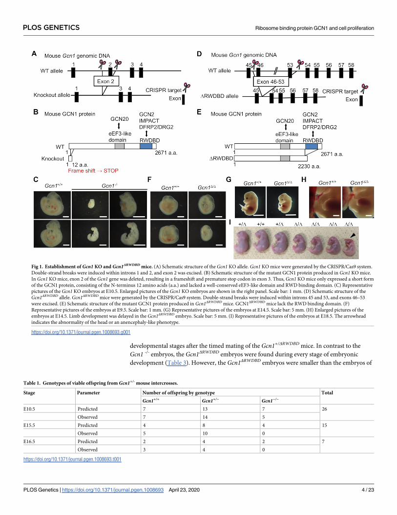
Fig 1. Establishment of Gcn1 KO and Gcn1ΔRWDBD mice. (A) Schematic structure of the Gcn1 KO allele. Gcn1 KO mice were generated by the CRISPR/Cas9 system.
Double-strand breaks were induced within introns 1 and 2, and exon 2 was excised. (B) Schematic structure of the mutant GCN1 protein produced in Gcn1 KO mice.
In Gcn1 KO mice, exon 2 of the Gcn1 gene was deleted, resulting in a frameshift and premature stop codon in exon 3. Thus, Gcn1 KO mice only expressed a short form
of the GCN1 protein, consisting of the N-terminus 12 amino acids (a.a.) and lacked a well-conserved eEF3-like domain and RWD binding domain. (C) Representative
pictures of the Gcn1 KO embryos at E10.5. Enlarged pictures of the Gcn1 KO embryos are shown in the right panel. Scale bar: 1 mm. (D) Schematic structure of the
Gcn1ΔRWDBD allele. Gcn1ΔRWDBD mice were generated by the CRISPR/Cas9 system. Double-strand breaks were induced within introns 45 and 53, and exons 46–53
were excised. (E) Schematic structure of the mutant GCN1 protein produced in Gcn1ΔRWDBD mice. GCN1ΔRWDBD mice lack the RWD binding domain. (F)
Representative pictures of the embryos at E9.5. Scale bar: 1 mm. (G) Representative pictures of the embryos at E14.5. Scale bar: 5 mm. (H) Enlarged pictures of the
embryos at E14.5. Limb development was delayed in the Gcn1ΔRWDBD embryo. Scale bar: 5 mm. (I) Representative pictures of the embryos at E18.5. The arrowhead
indicates the abnormality of the head or an anencephaly-like phenotype.
https://doi.org/10.1371/journal.pgen.1008693.g001
Table 1. Genotypes of viable offspring from Gcn1+/- mouse intercrosses.
Stage Parameter Number of offspring by genotype Total
Gcn1+/+ Gcn1+/− Gcn1−/−
E10.5 Predicted 7 13 7 26
Observed 7 14 5
E15.5 Predicted 4 8 4 15
Observed 5 10 0
E16.5 Predicted 2 4 2 7
Observed 3 4 0
https://doi.org/10.1371/journal.pgen.1008693.t001
PLOS GENETICS Ribosome binding protein GCN1 and cell proliferation
PLOS Genetics | https://doi.org/10.1371/journal.pgen.1008693 April 23, 2020 4 / 23

Gcn1+/ΔRWDBD and their Gcn1+/+ littermates between E9.5 and E18.5 (Fig 1F, 1G and 1I) and
showed a statistically significant decrease in body weight after E17.5 (Fig 2B). Although growth
retardation was observed, the Gcn1ΔRWDBD embryos were apparently normal, although some
of these embryos either showed abnormalities in the head, an anencephaly-like phenotype at
E14.5 (S2A Fig) or E18.5 (Fig 1I). They also showed a developmental delay in the limbs at
E14.5, which was not observed in the Gcn1+/+ embryos (Fig 1H). To determine the cause of
lethality, we then examined the Gcn1ΔRWDBD embryos at E18.5 (i.e., one day before birth) after
cesarean section. Of note, Gcn1ΔRWDBD mice at E18.5 did not respire and died within 20 min
after delivery by cesarean section, while almost all Gcn1+/+ and Gcn1+/ΔRWDBD embryos
respired and their color turned red. We next tried to prolong the gestational period of preg-
nant mothers by progesterone administration because progesterone maintains pregnancy (Fig
2A). The Gcn1ΔRWDBD embryos at E19.5 and E20.5 still showed lower body weight compared
to the Gcn1+/+ and Gcn1+/ΔRWDBD embryos, although they gradually increased their body
weight (Fig 2B). At E20.5, we observed 5 Gcn1ΔRWDBD embryos with normal appearance and 4
Gcn1ΔRWDBD embryos with mandibular hypoplasia and exophthalmos/hypoplasia of the eyelid
(Fig 2C and S2B and S2C Fig). The Gcn1ΔRWDBD embryos with abnormal appearance died
within 20 min after delivery by cesarean section, whereas 2 of 5 Gcn1ΔRWDBD embryos with
normal appearance respired, turned red, and survived for at least 4 h (Fig 2C). To further
examine the cause of the respiratory failure, we analyzed lung development in the Gcn1ΔRWDBD
embryos at E18.5 by a lung float test and histological analysis. Curiously, the lungs from the
Table 2. Genotypes of viable offspring from Gcn1+/ΔRWDBD mouse intercrosses.
Parameter at 2 weeks Number of offspring of genotype
Gcn1+/+ Gcn1+/Δ Gcn1Δ/Δ Total
Predicted 32 64 32 128
Observed 47 80 0 127
https://doi.org/10.1371/journal.pgen.1008693.t002
Table 3. Genotypes of viable offspring from Gcn1+/ΔRWDBD mouse intercrosses.
Stage Parameter Number of offspring by genotype Total
Gcn1+/+ Gcn1+/Δ Gcn1Δ/Δ
E9.5 Predicted 2 4 2 8
Observed 1 4 3
E11.5 Predicted 4 9 4 17
Observed 6 8 3
E14.5 Predicted 7 14 7 27
Observed 7 16 4
E16.5 Predicted 2 3 2 7
Observed 3 2 1
E17.5 Predicted 2 5 2 9
Observed 2 3 4
E18.5 Predicted 12 24 12 47
Observed 12 28 7
E19.5 Predicted 5 10 5 19
Observed 7 8 4
E20.5 Predicted 10 19 10 38
Observed 4 27 9
https://doi.org/10.1371/journal.pgen.1008693.t003
PLOS GENETICS Ribosome binding protein GCN1 and cell proliferation
PLOS Genetics | https://doi.org/10.1371/journal.pgen.1008693 April 23, 2020 5 / 23
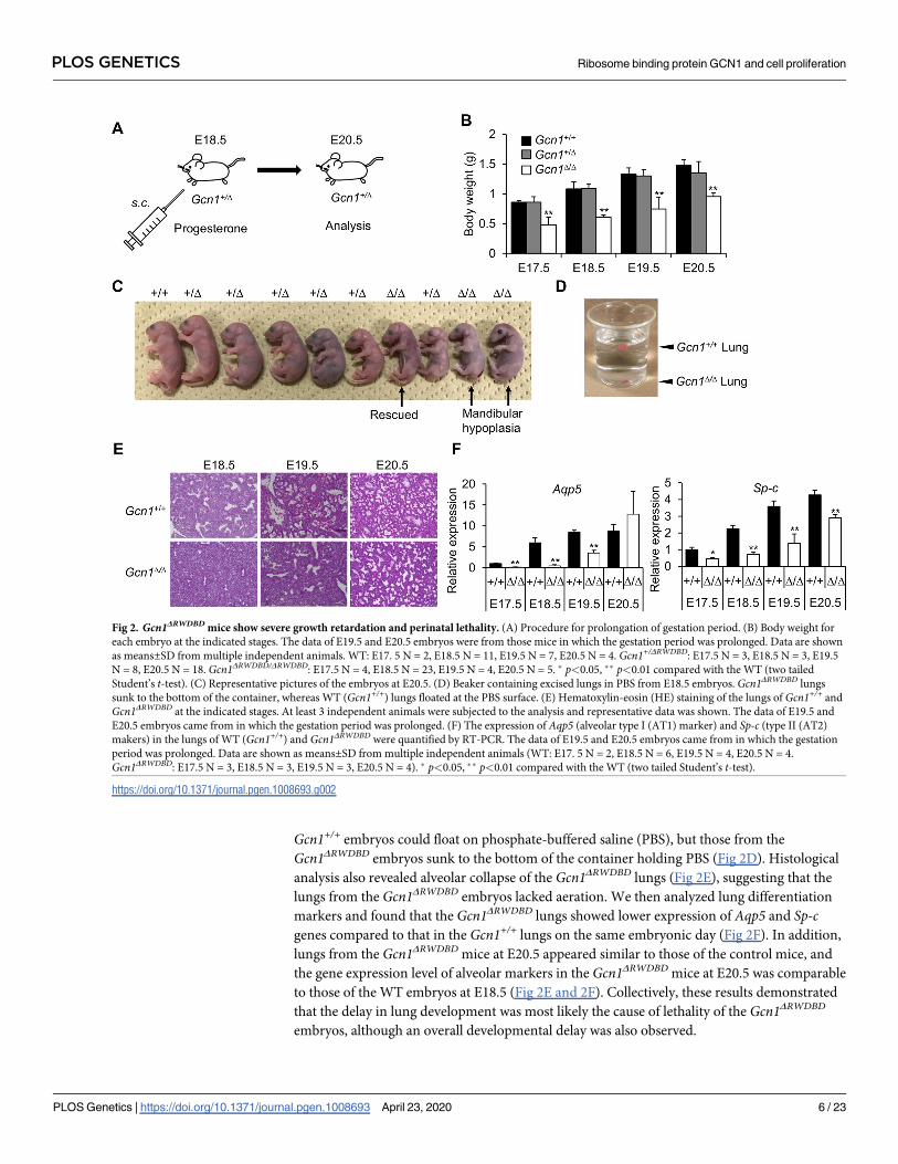
Gcn1+/+ embryos could float on phosphate-buffered saline (PBS), but those from the
Gcn1ΔRWDBD embryos sunk to the bottom of the container holding PBS (Fig 2D). Histological
analysis also revealed alveolar collapse of the Gcn1ΔRWDBD lungs (Fig 2E), suggesting that the
lungs from the Gcn1ΔRWDBD embryos lacked aeration. We then analyzed lung differentiation
markers and found that the Gcn1ΔRWDBD lungs showed lower expression of Aqp5 and Sp-cgenes compared to that in the Gcn1+/+ lungs on the same embryonic day (Fig 2F). In addition,
lungs from the Gcn1ΔRWDBD mice at E20.5 appeared similar to those of the control mice, and
the gene expression level of alveolar markers in the Gcn1ΔRWDBD mice at E20.5 was comparable
to those of the WT embryos at E18.5 (Fig 2E and 2F). Collectively, these results demonstrated
that the delay in lung development was most likely the cause of lethality of the Gcn1ΔRWDBD
embryos, although an overall developmental delay was also observed.
Fig 2. Gcn1ΔRWDBD mice show severe growth retardation and perinatal lethality. (A) Procedure for prolongation of gestation period. (B) Body weight for
each embryo at the indicated stages. The data of E19.5 and E20.5 embryos were from those mice in which the gestation period was prolonged. Data are shown
as means±SD from multiple independent animals. WT: E17. 5 N = 2, E18.5 N = 11, E19.5 N = 7, E20.5 N = 4. Gcn1+/ΔRWDBD: E17.5 N = 3, E18.5 N = 3, E19.5
N = 8, E20.5 N = 18. Gcn1ΔRWDBD/ΔRWDBD: E17.5 N = 4, E18.5 N = 23, E19.5 N = 4, E20.5 N = 5. � p<0.05, �� p<0.01 compared with the WT (two tailed
Student’s t-test). (C) Representative pictures of the embryos at E20.5. (D) Beaker containing excised lungs in PBS from E18.5 embryos. Gcn1ΔRWDBD lungs
sunk to the bottom of the container, whereas WT (Gcn1+/+) lungs floated at the PBS surface. (E) Hematoxylin-eosin (HE) staining of the lungs of Gcn1+/+ and
Gcn1ΔRWDBD at the indicated stages. At least 3 independent animals were subjected to the analysis and representative data was shown. The data of E19.5 and
E20.5 embryos came from in which the gestation period was prolonged. (F) The expression of Aqp5 (alveolar type I (AT1) marker) and Sp-c (type II (AT2)
makers) in the lungs of WT (Gcn1+/+) and Gcn1ΔRWDBD were quantified by RT-PCR. The data of E19.5 and E20.5 embryos came from in which the gestation
period was prolonged. Data are shown as means±SD from multiple independent animals (WT: E17. 5 N = 2, E18.5 N = 6, E19.5 N = 4, E20.5 N = 4.
Gcn1ΔRWDBD: E17.5 N = 3, E18.5 N = 3, E19.5 N = 3, E20.5 N = 4). � p<0.05, �� p<0.01 compared with the WT (two tailed Student’s t-test).
https://doi.org/10.1371/journal.pgen.1008693.g002
PLOS GENETICS Ribosome binding protein GCN1 and cell proliferation
PLOS Genetics | https://doi.org/10.1371/journal.pgen.1008693 April 23, 2020 6 / 23

Expression of GCN1 in WT and Gcn1ΔRWDBD MEFs
To investigate the effect of RWDBD deletion of GCN1 on cellular function, we established
mouse embryonic fibroblasts (MEFs) from the Gcn1ΔRWDBD embryos. As expected, the
Gcn1ΔRWDBD MEFs expressed a truncated type of GCN1 protein, but the level of the
ΔRWDBD GCN1 protein decreased to approximately 30% compared to that of the WT MEFs
(Fig 3A). As previous results indicated a role for GCN1 in the nucleus [26], we examined the
subcellular localization of GCN1. Immunocytochemical analysis in HeLa cells showed that
GCN1 mainly localized to the cytoplasm (S3A Fig); however, GCN1 exactly colocalized neither
with the ER marker calnexin nor with the mitochondrial marker pyruvate dehydrogenase (S3B
and S3C Fig). Cell extract fractionation analysis also showed that GCN1 localized to the cyto-
plasm and rarely localized to the nucleus in HeLa cells (S3D Fig), and both in WT and
Gcn1ΔRWDBD MEFs (S3E Fig).
Fig 3. GCN1 is necessary for GCN2-mediated ATF4 activation. (A) Whole cell protein extracted from the Gcn1+/+ and Gcn1ΔRWDBD MEFs was subjected to
immunoblot analysis to detect GCN1. (B) The WT (Gcn1+/+) and Gcn1ΔRWDBD MEFs were exposed to leucine starvation for 4 h, and the phosphorylation level of eIF2αwas examined by immunoblot. (C) The Gcn1+/+ and Gcn1ΔRWDBD MEFs were exposed to leucine starvation for the indicated times, and the mRNA levels of the ATF4
target genes Asns and Psat1 were quantified by RT-PCR. The value at 0 h for WT cells was set to 1, and the results were shown as the relative folds±SD from multiple
independent experiments (N = 4). � p<0.05, �� p<0.01 compared with the WT (two tailed Student’s t-test). # p<0.05, ## p<0.01 compared with the value at 0 h for the
WT cells (one-way ANOVA with with Bonferroni post hoc test). (D) The WT (Gcn1+/+) and Gcn1ΔRWDBD MEFs were exposed to leucine (Leu), methionine (Met), serine
(Ser) or cystine (Cys) starvation for 4 h or cultured in the control (Ctrl) medium and cells were fractionated into cytosol, nuclear fractions and subjected to immunoblot
analysis to detect the GCN1, phosphorylated GCN2 (P-GCN2), GCN2, phosphorylated eIF2α (P-eIF2α), eIF2α, HSP90, ATF4 and Lamin B. (E) The WT (Gcn1+/+) and
Gcn1ΔRWDBD MEFs lacking the indicated amino acids were exposed to AAS for 8 h, and the mRNA levels of Asns and Psat1 were quantified by RT-PCR. The value for
WT cells cultured in the control (Ctrl) medium was set to 1, and the results were shown as the relative folds±SD from multiple independent experiments (N = 4). �
p<0.05, �� p<0.01 compared with the WT (two tailed Student’s t-test). (F) The WT (Gcn2+/+) and Gcn2 KO (Gcn2-/-) MEFs were exposed to AAS lacking the indicated
amino acids for 8 h, and the mRNA levels of Asns and Psat1 were quantified by RT-PCR. The value for WT cells cultured in the control (Ctrl) medium was set to 1, and
the results were shown as the relative folds±SD from multiple independent experiments (N = 4). � p<0.05, �� p<0.01 compared with the WT (two tailed Student’s t-test).
https://doi.org/10.1371/journal.pgen.1008693.g003
PLOS GENETICS Ribosome binding protein GCN1 and cell proliferation
PLOS Genetics | https://doi.org/10.1371/journal.pgen.1008693 April 23, 2020 7 / 23

GCN1 was necessary for the AAS response of MEFs
Next, we examined the role of GCN1 in the GCN2-eIF2α pathway in the MEFs. Under basal
conditions, the global translation level in the Gcn1ΔRWDBD MEFs was comparable to that in the
WT MEFs (S4 Fig). Then, we analyzed the role of GCN1 in response to AAS. eIF2α phosphor-
ylation was increased in the WT MEFs but was unchanged in the Gcn1ΔRWDBD MEFs (Fig 3B
and quantified in S5A Fig). Upon leucine starvation, the expression of ATF4 target genes
asparagine synthetase (Asns) and phosphoserine aminotransferase 1 (Psat1) was activated by
leucine starvation in the WT MEFs, while the induction of these genes was suppressed in the
Gcn1ΔRWDBD MEFs (Fig 3C). We further explored the role of GCN1 in activating the GCN2/
ATF4 pathway by depleting amino acids other than leucine. Phosphorylation of GCN2 and
eIF2α and nuclear accumulation of ATF4 were induced by depletion of leucine, methionine,
serine or cystine in WT MEFs, which was not observed in Gcn1ΔRWDBD MEFs (Fig 3D and S5B
Fig). Both Asns and Psat1 genes were induced by depletion of methionine, serine or cystine in
the WT MEFs, whereas the induction was largely suppressed in both Gcn1ΔRWDBD and Gcn2KO MEFs (Fig 3E and 3F). These data suggested that GCN1 is necessary for GCN2-dependent
ATF4 activation upon AAS in mammalian cells.
GCN1 was also necessary for the GCN2-mediated response to UV stress
UV radiation increases eIF2α phosphorylation in a GCN2-dependent manner [27]. Therefore,
we next examined the role of GCN1 in response to UV stress. In the WT MEFs, GCN2 and
eIF2α were phosphorylated after 30 min of UV irradiation as previously reported (Fig 4A and
4B and quantified in S6 Fig). On the other hand, inducible phosphorylation of GCN2 and
eIF2α was impaired in both Gcn1ΔRWDBD MEFs and Gcn2 KO MEFs (Fig 4A and 4B and quan-
tified in S6 Fig), indicating that GCN1 is also involved in the UV stress response.
Fig 4. GCN1 and GCN2 dependency in response to UV exposure. (A)(B) The WT and Gcn1ΔRWDBD (A) or Gcn2 KO
(Gcn2-/-) MEFs (B) were irradiated by 80 J/m2 UV and were allowed to recover in fresh medium for 30 min or 4 h
before harvest. Whole cell protein extracts of the MEFs were subjected to immunoblot analysis to detect phosphorylated
GCN2 (P-GCN2), GCN2, phosphorylated eIF2α (P-eIF2α), eIF2α and β-actin.
https://doi.org/10.1371/journal.pgen.1008693.g004
PLOS GENETICS Ribosome binding protein GCN1 and cell proliferation
PLOS Genetics | https://doi.org/10.1371/journal.pgen.1008693 April 23, 2020 8 / 23

The role of GCN1 in eIF2α phosphorylation by HRI, PERK and PKR
We further explored other stimulations that activate other eIF2α kinases, HRI, PERK and
PKR. It has been reported that H2O2 and the proteasome inhibitor MG132 activate HRI and
phosphorylate eIF2α in MEFs [28, 29]. H2O2 treatment induced phosphorylation of eIF2α in
WT, Gcn1ΔRWDBD and Gcn2 KO MEFs (S7A–S7B Fig). It is well known that the ER stressor
tunicamycin (Tm) induces eIF2α phosphorylation via PERK activation [30]. Tm induced
eIF2α phosphorylation in WT, Gcn1ΔRWDBD and Gcn2 KO MEFs (S7C and S7D Fig). We fur-
ther analyzed ATF4 target genes, Asns and Psat1 upon Tm treatment (S6E Fig). Tm-induced
Asns and Psat1mRNA levels in Gcn1ΔRWDBD MEFs were comparable to those in WT MEFs.
We then examined the role of GCN1 in PKR activation. dsRNA mimic Poly(I:C) induced
eIF2α phosphorylation in the absence of GCN1 and GCN2 (S7F and S7G Fig). These results
indicate that GCN1 is dispensable for HRI, PERK and PKR activation.
Gcn1ΔRWDBD MEFs showed decreased growth rate and arrested in G2/M
As the Gcn1ΔRWDBD embryos exhibited growth retardation (Figs 1 and 2), we examined the
cell growth capacity of the Gcn1ΔRWDBD MEFs. As expected, the cell growth of the Gcn1ΔRWDBD
MEFs was significantly reduced compared to that of the WT MEFs in both primary (Fig 5A)
and immortalized MEFs (Fig 5B). Consistent with the decreased cell growth in the
Gcn1ΔRWDBD MEFs, BrdU incorporation was substantially reduced in the Gcn1ΔRWDBD MEFs
compared to the BrdU incorporation in the WT MEFs (Fig 5C). We also analyzed the levels of
cleaved PARP and Caspase-3, both of which indicate apoptosis, in the Gcn1ΔRWDBD MEFs
(S8A–S8C Fig). The levels of cleaved PARP and Caspase-3 both in the primary and immortal-
ized Gcn1ΔRWDBD MEFs were comparable to those of the WT MEFs (S8A–S8C Fig). Flow cyto-
metric analysis revealed that the Gcn1ΔRWDBD MEFs were significantly larger in size compared
to the WT MEFs (Fig 5D). Cell enlargement is often associated with cellular senescence [31];
however, β-galactosidase staining showed similar intensities in both WT and Gcn1ΔRWDBD
MEFs (S9 Fig). Next, we performed cell cycle analysis of the Gcn1ΔRWDBD MEFs by staining
them with propidium iodide (PI) and analyzing them with flow cytometry. The proportion of
cells in G2/M phase was increased in the Gcn1ΔRWDBD MEFs compared to that in the WT
MEFs (Fig 5E and 5F). On the other hand, the rate of cell growth and the cell cycle profile were
comparable between the Gcn2 KO MEFs and the WT MEFs (Fig 5G–5I).
Cdk1 decreased and p21 increased in Gcn1ΔRWDBD MEFs
The RWD domain-containing protein, DRG family-regulatory protein 2 (DFRP2), is a human
homolog of yeast Gir2, which heterodimerizes with developmentally regulated GTP-binding
protein 2 (DRG2), with which it is mutually stabilized in a manner similar to that of the yeast
Gir2/Rbg2 complex [32, 33]. It is noteworthy that DRG2 is involved in G2/M regulation such
that both the overexpression and knockdown of DRG2 increase the number of cells in the G2/
M phase [34, 35]. Therefore, we speculated that GCN1 might modulate the cell cycle through
the DFRP2/DRG complex. In DRG2-knockdown HeLa cells, CDK1 and Cyclin B1 are
decreased, while p21 is increased [34]. It is well known that the CDK1/Cyclin B1 complex
induces G2/M transition and that p21 inhibits CDKs/cyclins [36, 37]. Therefore, we analyzed
Cdk1, Cyclin B1 and p21 gene expression in the Gcn1ΔRWDBD and Gcn2 KO MEFs. Decreased
Cdk1 gene expression was observed in the Gcn1ΔRWDBD MEFs, but not in the Gcn2 KO MEFs,
compared to the levels in the corresponding WT MEFs (Fig 6A and 6B). In contrast, an
increase in p21 gene expression was observed both in the Gcn1ΔRWDBD and Gcn2 KO MEFs.
The increase in p21 and decrease in Cdk1 in the Gcn1ΔRWDBD MEFs were also observed at the
protein level, which was consistent with respective level of the mRNA (Fig 6C and quantified
PLOS GENETICS Ribosome binding protein GCN1 and cell proliferation
PLOS Genetics | https://doi.org/10.1371/journal.pgen.1008693 April 23, 2020 9 / 23

in S10 Fig). Interestingly, Cyclin B1 protein levels were decreased in the Gcn1ΔRWDBD MEFs
compared to those in the WT MEFs (Fig 6C and quantified in S10 Fig), although the mRNA
expression was comparable to that of the WT MEFs (Fig 6A). On the other hand, the Cyclin
B1 protein level in the Gcn2 KO MEFs was comparable to that in the control MEFs (Fig 6D
and quantified in S10 Fig). Thus, it is likely that GCN1 modulates the cell cycle through the
upregulation of Cyclin B1 and Cdk1. In addition, p21 inhibits the Cyclin B1/Cdk1 complex
[38]. Therefore, increased p21 in the Gcn1ΔRWDBD MEFs might be involved in Cyclin B1/Cdk1
inhibition.
Fig 5. Gcn1ΔRWDBD MEFs exhibited reduced cell proliferation. (A)(B)(G) After cells were cultured in IMDM for the indicated periods, the relative cell numbers of
primary Gcn1ΔRWDBD MEFs (A), immortalized Gcn1ΔRWDBD MEFs (B) and immortalized Gcn2 KO (Gcn2-/-) MEFs (G) were counted and are shown with those of the
corresponding WT (Gcn1+/+ or Gcn2+/+) cells. The initial cell number was set to 1 and the results are shown as the relative folds±SD from multiple independent
experiments ((A): N = 6, (B): N = 4, (C): N = 3). � p<0.05, �� p<0.01 compared with the WT (two tailed Student’s t-test). (C) Cell proliferation in the primary WT
(Gcn1+/+) and Gcn1ΔRWDBD MEFs was examined using BrdU incorporation. The results are presented as fold differences compared to the WT (Gcn1+/+) MEFs from
multiple independent experiments (N = 6). �� p<0.01 compared with the WT (two tailed Student’s t-test). (D) Relative cell sizes in the primary WT (Gcn1+/+) and
Gcn1ΔRWDBD MEFs were measured using forward light scatter (FSC) of flow cytometry. The results are presented as fold differences compared to the WT (Gcn1+/+)
MEFs from multiple independent experiments (N = 7). �� p<0.01 compared with the WT (two tailed Student’s t-test). (E) The WT (Gcn1+/+) and Gcn1ΔRWDBD
primary MEFs were stained by propidium iodide (PI) to assess the DNA contents in different phases of the cell cycle. The percentage of cells in each cell cycle was
calculated (F). The results are presented as fold differences compared to those of the WT (Gcn1+/+) MEFs from multiple independent experiments (N = 5). �� p<0.01
compared with the WT (two tailed Student’s t-test). (H) WT (Gcn2+/+) and immortalized Gcn2 KO (Gcn2-/-) MEFs were stained by PI, and the percentage of cells in
each cell cycle was calculated (I). Data are shown as means±SD from multiple independent experiments (N = 4).
https://doi.org/10.1371/journal.pgen.1008693.g005
PLOS GENETICS Ribosome binding protein GCN1 and cell proliferation
PLOS Genetics | https://doi.org/10.1371/journal.pgen.1008693 April 23, 2020 10 / 23

Originally, p21 was reported to inhibit Cyclin E/Cdk2 and Cyclin D/Cdk4/6, which regulate
the G1/S transition [39]. Therefore, we compared cell cycle progression between the
Gcn1ΔRWDBD MEFs and the WT MEFs. The MEFs were synchronized to the G0/G1 phase by
serum deprivation for 72 h and thereafter were released into the cell cycle by the replacement
of the medium with fresh medium. Replenishment with fresh medium resulted in more WT
MEFs in the S phase at 24 h, but at 28 h, fewer Gcn1ΔRWDBD MEFs were in the S phase com-
pared to WT MEFs (Fig 6E and quantified in S11 Fig). These results suggest that the
Gcn1ΔRWDBD MEFs are delayed at the G1/S transition. Accumulation of Cyclin B1 and Cdk1
was also observed in the Gcn1ΔRWDBD MEFs, although to a lesser extent than that in the WT
MEFs (Fig 6F). Interestingly, p21 expression was higher in the Gcn1ΔRWDBD MEFs at every
time point, suggesting that an increase in p21 might be one of the causes of the cell cycle delay.
Notably, serum starvation-induced eIF2α phosphorylation was decreased in the Gcn1ΔRWDBD
MEFs and was not recovered until 44 h after the medium was replaced (Fig 6F).
Fig 6. Expression of cell cycle regulation factors in Gcn1ΔRWDBD MEFs. (A) (B) Gene expression levels related to cell cycle progression in the Gcn1ΔRWDBD MEFs (A)
and Gcn2 KO (Gcn2-/-) MEFs (B) were quantified by RT-PCR. The value for wild-type cells was set to 1, and the results are shown as the relative folds±SD from
multiple independent experiments ((A): N = 4, (B): N = 4). � p<0.05, �� p<0.01 compared with the WT (two tailed Student’s t-test). (C) Whole cell proteins extracted
from the WT (Gcn1+/+) and Gcn1ΔRWDBD MEFs were subjected to immunoblotting to detect GCN1, Cdk1, Cyclin B1, p21 and β-actin. (D) Whole cell proteins
extracted from the WT (Gcn2+/+) and Gcn2 KO (Gcn2-/-) MEFs were subjected to immunoblot analysis to detect Cdk1, Cyclin B1, p21 and β-actin. (E) Cell cycle
analysis of the MEFs. WT (Gcn1+/+) and Gcn1ΔRWDBD MEFs were synchronized at G0-G1 by serum deprivation for 72 h. After serum stimulation at the indicated
times, the cells were treated with PI, and the cell content was analyzed using flow cytometry. (F) Whole cell protein extracts from the WT (Gcn1+/+) and Gcn1ΔRWDBD
MEFs obtained by the experiment in (E) were subjected to immunoblot analysis to detect GCN1, phosphorylated eIF2α (P-eIF2α), eIF2α, DRG2, Cyclin D1, p21,
Cyclin B1, Cdk1 and β-actin.
https://doi.org/10.1371/journal.pgen.1008693.g006
PLOS GENETICS Ribosome binding protein GCN1 and cell proliferation
PLOS Genetics | https://doi.org/10.1371/journal.pgen.1008693 April 23, 2020 11 / 23

Discussion
In this study, we clarified, for the first time, that GCN1 is essential for embryonic development
by generating two Gcn1mutant mouse lines (i.e., Gcn1 KO and Gcn1ΔRWDBD mice). We sum-
marize the developmental defects observed in Gcn1 KO and Gcn1ΔRWDBD mice in Fig 7A.
Gcn1ΔRWDBD mice showed growth defects as early as E9.5 and died perinatally, likely due to
respiratory failure caused by poor differentiation and insufficient lung inflation (Figs 1 and 2).
Simple developmental delay, but not the absolute requirement for GCN1 RWDBD, during
lung differentiation presumably underlies this phenotype, as prolonged gestation by progester-
one administration at least partially rescued the lung differentiation defects and, consequently,
the lethality observed immediately after the cesarean section. As Gcn2 KO mice did not show
defects in embryonic development and perinatal lethality [12], our results indicate that the role
of GCN1 in embryonic development is GCN2-independent. This is fully consistent with our
Fig 7. Graphical summary. (A) Summary of phenotypes of the Gcn1 KO and Gcn1ΔRWDBD embryos. (B) Schematic of the role of GCN1 in the control of cell
proliferation and cell cycles.
https://doi.org/10.1371/journal.pgen.1008693.g007
PLOS GENETICS Ribosome binding protein GCN1 and cell proliferation
PLOS Genetics | https://doi.org/10.1371/journal.pgen.1008693 April 23, 2020 12 / 23
in vitro results, which revealed that the Gcn2 KO MEFs neither showed proliferative defects
nor decreased Cdk1 and cyclin B1 protein levels, as observed in the Gcn1ΔRWDBD MEFs (Fig
6). Interestingly, GCN1mutation in human was recently suggested to lead to fetal akinesia that
is characterized by reduced or absent fetal movement, but often associate with multiple abnor-
malities including intra uterine growth retardation [40]. Although GCN1 protein levels were
decreased to approximately 30–40% compared to those of the wild-type MEFs, it is not clear at
this point to what extent the decrease in GCN1 protein levels contributes to the phenotypes of
Gcn1ΔRWDBD mice other than the lack of the RWDBD. Gcn1 heterozygous KO MEFs expressed
about half amount of mRNA and protein compared with WT MEFs (S12A–S12C Fig). And
heterozygous Gcn1 KO mice showed neither perinatal lethality nor embryonic growth defects
(S12D Fig), suggesting that at least one-half the amount of GCN1 is sufficient for normal
development.
Recent reports in C. elegans and A. thaliana have clarified the pleiotropic roles of GCN1,
which are independent of GCN2. Of note, GCN1 acts with GCN20 independently of GCN2 to
regulate translation that enhances the response to infection and mitochondrial dysfunction in
Arabidopsis [25]. Consistent with the severe phenotype of Gcn1 KO mice compared to
Gcn1ΔRWDBD mice, Arabidopsis gcn1mutants showed basic growth retardation that was more
severe in the mutants that lacked an extensive portion of the sequence toward the GCN1 N-
terminus. The importance of the central region of GCN1 spanning the GCN20 binding
domain for apoptosis regulation was reported for C. elegansmutants [24]. Therefore, it is pos-
sible that the phenotypic difference between the Gcn1 KO and Gcn1ΔRWDBD mice is attribut-
able to the GCN20 binding domain, since this domain is intact in the Gcn1ΔRWDBD mice (Fig
1E). Considering that the GCN1mutation in C. elegans and A. thaliana caused growth defects,
the role of GCN1 in developmental growth and cell cycle progression during the G2/M transi-
tion might be conserved among eukaryotes.
We observed growth defects and an increase in the G2/M population in both immortalized
and primary Gcn1ΔRWDBD MEFs (Fig 5A, 5B, 5E and 5F and S13 Fig). We also observed a
delay in G1/S progression in the cell cycle-synchronized cells (Fig 6E and quantified in S11
Fig) and decreased incorporation of BrdU in the primary Gcn1ΔRWDBD MEFs compared to the
that in WT MEFs (Fig 5C). Although only a slight increase in apoptotic markers was observed
in immortalized Gcn1ΔRWDBD MEFs, we consider that the decrease in cell growth in the
Gcn1ΔRWDBD MEFs is mainly due to decreased cell proliferation but not to increased cell
death. Although increase of p21mRNA and p21 protein levels were observed in Gcn2 KO
MEFs, we did not observe proliferation defects of the cells. One caveat in this study is that we
used Gcn2 KO MEF that was immortalized by large T antigen [41]. Therefore, proliferation
defects can be rescued by the expression of large T antigen. However, we speculate that Gcn2KO cells do not show proliferation defects in vivo as Gcn2 KO mice did not show overt growth
defects during embryonic development.
It is known that yeast Gir2 binds to Gcn1 via the Gir2 RWD domain. Gir2 heterodimerizes
with Rbg2 and is necessary for maximum proliferation under AAS [32]. Under AAS, the bind-
ing of Gcn1 with Gir2/Rbg2 is increased, and the Gir2/Rbg2 complex is stabilized in a GTP-
dependent manner. DFRP2, the human homolog of Gir2, heterodimerizes human DRG2 and
they stabilize each other in a manner similar to Gir2 and Rbg2 in yeast [33] (Fig 7B). The high
conservation of both amino acid sequences and the mutual stabilization mechanism between
the yeast Gir2/Rbg2 and human DFRP2/DRG2 complexes suggest their functional importance
in cell growth. Jang et al. showed that DRG2-deficient HeLa cells showed a defect in cell prolif-
eration and G2/M arrest associated with p21 induction and a decrease in Cdk1 and Cyclin B1
expression that were observed in the Gcn1ΔRWDBD MEFs (Fig 6A and 6C). The G2/M arrest
observed in the Gcn1ΔRWDBD MEFs might reflect a disrupted DRG2/DFRP2 function [34] (Fig
PLOS GENETICS Ribosome binding protein GCN1 and cell proliferation
PLOS Genetics | https://doi.org/10.1371/journal.pgen.1008693 April 23, 2020 13 / 23

7B). The only difference in this respect is that both the Cyclin B1mRNA expression and the
protein levels were decreased inDRG2-knockdown HeLa cells, while only the cyclin B1 protein
levels were decreased in the Gcn1ΔRWDBD MEFs (Fig 6A and 6C and quantified in S10 Fig). It
is well known that Cyclin B1mRNA is upregulated in the G2/M phase and that the Cyclin B1
protein is degraded by the APC/C-mediated ubiquitin-proteasome pathway before the onset
of mitosis [42]. Therefore, the Cyclin B1 protein level might be decreased by the premature
activation of APC/C. It is of note that Drg2 deficient mice were just recently reported and that
they show embryonic growth retardation, but survived to the adulthood [43]. The observation
strongly supports our contention that defect of DRG2 might be the cause of growth retardation
in Gcn1ΔRWDBD mice.
Notably, Ola1 KO mice show strikingly similar phenotypes as those of Gcn1ΔRWDBD mice
(i.e., embryonic growth retardation and perinatal lethality due to respiratory failure) [44].
OLA1 belongs to an Obg-family GTPase and reduces ternary complex formation by binding
to eIF2 and hydrolyzing GTP, which leads to the activation of ISR [45]. Ding et al. showed that
Ola1 KO mice exhibit growth retardation and high levels of embryonic lethality due to the
decreased cell proliferation associated with increased p21mRNA translation. They also
showed that the increased p21mRNA translation in the Ola1 KO MEFs is reversed by an eIF2
phosphatase inhibitor, salubrinal, indicating that translation of p21mRNA is sensitive to the
amount of ternary complex [44]. Although both OLA1 and GCN1 can mediate stress-induced
decreases in ternary complex formation, the reasons for similarity should be clarified in the
future. In contrast to their similar in vivo phenotypes, Ola1 KO MEFs show G1/S arrest associ-
ated with the decrease in Cyclin D1 and E1, but not Cyclin B1, which differs from the pheno-
types of the Gcn1ΔRWDBD MEFs. However, it is noteworthy that the Ola1 KO MEF population
is also increased in G2/M. The detailed molecular connections between OLA1 and GCN1
need to be explored in future studies.
The levels of the p21 protein and the p21mRNA were increased in the Gcn2 KO MEFs and
in the Gcn1ΔRWDBD MEFs (Fig 6A–6D and quantified in S10 Fig). Both increased transcription
and translation could potentially contribute to the increase in p21 protein levels in these cells.
As described above, p21 translation is increased by an increase in the ternary complex. Indeed,
the basal level of eIF2α phosphorylation appeared to decrease in both Gcn1ΔRWDBD and Gcn2KO MEFs (Fig 4). On the other hand, Nakamura et al. recently reported that GCN2 localizes
at the nucleolus and that p21mRNA transcription is increased by GCN2 knockdown in a
p53-dependent manner [46]. p53 activity is regulated by the signals from the nucleolus [47]
and the transcripts in the nucleolus such as ribosomal 5S RNA are proposed to activate p53
[48]. Nakamura et al. showed that accumulated small RNAs including 5S RNA lead to p53 acti-
vation in GCN2 knockdown cancer cells indicating that GCN2 may inhibit RNA polymerase
III-dependent transcription including 5S RNA. Although we failed to detect p53 accumulation
in Gcn1ΔRWDBD MEFs, but it is an interesting possibility that GCN1 regulates GCN2 localiza-
tion at the nucleolus and its function. As p21mRNA is also increased by theDRG2 knockdown
in HeLa cells [42], we surmise that both GCN2 and DRG2 are involved in p21mRNA upregu-
lation in Gcn1ΔRWDBD MEFs.
In conclusion, we confirmed that GCN1 is required for the GCN2-dependent response to
stress such as that induced by AAS and UV radiation, which is consistent with a previous
report [22] (Fig 7B). Furthermore, we clarified that GCN1 is necessary for cell proliferation
and development and that the GCN1 action is GCN2-independent. Thus, we propose that
GCN1 is a hub protein of cellular signaling that integrates cellular stress responses and growth.
This study established a molecular basis for the future clarification of mammalian GCN1 func-
tion (Fig 7B).
PLOS GENETICS Ribosome binding protein GCN1 and cell proliferation
PLOS Genetics | https://doi.org/10.1371/journal.pgen.1008693 April 23, 2020 14 / 23
Materials and methods
Ethics statement
Mice were maintained in temperature- and humidity-controlled rooms on a 12-h light-dark
cycle. All mouse experiments were approved by the Committee for the Ethics of Animal
Experimentation of Hirosaki University (#M14040) and carried out according to the Guideline
for Animal Experimentation of Hirosaki University.
Generation of mutant mouse by CRISPR/Cas9 system
Gcn1 KO mice. Gcn1 KO mice were generated similarly to the Gcn1ΔRWDBD mice. To con-
struct a Cas9/sgRNA expression vector, the following oligonucleotide DNAs were used: for
exon 2 KO (targeted to introns 1 and 2), 5’-TGC TGC TGA TGT GAG TGC GG-3’ and 5’-
GCC TGT AGG GAC TGT TCT CG-3’). Genotypes were determined by PCR with primer
sets for the WT allele: 5’-AGA TGG TAG CAG GTG GCG TCC AG-3’ and 5’-GGG ATG
GAA GGT AGG CCG GCT G-3’, and for the deleted allele: 5’-TGC TCT TGT GGG CCT
GTG CCA-3’ and 5’-GGG ATG GAA GGT AGG CCG GCT G-3’.
Gcn1ΔRWDBD mice. To construct a Cas9/single-guide RNA (sgRNA) expression vector,
oligonucleotide DNAs (for exons 46–53 KO (targeted to introns 45 and 53): 50-AGA GCC
CTC ACC TAT CCT AT-30 and 50-TCA AAT TCC TGG CAC ACC GA-30) were annealed
and inserted into the pX330 vector (Addgene plasmid # 42230). The pX330 vector was injected
into the pronuclei of C57BL/6 fertilized eggs. F0 mice were genotyped for the deletion between
exons 46 and 53 of the Gcn1 gene due to a double-strand break in both introns 45 and 53 and
nonhomologous end-joining (NHEJ). F0 mice were checked for the absence of the Cas9 trans-
gene and off-target effects. F0 mice were mated with the C57BL/6 mice to obtain F1 offspring.
Genotypes were determined by PCR with the following primer sets for the WT allele: 5’-TGT
CCC TTA GTG TGT TAG ATT CC-3’ and 5’- TGC AGC TGC TCA AAG GTC TTG G-3’,
and the following primer set for the deleted allele: 5’-TGT CCC TTA GTG TGT TAG ATT
CC-3’ and 5’-AGT CAA GCT GAC TCT TGA CTG C-3’. To avoid using mice with unex-
pected off-target effects, F1 mice were backcrossed at least 8 times with WT C57BL/6 mice
purchased from CLEA Japan Inc. and used for analyses.
Administration of progesterone
Gcn1+/ΔRWDBD pregnant mice at E18.5 were administered 4 mg of progesterone by subcutane-
ous (s.c.) injection. Progesterone-injected mice could maintain pregnancy, and embryos at
E20.5 were delivered by cesarean section.
Quantitative RT-PCR (q-RT-PCR)
For q-RT-PCR, total RNA was prepared by using TRIzol RNA Isolation Reagents (Thermo-
Fisher, Tokyo, Japan) following the manufacturer’s protocol. cDNA was synthesized from
total RNA using PrimeScript Reverse Transcriptase (TaKaRa Bio Inc., Shiga, Japan). Real-time
PCR was performed using primer sets in Table 4, TB Green Premix Ex Taq II (Takara Bio Inc)
and a CFX96 thermal cycler (Bio-Rad, Richmond, CA).
Cell culture
Human cervical carcinoma HeLa cells were maintained in high glucose Dulbecco’s modified
Eagle medium (DMEM, Sigma-Aldrich, St. Louis, MO) containing 10% fetal bovine serum,
penicillin-streptomycin (PS) (100 U/ml-0.1 mg/ml, ThermoFisher) and 2 mM L-glutamine
(Sigma-Aldrich). MEFs were prepared from individual embryos at E13.5-E15.5. After the head
PLOS GENETICS Ribosome binding protein GCN1 and cell proliferation
PLOS Genetics | https://doi.org/10.1371/journal.pgen.1008693 April 23, 2020 15 / 23

and internal organs were removed, the torso was minced and dispersed in 0.05% trypsin-
EDTA. MEFs were maintained in Iscove’s modified Dulbecco’s medium (IMDM, Sigma-
Aldrich) containing 10% fetal bovine serum (FBS, Life Technologies, Carlsbad, CA), PS and 2
mM L-glutamine (Sigma-Aldrich). The generation of Gcn2-/- MEFs and WT cells was previ-
ously described [12]. For AAS experiments, MEFs were maintained in DMEM lacking specific
amino acids (custom-made by Cell Science & Technology Institute, Sendai, Japan) containing
10% dialyzed FBS, 4.5 g/L glucose and PS. MEFs were treated either with H2O2 (Kanto Chemi-
cal, Tokyo, Japan), Tm (Wako Pure Chemicals, Osaka, Japan) or MG132 (Peptide Institute,
Osaka, Japan). MEFs were transfected by Poly(I:C) by using a Lipofectamine 2000 (Thermo-
Fisher). The cells were maintained at 37˚C in a 5% CO2 incubator.
Immunofluorescence
HeLa cells were fixed with 4% paraformaldehyde (PFA)/PBS and permeabilized with 0.25%
Triton X-100. The following antibodies were used: GCN1 (Abcam, Cambridge, UK), calnexin
(BD Biosciences, Tokyo, Japan), pyruvate dehydrogenase (PDH) subunit E1 antibody (Invitro-
gen, Carlsbad, CA). Detection was performed using Alexa Fluor 488 for GCN1 and Alexa
Fluor 594 for calnexin and PDH. 4,6-Diamidino-2-phenylindole (DAPI) was used for nuclear
counterstaining. The fluorescent images were observed using the C1si confocal imaging sys-
tem (Nikon, Tokyo, Japan).
Immunoblot analysis
The cytosolic or nuclear extracts were prepared as previously described [49].
The protein samples were boiled in Laemmli buffer and electrophoresed under reducing
conditions by SDS-PAGE. Protein concentrations were determined using a bicinchoninic acid
(BCA) protein assay kit (Pierce Biotechnology, Rockford, IL).
Proteins were transferred to polyvinylidene difluoride (PVDF) membranes (Millipore) and
subjected to immunoblot analysis using primary antibodies against GCN1 (Abcam), GCN2
(Cell Signaling Technology, Danvers, Massachusetts), phospho-Thr899 GCN2 (Abcam),
eIF2α (Cell Signaling Technology), phospho-Ser51 eIF2α (Cell Signaling Technology), DRG2
(Abcam), Cdk1 (Cell Signaling Technology), cyclin B1 (Abcam), cyclin D1 (Santa Cruz, Dallas,
Texas), p21 (Santa Cruz), PARP (Cell Signaling Technology), Caspase-3 (Cell Signaling Tech-
nology) and β-actin (Sigma) and HRP-labeled secondary antibodies. Immunoreactive bands
Table 4. Primer sets used to detect indicated genes by q-RT-PCR.
Gene Forward (5’-3’) Reverse (5’-3’)
CycA AAGACTGAATGGCTGGATGG AGCTGTCCACAGTCGGAAAT
Aqp5 TCTTGTGGGGATCTACTTCACC TGAGAGGGGCTGAACCGAT
Sp-c ATGGACATGAGTAGCAAAGAGGT CACGATGAGAAGGCGTTTGAG
Asns GGCCCTTGTTTAAAGCCATGA AAGGGAGTGGTGGAGTGTTTT
Psat1 CAGTGGAGCGCCAGAATAGAA CCTGTGCCCCTTCAAGGAG
Cdk1 AGGTACTTACGGTGTGGTGTAT CTCGCTTTCAAGTCTGATCTTCT
Cyclin B1 CTTGCAGTGAGTGACGTAGAC CCAGTTGTCGGAGATAAGCATAG
p21 CCTGGTGATGTCCGACCTG CCATGAGCGCATCGCAATC
Gcn1(S1A–S1C Fig)
CTCTGCTGGAGACACTCAGC GTGAAGGTGGTCTGAAGCTG
Gcn1 (S12A Fig) AACAGCCAGTGTGAAGGAGC CGATGCAGTGTCAAGCAGAAC
Gcn2 TACTTTGCGATGAACTCCAGAGA GCTCAGGTGTGTAGCCAGAG
Impact ACGCGCAGACTTATCGAACA TCTGGGTCTGGCTCGGTTAT
https://doi.org/10.1371/journal.pgen.1008693.t004
PLOS GENETICS Ribosome binding protein GCN1 and cell proliferation
PLOS Genetics | https://doi.org/10.1371/journal.pgen.1008693 April 23, 2020 16 / 23

were visualized by using ImmunoStar chemiluminescent reagent (Wako Pure Chemicals,
Tokyo, Japan) and intensities were measured by using Image J software 1.52q (NIH, Bethesda,
MD).
UV irradiation
MEFs were washed twice with PBS, and the PBS was removed from the plates and cells. The
cells were irradiated at a dose of 80 J/m2 and 254 nm by using a UV cross-linker (CL-1000,
UVP Inc.). After UV irradiation, fresh medium was added to each plate.
Histological analysis
Lungs were fixed with 10% formalin (Mildform 10 N; Wako Pure Chemicals) and embedded
in paraffin using standard procedures. Sections (5 μm) were stained with hematoxylin-eosin.
Cell cycle analysis
The cells were collected, washed with PBS, and fixed in 70% ethanol at -20˚C for at least 2 h.
The fixed cells were washed with PBS, stained with a Tali cell cycle kit (containing propidium
iodide (PI) and RNase A) (ThermoFisher) for 30 min, and analyzed using a Canto II flow
cytometer (BD Biosciences). The cell sizes were examined using a forward light scatter (FSC)
parameter. Data were analyzed using the FACSDiva Software (BD Biosciences), ModFit LT3.2
Software (BD Biosciences) and FlowJo software (BD Biosciences).
Cell viability analysis
MEFs were plated on 96-well plates at a density of 5.0 × 10⁴ cells/well. The number of viable
cell numbers was quantified with a Cell Counting Kit-8 (Dojindo Molecular Technologies,
Kumamoto, Japan) according to the manufacturer’s protocol.
BrdU incorporation
MEFs were plated on 96-well plates and incubated for 24 h. The cells were labeled with BrdU
for 2 h, and the incorporation of BrdU was analyzed using a BrdU cell proliferation kit (Merck
Millipore, Darmstadt, Germany) according to the manufacturer’s protocol. Each value was
normalized by cell number using a Cell Counting Kit-8.
Metabolic labeling
Metabolic labeling was examined as previously reported [49].
Senescence-associated β-galactosidase staining
MEFs were plated on glass bottom dish and incubated for 24 h in the culture medium and β-
galactosidase staining was performed with a Cellular Senescence Assay Kit (Cell Biolabs Inc.,
San Diego, CA) according to the manufacturer’s protocol.
Statistical analysis
Two tailed Student’s t-tests or one-way ANOVA with Bonferroni post hoc test were used for
analyses of differences between groups. The data are presented as means with standard devia-
tion (SD) values. Statistical significance was considered as P<0.05.
PLOS GENETICS Ribosome binding protein GCN1 and cell proliferation
PLOS Genetics | https://doi.org/10.1371/journal.pgen.1008693 April 23, 2020 17 / 23
Supporting information
S1 Fig. Expression patterns of Gcn1 mRNA in embryos. (A) Gcn1 expression in WT
embryos at each stage was quantified by RT-PCR. The values for E9.5 were set to 1, and the
results are shown as relative means±SD from multiple independent animals (N = 4). (B) Gcn1expression in each part of the WT embryos at E14.5 was quantified by RT-PCR. The value for
the head was set to 1, and the results are shown as relative means±SD from multiple indepen-
dent animals (E9.5-E14.5: N = 4). (C) Gcn1, Gcn2, Impact expression of each organ of the WT
embryos at E18.5 was quantified by RT-PCR. The value for the brain was set to 1, and the
results are shown as relative means±SD from multiple independent animals (N = 3).
(TIF)
S2 Fig. Developmental abnormalities that observed in Gcn1ΔRWDBD embryos. (A) Represen-
tative pictures of the embryos at E14.5. Scale bar: 5 mm. No.155 of Gcn1ΔRWDBD embryo
showed abnormalities in the head or had an anencephaly-like phenotype. (B) Representative
pictures of the embryos at E20.5. No.260 of Gcn1ΔRWDBD embryo was dead at the time of cesar-
ean section and showed abnormalities in the head or had an anencephaly-like phenotype. (C)
Pictures of face of Gcn1ΔRWDBD embryo at E20.5 in S2B Fig. No.259 and 260 of Gcn1ΔRWDBD
embryos showed mandibular hypoplasia and exophthalmos/hypoplasia of the eyelid.
(TIFF)
S3 Fig. Localization of GCN1 to the cytosol. (A) Immunofluorescence analysis of GCN1 in
HeLa cells. GCN1 localization is shown in green, and nuclear DAPI staining is shown in blue.
The merged images are also shown. (B)(C) Double immunofluorescence staining of GCN1
(green) and calnexin (red) (B) or PDH (red) (C) in HeLa cells. Nuclear DAPI staining is
shown (blue). The merged images are also shown. (D) HeLa cells were fractionated into cytosol
(C), nuclear (N) and whole cell (W) fractions and subjected to immunoblot analysis to detect
GCN1, Lamin B and β-actin. (E) MEFs were fractionated into cytosol (C), nuclear (N) and
whole cell (W) fractions and subjected to immunoblot analysis to detect GCN1, α-Tubulin
and Lamin B. Equal amounts of proteins were subjected to SDS-PAGE.
(TIFF)
S4 Fig. Metabolic labeling of newly synthesized proteins. (A) De novo synthesized proteins
in the Gcn1+/+ and Gcn1ΔRWDBD MEFs were measured using L-azidohomoalanine (AHA). (B)
Protein levels were also confirmed by protein staining on the same membrane.
(TIFF)
S5 Fig. GCN1 is necessary for GCN2-mediated ATF4 activation. (A) The data in Fig 3B was
quantified and shown. The value for the WT control was set to 1, and the results are shown as
relative means±SD from multiple independent experiments (N = 3). (B) The replicate of Fig
3D was shown. The WT (Gcn1+/+) and Gcn1ΔRWDBD MEFs were exposed to leucine (Leu),
methionine (Met), serine (Ser) or cystine (Cys) starvation for 4 h or cultured in the control
(Ctrl) medium and cells were fractionated into cytosol, nuclear fractions and subjected to
immunoblot analysis to detect the phosphorylated GCN2 (P-GCN2), GCN2, phosphorylated
eIF2α (P-eIF2α), eIF2α, HSP90, ATF4 and Lamin B.
(TIFF)
S6 Fig. GCN1 and GCN2 dependency in response to UV exposure. (A) The data in Fig 4A
was quantified and shown. The value for the WT control cells was set to 1, and the results are
shown as relative means±SD from multiple independent experiments (N = 3). (B) The data in
Fig 4B was quantified and shown. The value for the WT control cells was set to 1, and the
PLOS GENETICS Ribosome binding protein GCN1 and cell proliferation
PLOS Genetics | https://doi.org/10.1371/journal.pgen.1008693 April 23, 2020 18 / 23

results are shown as relative means±SD from multiple independent experiments (N = 3).
(TIFF)
S7 Fig. The role of GCN1 in eIF2α phosphorylation by HRI, PERK and PKR. (A)(B) The
WT and Gcn1ΔRWDBD (A) or Gcn2 KO (Gcn2-/-) MEFs (B) were treated by H2O2 for 1 hour
and subjected to immunoblot to detect phosphorylated GCN2 (P-GCN2), GCN2, phosphory-
lated eIF2α (P-eIF2α), eIF2α and β-actin. (C)(D) The WT and Gcn1ΔRWDBD (C) or Gcn2 KO
(Gcn2-/-) MEFs (D) were treated by 10 μM MG132 (MG) or 2 μg/mL Tm for 1 hour and sub-
jected to immunoblot to detect phosphorylated GCN2 (P-GCN2), GCN2, phosphorylated
eIF2α (P-eIF2α), eIF2α and β-actin. (E) The WT and Gcn1ΔRWDBD MEFs were treated by 2 μg/
mL Tm for 16 hours, and the mRNA levels of the ATF4 target genes Asns and Psat1 were
quantified by RT-PCR. The value for WT control cells was set to 1, and the results were shown
as the relative folds±SD from multiple independent experiments (N = 4). � p<0.05, �� p<0.01
compared with the WT (two tailed Student’s t-test). (F)(G) The WT and Gcn1ΔRWDBD (F) or
Gcn2 KO (Gcn2-/-) MEFs (G) were transfected with Poly(I:C) and incubated for 4 hour and
subjected to immunoblot to detect phosphorylated GCN2 (P-GCN2), GCN2, phosphorylated
eIF2α (P-eIF2α), eIF2α.
(TIF)
S8 Fig. Detection of apoptosis in Gcn1ΔRWDBD MEFs. (A) Whole cell proteins extracted from
WT (Gcn1+/+) and primary (A) or immortalized (B) Gcn1ΔRWDBD MEFs were subjected to
immunoblot analysis to detect PARP, Caspase-3 and β-actin. Intact and cleaved forms of
PARP and Caspase-3 are indicated with filled and open arrowheads, respectively. WT MEFs
were treated with 2 μM doxorubicin (DXR) for 16 h and loaded as a positive control during
the analysis of apoptotic cells. (C) The data in S8B Fig was quantified and shown. The results
are shown as relative means±SD from multiple independent experiments (N = 4).
(TIFF)
S9 Fig. Analysis of senescence marker, β-galactosidase in Gcn1ΔRWDBD MEFs. Primary WT
(Gcn1+/+) and Gcn1ΔRWDBD MEFs were subjected to β-galactosidase staining and representa-
tive data was shown. Independent mouse lines were used and analyzed. The arrowheads indi-
cate β-galactosidase positive cells.
(TIFF)
S10 Fig. The expression of Cdk1, Cyclin B1 and p21 in Gcn1ΔRWDBD and Gcn2 KO MEFs.
The data in Fig 6C and 6D was quantified and shown. The value for the WT was set to 1, and
the results are shown as relative means±SD from multiple independent experiments (N = 3).�� p<0.01 compared with the WT (two tailed Student’s t-test).
(TIF)
S11 Fig. Cell synchronization by serum deprivation using Gcn1ΔRWDBD MEFs. The data in
Fig 6E was quantified and shown. The results are shown as relative means±SD from multiple
independent experiments (28 h: N = 2, 0 h, 20 h, 24 h and 44 h: N = 3). � p<0.05 compared
with the WT (two tailed Student’s t-test) (statistical analysis was not performed at 28 h).
(TIFF)
S12 Fig. GCN1 expression analysis and cell growth in heterozygous Gcn1 KO MEFs. (A)
Gcn1 expression levels in the WT (Gcn1+/+) and heterozygous Gcn1 KO MEFs were quantified
by RT-PCR. Primers (spanning exon 2 to 3) were designed to detect WT mRNA but not
mRNA from Gcn1 KO allele. The value for wild-type cells was set to 1, and the results are
shown as the relative folds±SD from multiple independent experiments (N = 3). �� p<0.01
compared with the WT (two tailed Student’s t-test). (B)(C) Whole cell protein extracted from
PLOS GENETICS Ribosome binding protein GCN1 and cell proliferation
PLOS Genetics | https://doi.org/10.1371/journal.pgen.1008693 April 23, 2020 19 / 23
the WT (Gcn1+/+) and heterozygous Gcn1 KO MEFs was subjected to immunoblot analysis to
detect GCN1 and β-actin. The data in S12B Fig was quantified and shown in (C). The value for
wild-type cells was set to 1, and the results are shown as the relative folds±SD from multiple
independent experiments (N = 3). �� p<0.01 compared with the WT (two tailed Student’s t-test). (D) After cells were cultured in IMDM for the indicated periods, the relative cell num-
bers of primary heterozygous Gcn1 KO MEFs were counted and are shown with the corre-
sponding WT (Gcn1+/+) cells. The initial cell number was set to 1 and the results are shown as
the relative folds±SD from multiple independent experiments (N = 3). � p<0.05 compared
with the WT (two tailed Student’s t-test).
(TIF)
S13 Fig. Cell cycle analysis in Gcn1ΔRWDBD primary MEFs. The WT (Gcn1+/+) and
Gcn1ΔRWDBD immortalized MEFs were stained by propidium iodide (PI) to assess the DNA
contents in different phases of the cell cycle and the percentage of cells in each cell cycle was
calculated. The results are presented as fold differences compared to those of the WT (Gcn1+/+) MEFs from multiple independent experiments (N = 6). �� p<0.01 compared with the WT
(two tailed Student’s t-test).
(TIFF)
Acknowledgments
We thank Drs. Masanobu Morita and Fumiki Katsuoka for critical advice. We also thank Siori
Osanai, Fumiko Tsukidate, Yuko Tsushima, Michiko Nakata, Ayano Ono, Shun Igarashi and
Dr. Ryo Ito for technical assistance and laboratory members for useful discussions.
Author Contributions
Conceptualization: Hiromi Yamazaki, Shuya Kasai, Junsei Mimura, Peng Ye, Atsushi Inose-
Maruyama, Tsubasa Sato, Ken Itoh.
Data curation: Hiromi Yamazaki.
Formal analysis: Hiromi Yamazaki.
Funding acquisition: Hiromi Yamazaki, Ken Itoh.
Investigation: Hiromi Yamazaki, Shuya Kasai, Junsei Mimura, Peng Ye, Atsushi Inose-Mar-
uyama, Kunikazu Tanji, Koichi Wakabayashi, Seiya Mizuno, Fumihiro Sugiyama, Satoru
Takahashi, Tsubasa Sato, Taku Ozaki, Douglas R. Cavener, Masayuki Yamamoto, Ken Itoh.
Resources: Seiya Mizuno, Fumihiro Sugiyama, Satoru Takahashi, Douglas R. Cavener.
Supervision: Ken Itoh.
Writing – original draft: Hiromi Yamazaki, Ken Itoh.
Writing – review & editing: Shuya Kasai, Junsei Mimura, Peng Ye, Atsushi Inose-Maruyama,
Kunikazu Tanji, Koichi Wakabayashi, Seiya Mizuno, Fumihiro Sugiyama, Satoru Takaha-
shi, Tsubasa Sato, Taku Ozaki, Douglas R. Cavener, Masayuki Yamamoto.
References1. Castilho BA, Shanmugam R, Silva RC, Ramesh R, Himme BM, Sattlegger E. Keeping the eIF2 alpha
kinase Gcn2 in check. Biochim Biophys Acta. 2014; 1843(9):1948–68. https://doi.org/10.1016/j.
bbamcr.2014.04.006 PMID: 24732012
PLOS GENETICS Ribosome binding protein GCN1 and cell proliferation
PLOS Genetics | https://doi.org/10.1371/journal.pgen.1008693 April 23, 2020 20 / 23

2. Baird TD, Wek RC. Eukaryotic initiation factor 2 phosphorylation and translational control in metabo-
lism. Adv Nutr. 2012; 3(3):307–21. https://doi.org/10.3945/an.112.002113 PMID: 22585904
3. Ameri K, Harris AL. Activating transcription factor 4. Int J Biochem Cell Biol. 2008; 40(1):14–21. https://
doi.org/10.1016/j.biocel.2007.01.020 PMID: 17466566
4. Hai T, Hartman MG. The molecular biology and nomenclature of the activating transcription factor/
cAMP responsive element binding family of transcription factors: activating transcription factor proteins
and homeostasis. Gene. 2001; 273(1):1–11. https://doi.org/10.1016/s0378-1119(01)00551-0 PMID:
11483355
5. Krishna KH, Kumar MS. Molecular evolution and functional divergence of eukaryotic translation initia-
tion factor 2-alpha kinases. PloS one. 2018; 13(3):e0194335. https://doi.org/10.1371/journal.pone.
0194335 PMID: 29538447
6. Garcia-Barrio M, Dong J, Ufano S, Hinnebusch AG. Association of GCN1-GCN20 regulatory complex
with the N-terminus of eIF2alpha kinase GCN2 is required for GCN2 activation. EMBO J. 2000; 19
(8):1887–99. https://doi.org/10.1093/emboj/19.8.1887 PMID: 10775272
7. Sattlegger E, Hinnebusch AG. Polyribosome binding by GCN1 is required for full activation of eukaryotic
translation initiation factor 2{alpha} kinase GCN2 during amino acid starvation. J Biol Chem. 2005; 280
(16):16514–21. https://doi.org/10.1074/jbc.M414566200 PMID: 15722345
8. Qiu H, Hu C, Dong J, Hinnebusch AG. Mutations that bypass tRNA binding activate the intrinsically
defective kinase domain in GCN2. Genes Dev. 2002; 16(10):1271–80. https://doi.org/10.1101/gad.
979402 PMID: 12023305
9. Anda S, Zach R, Grallert B. Activation of Gcn2 in response to different stresses. PloS one. 2017; 12(8):
e0182143. https://doi.org/10.1371/journal.pone.0182143 PMID: 28771613
10. Baker BM, Nargund AM, Sun T, Haynes CM. Protective coupling of mitochondrial function and protein
synthesis via the eIF2alpha kinase GCN-2. PLoS Genet. 2012; 8(6):e1002760. https://doi.org/10.1371/
journal.pgen.1002760 PMID: 22719267
11. Borch Jensen M, Qi Y, Riley R, Rabkina L, Jasper H. PGAM5 promotes lasting FoxO activation after
developmental mitochondrial stress and extends lifespan in Drosophila. Elife. 2017; 6. https://doi.org/
10.7554/eLife.26952 PMID: 28891792
12. Zhang P, McGrath BC, Reinert J, Olsen DS, Lei L, Gill S, et al. The GCN2 eIF2alpha kinase is required
for adaptation to amino acid deprivation in mice. Mol Cell Biol. 2002; 22(19):6681–8. https://doi.org/10.
1128/MCB.22.19.6681-6688.2002 PMID: 12215525
13. Anthony TG, McDaniel BJ, Byerley RL, McGrath BC, Cavener DR, McNurlan MA, et al. Preservation of
liver protein synthesis during dietary leucine deprivation occurs at the expense of skeletal muscle mass
in mice deleted for eIF2 kinase GCN2. J Biol Chem. 2004; 279(35):36553–61. https://doi.org/10.1074/
jbc.M404559200 PMID: 15213227
14. Chaveroux C, Lambert-Langlais S, Parry L, Carraro V, Jousse C, Maurin AC, et al. Identification of
GCN2 as new redox regulator for oxidative stress prevention in vivo. Biochem Biophys Res Commun.
2011; 415(1):120–4. https://doi.org/10.1016/j.bbrc.2011.10.027 PMID: 22020073
15. Xia X, Lei L, Qin W, Wang L, Zhang G, Hu J. GCN2 controls the cellular checkpoint: potential target for
regulating inflammation. Cell Death Discov. 2018; 4:20. https://doi.org/10.1038/s41420-017-0022-5
PMID: 29531817
16. Kwon NH, Kang T, Lee JY, Kim HH, Kim HR, Hong J, et al. Dual role of methionyl-tRNA synthetase in
the regulation of translation and tumor suppressor activity of aminoacyl-tRNA synthetase-interacting
multifunctional protein-3. Proc Natl Acad Sci U S A. 2011; 108(49):19635–40. https://doi.org/10.1073/
pnas.1103922108 PMID: 22106287
17. Kim JM, Seok OH, Ju S, Heo JE, Yeom J, Kim DS, et al. Formyl-methionine as an N-degron of a eukary-
otic N-end rule pathway. Science. 2018; 362(6418). https://doi.org/10.1126/science.aat0174 PMID:
30409808
18. Wang SF, Chen MS, Chou YC, Ueng YF, Yin PH, Yeh TS, et al. Mitochondrial dysfunction enhances
cisplatin resistance in human gastric cancer cells via the ROS-activated GCN2-eIF2alpha-ATF4-xCT
pathway. Oncotarget. 2016; 7(45):74132–51. https://doi.org/10.18632/oncotarget.12356 PMID:
27708226
19. Chaveroux C, Sarcinelli C, Barbet V, Belfeki S, Barthelaix A, Ferraro-Peyret C, et al. Nutrient shortage
triggers the hexosamine biosynthetic pathway via the GCN2-ATF4 signalling pathway. Sci Rep. 2016;
6:27278. https://doi.org/10.1038/srep27278 PMID: 27255611
20. Eyries M, Montani D, Girerd B, Perret C, Leroy A, Lonjou C, et al. EIF2AK4 mutations cause pulmonary
veno-occlusive disease, a recessive form of pulmonary hypertension. Nat Genet. 2014; 46(1):65–9.
https://doi.org/10.1038/ng.2844 PMID: 24292273
PLOS GENETICS Ribosome binding protein GCN1 and cell proliferation
PLOS Genetics | https://doi.org/10.1371/journal.pgen.1008693 April 23, 2020 21 / 23

21. Van de Velde LA, Guo XJ, Barbaric L, Smith AM, Oguin TH 3rd, Thomas PG, et al. Stress Kinase
GCN2 Controls the Proliferative Fitness and Trafficking of Cytotoxic T Cells Independent of Environ-
mental Amino Acid Sensing. Cell Rep. 2016; 17(9):2247–58. https://doi.org/10.1016/j.celrep.2016.10.
079 PMID: 27880901
22. Cambiaghi TD, Pereira CM, Shanmugam R, Bolech M, Wek RC, Sattlegger E, et al. Evolutionarily con-
served IMPACT impairs various stress responses that require GCN1 for activating the eIF2 kinase
GCN2. Biochem Biophys Res Commun. 2014; 443(2):592–7. https://doi.org/10.1016/j.bbrc.2013.12.
021 PMID: 24333428
23. Pereira CM, Sattlegger E, Jiang HY, Longo BM, Jaqueta CB, Hinnebusch AG, et al. IMPACT, a protein
preferentially expressed in the mouse brain, binds GCN1 and inhibits GCN2 activation. J Biol Chem.
2005; 280(31):28316–23. https://doi.org/10.1074/jbc.M408571200 PMID: 15937339
24. Hirose T, Horvitz HR. The translational regulators GCN-1 and ABCF-3 act together to promote apopto-
sis in C. elegans. PLoS Genet. 2014; 10(8):e1004512. https://doi.org/10.1371/journal.pgen.1004512
PMID: 25101958
25. Izquierdo Y, Kulasekaran S, Benito P, Lopez B, Marcos R, Cascon T, et al. Arabidopsis nonresponding
to oxylipins locus NOXY7 encodes a yeast GCN1 homolog that mediates noncanonical translation regu-
lation and stress adaptation. Plant Cell Environ. 2018; 41(6):1438–52. https://doi.org/10.1111/pce.
13182 PMID: 29499090
26. Knuesel MT, Meyer KD, Donner AJ, Espinosa JM, Taatjes DJ. The human CDK8 subcomplex is a his-
tone kinase that requires Med12 for activity and can function independently of mediator. Mol Cell Biol.
2009; 29(3):650–61. https://doi.org/10.1128/MCB.00993-08 PMID: 19047373
27. Deng J, Harding HP, Raught B, Gingras AC, Berlanga JJ, Scheuner D, et al. Activation of GCN2 in UV-
irradiated cells inhibits translation. Curr Biol. 2002; 12(15):1279–86. https://doi.org/10.1016/s0960-
9822(02)01037-0 PMID: 12176355
28. Taniuchi S, Miyake M, Tsugawa K, Oyadomari M, Oyadomari S. Integrated stress response of verte-
brates is regulated by four eIF2alpha kinases. Sci Rep. 2016; 6:32886. https://doi.org/10.1038/
srep32886 PMID: 27633668
29. Yerlikaya A, Kimball SR, Stanley BA. Phosphorylation of eIF2alpha in response to 26S proteasome inhi-
bition is mediated by the haem-regulated inhibitor (HRI) kinase. Biochem J. 2008; 412(3):579–88.
https://doi.org/10.1042/BJ20080324 PMID: 18290760
30. Teske BF, Wek SA, Bunpo P, Cundiff JK, McClintick JN, Anthony TG, et al. The eIF2 kinase PERK and
the integrated stress response facilitate activation of ATF6 during endoplasmic reticulum stress. Mol
Biol Cell. 2011; 22(22):4390–405. https://doi.org/10.1091/mbc.E11-06-0510 PMID: 21917591
31. Phillip JM, Wu P-H, Gilkes DM, Williams W, McGovern S, Daya J, et al. Biophysical and biomolecular
determination of cellular age in humans. Nat Biomed Eng. 2017; 1:0093. https://doi.org/10.1038/
s41551-017-0093 PMID: 31372309
32. Ishikawa K, Ito K, Inoue J, Semba K. Cell growth control by stable Rbg2/Gir2 complex formation under
amino acid starvation. Genes Cells. 2013; 18(10):859–72. https://doi.org/10.1111/gtc.12082 PMID:
23899355
33. Ishikawa K, Akiyama T, Ito K, Semba K, Inoue J. Independent stabilizations of polysomal Drg1/Dfrp1
complex and non-polysomal Drg2/Dfrp2 complex in mammalian cells. Biochem Biophys Res Commun.
2009; 390(3):552–6. https://doi.org/10.1016/j.bbrc.2009.10.003 PMID: 19819225
34. Jang SH, Kim AR, Park NH, Park JW, Han IS. DRG2 Regulates G2/M Progression via the Cyclin B1-
Cdk1 Complex. Mol Cells. 2016; 39(9):699–704. https://doi.org/10.14348/molcells.2016.0149 PMID:
27669826
35. Song H, Kim SI, Ko MS, Kim HJ, Heo JC, Lee HJ, et al. Overexpression of DRG2 increases G2/M
phase cells and decreases sensitivity to nocodazole-induced apoptosis. J Biochem. 2004; 135(3):331–
5. https://doi.org/10.1093/jb/mvh040 PMID: 15113831
36. Niehrs C, Acebron SP. Mitotic and mitogenic Wnt signalling. EMBO J. 2012; 31(12):2705–13. https://
doi.org/10.1038/emboj.2012.124 PMID: 22617425
37. Lim S, Kaldis P. Cdks, cyclins and CKIs: roles beyond cell cycle regulation. Development. 2013; 140
(15):3079–93. https://doi.org/10.1242/dev.091744 PMID: 23861057
38. Baus F, Gire V, Fisher D, Piette J, Dulic V. Permanent cell cycle exit in G2 phase after DNA damage in
normal human fibroblasts. EMBO J. 2003; 22(15):3992–4002. https://doi.org/10.1093/emboj/cdg387
PMID: 12881433
39. Coqueret O. New roles for p21 and p27 cell-cycle inhibitors: a function for each cell compartment?
Trends Cell Biol. 2003; 13(2):65–70. https://doi.org/10.1016/s0962-8924(02)00043-0 PMID: 12559756
PLOS GENETICS Ribosome binding protein GCN1 and cell proliferation
PLOS Genetics | https://doi.org/10.1371/journal.pgen.1008693 April 23, 2020 22 / 23
40. Pergande M, Motameny S, Ozdemir O, Kreutzer M, Wang H, Daimaguler HS, et al. The genomic and
clinical landscape of fetal akinesia. Genet Med. 2020; 22(3):511–523. https://doi.org/10.1038/s41436-
019-0680-1 PMID: 31680123
41. Jiang HY, Wek SA, McGrath BC, Scheuner D, Kaufman RJ, Cavener DR, et al. Phosphorylation of the
alpha subunit of eukaryotic initiation factor 2 is required for activation of NF-kappaB in response to
diverse cellular stresses. Mol Cell Biol. 2003; 23(16):5651–63. https://doi.org/10.1128/MCB.23.16.
5651-5663.2003 PMID: 12897138
42. Sivakumar S, Gorbsky GJ. Spatiotemporal regulation of the anaphase-promoting complex in mitosis.
Nat Rev Mol Cell Biol. 2015; 16(2):82–94. https://doi.org/10.1038/nrm3934 PMID: 25604195
43. Lim HR, Vo MT, Kim DJ, Lee UH, Yoon JH, Kim HJ, et al. DRG2 Deficient Mice Exhibit Impaired Motor
Behaviors with Reduced Striatal Dopamine Release. Int J Mol Sci. 2019; 21(1). https://doi.org/10.3390/
ijms21010060 PMID: 31861806
44. Ding Z, Liu Y, Rubio V, He J, Minze LJ, Shi ZZ. OLA1, a Translational Regulator of p21, Maintains Opti-
mal Cell Proliferation Necessary for Developmental Progression. Mol Cell Biol. 2016; 36(20):2568–82.
https://doi.org/10.1128/MCB.00137-16 PMID: 27481995
45. Chen H, Song R, Wang G, Ding Z, Yang C, Zhang J, et al. OLA1 regulates protein synthesis and inte-
grated stress response by inhibiting eIF2 ternary complex formation. Sci Rep. 2015; 5:13241. https://
doi.org/10.1038/srep13241 PMID: 26283179
46. Nakamura A, Kimura H. A new role of GCN2 in the nucleolus. Biochem Biophys Res Commun. 2017;
485(2):484–91. https://doi.org/10.1016/j.bbrc.2017.02.038 PMID: 28189689
47. Rubbi CP, Milner J. Disruption of the nucleolus mediates stabilization of p53 in response to DNA dam-
age and other stresses. EMBO J. 2003; 22(22):6068–77. https://doi.org/10.1093/emboj/cdg579 PMID:
14609953
48. Donati G, Peddigari S, Mercer CA, Thomas G. 5S ribosomal RNA is an essential component of a
nascent ribosomal precursor complex that regulates the Hdm2-p53 checkpoint. Cell Rep. 2013; 4
(1):87–98. https://doi.org/10.1016/j.celrep.2013.05.045 PMID: 23831031
49. Mimura J, Inose-Maruyama A, Taniuchi S, Kosaka K, Yoshida H, Yamazaki H, et al. Concomitant Nrf2-
and ATF4-activation by Carnosic Acid Cooperatively Induces Expression of Cytoprotective Genes. Int J
Mol Sci. 2019; 20(7). https://doi.org/10.3390/ijms20071706 PMID: 30959808
PLOS GENETICS Ribosome binding protein GCN1 and cell proliferation
PLOS Genetics | https://doi.org/10.1371/journal.pgen.1008693 April 23, 2020 23 / 23