rickettsial (spotted & typhus fevers) & related infections (anaplasmosis & ehrlichiosis)
TRANSCRIPT
Rickettsial (Spotted & Typhus Fevers) & Related Infections
(Anaplasmosis & Ehrlichiosis)

RICKETTSIACEAE FAMILY
General characteristicsConsists of 3 genera
Rickettsia Ehrlichia Coxiella
Obligate intracellular parasites.
Small Gram (-) coccobacilli (0.3-0.5 um)
Cell membrane similar to Gram (-) bacteria with LPS & peptidoglycan
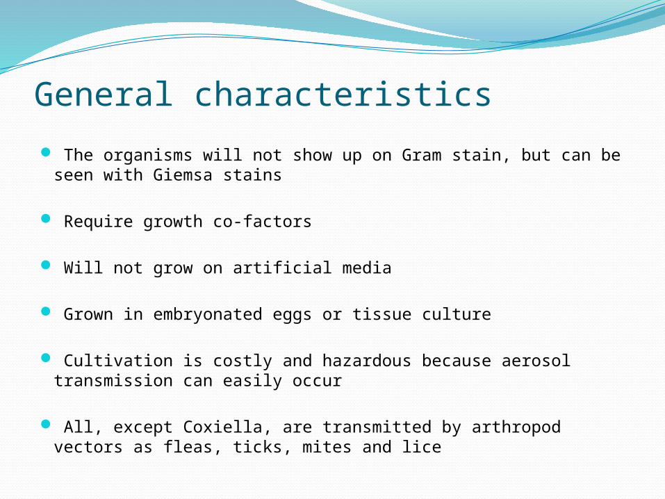
General characteristics
The organisms will not show up on Gram stain, but can be seen with Giemsa stains
Require growth co-factors
Will not grow on artificial media
Grown in embryonated eggs or tissue culture
Cultivation is costly and hazardous because aerosol transmission can easily occur
All, except Coxiella, are transmitted by arthropod vectors as fleas, ticks, mites and lice
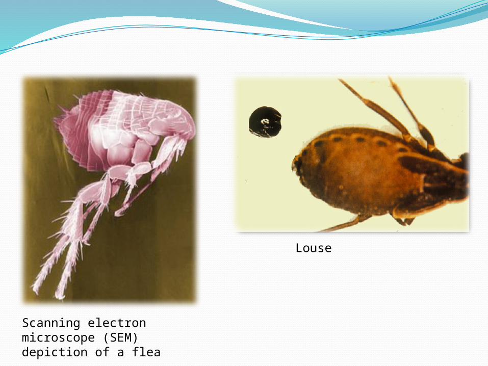
Scanning electron microscope (SEM) depiction of a flea
Louse

Electron micrograph of Rickettsia prowazekii in experimentally infected tick tissue
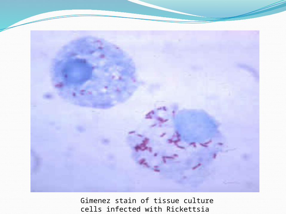
Gimenez stain of tissue culture cells infected with Rickettsia rickettsii

Rickettsia
TransmissionRickettsia are usually introduced into human skin by
the bite of an insect (flea or louse) or an arachnid (tick or mite)
R. rickettsii invades the endothelial cells that line the blood vessels
Incubation period: ~1 week
Virulence factors of Rickettsial specieschanges in the host cell phagocytosisbacterial surface protein

Castor bean tick, Ixodes ricinusEngorged tick attached to back of toddler's head. Adult thumb shown for scale.

Arthropod Vector
Rickettsia rickettsii

Pathogenesis
During the first few days of incubation periodlocal reaction caused by hypersensitivity to tick or vector
products
Bacteria multiply at the site & later disseminate via lymphatic system
Bacteria is phagocytosed by macrophages (1st barrier to rickettsial multiplication)
After 7-10 days
organisms disseminatereplicate in the nucleus or cytoplasm of endothelial cells
causing vasculitis

Infected cells show intracytoplasmic inclusions & intranuclear inclusions
Endothelial damage & vasculitis progress causingdevelopment of maculopapular skin rashesperivascular tissue necrosisthrombosis & ischemia
Disseminated endothelial lesion lead to increased capillary permeability, edema, hemorrhage & hypotensive shock
Endothelial damage can lead to activation of clotting system ---> Disseminated intravascular coagulation (DIC)

Pathogenesis: Rickettsia cell-to-cell spread

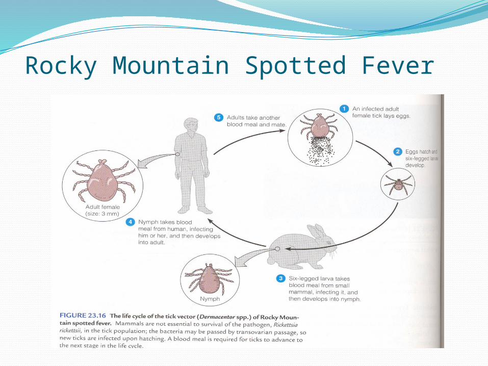
Rocky Mountain Spotted Fever
Etiologic agent: Rickettsia rickettsiae
Most common rickettsial disease
Individuals younger than 19 years old are usually at risk
Males affected twice as often as females
It is common during summer months

Serious disease with 35% mortality rate
Transmitted by ticks that must remain attached for hours in order to transmit the disease
Incubation of 2-6 days
Followed by a severe headache, chills, fever, aching, and nausea

After 2-6 days, a maculopapular rash develops, first on the extremities, including palms, foot soles, and spreading to the chest and abdomen
If left untreated, the rash will become
petechial with hemorrhages in the skin and mucous membranes due to vascular damage as the organism invades the blood vessels
Death may occur during the end of the second week due to kidney or heart failure
Rocky Mountain Spotted Fever
Endemic TyphusEtiologic agent: Rickettsia typhi
Incubation period: 5-18 days
Transmitted to man by rat fleas cat fleas and mouse fleas are less common
modes of transmission
The disease occurs sporadically

Symptoms: severe headache, chills, fever, and after a fourth day, a maculopapular rash caused by subcutaneous hemorrhaging as Rickettsia invade the blood vessels
The rash begins on the upper trunk and spread to involve the whole body except the face, palms of the hands, and the soles of the feet
The disease lasts about 2 weeks and the patient may have a prolonged convalescence
Ehrlichia
Disease: Ehrlichiosis
Transmitted via tick vectors
Etiologic agent: E. chaffeensis
Invade leukocytes and grow in cytoplasmic vacuoles making characteristic inclusions known as morulae
Symptoms resemble Rocky Mountain spotted fever

Clinically manifests as acute fever withleucopeniathrombocytopeniaelevations of aminotransferase levels
Rash is infrequent
Vasculitis is rare

Coxiella burnetiiThe only species of Coxiella genus
Causal agent of Q-fever
Found in infected animals, arthropods or humans and highly infectious
TransmissionInhalation of airborne organisms
infected dusts in farm and slaughterhousesContact with the milk, urine, feces, of infected animals
It has spore-like form that resists heat and dryness allowing it to survive in extracellular environment

Q feverQ for “query” or mysterious febrile illness
Occurs in veterinarians, ranchers, and animal researchers who are in contact with infected placenta from sheep, cattle, or goats (no arthropod vector for C. burnetii)
Incubation period: 10-28 days
Disease characterized by fever, influenza-like syndromes; but no skin rash
Some patients present with bronchopneumonia with patchy interstitial infiltrates
Rare complications: hepatitis, endocarditis, and meningoencephalitis

Q fever
Doughnut shaped non-caseating granuloma of Q fever

Laboratory Diagnosis of Rickettsiae1. Culture & isolation
Difficult & dangerous because of the highly infectious nature of rickettsiae
2. Serologic testA. Weil-Felix test: based on cross-reactivity between some
strains of Proteus & RickettsiaB. Complement fixation: not very sensitive & time
consumingC. Indirect fluorescence (EIA): more sensitive & specific;
allows discrimination between IgM & IgG antibodies which helps in early diagnosis
D. Direct immunofluorescence: the only serologic test that is useful for clinical diagnosis, 100% specific & 70% sensitive allowing diagnosis in 3-4 days into the illness