right portal vein ligation is as efficient as portal vein embolization to induce hypertrophy of the...
TRANSCRIPT
Right Portal Vein Ligation is as Efficient as Portal VeinEmbolization to Induce Hypertrophy of the Left Liver Remnant
B. Aussilhou & M. Lesurtel & A. Sauvanet & O. Farges &
S. Dokmak & N. Goasguen & A. Sibert & V. Vilgrain &
J. Belghiti
Received: 6 September 2007 /Accepted: 25 October 2007 /Published online: 30 November 2007# 2007 The Society for Surgery of the Alimentary Tract
AbstractBackground Aim of this retrospective study was to compare induction of left liver hypertrophy after right portal veinligation (PVL) and right portal vein embolization (PVE) before right hepatectomy for liver metastases.Materials and Methods Between 1998 and 2005, 18 patients underwent a PVE, whereas 17 patients underwent a PVLduring a first stage laparotomy.Results There was no complication related to PVE or PVL. After a similar interval time (7±3 vs 8±3 weeks), the increaseof the left liver volume was similar between the two groups (35±38 vs 38±26%). After PVE and PVL, right hepatectomywas performed in 12 and 14 patients, respectively. Technical difficulties during the right hepatectomy were similaraccording to duration of procedure (6.4±1 vs 6.7±1 h, p=0.7) and transfusion rates (33 vs 28%, p=0.7). Mortality was nilin both groups, and morbidity rates were respectively 58% for the PVE group and 36% for the PVL group (p=0.6).Conclusion Right PVL and PVE result in a comparable hypertrophy of the left liver. During the first laparotomy of a two-step liver resection, PVL can be efficiently and safely performed.
Keywords Portal vein occlusion . Portal vein ligation .
Liver metastasis . Liver hypertrophy
AbbreviationsPVL portal vein ligationPVE portal vein embolizationFLR future liver remnant
Introduction
In patients with primary liver tumors or selected livermetastases, complete resection is often the only chance ofpotential curative treatment to expect a long-term surviv-al.1,2 In case of extended hepatic lesions, liver resectionmay be hampered by the small volume of the residual liver,which is associated with a risk of postoperative life-threatening liver failure.3,4 Moreover, many patients withadvanced metastatic disease are referred to surgeons after aneoadjuvant chemotherapy, which has allowed downsizingof initially unresectable liver metastases,5 or when there is adocumented chemosensitivity.6,7 Now, it has been wellestablished that chemotherapy induces liver parenchymachanges, which may worsen postoperative morbidity.8,9 Inpatients considered for liver resection of metastasis, bothsmall volume of the future remnant liver and neoadjuvantchemotherapy increase the postoperative morbidity andmortality risk.
To overcome this risk and to increase resectability, moreaggressive surgical treatment procedures have been pro-posed using the regenerative capacity of the liver.10,11
Among them, occlusion of one branch of the portal vein
J Gastrointest Surg (2008) 12:297–303DOI 10.1007/s11605-007-0410-x
B. Aussilhou :M. Lesurtel :A. Sauvanet :O. Farges :S. Dokmak :N. Goasguen : J. Belghiti (*)Department of HPB Surgery,Beaujon Hospital (Assistance Publique-Hôpitaux de Paris),100, Bd Général Leclerc,92110 Clichy, Francee-mail: [email protected]
A. Sibert :V. VilgrainDepartment of Radiology,Beaujon Hospital (Assistance Publique-Hôpitaux de Paris),Clichy, France
B. Aussilhou :M. Lesurtel :A. Sauvanet :O. Farges :S. Dokmak :N. Goasguen :A. Sibert :V. Vilgrain : J. BelghitiUniversity Paris 7 Denis Diderot,Paris, France
results in the atrophy of the ipsilateral and hypertrophy ofthe contralateral liver segments. This phenomenon wasinitially observed in patients with cholangiocarcinoma,which induced portal vein occlusion by tumor invasion.12
Including portal vein occlusion in a strategy of scheduledsequential liver resections allowed to increase the numberof patients amenable to curative surgery, while they wereinitially deemed unresectable.10,11,13 Portal vein occlusionmay be achieved by either percutaneous embolization orsurgical ligation during a first-step laparotomy. Right portalvein ligation (PVL) has been considered to be less efficientthan right portal vein embolization (PVE) before a righthepatectomy.14,15
The present study aimed to compare PVL and PVEbefore a right hepatectomy in patients with liver metasta-ses in terms of safety, efficacy for hypertrophy of the leftliver remnant, resectability rates, and technical impact onliver resection.
Materials and Methods
Patients
Between 1998 and 2005, 35 patients with multiplecolorectal or neuroendocrine liver metastases underwent aright portal branch obstruction before “high risk” righthepatectomy because of a future liver remnant (FLR)volume less than 30% of the total liver volume or becauseof a post-chemotherapy liver parenchyma. Eighteen patientsunderwent a percutaneous PVE because metastases wereconsidered resectable in one stage. Seventeen patients had aPVL during a first-stage laparotomy when the metastaticdisease in the left liver was judged too extensive to besafely resected along with the right liver (n=10) and/orwhen the resection of the primary tumor was also required(n=10). Patients and tumors characteristics are given inTable 1. In the PVE group, patients were older than in thePVL group (51±10 vs 61±14 years, respectively, p=0.023), and all patients had colorectal metastases, whereas
ten (59%) patients had neuroendocrine metastases in thePVL group (p=0.02). There were more hepatic lesions, andthey were bigger in the PVL group. Liver function assessedby prothrombin time, and bilirubin was normal andcomparable in both groups (data not shown). Neoadjuvantchemotherapy consisting in the combined use of 5-fluorouracil and either oxaliplatin or irinotecan wasadministrated to all patients before PVE and to eight(47%) patients before PVL (p=0.001). Mean time betweenthe end of chemotherapy and portal vein occlusion was2.2±1.7 months and was not significantly differentbetween groups.
Right Portal Vein Embolization
Right PVE was performed using the contralateral trans-hepatic approach as previously described.16 In brief, acollateral vein of the left branch of the portal vein waspunctured under light general anesthesia and ultrasoundguidance. After control venous portography, the rightanterior and posterior portal branches were embolized witha mixture of cyanoacrylate (Histoacryle; Braun Lab,Hamburg, Germany) and lipiodol (Lipiodol Ultrafluide;Guerbert Lab, Paris, France). In none of them, branches tosegment 4 were embolized. Control portography wasperformed at the end of the procedure.
Right Portal Vein Ligation
Ligation of the right branch of the portal vein wasperformed as part of a two-stage procedure.10 During thefirst stage, the resection of the primary tumor wasperformed in ten patients (one left colectomy, two ileo-colic resections, and seven left pancreatectomies), andenucleation of the left-sided liver metastases, with at leasta 5-mm margin, was achieved in 16 patients. Extraparen-chymal ligation of the right portal branch was performedusing a nonabsorbable suture. Its efficacy was checked bypreoperative Doppler ultrasounds. Cholecystectomy wasperformed in the same time in ten patients.
Table 1 Characteristics ofPatients Who Underwent PVEor PVL Before Right Hepatec-tomy for Liver Metastases
Continuous variables expressedas mean±SD
PVE (n=18) PVL (n=17) p value
Gender (F/M) 7/11 10/7 0.3Age (year) 61±10 51±14 0.023Primary tumorAdenocarcinoma 18 7 0.001Neuroendocrine 0 10Hepatic tumor location unilobar/bilobar 12/6 1/16 0.03No. of tumors/patientRight liver 4.5±6 7±3 0.05Left liver 0.5±0.7 3.2±2 0.001Preoperative chemotherapy (%) 18 (100) 8 (47) 0.001
298 J Gastrointest Surg (2008) 12:297–303

Right Hepatectomy
Right hepatectomy was performed 7 to 8 weeks after portalvein occlusion. All patients underwent liver resection bythree senior liver surgeons, using a standardized techniquefor right hepatectomy.16 Parenchymal transection wasperformed by either the clamp-crush technique or with anultrasound aspiration dissector (Dissectron™; Satelec Med-ical, Merignac, France), with intermittent clamping of thehepatic pedicle. Patients were routinely transferred tothe intensive care unit and returned to the wards at thediscretion of the intensive care consultant. After righthepatectomy, the resected specimens were examined patho-logically, paying attention to the disease-free margins and tothe extent of necrosis of tumor. Tumor necrosis was definedas complete if no viable cells were observed in any nodule.
Follow-up and End Points
The primary end point of the analysis was the hypertrophyof the FLR induced by the right portal vein occlusion. Allpatients underwent volumetric helicoidal computed tomo-graphic (CT) scan estimation of their liver volumes beforethe obstruction and 4–6 weeks thereafter. Measurementswere performed for the whole liver and for the FLR usingthe middle hepatic vein, gallbladder bed, and umbilicalportion of the left portal vein as landmarks. The FLRvolume was expressed as a percentage of the total livervolume, excluding the tumor volume. Its hypertrophy afterportal vein occlusion was calculated as follows: (FLRvolume 4 to 6 weeks after portal vein obstruction−FLRvolume before portal vein obstruction)×100/FLR volumebefore portal vein obstruction.
The secondary end points of the analysis were theresectability rate and the postoperative course. Operativemortality was defined as death occurring within the samehospital stay or within 30 days of surgery. Postoperativecomplications, recorded prospectively, were defined asfollow: (a) liver failure was defined by a prothrombin timeof less than 50% (of normal) and serum bilirubin levelgreater than 50 μmol/l on postoperative day 5,17 (b)significant ascites (abdominal drain output more than500 ml/day), (c) biliary leak as the presence of bile in theabdominal drainage or abdominal collections greater thantwice the serum level, (d) postoperative pulmonary com-plications included all clinically symptomatic pleuraleffusions, atelectases, and infections, and (e) renal insuffi-ciency (serum creatinine level greater than 150 μmol/l).
Statistical Analysis
Summary statistics are expressed as mean±SD unlessotherwise stated. Continuous variables were compared
using the Fisher’s exact t test, and categorical variableswere compared using the Mann–Whitney test. A p value ofless than 0.05 was considered as statistically significant. Allthe calculations were performed with the Statistical Packagefor the Social Sciences (SPSS) 14.0 statistical package(SPSS, Chicago, IL, USA).
Results
Liver Hypertrophy
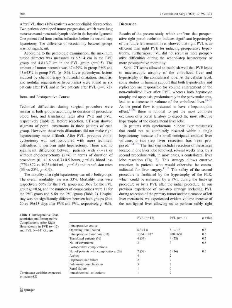
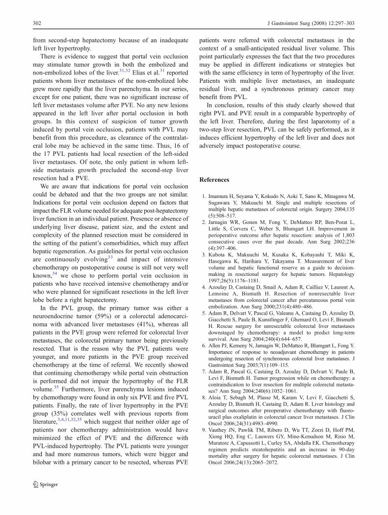
Right portal vein occlusion was complete in all the cases inboth groups. The mean interval time between portal veinocclusion and liver resection was similar in both groups(7±3 after PVE vs 8±3 weeks after PVL, p=0.6). The leftliver volume increased from 509±222 ml to 641±220 mlafter PVE (p<0.001) and from 477±179 to 638±192 mlafter PVL (p<0.001). After portal vein occlusion, theincrease of the left liver volume was not significantlydifferent between the two groups (35±38% after PVE vs38±26% after PVL, p=0.7; Fig. 1). None of the tumor butone in the left lobe increased until surgery (see below).
There was no complication after PVE and postoperativehospital stay was 2±1 days. In group PVL, four patientshad postoperative complications (one left pleural effusion,two pancreatic fistulae, and one intra-abdominal abscess),which were all related to primary tumor resection, andpostoperative hospital stay was 13±6 days.
Resectability
After PVE, six (30%) patients were not eligible for righthepatectomy because of insufficient hypertrophy of the leftliver (n=2) or tumor progression (n=4). Two patients hadperitoneal implants at laparotomy, one patient developedmediastinal metastatic lymph nodes, and in the last patient,diameter of the left lobe metastasis increased from 4 to 7.5 cm.
0
100
200
300
400
500
600
700
800
900
Before PVO 4-6 weeks after PVO
FL
R (
ml)
PVE
PVL
Figure 1 Volume of the future liver remnant (FLR) before and 4–6weeks after portal vein embolization (PVE) or portal vein ligation(PVL). PVO Portal vein occlusion.
J Gastrointest Surg (2008) 12:297–303 299299
After PVL, three (18%) patients were not eligible for resection.Two patients developed tumor progression, which were lungmetastases and metastatic lymph nodes in the hepatic ligament.One patient died from cardiac infarction before the second-steplaparotomy. The difference of resectability between groupswas not significant.
According to the pathologic examination, the maximumtumor diameter was measured as 6.5±4 cm in the PVEgroup and 4.8±3.7 cm in the PVL group (p=0.5). Theamount of tumor necrosis was 47±29% in group PVE and43±43% in group PVL (p=0.6). Liver parenchyma lesionsinduced by chemotherapy (sinusoidal dilatation, steatosis,and nodular regenerative hyperplasia) were found in sixpatients after PVE and in five patients after PVL (p=0.72).
Intra- and Postoperative Course
Technical difficulties during surgical procedure weresimilar in both groups according to duration of procedure,blood loss, and transfusion rates after PVE and PVL,respectively (Table 2). Before resection, CT scan showedstigmata of portal cavernoma in three patients of eachgroup. However, these vein dilatations did not make righthepatectomy more difficult. After PVL, previous chole-cystectomy was not associated with more technicaldifficulties to perform right hepatectomy. There was nosignificant difference between patients with (n=8) orwithout cholecystectomy (n=6) in terms of duration ofprocedure (6.1±1.6 vs 6.3±0.5 hours, p=0.8), blood loss(775±872 vs 1025±464 ml, p=0.6) and transfusion rates(33 vs 25%, p=0.9).
Themortality after right hepatectomywas nil in both groups.The overall morbidity rate was 33%. Morbidity rates wererespectively 58% for the PVE group and 36% for the PVLgroup (p=0.6), and the numbers of complications were 11 forthe PVE group and 8 for the PVL group (Table 2). Hospitalstay was not significantly different between both groups (24±20 vs 19±13 days after PVE and PVL, respectively, p=0.5).
Discussion
Results of the present study, which confirms that preoper-ative right portal occlusion induces significant hypertrophyof the future left remnant liver, showed that right PVL is asefficient than right PVE for inducing preoperative hyper-trophy. Furthermore, PVL did not result in more preoper-ative difficulties during the second-step hepatectomy ormore postoperative morbidity.
Serial CT scans allowed to establish well that PVE leadsto macroscopic atrophy of the embolized liver andhypertrophy of the contralateral lobe. At the cellular level,some studies in humans support that both hypertrophy andreplication are responsible for volume enlargement of thenon-embolized liver after PVE, whereas both hepatocyteatrophy and apoptosis, predominantly in the perivenular area,lead to a decrease in volume of the embolized liver.18–20
As the portal flow is presumed to have a hepatotrophiceffect,21,22 there is rational to get the most completeocclusion of a portal territory to expect the most effectivehypertrophy of the contralateral liver lobe.
In patients with synchronous bilobar liver metastasesthat could not be completely resected within a singlehepatectomy because of a small-anticipated residual livervolume, a two-step liver resection has been pro-posed.10,11,13 The first step includes resection of metastaseslocated in one liver lobe followed, several weeks later, by asecond procedure with, in most cases, a contralateral liverlobe resection (Fig. 2). This strategy allows curativeresection in patients who would otherwise be contra-indicated for liver surgery.11,13 The safety of the secondprocedure is facilitated by the hypertrophy of the FLR,which could be enhanced by a PVL during the first-stepprocedure or by a PVE after the initial procedure. In ourprevious experience of two-step strategy including PVLduring resection of the primary tumor and/or clearance of leftliver metastasis, we experienced evident volume increase ofthe non-ligated liver allowing us to perform safely right
Table 2 Intraoperative Char-acteristics and PostoperativeComplications After RightHepatectomy in PVE (n=12)and PVL (n=14) Groups
Continuous variables expressedas mean±SD
PVE (n=12) PVL (n=14) p value
Intraoperative courseOperating time (hours) 6.3±1.8 6.1±1.3 0.8Intraoperative blood loss (ml) 1354±1837 900±660 0.5Transfused patients (%) 4 (33) 4 (28) 0.7No. of cavernoma 3 3 0.8Postoperative complicationsNo. of patients with complications (%) 7 (58) 5 (36) 0.6Ascites 4 2Hepatocellular failure 2 2Pulmonary complications 3 2Renal failure 1 0Intraabdominal collections 1 2
300 J Gastrointest Surg (2008) 12:297–303
hepatectomy.10 As there is still a debate whether PVL is asefficient as PVE, this retrospective study aimed to comparePVL and PVE in terms of efficiency to induce hypertrophyof the FLR volume and impact on the planned liver resectionafter portal vein occlusion.
Broering et al.,15 comparing PVE and PVL for inductionof hypertrophy of the left lateral lobe before extended righthepatectomy, showed that PVE was more efficient. How-ever, in the latter study, 60% of patients who underwentPVE had a partial or complete occlusion of the segment IVbranches, whereas 29% in the PVL group (p=0.02). In ourexperience, we do not embolize segment IV branches toavoid migration of cyanoacrylate in the left portal branch,which would compromise the second-step hepatectomy.Results of the present study are consistent with those fromBouzari et al.23 Their results confirm that PVL is aseffective as PVE in inducing hypertrophy of the FLRvolume. PVL was supposed to be less efficient than PVEbecause it may induce the formation of intrahepatic porto-portal collaterals leading to failure of liver hypertrophy.14
However, in an experimental model, Krupski et al.24
showed that the increase of liver volume after PVL wasnot restrained by the formation of porto-portal collaterals.The fortnight normalization of increased portal blood flowinduced by portal vein occlusion in humans25 and the earlypeak of hepatocyte proliferation after portal occlusion inrodents26,27 suggest that liver hypertrophy is early inducedafter portal occlusion. Then, later formation of porto-portalcollaterals would not impact on the induced liver hyper-trophy. In this way, a recent experimental study in a non-
human primate model supports that even a reversibleportal vein occlusion may act as a starter for the liverhypertrophy.28
Another important result of the present study is that PVLdid not result in more perioperative difficulties during thesecond-step hepatectomy or more postoperative morbidity.The second-step right hepatectomy were performed with azero mortality rate and a 33% overall morbidity rate, whichare consistent with the literature.2,29,30 According tooperating time, blood loss, and transfusion rates, perioper-ative technical difficulties during second-step hepatectomyin the PVL group were not affected by the presence ofportal collaterals, previous liver resection, and cholecystec-tomy. We think that attention should be directed towardsafe and complete left liver resection without unnecessarydissection or mobilization that could impact the difficulty ofthe second step. Excessive dissection of the porta hepatitisshould be avoided to facilitate redissection at the secondprocedure. Cholecystectomy, which could be necessary toallow efficient control of the right branch of the portal vein,seems to have no impact on the technical difficulties.
In the present study, we were able to perform thescheduled second-step right hepatectomy in 74% of thepatients. This figure is comparable with other series oftwo-stage hepatectomy from the literature, which report55–85% resectability rates.13,15,31 This rate was 82% afterPVL and 67% after PVE, but the difference did not reachsignificance. This difference could be explained by a moreimportant severity of colorectal cancer than neuroendo-crine cancer. Interestingly, no PVL patient was precluded
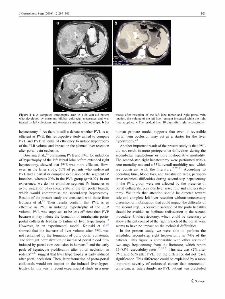
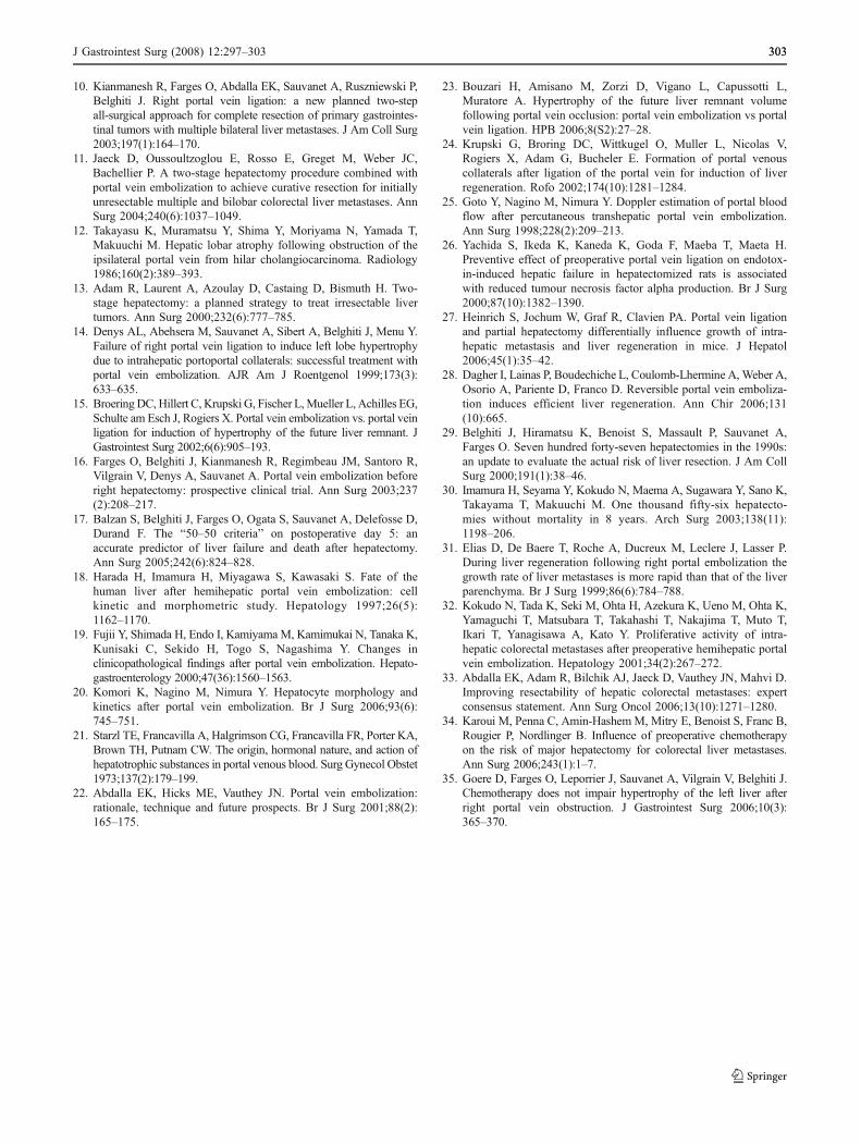
Figure 2 a A computed tomography scan in a 56-year-old patientwho developed synchronous bilobar colorectal metastases and wastreated by left colectomy and 6-month systemic chemotherapy. b Six
weeks after resection of the left lobe tumor and right portal veinligation, the volume of the left liver remnant increased while the rightliver atrophied. c The residual liver 10 days after right hepatectomy.
J Gastrointest Surg (2008) 12:297–303 301301
from second-step hepatectomy because of an inadequateleft liver hypertrophy.
There is evidence to suggest that portal vein occlusionmay stimulate tumor growth in both the embolized andnon-embolized lobes of the liver.31,32 Elias et al.31 reportedpatients whom liver metastases of the non-embolized lobegrew more rapidly that the liver parenchyma. In our series,except for one patient, there was no significant increase ofleft liver metastases volume after PVE. No any new lesionsappeared in the left liver after portal occlusion in bothgroups. In this context of suspicion of tumor growthinduced by portal vein occlusion, patients with PVL maybenefit from this procedure, as clearance of the contralat-eral lobe may be achieved in the same time. Thus, 16 ofthe 17 PVL patients had local resection of the left-sidedliver metastases. Of note, the only patient in whom left-side metastasis growth precluded the second-step liverresection had a PVE.
We are aware that indications for portal vein occlusioncould be debated and that the two groups are not similar.Indications for portal vein occlusion depend on factors thatimpact the FLR volume needed for adequate post-hepatectomyliver function in an individual patient. Presence or absence ofunderlying liver disease, patient size, and the extent andcomplexity of the planned resection must be considered inthe setting of the patient’s comorbidities, which may affecthepatic regeneration. As guidelines for portal vein occlusionare continuously evolving33 and impact of intensivechemotherapy on postoperative course is still not very wellknown,34 we chose to perform portal vein occlusion inpatients who have received intensive chemotherapy and/orwho were planned for significant resections in the left liverlobe before a right hepatectomy.
In the PVL group, the primary tumor was either aneuroendocrine tumor (59%) or a colorectal adenocarci-noma with advanced liver metastases (41%), whereas allpatients in the PVE group were referred for colorectal livermetastases, the colorectal primary tumor being previouslyresected. That is the reason why the PVL patients wereyounger, and more patients in the PVE group receivedchemotherapy at the time of referral. We recently showedthat continuing chemotherapy while portal vein obstructionis performed did not impair the hypertrophy of the FLRvolume.35 Furthermore, liver parenchyma lesions inducedby chemotherapy were found in only six PVE and five PVLpatients. Finally, the rate of liver hypertrophy in the PVEgroup (35%) correlates well with previous reports fromliterature,3,4,11,32,35 which suggest that neither older age ofpatients nor chemotherapy administration would haveminimized the effect of PVE and the difference withPVL-induced hypertrophy. The PVL patients were youngerand had more numerous tumors, which were bigger andbilobar with a primary cancer to be resected, whereas PVE
patients were referred with colorectal metastases in thecontext of a small-anticipated residual liver volume. Thispoint particularly expresses the fact that the two proceduresmay be applied in different indications or strategies butwith the same efficiency in term of hypertrophy of the liver.Patients with multiple liver metastases, an inadequateresidual liver, and a synchronous primary cancer maybenefit from PVL.
In conclusion, results of this study clearly showed thatright PVL and PVE result in a comparable hypertrophy ofthe left liver. Therefore, during the first laparotomy of atwo-step liver resection, PVL can be safely performed, as itinduces efficient hypertrophy of the left liver and does notadversely impact postoperative course.
References
1. Imamura H, Seyama Y, Kokudo N, Aoki T, Sano K, Minagawa M,Sugawara Y, Makuuchi M. Single and multiple resections ofmultiple hepatic metastases of colorectal origin. Surgery 2004;135(5):508–517.
2. Jarnagin WR, Gonen M, Fong Y, DeMatteo RP, Ben-Porat L,Little S, Corvera C, Weber S, Blumgart LH. Improvement inperioperative outcome after hepatic resection: analysis of 1,803consecutive cases over the past decade. Ann Surg 2002;236(4):397–406.
3. Kubota K, Makuuchi M, Kusaka K, Kobayashi T, Miki K,Hasegawa K, Harihara Y, Takayama T. Measurement of livervolume and hepatic functional reserve as a guide to decision-making in resectional surgery for hepatic tumors. Hepatology1997;26(5):1176–1181.
4. Azoulay D, Castaing D, Smail A, Adam R, Cailliez V, Laurent A,Lemoine A, Bismuth H. Resection of nonresectable livermetastases from colorectal cancer after percutaneous portal veinembolization. Ann Surg 2000;231(4):480–486.
5. Adam R, Delvart V, Pascal G, Valeanu A, Castaing D, Azoulay D,Giacchetti S, Paule B, Kunstlinger F, Ghemard O, Levi F, BismuthH. Rescue surgery for unresectable colorectal liver metastasesdownstaged by chemotherapy: a model to predict long-termsurvival. Ann Surg 2004;240(4):644–657.
6. Allen PJ, Kemeny N, Jarnagin W, DeMatteo R, Blumgart L, Fong Y.Importance of response to neoadjuvant chemotherapy in patientsundergoing resection of synchronous colorectal liver metastases. JGastrointest Surg 2003;7(1):109–115.
7. Adam R, Pascal G, Castaing D, Azoulay D, Delvart V, Paule B,Levi F, Bismuth H. Tumor progression while on chemotherapy: acontraindication to liver resection for multiple colorectal metasta-ses? Ann Surg 2004;240(6):1052–1061.
8. Aloia T, Sebagh M, Plasse M, Karam V, Levi F, Giacchetti S,Azoulay D, Bismuth H, Castaing D, Adam R. Liver histology andsurgical outcomes after preoperative chemotherapy with fluoro-uracil plus oxaliplatin in colorectal cancer liver metastases. J ClinOncol 2006;24(31):4983–4990.
9. Vauthey JN, Pawlik TM, Ribero D, Wu TT, Zorzi D, Hoff PM,Xiong HQ, Eng C, Lauwers GY, Mino-Kenudson M, Risio M,Muratore A, Capussotti L, Curley SA, Abdalla EK. Chemotherapyregimen predicts steatohepatitis and an increase in 90-daymortality after surgery for hepatic colorectal metastases. J ClinOncol 2006;24(13):2065–2072.
302 J Gastrointest Surg (2008) 12:297–303
10. Kianmanesh R, Farges O, Abdalla EK, Sauvanet A, Ruszniewski P,Belghiti J. Right portal vein ligation: a new planned two-stepall-surgical approach for complete resection of primary gastrointes-tinal tumors with multiple bilateral liver metastases. J Am Coll Surg2003;197(1):164–170.
11. Jaeck D, Oussoultzoglou E, Rosso E, Greget M, Weber JC,Bachellier P. A two-stage hepatectomy procedure combined withportal vein embolization to achieve curative resection for initiallyunresectable multiple and bilobar colorectal liver metastases. AnnSurg 2004;240(6):1037–1049.
12. Takayasu K, Muramatsu Y, Shima Y, Moriyama N, Yamada T,Makuuchi M. Hepatic lobar atrophy following obstruction of theipsilateral portal vein from hilar cholangiocarcinoma. Radiology1986;160(2):389–393.
13. Adam R, Laurent A, Azoulay D, Castaing D, Bismuth H. Two-stage hepatectomy: a planned strategy to treat irresectable livertumors. Ann Surg 2000;232(6):777–785.
14. Denys AL, Abehsera M, Sauvanet A, Sibert A, Belghiti J, Menu Y.Failure of right portal vein ligation to induce left lobe hypertrophydue to intrahepatic portoportal collaterals: successful treatment withportal vein embolization. AJR Am J Roentgenol 1999;173(3):633–635.
15. BroeringDC, Hillert C, Krupski G, Fischer L,Mueller L, Achilles EG,Schulte am Esch J, Rogiers X. Portal vein embolization vs. portal veinligation for induction of hypertrophy of the future liver remnant. JGastrointest Surg 2002;6(6):905–193.
16. Farges O, Belghiti J, Kianmanesh R, Regimbeau JM, Santoro R,Vilgrain V, Denys A, Sauvanet A. Portal vein embolization beforeright hepatectomy: prospective clinical trial. Ann Surg 2003;237(2):208–217.
17. Balzan S, Belghiti J, Farges O, Ogata S, Sauvanet A, Delefosse D,Durand F. The “50–50 criteria” on postoperative day 5: anaccurate predictor of liver failure and death after hepatectomy.Ann Surg 2005;242(6):824–828.
18. Harada H, Imamura H, Miyagawa S, Kawasaki S. Fate of thehuman liver after hemihepatic portal vein embolization: cellkinetic and morphometric study. Hepatology 1997;26(5):1162–1170.
19. Fujii Y, Shimada H, Endo I, Kamiyama M, Kamimukai N, Tanaka K,Kunisaki C, Sekido H, Togo S, Nagashima Y. Changes inclinicopathological findings after portal vein embolization. Hepato-gastroenterology 2000;47(36):1560–1563.
20. Komori K, Nagino M, Nimura Y. Hepatocyte morphology andkinetics after portal vein embolization. Br J Surg 2006;93(6):745–751.
21. Starzl TE, Francavilla A, Halgrimson CG, Francavilla FR, Porter KA,Brown TH, Putnam CW. The origin, hormonal nature, and action ofhepatotrophic substances in portal venous blood. Surg Gynecol Obstet1973;137(2):179–199.
22. Abdalla EK, Hicks ME, Vauthey JN. Portal vein embolization:rationale, technique and future prospects. Br J Surg 2001;88(2):165–175.
23. Bouzari H, Amisano M, Zorzi D, Vigano L, Capussotti L,Muratore A. Hypertrophy of the future liver remnant volumefollowing portal vein occlusion: portal vein embolization vs portalvein ligation. HPB 2006;8(S2):27–28.
24. Krupski G, Broring DC, Wittkugel O, Muller L, Nicolas V,Rogiers X, Adam G, Bucheler E. Formation of portal venouscollaterals after ligation of the portal vein for induction of liverregeneration. Rofo 2002;174(10):1281–1284.
25. Goto Y, Nagino M, Nimura Y. Doppler estimation of portal bloodflow after percutaneous transhepatic portal vein embolization.Ann Surg 1998;228(2):209–213.
26. Yachida S, Ikeda K, Kaneda K, Goda F, Maeba T, Maeta H.Preventive effect of preoperative portal vein ligation on endotox-in-induced hepatic failure in hepatectomized rats is associatedwith reduced tumour necrosis factor alpha production. Br J Surg2000;87(10):1382–1390.
27. Heinrich S, Jochum W, Graf R, Clavien PA. Portal vein ligationand partial hepatectomy differentially influence growth of intra-hepatic metastasis and liver regeneration in mice. J Hepatol2006;45(1):35–42.
28. Dagher I, Lainas P, Boudechiche L, Coulomb-Lhermine A, Weber A,Osorio A, Pariente D, Franco D. Reversible portal vein emboliza-tion induces efficient liver regeneration. Ann Chir 2006;131(10):665.
29. Belghiti J, Hiramatsu K, Benoist S, Massault P, Sauvanet A,Farges O. Seven hundred forty-seven hepatectomies in the 1990s:an update to evaluate the actual risk of liver resection. J Am CollSurg 2000;191(1):38–46.
30. Imamura H, Seyama Y, Kokudo N, Maema A, Sugawara Y, Sano K,Takayama T, Makuuchi M. One thousand fifty-six hepatecto-mies without mortality in 8 years. Arch Surg 2003;138(11):1198–206.
31. Elias D, De Baere T, Roche A, Ducreux M, Leclere J, Lasser P.During liver regeneration following right portal embolization thegrowth rate of liver metastases is more rapid than that of the liverparenchyma. Br J Surg 1999;86(6):784–788.
32. Kokudo N, Tada K, Seki M, Ohta H, Azekura K, Ueno M, Ohta K,Yamaguchi T, Matsubara T, Takahashi T, Nakajima T, Muto T,Ikari T, Yanagisawa A, Kato Y. Proliferative activity of intra-hepatic colorectal metastases after preoperative hemihepatic portalvein embolization. Hepatology 2001;34(2):267–272.
33. Abdalla EK, Adam R, Bilchik AJ, Jaeck D, Vauthey JN, Mahvi D.Improving resectability of hepatic colorectal metastases: expertconsensus statement. Ann Surg Oncol 2006;13(10):1271–1280.
34. Karoui M, Penna C, Amin-Hashem M, Mitry E, Benoist S, Franc B,Rougier P, Nordlinger B. Influence of preoperative chemotherapyon the risk of major hepatectomy for colorectal liver metastases.Ann Surg 2006;243(1):1–7.
35. Goere D, Farges O, Leporrier J, Sauvanet A, Vilgrain V, Belghiti J.Chemotherapy does not impair hypertrophy of the left liver afterright portal vein obstruction. J Gastrointest Surg 2006;10(3):365–370.
J Gastrointest Surg (2008) 12:297–303 303303