(riso, mg9l2, - pnas.org · proc. natl. acad.sci. usa73(1976) 2795 (3. trnaty, 4-4-4--fig. 2....
TRANSCRIPT

Proc. Natl. Acad. Sci. USAVol. 73, No. 8, pp. 2793-2797, August 1976Cell Biology
Transfer of tRNAs to somatic cells mediated bySendai-virus-induced fusion
(loading of red blood cells/microinjection/yeast tRNAPhC/suIII+ tRNAiTYr/suppression)
KELD KALTOFT*, JESPER ZEUTHENt, FRODE ENGBAEKO, PETER W. PIPERO, AND JULIO E. CELIS**Division of Biostructural Chemistry, Institute of Chemistry, tInstitute of Human Genetics, and *Department of Molecular Biology, University of Aarhus,DK-8000 Aarhus C., Denmark
Communicated by F. H. C. Crick, March 18,1976
ABSTRACT tRNAs from yeast (tRNAPhe and 4S RNA) andEscherichia coli (suIII+ tRNAITYr) have been transferred tomouse cells by means of a two-step transfer procedure [Loyter,Zakai, and Kulka (1975)1. Cell Biol. 66,292-304; Schlegel andRechsteiner (1975) Cell 5, 371-379]. In the first stage of thetransfer tRNAs were incorporated into rabbit red blood cells(RBCs). Thereafter, the loaded erythrocytes were fused withrecipient mouse cells by means of Sendai virus. At least 0.3-0.4% of the total input of tRNA used to load the RBCs could betransferred to mouse cells. Of the tRNA incorporated in themouse cells, at least 50% could be recovered in the form of in-tact tRNA molecules when yeast 4S RNA was used. With E colisuIII+ tRNAITYr a rather smaller proportion of the tRNA re-mained intact (33%).Although the loading of tRNA into RBCs is not essential for
its uptake, we find that the transfer is four times more efficientwith RBCs as a vehicle for the injection. Sifnificantly, a fraction(2%) of the recipient cells possessed much more incorporatedtRNA than the average cell when Sendai virus and loaded RBCswere used. Such cells were not found in control experiments inwhich tRNA uptake was induced by Sendai virus alone.
It is important to develop techniques for the injection of mac-romolecules into mammalian cells in order to study the bio-logical function of such molecules under conditions which re-semble the in vivo situation. Gurdon and coworkers (1-3) haveshown that it is possible to inject macromolecules into the cy-toplasm of large cells like the Xenopus oocyte by physical mi-croinjection. Unfortunately this technique is difficult and la-borious when applied to normal size somatic cells (4, 5). Re-cently a two-step procedure has been introduced by Loyter etal. (6) and by Schlegel and Rechsteiner (7), by which it is pos-sible to incorporate proteins into a wide range of mammaliancell types. This transfer procedure can be used with cells havingreceptors for Sendai virus and comprises two separate steps. Theproteins are initially incorporated into red blood cells (RBCs)(8, 9) and the loaded RBCs are subsequently induced to fuse,through the action of Sendai virus, with recipient culture cells.By means of this procedure proteins such as ferritin (6), albu-min, and thymidine kinase (7) have been injected into mam-malian cells. Moreover, the transfer of thymidine kinase intoa thymidine-kinase-deficient cell line by this procedure canresult in a correction of the metabolic deficiency of these cells(7).Our aim in using this technique has been to develop an in
vwo system that could be used to assay the presence of nonsensemutations in mammalian cells, through the injection of yeastsuppressor tRNAs. We describe here experiments in whichdefined species of tRNA derived from yeast or Escherichla coli
Abbreviations: Buffer Na, 160 mM NaCI, 20 mM N-[tris(hydroxy-methyl)methyllglycine (Tricine), pH 7.4; BSS, (balanced salt solution)137 mM NaCI, 5.3mM KCl, 10mM Tris-HCl at pH 7.3, 1 mM CaC12,0.5 mM MgCl2, 0.5 mM MgSO4; MEM, Eagle's minimal essentialmedium; RBCs, red blood cells.
2793
have been transferred to cultured mouse cells. Through use ofelectrophoresis in denaturing polyacrylamide gels and fin-gerprinting of T1 RNase digests, it was shown that a substantialproportion of the tRNA transferred to the cells was structurallyintact. The loading of tRNA into erythrocytes is not essentialfor its transfer to mouse cells, since these cells took up tRNAfrom the medium in the presence of Sendai virus alone. How-ever, the treatment of the mouse cells with Sendai virus andloaded erythrocytes resulted in a more efficient tRNA uptakeand in the formation of some recipient cells that containedconsiderably more tRNA than the average injected cell.
MATERIALS AND METHODSPreparation of Labeled tRNAs. 32P-Labeled Saccharomyces
cerevssiae 4S RNA was prepared from strain L-133. Cultureswere grown for several generations in low-phosphate mediumat 300 (10) with 0.05 mCi/ml of [32P]orthophosphate (Riso,Denmark). The cells were recovered by pelleting and washedtwice with 10 mM Tris-HCl, 10 mM Mg9l2, 200 mM sodiumacetate at pH 5.0. After phenol extraction the RNA was pre-cipitated with 2 volumes of ethanol at -200, dried by meansof lyophilization, and applied to a 12% polyacrylamide gel (11).After autoradiography, the portion of the gel containing 4SRNA was cut out. The 4S RNA was eluted, precipitated with2 volumes of ethanol at -200, dried, and resuspended in 10mMTris-HCI, 10 mM MgCl2 at pH 7.4.
32P-Labeled suIII+ tRNA1TYr was prepared from E. coli1000B cells infected with 080 psuIII+ (12, 13). A 12% poly-acrylamide gel was used to purify the suppressor tRNA fromall other tRNAs (14).
'25I-Labeled yeast tRNAPhe was prepared by iodination ofpurified tRNAPhe (batch control no. 7285420, BoehringerMannheim GmbH) at 370 (15). tRNAPhe was purified from thereaction mixture by electrophoresis on a 12% gel.Loading of Red Blood Cells. Blood was collected from the
ear vein of rabbits into Ca2+_ and Mg2+-free Hanks' salt solutioncontaining 1 mg/ml of EDTA and 25 international units (IU)/ml of heparin. The red cells were washed three times in bal-anced salt solution (BSS; 137 mM NaCI, 5.3 mM KCI, 10 mMTris-HCl at pH 7.3, 1 mM CaC12, 0.5 mM MgCl2, 0.5 mMMgSO4). White blood cells were carefully removed during thewashings and the RBCs were used the same day. The procedurefor loading RBCs with tRNA was essentially as described byRechsteiner (7, 9) for loading proteins, except that BSS was usedinstead of Hanks' salt solution. Sufficient 10 X BSS was addedto make the suspension of red blood cells with tRNA 137 mMin NaCl during the resealing step. In a typical experiment weused 0.2 ml of tRNA solution (containing 0.5-1 mg of tRNA)in 10 mM Tris-HCI at pH 7.4, 10 mM MgCI2, and 0.5 ml ofpelleted RBCs. After resealing, cells were either washed three

Proc. Natl. Acad. Sci. USA 73 (1976)
.. .~...-w
Ri z !z
*?_4,.,4%i
A D
t I
A} _In _
,&;i4' __ a_ z_ t -- < .. ,
j {^4_
_l:I'.s .-
.= * t
z As.::* .:
B E~sil:.; b~~i 8 ,a,
.4b.
Pi
CFIG. 1. Yeast l2sI-tRNAPhe transferred to mouse Cl-lD celis as observed by autoradiography. Transfer of l25I-tRNAPhe to mouse cells after
concomitant exposure to 125I-tRNAPhe and Sendai virus (A, B, and C) and by fusion with l25I-tRNAPhe-loaded RBCs (D, E, and F). (X290;Wright-Giemsa stain).
times in BSS and resuspended in 160 mM NaCl, 20 mM N-[tris(hydroxymethyl)methyl]glycine (Tricine), pH 7.4 (bufferNa), or diluted with buffer Na and used directly for cell fu-sion.
Cell Fusion. A thymidine-kinase-deficient derivative Cl-1Dderived from the LM subclone of C3H mouse cells (16) was usedthroughout these experiments. Cells were maintained in Eagle'sminimal essential medium (MEM) with 10% calf serum andantibiotics (penicillin, 100 IU/ml; streptomycin, 50 ,g/ml). Cellfusion was carried out essentially as described by Loyter et al.(6). Cl-iD cells were detached by treatment with 0.05% Try-pure® (Novo, Denmark) at 370 and resuspended in 128 mM
KC1, 32 mM NaCl, 20 mM Tricine, pH 7.4 (buffer K) con-taining 1 mM MnCl2. UV-inactivated Sendai virus (1000 he-magglutinating units (HAU) prepared according to Giles andRuddle (17) and tRNA-loaded RBCs were added and the sus-pension incubated in the cold for 15 min to allow agglutination.To obtain fusion, the mixture was then incubated with gentleshaking at 370 for 30 min. In control experiments the loadedRBCs were omitted and an equivalent amount of labeled tRNAwas added to the suspension.
After the termination of fusion the cells were gently spundown, resuspended in medium, and reseeded on Falcon flasksor on glass coverslips in plastic petri dishes. Cells were generally
2794 Cell Biology: Kaltoft et al.
,k.,.frI
... ... .":f-p".
I
F A
14.1.
N
J
vA
,4qI%I- -,q
- I. a
. 14 .
. .. I A "W.1 ,;Om.
.* 1, kl0 0,

Proc. Natl. Acad. Sci. USA 73 (1976) 2795
(3
. tRNATy,
4-
4-
4--
FIG. 2. Mouse Cl-1D cells fused with loaded RBCs, photographedafter the termination at the incubation at 37°. Mouse cells can beobserved to fuse with several RBCs (arrows). (X390; phase contrast).
harvested 6 hr after the termination of fusion. Where it wasdesired to eliminate RBCs from the cells in monolayer, thecultures were incubated with 0.83% NH4Cl for 10 min. Thistreatment was repeated once to ensure complete lysis of allRBCs and then the monolayer was washed twice with Hanks'salt solution.
Autoradiography. Monolayer cultures on glass coverslips orsmears on object slides were fixed in methanol, immersed incold 5% trichloroacetic acid for 5 min to remove low-molecu-lar-weight material, rinsed in cold water, and then dried. Theglass coverslips were mounted on object slides with DepexO.After coating with gelatin the slides were covered with IlfordK2 liquid emulsion and the autoradiograms were developedafter a 15-day exposure using standard procedures and stainedwith Wright-Giemsa stain.Formamide Polyacrylamide Gel Electrophoresis. Samples
were taken at different stages of the transfer procedure andextracted at room temperature with an equal volume of phenolin the presence of 0.2 M Na acetate, pH-5.0, and 0.1% sodium
4S RNA -*4-
4*-
4*-
a b c d e f
FIG. 3. Gel electrophoresis of 32P-labeled 4S yeastRNA recoveredat different stages of the transfer procedure. Samples for gel elec-trophoresis were processed as described in Material and Methods.(a) Control 4S RNA. RNAs recovered from: (b) red blood cells, (c)
supernatant after fusion, (d) supernatant after 6 hr incubation at 370,(e) NH4C1 wash, (f) monolayers. The gels were dried and exposedovernight to Kodak blue brand x-ray film. Similar results have beenobtained by running longer gels.
a b c d eFIG. 4. Gel electrophoresis of 32P-labeled suIII+ tRNAITYr re-
covered at different stages of the transfer. The RNAs were recoveredas described in Materials and Methods. (a and b) Control tRNA.tRNAs recovered from (c) loaded red blood cells, (d) supernatant afterfusion, (e) supernatant after 6 hr incubation at 370, (f) monolayers.The gels were dried and exposed overnight to Kodak blue brand x-rayfilm. Similar results have been obtained by running longer gels.
dodecyl sulfate. The RNA in the aqueous phase was precipi-tated with 2 volumes of ethanol, dried, and resuspended in abuffer containing 40 mM Tris-acetate at pH 8.3, 20 mM Naacetate, 2mM EDTA, 10% (vol/vol) glycerol, and 90% (vol/vol)formamide. The samples were heated at 80° for 1 min beforeapplication to a 12% polyacrylamide gel prepared in 90%formamide.
Fingerprinting. Samples for fingerprinting were processedin the same way as the samples for gel electrophoresis but notdried. They were digested with T1 RNase and the digestionproducts were separated by two-dimensional fingerprintingtechniques (18, 19).
RESULTSTransfer of yeast 125I-tRNAPhe to mouse cellsWith the procedure described for incorporating labeled tRNAinto rabbit RBCs, about 10% of the total input of labeled tRNAwas generally taken up by the RBCs. In one typical experimentthat utilized 43 X 106 cpm of l25I-tRNAPhe, 4 X 10⁶ cpm be-came incorporated into RBC ghosts. l25I-tRNAPhe-loaded RBCswere fused with mouse cells and after fusion the cells were re-covered by centrifugation, resuspended in MEM, and seededon glass coverslips in petri dishes. Six hours later the slides werewashed twice with 0.83% NH4Cl to eliminate remaining RBCs.Complete lysis of the loaded RBCs was confirmed by micro-scopic examination of the cultures. The slides were counteddirectly and subsequently covered with autoradiographicemulsion and exposed. A control experiment in which thetRNA-loaded RBCs were substituted by an equivalent amountof l25I-tRNAPhe (4 X 10⁶ cpm) was also analyzed as describedabove. Autoradiographic analysis of cells from these experi-ments clearly demonstrated transfer of 125I-tRNAPhe to mousecells despite the omission of the loading step (Fig. 1A, B, andC), in agreement with the results of Tanaka et al. (20), whichshow that human fibroblasts will incorporate phage T4 endo-
Cell Biology: Kaltoft et al.
f
T,,tRNA ___). 4ftdo ft
Proc. Natl. Acad. Sci. USA 73 (1976)
#4 Anticodon
* 3'end
ACUUCGTYCG UCAUCG
UUCCCG
PG p
a b4 $
UGCCAAAG
CGmGAAG
AGCAG
CCG
CG
G
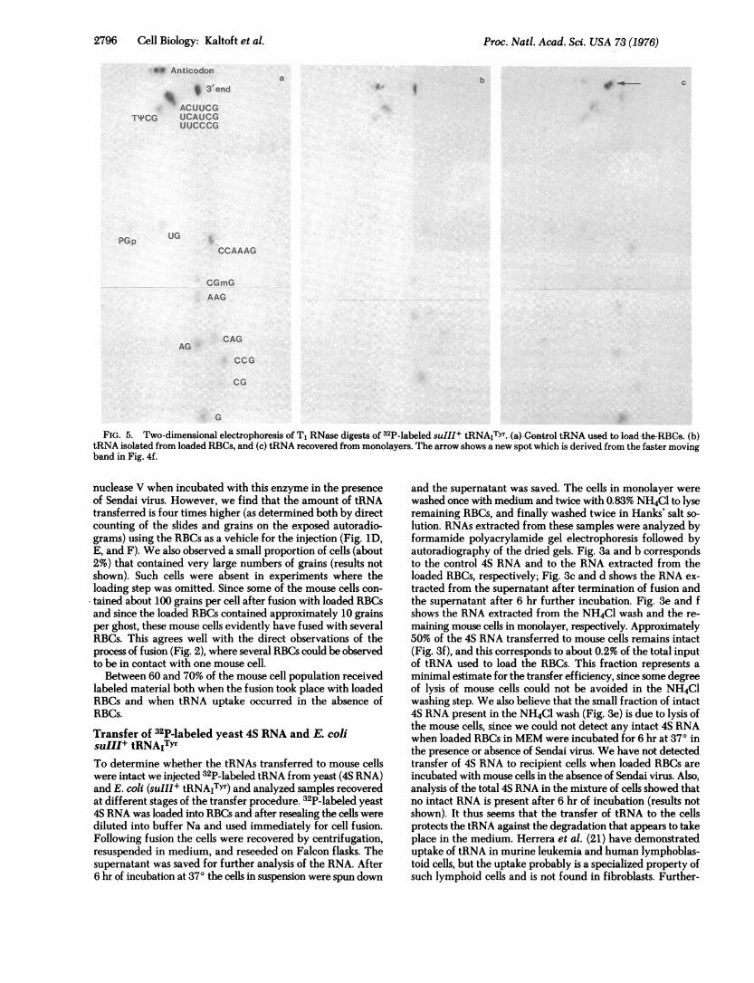
FIG. 5. Two-dimensional electrophoresis of T1 RNase digests of 32P-labeled suIIIt tRNAiTYr.-(a) Control tRNA used to-load-the-RBCs. (b)tRNA isolated from loaded RBCs, and (c) tRNA recovered from monolayers. The arrow shows a new spot which is derived from the faster movingband in Fig. 4f.
nuclease V when incubated with this enzyme in the presenceof Sendai virus. However, we find that the amount of tRNAtransferred is four times higher (as determined both by directcounting of the slides and grains on the exposed autoradio-grams) using the RBCs as a vehicle for the injection (Fig. 1D,E, and F). We also observed a small proportion of cells (about2%) that contained very large numbers of grains (results notshown). Such cells were absent in experiments where theloading step was omitted. Since some of the mouse cells con-tained about 100 grains per cell after fusion with loaded RBCsand since the loaded RBCs contained approximately 10 grainsper ghost, these mouse cells evidently have fused with severalRBCs. This agrees well with the direct observations of theprocess of fusion (Fig. 2), where several RBCs could be observedto be in contact with one mouse cell.
Between 60 and 70% of the mouse cell population receivedlabeled material both when the fusion took place with loadedRBCs and when tRNA uptake occurred in the absence ofRBCs.
Transfer of 32P-labeled yeast 4S RNA and E. coilsuIII+ tRNAITYrTo determine whether the tRNAs transferred to mouse cellswere intact we injected 32P-labeled tRNA from yeast (4S RNA)and E. coli (suIII+ tRNAITYr) and analyzed samples recoveredat different stages of the transfer procedure. 32P-labeled yeast4S RNA was loaded into RBCs and after resealing the cells werediluted into buffer Na and used immediately for cell fusion.Following fusion the cells were recovered by centrifugation,resuspended in medium, and reseeded on Falcon flasks. Thesupernatant was saved for further analysis of the RNA. After6 hr of incubation at 370 the cells in suspension were spun down
and the supernatant was saved. The cells in monolayer werewashed once with medium and twice with 0.83% NH4Cl to lyseremaining RBCs, and finally washed twice in Hanks' salt so-lution. RNAs extracted from these samples were analyzed byformamide polyacrylamide gel electrophoresis followed byautoradiography of the dried gels. Fig. 3a and b correspondsto the control 4S RNA and to the RNA extracted from theloaded RBCs, respectively; Fig. 3c and d shows the RNA ex-tracted from the supernatant after termination of fusion andthe supernatant after 6 hr further incubation. Fig. 3e and fshows the RNA extracted from the NH4C1 wash and the re-maining mouse cells in monolayer, respectively. Approximately50% of the 4S RNA transferred to mouse cells remains intact(Fig. 3f), and this corresponds to about 0.2% of the total inputof tRNA used to load the RBCs. This fraction represents aminimal estimate for the transfer efficiency, since some degreeof lysis of mouse cells could not be avoided in the NH4Clwashing step. We also believe that the small fraction of intact4S RNA present in the NH4C1 wash (Fig. 3e) is due to lysis ofthe mouse cells, since we could not detect any intact 4S RNAwhen loaded RBCs in MEM were incubated for 6 hr at 370 inthe presence or absence of Sendai virus. We have not detectedtransfer of 4S RNA to recipient cells when loaded RBCs areincubated with mouse cells in the absence of Sendai virus. Also,analysis of the total 4S RNA in the mixture of cells showed thatno intact RNA is present after 6 hr of incubation (results notshown). It thus seems that the transfer of tRNA to the cellsprotects the tRNA against the degradation that appears to takeplace in the medium. Herrera et al. (21) have demonstrateduptake of tRNA in murine leukemia and human lymphoblas-toid cells, but the uptake probably is a specialized property ofsuch lymphoid cells and is not found in fibroblasts. Further-
2796 Cell Biology: Kaltoft et al.

Proc. Natl. Acad. Sci. USA 73 (1976) 2797
more, they have demonstrated that a fraction of the tRNA takenup is functional as judged by aminoacylation in vitro.
Similar results were obtained when purified E. coli suIII +tRNAITYr was transferred to cultured mouse cells. In this casethe loaded RBCs were washed three times with BSS prior tofusion. Six hours after fusion the tRNA was recovered from theNH4Cl-washed cells and analyzed by formamide polyacryl-amide gel electrophoresis and fingerprinting of T1 RNase di-gests. In this case we found approximately 33% of the radio-activity applied to the gel corresponded to intact tRNA as de-termined by slicing and counting the gel (Fig. 4f). This corre-
sponded to about 0.1% of the total input tRNA used to load theRBCs. We were not able to recover intact material from thesupernatant after 6 hr incubation at 370, indicating once againthat material outside the mouse cells is not protected from nu-
clease activity. Fingerprinting of T, RNase digests of an aliquotof the sample applied to the gel (Fig. 4f) demonstrated that thismaterial is intact (Fig. 5c). We found, however, one major spotnot corresponding to the normal fingerprints of suIlI +tRNATYr (Fig. 5c, arrow) and that we have demonstratedoriginates from the major fragment observed in the formamidegel (data not shown). Further analysis of the other spots afterdigestion with pancreatic RNase indicates that they all corre-spond to the normal sequence. A fingerprint of the tRNA ex-
tracted from the loaded RBCs is shown in Fig. 5b, and that ofthe control suIII + tRNATYr is in Fig. 5a.
DISCUSSIONThe data presented in this article clearly show that tRNAs can
be transferred to cultured mouse cells by using the two-steptransfer procedure mediated by Sendai virus (6, 7). Althoughwe have found the loading of RBCs not to be an essential stepin the transfer procedure, in agreement with the results ofTanaka et al. (20), we observed that four times more tRNA istransferred when loaded RBCs are used as vehicles for the in-jection. Furthermore, we observed that when loaded RBCs were
used a small fraction of the recipient cells contained much more
tRNA than the average cell. We could not detect such heavilyloaded recipient cells in experiments using Sendai virus alone.It seems that the loading of RBCs with tRNA produces a con-
centration effect around the recipient cells.An important fraction of the injected tRNAs remained intact
after 6 hr of incubation as determined by denaturing polyac-rylamide gel electrophoresis and fingerprinting of T1 RNasedigests. Some related results have been described by Allendeet al. (22) for tRNAs injected into Xenopus oocytes. Further-more, it has been shown that the injected tRNAs are functionalin the sense that they are (23) aminoacylated in the Xenopusoocyte.
At present there is a considerable amount of evidence indi-cating that nonsense mutations might exist in eukaryotic cells(24-26) and in their viruses (27). Recently Secher et al. (28) havedemonstrated that mRNA from one mutant myeloma cell linesynthesizing a shortened immunoglobulin H chain could bemade to synthesize a longer H chain in vitro in the presence ofa UAA ("ochre") suppressing tRNA from yeast. We believe that
the present method for transferring tRNAs to cells can be usedas an in vivo assay for the existence of nonsense mutations ineukaryotic cells.
We wish to thank Mrs. A. Celis and E. Riisom for expert assistanceand Profs. B. F. C. Clark, K. A. Marcker, and A. J. Therkelsen for en-couragement. We would also like to thank Dr. S. W. Liebman forsending us the yeast strain L-133. P.W.P. held a postdoctoral fellowshipfrom the European Molecular Biology Organization. This work wassupported by Grants no. 2137/1975 from the Landsforeningen tilKraeftens Bekaempelse and no. 521/18 and 511/6606 from the DanishNatural Science Research Council.
1. Gurdon, J. B., Lane, C. D., Woodland, H. R. & Marbaix, G. (1971)Nature 233, 177-182.
2. Lane, C. D., Marbaix, G. & Gurdon, J. B. (1971) J. Mol. Biol. 61,73-91.
3. Moar, V., Gurdon, J. B. & Lane, C. D. (1971) J. Mol. Biol. 61,93-103.
4. Feldherr, C. M. (1969) J. Cell Biol. 42, 841-845.5. Graessmann, M., Graessmann, A., Hoffmann, E., Niebel, J. &
Pilaski, K. (1973) Mol. Biol. Rep. 1, 233-241.6. Loyter, A., Zakai, N. & Kulka, R. G. (1975) J. Cell Biol. 66,
292-304.7. Schlegel, R. A. & Rechsteiner, M. C. (1975) Cell 5,371-379.8. Ihler, G., Glen, R. & Schnurre, F. (1973) Proc. Natl. Acad. Sci.
USA 70, 2663-2666.9. Rechsteiner, M. C. (1975) Exp. Cell Res. 93, 487-492.
10. Rubin, G. (1973) J. Biol. Chem. 248,3860-3875.11. Peacock, A. C. & Dingman, C. W. (1967) Biochemistry 6,
1818-1827.12. Abelson, J. N., Gefter, M. L., Barnett, L., Landy, A., Russell, R.
& Smith, J. D. (1970) J. Mol. Biol. 47, 15-28.13. Anderson, K. W. & Smith, J. D. (1972) J. Mol. Biol. 69, 349-
356.14. Smith, J. D., Barnett, L., Brenner, S. & Russell, R. (1970) J. Mol.
Biol. 54, 1-14.15. Batey, I. I. & Brown, D. M. (1975) Mol. Biol. Rep. 2, 65-72.16. Kit, S., Dubbs, D. R., Piekarski, L. I. & Hsu, T. C. (1963) Exp. Cell
Res. 31, 297-312.17. Giles, R. E. & Ruddle, F. H. (1973) In Vitro 9, 103-107.18. Sanger, F., Brownlee, G. G. & Barrel, B. G. (1965) J. Mol. Biol.
13,373-398.19. Brownlee, G. G. & Sanger, F. (1976) J. Mol. Biol. 23, 337-
353.20. Tanaka, K., Sekiguchi, M. & Okada, Y. (1975) Proc. Natl. Acad.
Sci. USA 72,4071-4075.21. Herrera, F., Adamson, R. H. & Gallo, R. C. (1970) Proc. Natl.
Acad. Sci. USA 67, 1943-1950.22. Allende, J. E., Allende, C. C. & Firtel, R. A. (1974) Cell 2, 189-
196.23. Gatica, M., Tarrago, A., Allende, C. C. & Allende, J. E. (1975)
Nature 256,675-678.24. Kuehl, W. M. & Scharff, M. D. (1974) J. Mol. Biol. 89, 409-
421.25. Epstein, H. F., Waterson, R. H. & Brenner, S. (1974) J. Mol. Biol.
90,291-300.26. Cowan, N. J., Secher, D. S. & Milstein, C. (1974) J. Mol. Biol. 90,
691-701.27. Summers, W. P., Wagner, M. & Summers, W. C. (1975) Proc.
Natl. Acad. Sci. USA 72,4081-4084.28. Secher, D. S. Gesteland, R. F. & Milstein, C. (1975) Biochem. Soc.
Trans. 3, 873-875.
Cell Biology: Kaltoft et al.