role of proopiomelanocortin mediating leptin's action in
TRANSCRIPT
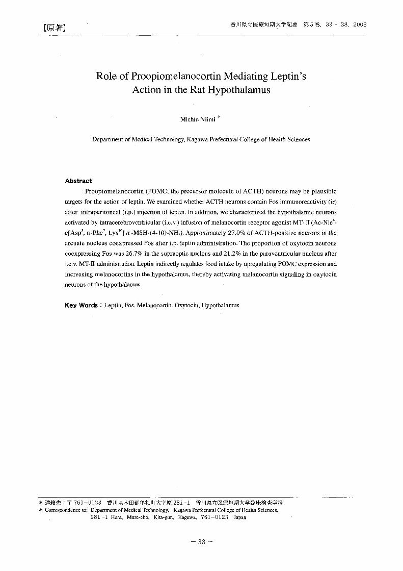
【原著]香川県立医療短期大学紀要第5巻, 33 -38, 2003
Role of Proopiomelanocortin Mediating Leptin's
Action in the Rat Hypothalamus
Michio Niimi *
Department of Medical Technology, Kagawa Prefectural College of Health Sciences
Abstract
Proopiomelanocortin (POMC; the precursor molecule of ACTH) neurons may be plausible
targets for the action of leptin. We examined whether ACTH neurons contain Fos immunoreactivity (ir)
after intraperitoneal (i.p.) injection of leptin. In addition, we characterized the hypothalamic neurons
activat.巴dby intracerebroventricular (i.c.v.) infusion of melanocortin receptor agonist MT町 11(Ac-Nle4-
C[ASp5, D-Phe7, LyslO]α-MSH-(4-1O)-NHz)' Approximately 27.0% of ACTH-positive neurons in the
arcuate nucl巴uscoexpress巴dFos after i.p. leptin administration. Th巴 proportionof oxytocin neurons
coexpressing Fos was 26.7% in the supraoptic nucleus and 21.2% in the parav巴ntricularnucleus after
i.c.v. MT-I1 administration. Leptin indirectly regulates food intake by upregulating POMC expr巴ssionand
increasing melanocortins in the hypothalamus, thereby activating m巴lanocortinsignaling in oxytocin
neurons of the hypothalamus.
Key Words : Leptin, Fos, Melanocortin, Oxytocin, Hypothalamus
牢連絡先:干 761-0123 香川県木田郡牟礼町大字原 281-1 香川県立医療短期大学臨床検査学科
* Correspond巴nc巴to:Department of Medical1i巴C加lology,Kagawa Prefectura1 College of Health Sciences,
281-1 Hara, Mure-cho, Kita-gun, Kagawa, 761-0123, Japan
内〈
uqtu

Introduction
Leptin is a hormone synthesized in white adipose
tissue and released to the circulation, participating in
the regulation of food intake and metabolism 1). The
food reducing effects of leptin are mediated via specific
hypothalamic neurons with leptin receptors (Ob-R) 1.4) •
Recently, it has been shown that leptin may reduce food
intake and body weight by stimulating the hypothalamic
melanocortin (MC) system5•6
). Targeted deletion of the
MC4 receptor gene, as well as antagonism of ectopically
ov巴r巴xpressedAgouti peptide at the MC4 receptor as
shown in agouti obesity syndrome, resulted in stimulation
of food intake and severe obesity in mice 7.8). Fan et al
9) have also shown that MT四 II, a potent agonist of the
neural MC3 and MC4 receptors, inhibits food intake in
normal and obese animals, while MC r巴ceptorantagomst
SHU司 9119has the opposite巴ffect.In a previous study we
demonstrated that i.p. or i.c.v. inj巴ctionof leptin markedly
stimulated Fos巴xpressionin specific hypothalamic nuc1ei,
particularly in the ventrolateral part of the arcuat巴nuc1eus
(ARC)lO). Schwartz et al. 6) r巴portedthat central infusion
of leptin increased preproopiomelanocortin mRNA
expression in the ARC of the hypothalamus. Therefore,
leptin may induce Fos mRNA in the POMC neurons.
We examined whether ACTH neurons contain Fos-ir
after i.p. injection of leptin. In addition, we characterized
some hypothalamic neurons activated by i.c.v. infusion
of the勘1Cr巴ceptoragonist MT-II by a dual labeling
immunohistochemical technique.
民lethodsand Protocols
All experiments were performed on adult male Sprague-
Dawly (SD) rats, weighing 250-300g. They were divid巴d
into two groups. In the first experimental group (n=3), th巴
rats were individually housed in cages and wer巴handled
for 10 min/day for 7 days. The animals were divided into
two groups and food was withdrawn at 15:00. The rats
received i.p. inj巴ctionof r巴combinantmouse leptin 2.0
mg/kg dissolved in vehic1e (0.05 M Tris前 HC1,pH 8.0)
or v巴hic1ealone at 16:00. Two hours later, the rats were
anesthetized with sodium pentobarbital (40 mg/kg i.p.)
and transcardially perfused with 4% paraformaldehyde
in 0.12 M phosphate buffer. The brains were removed,
postfixed overnight in the same fresh fixative and then
placed in 15% sucrose in phosphate buffer巴dsaline
(PBS) for 24h at 4 oC . The brains were frozen, and 25-Jlm
transverse sections were cut on a cryostat and collected in
PBS. For Fos and ACTH ( a marker for POMC n巴urons)
double immunohistochemistry, sections were first stained
for Fos (rabbit polyc1onal IgG, Oncog巴nescienc巴, NY;
1:2000) for 48h at 4 oC. The subsequ巴ntprocedure for the
immunohistochemistry followed a Vector's protocol
(Vectastain ABC kit, Vector), as previously described
els巴where10,11) Fos-ir was visualized by nickel-
diaminobenzidine (Ni-DAB) choromogen (10 mg nickel
ammonium sulfate, 10 mg DAB, 10μ10.3% hydrogen
peroxide in 10 ml 0.05 M Tris-HC1, pH 7.6). Before
immunostaining for ACTH, the sections were incubated
in 3% hydrogen peroxide in PBS to eliminate Fos-linked
peroxidase activity. The sections were then incubated
with rabbit anti-human ACTH IgG (NIDDK, 1:1000)
containing 4% normal goat serum. After approximately
24-hour incubation with th巴primaryantibody, the sections
were rins巴din PBS and the subsequent procedure was
carried out according to the Vector's protocol above.
Peroxidase activity link巴dto ACTH was visualized
with 3,3' -diaminobenzidine and hydrogen peroxide.
Preadsorption of ACTH antibody with 10μ恥1{ratACTH
(Phoenix pharmaceuticals, Inc., CA) complete1y blocked
ACTH-ir.
In the second experimental group, animals were
anesthetized with sodium pentobarbital (40 mg/kg i.p.) and
ani心 v.cannula was implanted according to the previously
described m巴thodlO, 11). After surgery, all rats were placed
in individual cages and were handled for about 10 minJday.
Seven days aft巴rthe operation, the animals were injected
MT-II (1 nmol/1 0μ1 in saline, Sigma Chemical Co. ST.
Louis, MO) or saline at 16:00. Two hours later, the rats
were anesthetized with sodium pentobarbital (40 mg/kg
i.p.) and perfused transcardially with paraformaldehyde in
phosphate buffer.
Immunohistochemical detection of Fos was carried out
as d巴scribedabove. In addition, some sections were
stained for Fos and oxytocin (OT) in the manner described
above. Anti-OT antibody raised in rabbit was obtained
commercially (Chemicon International INC., CA) and
used at 1:1000 dilution. Preadsorption of anti-OT antibody
with 10μM OT blocked OT-ir.
Statistic Analysis
Cell count data in the first experimental group were
4
qJ

香川県立医療短期大学紀要第5巻, 33 -38, 2003
analyzed with unpaired Student's t-test. Cell count data in
the second experimental group were analyzed with one-way
ANOVA followed by Fisher's PLSD test. Data are presented
as mean:tSEM.
Results
appe紅巳din the ventromedial hypothalamic (VMH), the
posterior part of the dorsomedial hypothalamic (DMH),
the arcuate (ARC) nuclei, and in the retrochiasmatic
area (RCh). ACTH neurons were widely distributed
throughout the ARC and the RCh of th巴 hypothalamus.
Double immunostaining revealed that Fos-ir was present
in many ACTH neurons of the ventrolateral part of the
ARC and the RCh (Fig. lA, B). ln ARC the p巴rcentageln rats with i.p. injection of leptin, many Fos-positive cells
Fig.1 A-B: Representatives of the rostral arcuate (ARC) nucleus stained immunocytochemically for ACTH
(cytoplasm) and Fos (nucleus) in leptin injected rats. Black arrows indicate double-Iabeled cells; open
arrows indicate ACTH neurons without Fos signals. 3V; third ventricle, Scale bars, 50μm.
C-O: Many oxytocin (OT) cells in the SON (C) and the PVN (0) expressed Fos in MT-II ・infusedrats. Black
arrows indicate double-Iabeled cells; open arrows indicate OT neurons without Fos signals. OX; optic
chiasm, Scale bars, 50μm
FiD
円
δ

A B 30 -,
**
** ω o u. 。 vl
.5; 20 O LL c
E 伺
+C 。υ J 'c 20
+同c 。υ d 的
ω ω10 同
ヱトυ 〈 ~ 10 ド。
崎。ー"。ー
~ まO O
ARC SON PVN
口 vehicle 図 leptin 口 vehicle 臼 MT-II
Fig.2 A, The percentage of ACTH-immunoreactive neurons in the ARC with nuclear Fos immunostaining. Data
are expressed as mean:t:SEM (n=3). **P<O‘0001. 8, The percentage of OT-immunoreactive neurons in the
SON and PVN with nuclear Fos immunostaining. Data are expressed as mean:t:SEM (n=3). **P<O.0001,
*P<O.001.
of ACTH neurons double町 label巴dfor Foトirwere 27.0土
1.4 and 2.7:1:0.3 in th巴leptin-tr巴at巴dand vehicle同 treated
groups, respectively (mean :t:SEM; n=3; Fig.2A).
Intracerebroventricular administration of 1 nmol MT-
II induced expression of Fos in the suprachiasmatic (SCh),
supraoptic (SON), and paraventricular (PVN) nuclei of th巴
hypothalamus, the central nucleus of the amygdala (CeA),
and in the lateral parabrachial nucl巴us(PBN). OT-ir was
clearly observed in p巴rikaryain the PVN and the SON and
in axons in the n巴urohypophysealtract. The proportion of
oxytocinergic neurons coexpressing Fos was 26.7:t:3.1 %
in the SON and 21.2:t:2.0% in the PVN (maen:t:SEM; n=3;
Fig. 1C,D, Fig. 2B).
Discussion
The present study demonstrated that i.p. injection of
leptin triggered Fos巴xpr巴ssionin some ACTH-containing
perikarya in the RCh and th巴ventrolateralpart of ARC.
This observation suggests that 1巴ptinacts directly on
its r巴ceptorof a subpopulation of POMC neurons and
activates an intracellular signaling pathway that regulates
the POMC gene. This concept is consistent with recent
evidence that leptin rec巴ptormRNA is found to be highly
colocalized with POMC mRNA in neurons in th巴ARC5).
POMC凶 derivedp巴ptidessuch as α-melanocyte stimulating
hormone (α-MSH) and β-endorphin have been implicated
in the regulation of feeding and r巴production12-14).
A1pha-MSH binds to the MC4-rec巴ptor.It is interesting to
identify the oth巴rphenotyp巴sof cells expressing th巴お1C4-
receptor. Therefore, the distribution and quantity of恥1C
receptor agonist (お1T-II )-induced Foトirwere evaluated in
the rat brain. A dramatic increase of Fos-positive n巴urons
was noted in the PVN and SON, and in the CeA and PBN.
These results are consistent with thos巴ofThiele et al. 15)
and Mountjoy et al. 16).
In addition, we demonstrated that MT-II activated Fos in
OT neurons in the SON and PVN. Interrestingly, central
infusion of OT has be巴nshown to reduce food intakel7, 18).
It has been estimated that OT n巴uronscomprise 11同 169も
of the PVN projection to the medulla and spinal cord in
ratsI9). W I巴considerthat MC receptor agonist mediates its
inhibitory action on feeding via brainstem-projecting OT
neurons.
On the other hand, the ventromedial part of the ARC
has also a high 1巴V巴1of leptin receptor gene expression
and contains neuropeptide Y (NPY) neurons stimulating
appetite4). Since leptin is known to suppress synthesis and
円
bqペU

release of NPy20>, these neurons would be inhibited by
leptin and would not induce Fos-ir. Leptin is hypoth巴sized
to affect food intake not only by stimulating cataboric
effector pathways such as the melanocortin system, but
also by inhibiting anabolic signaling systems such as NPY
neurons in the ARC. Friedman suggests that NPY is an
important mediator in r巴sponseto starvation, whereas
MSH and the MC4・receptorare key components of the
hypothalamic r回 ponseto obesity2I).
In summary, we have identified POMC neurons
in the ARC as a target for the action of leptin. The
melanocortins, which are derived from the precursor
POMC, activate melanocortin signaling in oxytocin
neurons of the hypothalamus.
Acknowledgements
The author wishes to thank NIDDK for supplying the
ACTH antibody.
Abbreviation
ACTH; adrenocorticotropic hormone, Fos; Finkel-
Biskis-Jinkins murine osteosarcoma, MSH; melanocyte-
stimulating hormone, NPY; n巴uropeptideY, OT; oxytocin,
POMC; proopiome1anocortin
References
1) Zhang Y, Pro巴ncaR, Maffei M, Baron巴 M,Leopold L,
Friedman JM (1994 )Posisional c10ning of the mouse obese
gene and its human homologue. Nature 372: 425-432.
2) Tartaglia LA, Dembski M, Weng X, Deng N, Culpepper
J, Devos R, Richads GJ, et al. (1995) Identification and
expression cloning of a leptin receptor, OB-R. Cell 83:
1263-1271
3) Fei H, Okano HJ, Li C, Lee GH, Zhao C, Darnell R,
Friedman JM (1997) Anatomicallocalization of altematively
splic巴d1巴ptinreceptors (Ob-R) in mouse brain and other
tissues. Proc Natl Acad Sci USA 94: 7001-7005.
4) Hakansson M-L, Brown H, Ghilardi N, Skoda RC, Meister
B (1998) Leptin receptor immnoreactivity in chemically
defined target neurons of the hypothalamus. J Neurosci 18:
559-572.
5) Cheung CC, Clifton DK, Steiner RA (1997)
Proopiomelanocortin neurons are direct targets for leptin in
香川県立医療短期大学紀要第5巻, 33 -38, 2003
the hypothalamus. Endocrinology 138: 4489-4492.
6) Schwartz W W, Seeley RJ, Woods SC, Weigle DS,
Campfield LA, Bum P, Baskin DG (1997) Leptin increases
hypothalamic proopiomelanocortin mRNA expression in the
rostral arcuate nuc1eus. Diabetes 46: 2119-2123.
7) Huszar D, Lynch CA, Fairchild-Huntress V, Dunmore JH,
Fang Q, Berkemei巴rLR, Gu W, et al. (1997) Targeted
disruption of the melanocortirト4receptor results in obesity
inmice. Ce1l88: 131-141.
8) Lu D, Willard D, Pater IR, Kadwell S, Overton L, Kost T,
Luth巴rM, et al. (1994) Agouti protein is an antagonist of
the m巴lanocyte-stimulatinghormon巴receptor.Nature 371:
799-802.
9) Fan W, Boston BA, Kesterson RA, Hruby VJ, Cone RD
(1997) Role of melanocortinergic neurons in feeding and the
agouti obesity syndrome. Nature 385: 165-168
10) Niimi M, Sato M, Yokote R, Tada S, Takahara J (1999)
Effects of central and peripheral injection of leptin on
food intake and on brain c-fos expression in the Otsuka
Long-Evans Tokushima Fatty Rat with hyperleptin巴mia.J
Neuroendocrinol11: 605-611.
11) Niimi M, Sato M, Taminato T (2001) Neuropeptide Y in
central control of fe巴dingand interactions with orexin and
leptin. Endocrine 14: 269-273.
12) Poggioli RA, Vergoni AV, Bertolini A (1986) ACTH-(1-24)
and alpha-MSH antagonize feeding b巴haviorstimulated by
kappa opiate agonists. Peptides 7: 843-848.
13) Ts吋iiS, Bray GA (1989) Acetylation alters the fe巴ding
response to MSH and beta-endorphin. Brain Res Bull 23・
165-169.
14) Caballero C, Celis ME (1993) The effect of the blockade of
alpha-melanocyte-stimulating hormone on LH release in the
rat. J Endocrinol 137・197-202.
15) Thiele TE, van Dijk G, Yagaloff KA, Fisher SL, Schwartz M,
Bum P, Seely RJ (1998) Central infusion of melanocortin
agonist MT-Il in rats: assessment of c-fos expression and taste
aversion. Am J Physiol 274・R248-R254.
16) MounりoyKG, Mortrud MT, Low MJ, Simerly RB, Cone RD
(1994) Localization of the melanocortin-4 r巴ceptor(MC4・R)
in neuroendocrine and autonomic control circuits in the
brain. Mol Endocrinol 8: 1298-1308.
17) Olson BR, Drutarosky MD, Chow M-S, Hruby VJ, Stricker
EM, Verbalis JG (1991) Oxytocin and an oxytocin agonist
adrr泊nisteredcentrally decrease food intake in ra臼.Peptides
12: 113-118
18) Olson BR, Drutarosky MD, Stricker EM, Verbalis JG (1991)
門
iqu

Brain oxytocin receptor antagonism blunts the effects of
anorexigenic treatments in rats: Evidence for central oxytocin
inhibition of food intake. Endocrinol129: 260-272.
19) Sawchenko PE, Swanson LW (1982) Immunohistochemical
identification of neurons in the paraventricular nuc1eus of白e
hypothalamus that project to the medulla or to th巴spinalcord
in the rat. J Comp Neurol 205: 260-272.
20) Stephens TW, Basinski M, Bristow PK, Bue-VaUeskey
JM, Burgett SG, Craft L, Hale J et al. (1995) The role of
neuropeptide Y in the antiobesity action of the obese gene
product. Nature 377: 530-532.
21) Friedman JM (1997) The alphabet of weight control. Nature
385・119-120.
受付日 2003年 8月 12日
。。円
δ