role of proteolysis in physiological and pathological processes zdenka kučerová
TRANSCRIPT
Role of proteolysis in physiological and pathological processes
Zdenka Kučerová

Proteolytic enzymes :
• cleave peptide bonds ( proteins or peptides)• control of the cell cycle• key roles in many pathophysiological processes• targets for therapeutic strategies • speed of the cleavage depends on the strategically
located positions of cleaved bonds

Activity of proteolytic enzymes
• determined by many factors at the post-translational level
• allosteric nature physiologically important interactions explains why the enzymes carry a large body of structural and functional information
degradom
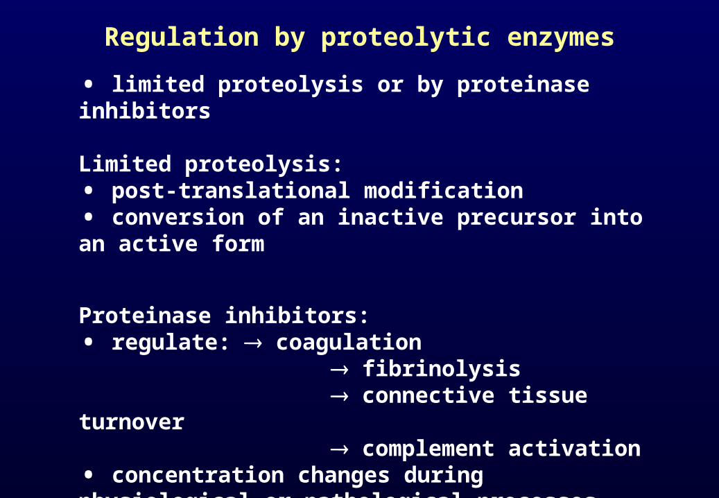
Regulation by proteolytic enzymes
• limited proteolysis or by proteinase inhibitors
Limited proteolysis:• post-translational modification• conversion of an inactive precursor into an active form
Proteinase inhibitors:• regulate: coagulation fibrinolysis connective tissue turnover complement activation• concentration changes during physiological or pathological processes
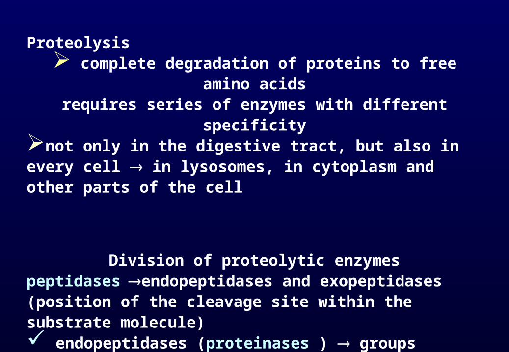
Proteolysis complete degradation of proteins to free amino acids
requires series of enzymes with different specificitynot only in the digestive tract, but also in every cell in lysosomes, in cytoplasm and other parts of the cell
Division of proteolytic enzymespeptidasesendopeptidases and exopeptidases (position of the cleavage site within the substrate molecule) endopeptidases (proteinases ) groups according to similar reaction mechanisms (e.g. serine or cysteine proteinases)

Mechnism of interaction
• enzymes recognise and bind to a short amino acid sequences and then specifically hydrolyse bonds between particular aminoacid residues


Classification of proteinases
A) endopeptidases – hydrolysis within the peptide chain1) serine proteinases – serine or histidine residues in active sites, pH optimum 7 – 9 trypsin, chymotrypsin, thrombin, plasmin2) cysteine proteinases – cysteine residue in active site, pH optimum 4 – 7calpain, caspases3) aspartic proteinases – two aspartic residues in active site, pH optimum below 5 pepsin, gastricsin, rennin, HIV proteinase4) metaloproteinases – metal ion important for catalysis, pH opt. 7 – 9matrix metaloproteinases, collagenase, gelatinase
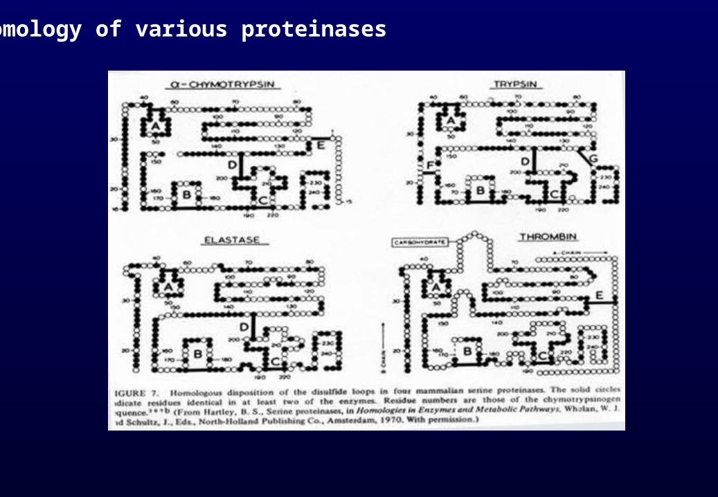
Homology of various proteinases

B) exopeptidases - cleave off single amino acid one after another1) aminopeptidases – cleave off a single amino acid from amino terminus2) dipeptidyldipeptidases – cleave off dipeptide from amino terminus3) carboxypeptidases – cleave off a single amino acid from carboxy terminus4) dipeptidases – hydrolyse dipeptides in two single amino acids
• Individual proteinases differ in their substrate specifity
• For complete degradation of a protein, enzymes from each of the above-named groups are required

Characterization of some proteinases
enzym m.wt isoelektric point (pH)chymotrypsino-gen 25.000 8.1 trypsin 24.000 10.8 plasmin 75.000 - elastase 26.000 9.5 papain 21.000 8.7 pepsin 33.000 1.0 carboxypepti- 34.000 6.0 dase A

Inhibitors
differ according to individual groups of proteinases
•Serine proteinases…..chlormethylketons,
•Cysteine proteinases…. dithiothreitol, mercaptans,
•Aspartic proteinasess… epoxy- and diazo- compounds, pepstatin
• Metalloproteinases….. chelates

Serine proteinases
Proteinases of digestive tract – trypsin, chymotrypsin
important for: fibrinolysis blood coagulation complement activation
produced as inactive zymogens
activated by limited proteolysis by another proteinase

Blood clotting
• enzymatic conversion of a soluble plasmatic fibrinogen into a fibrous network of insoluble fibrin polymers
• fibrinogen cleaved by thrombin and fibrin aggregate into insoluble fibrin polymer
• most of the coagulation factors are proteinases

two different ways• extravascular pathway - triggered by an injury of tissue • intravascular pathway - initiated by contact with a damaged inner surface of blood vessel
• cascade of proteolytic cleavages
• requires calcium and phospholipids
• conversion of inactive prothrombin to active thrombin
• series of zymogens of serine proteinases are sequentially activated (active serine proteinases arise from inactive precursors, the active enzymes then activate other inactive proteins)
Initiation of blood clotting

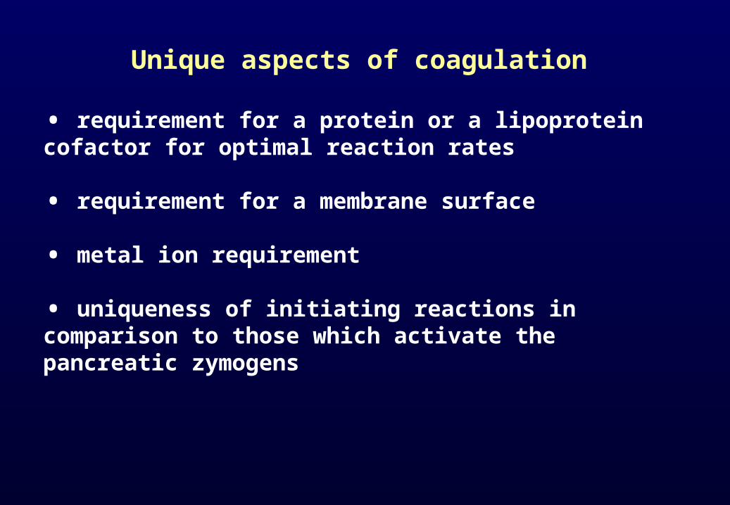
Unique aspects of coagulation
• requirement for a protein or a lipoprotein cofactor for optimal reaction rates
• requirement for a membrane surface
• metal ion requirement
• uniqueness of initiating reactions in comparison to those which activate the pancreatic zymogens


Blood coagulation cascade
• ended by dissociation of the membrane complex• regulated by activators and inhibitors in dynamic equilibrium
Antithrombin III
• inhibits activity of thrombin an anticoagulant factor• anticoagulant therapy reduction of formation or reduction of function of thrombin and other serine proteinases
Protein C
•Serine proteinase…inactivation of factor Va and VIIIa

Fibrinolysis
• fibrin networks are degraded by plasmin
• plasmin is serine proteinase
• plasmin evolves from precursor plasminogen after activation by plasminogen activator
• plasmin activity inhibited by α-2 antiplasmin
• therapy by fibrinolytic proteinases thrombus degradation

Characterization of plasmin, plasminogen and plasminogen activator
Plasminogen is one-chain glykoprotein hydrolyse bounds Arg67-Met68, Lys76-Lys77, Lys77-Val78 Plasmin is two chain serine protease (with disulfidic bound)
active site His-603, Asp-646, Ser-741its main target is fibrin Human plasminogen activator [PA] is serine protease (tissue and urokinase)one polypeptide chain ( single chain-ScuPA)hydrolyse bounds Arg275-Ileu276 (two chain-TctPA)
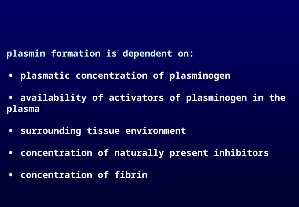
plasmin formation is dependent on:
• plasmatic concentration of plasminogen
• availability of activators of plasminogen in the plasma
• surrounding tissue environment
• concentration of naturally present inhibitors
• concentration of fibrin

Pathological fibrinolysis
• abnormal fibrinolysis, bleeding or thrombus formation
Higher fibrinolysis • increased level of plasminogen activator (PA)• deficiency of plasminogen activator inhibitor (PAI)• deficiency of antiplasmin
Thrombus formation • defective synthesis of plasmin or PA• PA used to pharmacological digestion of thrombus
Malignant tumors • disorders of hemostasis - ability of the tumor to alter the coagulation system

• Tumor malignancies connected with invasive growth and metastasis enzymatic breakdown of two proteolytic systems (fibrinolytic s. and matrix metaloproteinases s.)
Methastatic disease
• disintegrated regulation• irreversible progression of cell cycle• resistance to a cell signalling• understanding of the mechanism preventing tumor cell spread• proteases have proteolytic activity that may affect cancer progression

Proteinases - Biomarkers
• correspond to changes in the whole organismchanges: - biochemical - histological - morphological - physiological • very important for early diagnosis

Complement
This system collaborates in recognition and elimination of pathogens as a part of both the innate and acquired
immune systems.
•The activation of complement pathway - through attached serine proteases
•the complement system is one of the most highly organized innate immune systems
• serine proteinases are responsible for regulation of the early event of complement

Kallikrein
• plasmatic serine proteinases (15 )
• kalikrein - kinin is a complex system produced in various organs
•the kallikreins must now be considered as important 'hormonal' regulators of tissue function
• important for initiation of cellular fibrinolysis, which is independent on plasmin, fibrin and tissue plasminogen activator
•predictive marker for cancer

Cysteine proteinases
lysosomal - cysteine cathepsins
• implicated in tumor spread and metastasis
• prognostic factors for tumor recurrence in human breast cancer
non-lysosomal – calpain
• cleave cell-cycle proteins, cytoskeletal and myofibrilar proteins
• cytoplasmatic enzyme

Apoptosis
• evolutionary conserved form of cell suicide
• requires a proteolytic system - mainly caspases
•caspase family of proteases can be divided into pro-apoptotic and pro-inflammatory members based on their substrate specificity and participation in separate signalling cascades
• caspase regulation therapeutic purposes
• caspases - minimally requires a tetrapeptide substrate in which Asp is in absolute requirement in P1 position, the P4 substrate residues is unique to each homologue.

• defective control of apoptosis pathogenesis of diseases
• cancer chemotherapy and radiation cancer cell death by apoptosis
• Caspases are responsible for crucial aspects of inflammation and immune-cell death that are disrupted in a number of genetic autoimmune and autoinflammatory diseases.
• caspases proteolytic devitalizing and remodelling of tumor cells suppress tumor cell growth invasion and metastasis
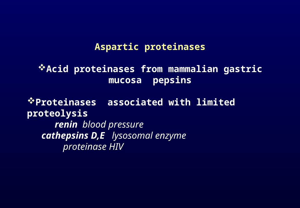
Aspartic proteinases
Acid proteinases from mammalian gastric mucosa pepsins
Proteinases associated with limited proteolysis renin blood pressure cathepsins D,E lysosomal enzyme proteinase HIV

Renin
• aspartic proteinase
• formed as a precursor (prorenin) in kidneys
• active form (renin) released into the blood
• renin cleaves angiotensinogen to form angiotensin I
• angiotensin-converting enzyme (ACE) then converts angiotensin I to angiotensin II

Renin
• highly specific
• synthesized in response to decreasing levels of sodium ions and declining blood pressure
• renin angiotensin II affects: - kidney - brain stem - hypophysis - adrenal cortex - blood vessel walls - heart


Cathepsins D and E
Cathepsin D • aspartic proteinase localized in lysosomes• in most tissues and cells of human organism• widely distributed among biological species• influences: - intracellular protein catabolism - hormone and antigen processing - pathological processes (neoplasia and neurodegenerative changes)
Cathepsin E• in human stomach cells (another stomach cells than pepsinogen and progastricsin)
Both is over- expressed by cancer cells
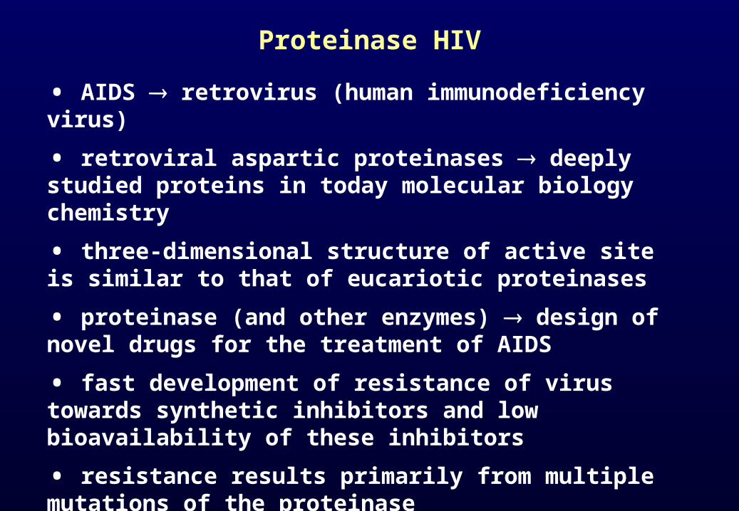
Proteinase HIV
• AIDS retrovirus (human immunodeficiency virus)
• retroviral aspartic proteinases deeply studied proteins in today molecular biology chemistry
• three-dimensional structure of active site is similar to that of eucariotic proteinases
• proteinase (and other enzymes) design of novel drugs for the treatment of AIDS
• fast development of resistance of virus towards synthetic inhibitors and low bioavailability of these inhibitors
• resistance results primarily from multiple mutations of the proteinase

• drug-resistant HIV proteinase → altered substrate specifity
• altered substrate specifity understanding → valuable in the design of new protease inhibitors
•active antiretroviral therapy (HAART) combining potent drugs that can inhibit reverse transcriptase, integrase and protease activities


Metaloproteinases
• matrix metaloproteinases (MMPs)
• zinc-dependent enzymes
• regulate tissue remodelling in physiological and pathological conditions
• MMPs are key enzymes for tumor progression cell carcinoma
• individual MMPs found in most tumor types
• presence of specific MMPs has been shown to be a prognostic marker at tumor invasion
• tumor invasion and metastasis → degradation of the extracellular matrix around tumor cells and cell migration
• cancer metastasis → imbalance between MMPs and its inhibitor
• MMPs → regulatory effects on both primary and secondary tumors
• inhibitors of MMPs were assessed for anticancer properties
• proteolysis has a central role for cancer metastasis
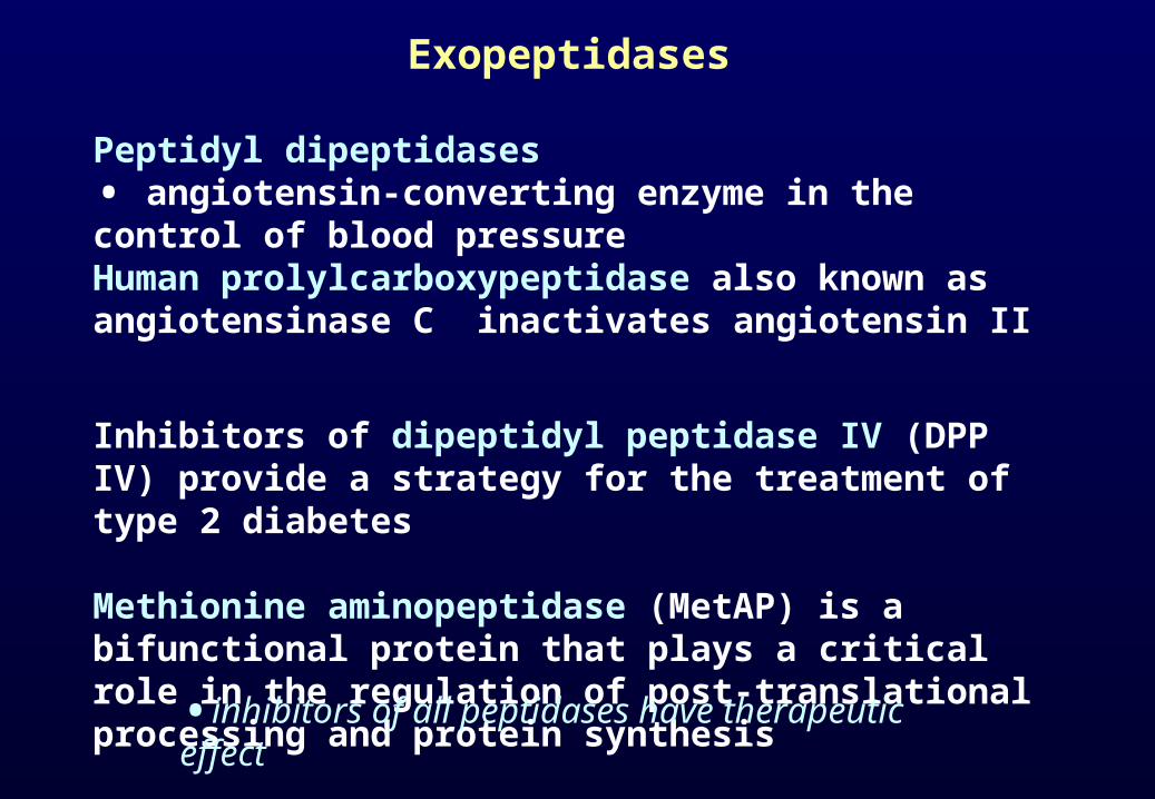
Exopeptidases
Peptidyl dipeptidases • angiotensin-converting enzyme in the control of blood pressureHuman prolylcarboxypeptidase also known as angiotensinase C inactivates angiotensin II
Inhibitors of dipeptidyl peptidase IV (DPP IV) provide a strategy for the treatment of type 2 diabetes
Methionine aminopeptidase (MetAP) is a bifunctional protein that plays a critical role in the regulation of post-translational processing and protein synthesis
•inhibitors of all peptidases have therapeutic effect
Human gastric proteinases
• digestive proteinases
• adult mammalian gastric mucosa contains the highest concentration and greatest diversity of gastric proteinases
• human gastric mucosa contains four aspartic proteinases: - pepsinogen A (PGA) - pepsinogen C (PGC) - chymosin - cathepsin E
• common biological properties (the ability to hydrolyze proteins at acid pH) but each is distinguishable by immunologic technique
• serum contains both PGA and PGC, but only PGA is detectable in urine

Schematic view of pepsinogen A

Pepsinogen A and pepsinogen C
• aspartic proteinases
• differ in charge → distinguishable by electrophoresis • heterogeneity of pepsinogens → multiple genes → posttranslational modifications of the primary gene product

Duodenal ulcer
• high level of PGA → subclinical marker of duodenal ulcer
Superficial gastritis
• acute or chronic superficial gastritis → high serum PGA and PGC levels
• inflammation of the mucosa → increased rates of release of both zymogens into the circulation → elevated levels of PGA and PGC
• level of PGC increased much more than level of PGA

Pepsinogen A / pepsinogen C ratio in peptic ulcer, gastric cancer and control


Gastric ulcer
• changed levels of PGA and PGC
• gastric ulcer usually develops within areas of chronic gastritis
• pepsinogen A / pepsinogen C ration decreases

Gastric cancer
• very low serum levels of PGA
• low serum PGA level → subclinical marker of increased risk for gastric cancer → it is possible to identify high-risk subjects → more effective approaches to diagnosis and treatment
• serum PGA and PGC levels → subclinical markers of disorders of gastric mucosal structure and function

Importance of monitorig serum PGA level
• apparently healthy subjects, but:
→ low serum PGA level
→ subclinical marker of increased risk for gastric cancer
→ better identification of high-risk subjects
→ development of more effective diagnosis and treatment