root surface areas of maxillary permanent teeth in anterior … · 2017-12-20 · sn to palatal...
TRANSCRIPT
- 241 -
Imaging Science in Dentistry 2017; 47: 241-6https://doi.org/10.5624/isd.2017.47.4.241
IntroductionRoot surface area, or the area of the dental root in con-
tact with the surrounding bone, plays an important role in orthodontic treatment, as well as in prosthodontic treatment and periodontal therapy. The root surface area of a tooth to be moved is vital in determining the proper force magnitude and in predicting the biological response during orthodontic tooth movement, especially during or-thodontic intrusion. Moreover, the root surface area has also been associated with the anchorage value of teeth.
Previous root surface area measuring methods have been categorized as follows: 1) the membrane method, 2) the weighting conversion method, and 3) the division planimetry method. Those methods have some disadvan-tages, such as inaccuracy, imprecision, complicated mea-suring procedures, and the need for tooth extraction prior to root surface area measurements.1 In recent years, mi-cro-computed tomography (micro-CT) has been applied to measure the root surface area of extracted permanent teeth because of its micrometer resolution and nonde-structive nature.2 Nevertheless, the root surface area of non-extracted teeth, which is important for orthodontic treatment, cannot be measured using any of these meth-ods. Hence, Tasanapanont et al.3 presented a novel cone-beam computed tomography (CBCT) approach for mea-suring root surface area, and suggested that this technique
Root surface areas of maxillary permanent teeth in anterior normal overbite and anterior open bite assessed using cone-beam computed tomography
Piyadanai Suteerapongpun1, Supassara Sirabanchongkran2, Tanapan Wattanachai2, Patiyut Sriwilas3, Dhirawat Jotikasthira2,*1Department of Orthodontics, Faculty of Dentistry, Chiang Mai University, Chiang Mai, Thailand
2Department of Orthodontics and Pediatric Dentistry, Faculty of Dentistry, Chiang Mai University, Chiang Mai, Thailand 3Department of Radiology, Faculty of Medicine Siriraj Hospital, Mahidol University, Bangkok, Thailand
AbstRAct
Purpose: The aim of this study was to compare the root surface areas of the maxillary permanent teeth in Thai patients exhibiting anterior normal overbite and in those exhibiting anterior open bite, using cone-beam computed tomography (CBCT).Materials and Methods: CBCT images of maxillary permanent teeth from 15 patients with anterior normal overbite and 18 patients with anterior open bite were selected. Three-dimensional tooth models were constructed using Mimics Research version 17.0. The cementoenamel junction was marked manually. The root surface area was calculated automatically by 3-Matic Research version 9.0. The root surface areas of each tooth type from both types of bite were compared using the independent t-test (P<.05). The intraclass correlation coefficient was used to assess intraobserver reliability.Results: The mean root surface areas of the maxillary central and lateral incisors in individuals with anterior open bite were significantly less than those in those with normal bite. The mean root surface area of the maxillary second premolar in individuals with anterior open bite was significantly greater than in those with normal bite.conclusion: Anterior open-bite malocclusion might affect the root surface area, so orthodontic force magnitudes should be carefully determined. (Imaging Sci Dent 2017; 47: 241-6)
Key woRds: Root Surface Area, Anterior Open Bite, Cone-Beam Computed Tomography, Three-Dimensional
Copyright ⓒ 2017 by Korean Academy of Oral and Maxillofacial RadiologyThis is an Open Access article distributed under the terms of the Creative Commons Attribution Non-Commercial License (http://creativecommons.org/licenses/by-nc/3.0)
which permits unrestricted non-commercial use, distribution, and reproduction in any medium, provided the original work is properly cited.Imaging Science in Dentistry·pISSN 2233-7822 eISSN 2233-7830
Received August 16, 2017; Accepted September 4, 2017*Correspondence to : Prof. Dhirawat JotikasthiraDepartment of Orthodontics and Pediatric Dentistry, Faculty of Dentistry, Chiang Mai University, Suthep Road, Muang, Chiang Mai 50200, ThailandTel) 66-53-944464, Fax) 66-53-222844, E-mail) [email protected]

Root surface areas of maxillary permanent teeth in anterior normal overbite and anterior open bite assessed using cone-beam computed tomography
- 242 -
could be used to assess the root surface area of non-ex-tracted teeth.
Normal occlusal loading and function are responsi-ble for the normal development of alveolar bone and dental roots. Occlusal hypofunction decreases alveolar bone mass, accelerates bone resorption, leads to atrophic changes in the periodontal ligament, causes deficient root development, and affects root surface area.4-6 Some previ-ous studies7,8 have reported that individuals with anterior open bite had a tendency to develop short dental roots from the incisors to the premolars. Occlusal hypofunction due to anterior open-bite malocclusion plays an important role in dental root morphology, leading to the risk of root resorption during tooth movement.9,10 Root morphology, such as dental root length, root size and root shape, might also be associated with root surface area.
The aim of our investigation was to compare the root surface areas of the maxillary permanent teeth in patients exhibiting anterior normal overbite and in those exhibit-ing anterior open bite, using CBCT.
Materials and MethodsSubjects and image acquisition
This study was approved by the Human Experimenta-tion Committee of the Faculty of Dentistry, Chiang Mai University, Thailand (No. 53/2016). Informed consent was provided by all patients before CBCT images were obtained. The subjects were Thai orthodontic patients who required pretreatment CBCT images and met the following inclusion criteria: 1) age from 15 to 30 years; 2) class I sagittal skeletal relationship (A point, nasion, B point angle of 2°±2°); 3) complete root formation of the permanent teeth (except for the third molars); and 4) no history of previous orthodontic treatment. The exclusion criteria were 1) variations of tooth morphology (such as peg-shaped lateral incisors); 2) root resorption; 3) radio-graphic signs of severe periodontitis or periapical lesions; and 4) the presence of craniofacial anomalies.
The subjects were divided into 2 groups according to the following 6 cephalometric measurements: 1) the sel-la-nasion (SN) to gonion-gnathion (GoGn) angle; 2) the SN to palatal plane (PP) angle; 3) the PP-GoGn angle; 4) the gonial angle; 5) the facial index; and 6) the ratio of posterior to anterior face height. If 3 or more of the parameters indicated an open configuration, the subjects were categorized as having an open vertical skeletal con-figuration. The normal bite group (15 patients; 4 males and 11 females; mean age 20.4±3.7 years) had anterior
normal overbite (overbite = 0-2 mm) and a normal verti-cal skeletal configuration. The anterior open bite group
(18 patients; 5 males and 13 females; mean age 19.0±3.1 years) had anterior open bite (overbite<0 mm) and an open vertical skeletal configuration.
CBCT images of maxillary permanent teeth, produced using a ProMax 3D (Planmeca OY, Helsinki, Finland) ma-chine at 84 kVp, 10 mA, an 8- × 8-cm field of view, and a voxel size of 0.16 mm, were categorized by tooth type as follows: central incisor, lateral incisor, canine, first premolar, second premolar, first molar, and second molar
(maxillary third molars were excluded from the study).
Measurement of the root surface area
Following the method of Tasanapanont et al.,3 the pa-tients’ Digital Imaging and Communications in Medicine files, obtained by CBCT, was converted to the stereoli-thography (STL) format using Mimics Research version 17.0 (Materialise, Leuven, Belgium). To reconstruct the areas of interest, the threshold value for the tooth region was predefined. The outer boundaries of the tooth mor-phology in 2-dimensional (2-D) images were identified by each slice orientation and each slice in each orientation manually (Fig. 1). To easily identify the cementoenamel junction (CEJ), intentional extension spine markings were constructed from the 2-D images. The CEJ on each sec-tion was marked (Fig. 2). When the “Calculate 3-D” func-tion was selected, the 3-D tooth models were constructed
(Fig. 3). The CEJ was marked, and the root surface area of each tooth was calculated automatically by 3-Matic Research version 9.0 (Materialise) (Fig. 4).
To test the intraobserver reliability, all CBCT images were re-measured by the same examiner after a 4-week interval.
Statistical analysis
All data were tested for normality using the Shapiro- Wilk test. The root surface areas were compared between the normal bite group and the anterior open bite group using the independent t-test. The results were considered statistically significant at P<.05. The intraclass correla-tion coefficient was used to assess the intraobserver reli-ability.
ResultsThe intraobserver reliability test for measurement of
the root surface area showed a high intraclass correlation
(r = 0.999), suggesting high reliability in the measure-

- 243 -
Piyadanai Suteerapongpun et al
ments. The root surface area measurements were nor-mally distributed, and showed no statistically significant difference between the left and right sides. Therefore, the measurements from both sides were pooled for the statis-tical analysis.
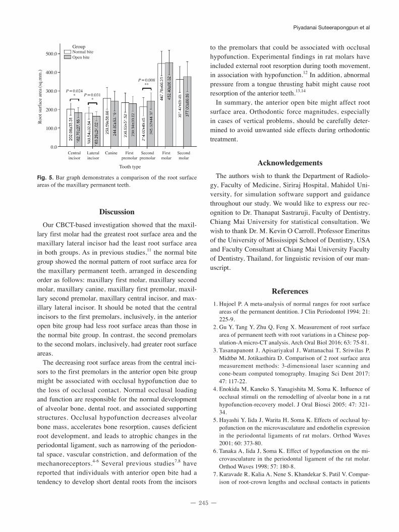
The means and standard deviations of the root surface areas of the teeth ranging from the maxillary central in-cisor to the maxillary second molar in the normal bite group were 202.08±33.31, 180.54±32.54, 259.59±58.86, 236.92±51.52, 214.02±49.45, 447.78±66.31,
Fig. 1. Identification of tooth morphology on 2-dimensional images of each slice orientation. A. Coronal view. B. Axial view. C. Sagittal view.
A B
C
Fig. 2. Construction of intentional extension spine markings by marking the cementoenamel junction on each section.

Root surface areas of maxillary permanent teeth in anterior normal overbite and anterior open bite assessed using cone-beam computed tomography
- 244 -
and 361.47±69.85 mm2, respectively. The means and standard deviations of the root surface areas of the teeth ranging from the maxillary central incisor to the maxil-lary second molar in the anterior open bite group were 182.70±27.48, 163.29±24.02, 244.80±53.19, 230.94±39.62, 245.52±44.37, 452.40±66.32 and 377.00±80.89
mm2, respectively (Fig. 5). In both groups, the maxillary first molar had the greatest root surface area and the max-illary lateral incisor had the least. The mean values of the
root surface areas of the maxillary central and lateral in-cisors in the anterior open bite group were significantly less than those in the normal bite group. The mean value of the root surface areas of the maxillary second premolar in the anterior open bite group was significantly greater than in the normal bite group. No statistically significant difference between the 2 groups was found in the canines, first premolars, first molars, or second molars.
Fig. 4. Identification of the cementoenamel junction and automatic calculation of the root surface area using 3-Matic Research version 9.0.
Fig. 3. Construction of the 3-dimensional tooth models.
- 245 -
Piyadanai Suteerapongpun et al
discussionOur CBCT-based investigation showed that the maxil-
lary first molar had the greatest root surface area and the maxillary lateral incisor had the least root surface area in both groups. As in previous studies,11 the normal bite group showed the normal pattern of root surface area for the maxillary permanent teeth, arranged in descending order as follows: maxillary first molar, maxillary second molar, maxillary canine, maxillary first premolar, maxil-lary second premolar, maxillary central incisor, and max-illary lateral incisor. It should be noted that the central incisors to the first premolars, inclusively, in the anterior open bite group had less root surface areas than those in the normal bite group. In contrast, the second premolars to the second molars, inclusively, had greater root surface areas.
The decreasing root surface areas from the central inci-sors to the first premolars in the anterior open bite group might be associated with occlusal hypofunction due to the loss of occlusal contact. Normal occlusal loading and function are responsible for the normal development of alveolar bone, dental root, and associated supporting structures. Occlusal hypofunction decreases alveolar bone mass, accelerates bone resorption, causes deficient root development, and leads to atrophic changes in the periodontal ligament, such as narrowing of the periodon-tal space, vascular constriction, and deformation of the mechanoreceptors.4-6 Several previous studies7,8 have reported that individuals with anterior open bite had a tendency to develop short dental roots from the incisors
to the premolars that could be associated with occlusal hypofunction. Experimental findings in rat molars have included external root resorption during tooth movement, in association with hypofunction.12 In addition, abnormal pressure from a tongue thrusting habit might cause root resorption of the anterior teeth.13,14
In summary, the anterior open bite might affect root surface area. Orthodontic force magnitudes, especially in cases of vertical problems, should be carefully deter-mined to avoid unwanted side effects during orthodontic treatment.
AcknowledgementsThe authors wish to thank the Department of Radiolo-
gy, Faculty of Medicine, Siriraj Hospital, Mahidol Uni-versity, for simulation software support and guidance throughout our study. We would like to express our rec-ognition to Dr. Thanapat Sastraruji, Faculty of Dentistry, Chiang Mai University for statistical consultation. We wish to thank Dr. M. Kevin O Carroll, Professor Emeritus of the University of Mississippi School of Dentistry, USA and Faculty Consultant at Chiang Mai University Faculty of Dentistry, Thailand, for linguistic revision of our man-uscript.
References 1. Hujoel P. A meta-analysis of normal ranges for root surface
areas of the permanent dentition. J Clin Periodontol 1994; 21: 225-9.
2. Gu Y, Tang Y, Zhu Q, Feng X. Measurement of root surface area of permanent teeth with root variations in a Chinese pop-ulation-A micro-CT analysis. Arch Oral Biol 2016; 63: 75-81.
3. Tasanapanont J, Apisariyakul J, Wattanachai T, Sriwilas P, Midtbø M, Jotikasthira D. Comparison of 2 root surface area measurement methods: 3-dimensional laser scanning and cone-beam computed tomography. Imaging Sci Dent 2017; 47: 117-22.
4. Enokida M, Kaneko S, Yanagishita M, Soma K. Influence of occlusal stimuli on the remodelling of alveolar bone in a rat hypofunction-recovery model. J Oral Biosci 2005; 47: 321-34.
5. Hayashi Y, Iida J, Warita H, Soma K. Effects of occlusal hy-pofunction on the microvasculature and endothelin expression in the periodontal ligaments of rat molars. Orthod Waves 2001; 60: 373-80.
6. Tanaka A, Iida J, Soma K. Effect of hypofunction on the mi-crovasculature in the periodontal ligament of the rat molar. Orthod Waves 1998; 57: 180-8.
7. Karavade R, Kalia A, Nene S, Khandekar S, Patil V. Compar-ison of root-crown lengths and occlusal contacts in patients
Roo
t sur
face
are
a (sq.
mm
.)
500.0
400.0
300.0
200.0
100.0
0.0
GroupNormal biteOpen bite
P = 0.024* P = 0.031
*
P = 0.008**
Central Lateral Canine First Second First Second incisor incisor premolar premolar molar molar
Tooth type
Fig. 5. Bar graph demonstrates a comparison of the root surface areas of the maxillary permanent teeth.

Root surface areas of maxillary permanent teeth in anterior normal overbite and anterior open bite assessed using cone-beam computed tomography
- 246 -
with class-III skeletal relationship, anterior open-bite and high mandibular plane angle. Int J Dent Med Spec 2015; 2: 7-13.
8. Uehara S, Maeda A, Tomonari H, Miyawaki S. Relationships between the root-crown ratio and the loss of occlusal contact and high mandibular plane angle in patients with open bite. Angle Orthod 2012; 83: 36-42.
9. de Freitas MR, Beltrão RT, Janson G, Henriques JF, Cançado RH. Long-term stability of anterior open bite extraction treat-ment in the permanent dentition. Am J Orthod Dentofacial Orthop 2004; 125: 78-87.
10. Harris EF, Butler ML. Patterns of incisor root resorption be-fore and after orthodontic correction in cases with anterior
open bites. Am J Orthod Dentofacial Orthop 1992; 101: 112-9.
11. Cobourne MT, DiBiase AT. Handbook of orthodontics. 2nd ed. New York: Elsevier; 2015.
12. Shimomoto Y, Chung C, Iwasaki-Hayashi Y, Muramoto T, Soma K. Effects of occlusal stimuli on alveolar/jaw bone for-mation. J Dent Res 2007; 86: 47-51.
13. Linge BO, Linge L. Apical root resorption in upper anterior teeth. Eur J Orthod 1983; 5: 173-83.
14. Linge L, Linge BO. Patient characteristics and treatment vari-ables associated with apical root resorption during orthodontic treatment. Am J Orthod Dentofacial Orthop 1991; 99: 35-43.