s viral encephalitis - iowa state university · viral encephalitis • western equine ... • no...
TRANSCRIPT

Slide 1
Viral Encephalitis
Slide 2
Center for Food Security and Public Health Iowa State University - 2004
Viral EncephalitisViral Encephalitis
• Western equine encephalitis (WEE)• Eastern equine encephalitis (EEE)• St. Louis encephalitis (SLE)• La Crosse encephalitis (LAC)• Venezuelan equine encephalitis (VEE)• West Nile virus (WNV)
More than 58 arboviruses (viruses that are transmitted by arthropods) have been identified in the United States and five more have been isolated in Canada. The viruses that are thought to cause significant human illness in the U.S. are western and eastern equine encephalitis (WEE, EEE), St. Louis encephalitis (SLE), La Crosse encephalitis (LAC), Venezuelan equine encephalitis (VEE) and West Nile virus (WNV; covered in a separate talk).
Slide 3
History
Slide 4
Center for Food Security and Public Health Iowa State University - 2004
HistoryHistory
• 1925−First arbovirus identified in the U.S.
Vesicular stomatitis Indiana virus
• 1930−WEE virus isolated in California
Karl Meyer isolated agent from horse brainCoincided with human polioencephalomyelitis cases
The first arbovirus identified in the United States was vesicular stomatitis Indiana virus, which was isolated in 1925. It is thought that arboviruses existed in natural cycles long before they were recognized. In 1930, Karl Meyer of the Hooper Foundation in California isolated what was later to be recognized as WEE from the brain of a horse afflicted with equine encephalitis. The equine outbreak seemed to coincide with several cases of human polioencephalomyelitis in the San Joaquin Valley, CA.
Slide 5
Center for Food Security and Public Health Iowa State University - 2004
HistoryHistory
• 1932− Aedes aegypti replicate and transmit WEE in
the laboratory− St. Louis encephalitis identified in causing
human disease
• 1933− St. Louis encephalitis virus isolated from
human brain− Eastern equine encephalitis virus
Isolated from equine brains Along eastern seaboard of the U.S.
In the unusually cool summer in 1932, more encephalitis outbreaks followed the outbreak of WEE in California in 1930. The virus was found to replicate and be transmitted by Aedes aegypti mosquitoes in the laboratory. Several birds and other wild animals were found to be serologically positive for the virus. In 1932, St. Louis encephalitis (SLE) virus was first identified as a cause of human disease in Paris, Illinois. SLE was isolated from human brain tissue in the St. Louis, Missouri outbreak in 1933. Also in 1933, eastern equine encephalitis (EEE) was isolated from equine brains along the eastern seaboard.

Slide 6
Center for Food Security and Public Health Iowa State University - 2004
HistoryHistory
• 1938−WEE and EEE isolated
from human brain tissue
• 1941−Culex tarsalis mosquitoes
found to be naturally infected with WEE
In 1938, WEE and EEE were isolated from human brain tissue. In 1941, WEE was isolated from naturally infected Culex tarsalis mosquitoes in Washington and California.
Slide 7
Transmission
Slide 8
Center for Food Security and Public Health Iowa State University - 2004
TransmissionTransmission
Virus Particles Dead End Hosts
Vertebrate Hosts
Mosquito Vector
Transovarial & Venereal
Transmission for arboviruses begins when the female mosquito takes a bloodmeal from a reservoir vertebrate host, which in most cases is a bird. A viremia then sets in and is of a sufficient level and duration to affect other mosquitoes, thus propagating the cycle. The virus particles can then replicate in the salivary glands of the mosquito to be passed onto other vertebrate hosts, or dead end hosts, such as humans and horses where overt disease occurs. In the California serogroup, the virus can also be transmitted transovarially and venereally between vectors.
Slide 9
Center for Food Security and Public Health Iowa State University - 2004
• 4 stages− Egg, larva, pupa, adult
• Aedes species − Lay single eggs− Damp soil, later flooded
• Culex species− 100-300 eggs in raft− Lay eggs at night on water surface
• Survival requires wind protection• Overwinter in egg stage
Mosquito Life CycleMosquito Life Cycle
There are about 200 different species of mosquitoes in the United States, all of which live in specific habitats, exhibit unique behaviors and bite different species of animals. Despite these differences, all mosquitoes share some common traits, such as a four-stage life cycle. The top image depicts the first stage of the life cycle of a mosquito, the egg. For Aedes species, an individual egg laid one at a time on vegetation or damp soil that is later flooded by water. In the case of Culex species, an egg raft consisting of 100 to 300 eggs is laid at night on the water’s surface (bottom image). It is essential for survival that the eggs are laid in an area that is sheltered from the wind. If a species overwinters, they do so in the egg stage. Eggs typically hatch within 48 hours to the larval stage. Egg photo http://www.mosquitobuzz.com/facts/mosquitolifecycle.html Egg raft http://www.mosquito.org/mosquito.html
Slide 10
Center for Food Security and Public Health Iowa State University - 2004
Mosquito Life CycleMosquito Life Cycle
• Larvae live upside down in water; “wriggler”− Breathe via siphon tube− Molt 4 times
• Pupal stage is restful, non-feeding; “tumbler”− Breathe via “trumpets”− Splits to allow adult to
emerge
Larva
Pupa
Larvae live in the water and come to the surface to breathe (“wriggler”) by utilizing a siphon tube while hanging upside down from the surface of the water. The larva require large amounts of nutrients for maturation and feed on organic matter in the water. Over a 4 to 14 day period, depending on water temperature, they molt four times. The stages between molts are referred to as instars, and the larvae grow larger each stage, finally becoming a pupa after the 4th instar. The pupal stage can last 1 to 4 days, again dependent on water temperature, and resembles the butterfly in the cocoon stage, because this is where the mosquito develops into an adult. It is a very restful, non-feeding stage and the only movement is when it “tumbles” to protect itself. It utilizes two breathing tubes called “trumpets” and floats at the water’s surface. During the summer, Culex species in the southern United States become an adult after two days in the pupal stage.

Slide 11
Center for Food Security and Public Health Iowa State University - 2004
Photo depicting the larval stage of mosquito development.
Slide 12
Center for Food Security and Public Health Iowa State University - 2004
• Newly emerged adult rests −Dry off wings in order to fly−Harden body parts
• Takes blood meal• Mates a few days after flight−Attractants for biting
Carbon dioxide, temperature, moisture, smell, color, movement
• Lifespan varies from 4-30 days
Mosquito Life CycleMosquito Life Cycle
After the pupal skin splits, the newly emerged adult rests on the water surface long enough to dry off its wings in order to fly and harden its body parts. After a few days, the adult begins to take blood meals and mate. The entire lifespan varies with temperature and species and can range from four days to one month. Only female mosquitoes bite animals and humans and require a protein found in blood for egg production. Male mosquitoes are nectar feeders and do not bite humans. Carbon dioxide, temperature, moisture, smell, color and movement are all attractants for biting mosquitoes and humans usually are not their first choice. Aedes species are strong fliers, as well as persistent and painful biters, feeding early morning, dusk and early evening. Culex are also painful and persistent biters but cannot fly as well and feed at dusk and after dark. They only live a few weeks during the warm summer months and hibernate until spring.
Slide 13
Center for Food Security and Public Health Iowa State University - 2004
Arboviruses Indigenous to the United States
Arboviruses Indigenous to the United States
Culex (Melanoconion) spp.
Ochleratatus triseriatus
Culex pipiens, Cx. quinquefasciatus, Cx. nigrapalpus, Cx. tarsalis
Culex tarsalis, Aedes melanimon, Aedes dorsalis, Aedes campestris
Culiseta melanura, Aedes spp., Culex (Cx.) nigrapalpus, Coquilletidia spp.
Mosquito Vector
VEE
LAC
SLE
WEE
EEEDisease
The various arboviruses that are indigenous to the United States are listed in the table, along with the mosquito vector that is responsible for their transmission. Eastern equine encephalitis (EEE), western equine encephalitis (WEE), St. Louis encephalitis (SLE), La Crosse encephalitis (LAC), Venezuelan equine encephalitis (VEE).
Slide 14
Center for Food Security and Public Health Iowa State University - 2004
Human Clinical SignsHuman Clinical Signs
• Most cases are asymptomatic• Flu-like illness in some−Sudden fever, headache, myalgia,
malaise, prostration
• Small proportion develop encephalitis−Permanent neurological damage−Death
The human clinical signs associated with mosquito-borne viral infections are very similar from one virus to the next. Many times patients are asymptomatic or have a flu-like illness; these signs can progress over time or be sudden in nature, such as fever, headache, myalgia, malaise and occasionally prostration. In a small proportion of cases viral encephalitis can occur and lead to permanent neurological damage or death.
Slide 15
Center for Food Security and Public Health Iowa State University - 2004
Human TreatmentHuman Treatment
• Manage symptoms−Reduce fever−Maintain hydration and electrolytes−Maintain blood oxygen levels−Anticonvulsants−Osmotic diuretics for intracranial
pressure−Physical therapy
• No effective anti-virals available
Due to the viral nature of the mosquito-borne diseases, treatment is an attempt to manage symptoms. Supportive care includes reducing the fever, maintaining hydration, electrolytes, and blood oxygen levels, utilizing anticonvulsants and osmotic diuretics to decrease intracranial pressure, and physical therapy once the patient has survived the encephalitis. Antibiotics are not effective for treatment and there are no effective antiviral drugs available yet.

Slide 16
Summary of Encephalitis Viruses Within the U.S.
Slide 17
Center for Food Security and Public Health Iowa State University - 2004
Arboviruses Indigenous to the United States
Arboviruses Indigenous to the United States
Togaviridae, Alphavirus
Bunyaviridae, Bunyavirus
Flaviviridae, Flavivirus
Togaviridae, Alphavirus
Togaviridae, Alphavirus
Family, Genus
United StatesVEE
Midwest, Eastern, Southern U.S.
LAC
United StatesSLE
Western U.S.WEE
Eastern U.S.EEE
DistributionDz
The various arboviruses that are indigenous to the United States are listed in the table, along with their virus family and genus and their geographic distribution. Eastern equine encephalitis (EEE), western equine encephalitis (WEE), St. Louis encephalitis (SLE), La Crosse encephalitis (LAC), Venezuelan equine encephalitis (VEE).
Slide 18
Center for Food Security and Public Health Iowa State University - 2004
Human Risks and OutcomesHuman Risks and Outcomes
• St. Louis Encephalitis (SLE)− Most common− Elderly most at risk− Case fatality rate:
5-15%• La Crosse
Encephalitis (LAC)− Children <16 years
most at risk− Human fatalities
less than 1%Average 73 cases/year
4478 confirmed cases
Before the West Nile virus outbreak of 1999, the most common cause of flavivirus encephalitis cases and the most common mosquito-transmitted human pathogen in the United States was St. Louis encephalitis (SLE) virus. The elderly are most at risk, but children with the disease have a high rate of encephalitis, with an overall case-fatality rate of 5-15%. SLE has a very broad distribution across the country. La Crosse encephalitis (LAC) cases most often occur in children under the age of 16 and clinical cases have a case fatality rate of less than 1%. Most of the LAC cases have been in the eastern half of the U.S.
Slide 19
Center for Food Security and Public Health Iowa State University - 2004
Human Risks and OutcomesHuman Risks and Outcomes
• Eastern Equine Encephalitis (EEE)− Elderly most at risk− Case fatality rate: 33%
• WEE− Children younger than 1
year most at risk− Case fatality rate
approximately 3%
• VEE− Children most often
affected− Fatalities are rare
Average 5 cases/year
Average 19 cases/year; < 1/year last 10 years
Eastern equine encephalitis (EEE) cases also put the elderly most at risk and have a case fatality rate of 33%. As expected, EEE has occurred mostly in the eastern regions of the U.S., while WEE is reported in the western and central U.S. Western equine encephalitis (WEE) causes severe disease in children under the age of one and the mortality rate is about 3%. Venezuelan equine encephalitis (VEE) cases are less severe than WEE and EEE and usually occur in children, but fatalities are rare.
Slide 20
Center for Food Security and Public Health Iowa State University - 2004
Animal Risks and OutcomesAnimal Risks and Outcomes
• Horse - Case-fatality rate−EEE ~ 90% −VEE ~ 40-80%−WEE ~ 20-50%
• Vaccine available in the U.S.−Trivalent formalin-inactivated
• SLE, LAC do not cause disease in horses or other non-human mammals
The case-fatality rate for horses varies depending on the specific encephalitic disease. EEE is the most fatal with approximately 90% case-fatality. Next is VEE ranging from 40-80%, and lastly WEE causing death in 20-50% of the cases. La Crosse and St. Louis encephalitis do not cause disease in horses or other non-human mammals. Fortunately there is a trivalent, formalin-inactivated vaccine available for horses for WEE, EEE, VEE in the United States.

Slide 21
California Serogroup(CAL)
La Crosse virusJamestown Canyon virusCache ValleyOthers
Slide 22
Center for Food Security and Public Health Iowa State University - 2004
California SerogroupCalifornia Serogroup
• First isolated in 1943 −Approximately 14 known viruses
10 known to cause human disease
• La Crosse virus−Only member known to cause
human mortality−Ochleratatus (Aedes) triseriatus
(treehole mosquito) vector• No two field isolates the same−Genetic change constantly occurring
The first viruses of the California serogroup (CAL) were isolated in Kern County, California from Aedes melanimon mosquitoes in 1943. Additional isolates also from Culex tarsalis were made in 1944 and three California encephalitis cases that occurred in 1945 were due to this virus. There are currently 14 known viruses in the CAL group, of which only 10 are known to cause human disease. The most serious disease results from La Crosse encephalitis (LAC) virus infection, which is the only CAL member to cause human mortality. It is transmitted by Ochleratatus (formerly Aedes) triseriatus, the treehole mosquito. Jamestown Canyon and Cache Valley viruses are related to LAC and are found in the United States, but rarely cause encephalitis. The etiology of CAL group viruses indicates that genetic change and evolution continue to occur. It is thought that the field isolates are forever “mixing” genetic material between the mosquito and the vertebrate host and that no two isolates obtained from nature are identical.
Slide 23
Center for Food Security and Public Health Iowa State University - 2004
CAL in the U.S.: 1993-2002
50
40
30
20
10
0
Report
ed C
ase
s
1993 1994 1995 1996 1997 1998 1999 2000 2001 2002Year (Month)
MMWR
Encephalitis/Meningitis, California Serogroup Viral. Reported cases in U.S., 1993-2002
This graph illustrates the number of reported cases of California serogroup (CAL) viruses in the U.S. from 1993 to 2002. Note the cyclical nature of reported cases as the primary vector, Aedes triseriatus, breeds in the summer months, thus causing human clinical disease from July through September. There were a total of 167 human cases reported to the CDC from 16 states in 2002, the highest number of cases in the 1964-2002 time period. This may be due to improved surveillance and increased disease reporting of CAL serogroup cases because of the West Nile surveillance. Data from the Summary of Notifiable Diseases 2002, CDC website.
Slide 24
Center for Food Security and Public Health Iowa State University - 2004
CAL EpidemiologyCAL Epidemiology
• Primarily in Western Hemisphere• Can occur in Africa, Asia, Europe• Virus transmission and amplification−Occurs in wild vertebrate hosts
Rodents, chipmunks, deer, reindeer
−Domestic animals are sentinels−Mosquitoes are largest reservoir
Ochleratatus (Aedes) species
The majority of CAL group viruses are found in the western hemisphere but several also occur in Africa, Asia and Europe. Transmission and amplification of the virus relies on a variety of wild vertebrate species, from small rodents to large ungulates. Domestic animals act only as sentinels for transmission. Ochleratatus spp. mosquitoes play the biggest role as being the reservoirs for the virus, as vertebrates cannot sustain a viremia.

Slide 25
Center for Food Security and Public Health Iowa State University - 2004
La Crosse Encephalitis: HistoryLa Crosse Encephalitis: History
• 1963−Discovered in La Crosse, WI−Causes human mortality−4-year-old girl died of acute encephalitis
• Cases since reported in other Midwestern and Mid-Atlantic states
• Bunyavirus• Ochleratatus (Aedes) triseriatus
La Crosse (LAC) encephalitis was first discovered when a 4-year old Minnesota girl died in La Crosse, Wisconsin in 1963 of acute encephalitis. Several cases have since been identified in other Midwestern and Mid-Atlantic states. LAC is in the family Bunyaviridae under the genus Bunyavirus. La Crosse encephalitis virus infection is the only CAL member to cause human mortality and is transmitted by Ochleratatus (formerly Aedes) triseriatus, the treehole mosquito.
Slide 26
Center for Food Security and Public Health Iowa State University - 2004
transovarial
Ochleratatus(Aedes) triseriatus
Virus present in new adult
Vertebrate host
Dead end host
LAC TransmissionLAC Transmission
Newly infected
transmits to vertebrate
host
La Crosse encephalitis virus is transmitted by Ochleratatus triseriatus, the daytime-biting treehole mosquito, which utilizes vertebrate hosts such as chipmunks and tree squirrels for amplification of the virus. Vertical transmission from the female mosquito to her eggs enables LAC virus to survive the winter. The adults coming from those eggs are able to transmit the virus to chipmunks, tree squirrels and humans, thus perpetuating the cycle. Humans are dead-end hosts for LAC. The most severe disease is found in children.
Slide 27
Center for Food Security and Public Health Iowa State University - 2004
LAC EpidemiologyLAC Epidemiology
• Human cases−75 cases reported each year− In 27 states
• Greatest risk for clinical disease−Children less than 16 years old
• Cases often un- or misdiagnosed• Case-fatality rate: < 1%
There are about 75 cases of LAC encephalitis reported to the CDC each year on average, and most cases of those occur in children under 16 years of age. In young adults less than 16 years old, a more severe disease characterized by seizures, coma and paralysis is found. Other neurological sequelae can occur after recovery, and death from encephalitis occurs less than 1% of the time. Due to the CNS signs, pediatricians routinely screen for herpes or enteroviral etiologies but not LAC, so the cases are reported as aseptic meningitis or viral encephalitis of unknown etiology.
Slide 28
Center for Food Security and Public Health Iowa State University - 2004
Average 73 cases/year
In the United States, most cases of LAC have historically occurred in the upper Midwestern states of Minnesota, Wisconsin, Iowa, Illinois, Indiana and Ohio. More cases are recently being reported from states in the Mid-Atlantic region including West Virginia, Virginia, North Carolina and in the southeastern region such as in Alabama and Mississippi. LAC is thought to be under-reported in the eastern U.S. because the etiologic agent is not always specifically identified. Map from CDC.
Slide 29
Center for Food Security and Public Health Iowa State University - 2004
La Crosse in Humans La Crosse in Humans
• Incubation: 2-7 days• Summertime illness• Fever, headache, nausea, vomiting,
lethargy• More severe disease in children <16 −Seizures, coma, paralysis, neurological
sequelae−Death less than 1% of cases−Not often correctly diagnosed
The incubation period for LAC is hard to pinpoint exactly but appears to be 2 to 7 days. This summertime illness often results in fever, headache, nausea, vomiting and lethargy. In young adults less than 16 years old, a more severe disease characterized by seizures, coma and paralysis can be seen. Prodromal symptoms often occur and electroencephalography (EEG) shows abnormal results 90% of the time. During the acute phase, frequent seizures often occur and can recur 20-50% of the time. Other neurological sequelae can occur after recovery. Death from encephalitis occurs less than 1% of the time. Due to the CNS signs, pediatricians routinely screen for herpes or enteroviral etiologies but not LAC specifically, so the cases are reported as aseptic meningitis or viral encephalitis of unknown etiology.

Slide 30
Center for Food Security and Public Health Iowa State University - 2004
La Crosse in Humans La Crosse in Humans
• Diagnosis− Hemagglutination inhibition− Paired sera monitoring for rise in
antibody titer• Treatment
− SupportiveManage seizures and increased intracranial pressure
• Prognosis poor with severe clinical disease
• No vaccine available
Diagnosis requires a specific hemagglutination inhibition (HI) test or paired sera monitoring a rise in antibody titer. Treatment involves supportive care based on managing seizures and the increased intracranial pressure. Prognosis for acute, severe encephalitis is poor. If the patient survives, behavioral, emotional, and intellectual effects can be seen. Some require long term care. No commercial vaccine is currently available.
Slide 31
Center for Food Security and Public Health Iowa State University - 2004
Animals and LACAnimals and LAC
• Incubation period: 24-48 hours• Short-lived viremia• Many wildlife species seroconvert• Asymptomatic• No known protocols for treatment,
prevention or control
Serological samples have identified LAC virus in many wildlife species and the incubation period is quite short, only 24 to 48 hours. Many times the animal will seroconvert without showing signs of viremia or clinical disease. Only in experimental situations has LAC resulted in clinical signs of disease, such as in suckling mice and fetal rabbits. Diagnosis is generally by virus isolation during viremia or a rise in the titer by complement fixation. Since none of the agents appear to be of much consequence to domestic or wild animals, no protocols for treatment, control or prevention have been established.
Slide 32
Eastern Equine Encephalitis
Slide 33
Center for Food Security and Public Health Iowa State University - 2004
EEE HistoryEEE History
• 1831−Massachusetts horses afflicted with
unknown encephalitis virus
• 1933−First isolated from a horse
• 1942-1943−Michigan epidemic
• Most epidemics along eastern seaboard and gulf states
Eastern equine encephalitis was first isolated from a horse with encephalomyelitis in 1933, but it is thought that the disease dates back to 1831 to horses in Massachusetts. Most epidemics since then tend to occur along the eastern seaboard, from New Hampshire along the Atlantic Coast to the Gulf of Mexico states. However, Michigan had an epidemic in 1942 and 1943, demonstrating that the vector is not restricted to the east coast states.
Slide 34
Center for Food Security and Public Health Iowa State University - 2004
EEE HistoryEEE History
• 1947: Southern LA and TX−14,000 horses, mules affected
83% fatality
• 1951− Isolated from Culiseta melanura
mosquito• Last 25 years−Most horse cases in Florida−1982 and 1983: over 500 cases−1991: 159 cases
The largest known epidemic occurred in 1947 in southern Louisiana and Texas. Fourteen thousand horses and mules were affected, and nearly 12,000 died. This 83% fatality rate reflects the typical disease in horses in North America. Most outbreaks occur in the late summer and early fall in North America, so investigators suspected it was transmitted by mosquitoes. The disease was first thought to be transmitted by an Aedes species mosquito, but in 1951 EEE was isolated from Culiseta melanura. Clinical disease is usually seen in horses before human cases. In the last 25 years most equine cases have occured in Florida, with smaller outbreaks in states east of the Mississippi River. Florida reported over 500 cases in 1982 and 1983, and 159 in 1991.

Slide 35
Center for Food Security and Public Health Iowa State University - 2004
EEE HistoryEEE History
• Human cases not as prevalent−1964-2002: 182 cases
• 1937−Disease identified in ring-necked
pheasants−Also occurs in sparrows, pigeons,
partridges, emus and ostriches
The number of human cases of EEE reported is much less than that for horses. From 1964-2002, only 182 human cases were reported. Birds are also susceptible to EEE as was discovered in ring-necked pheasants in 1937 in Connecticut. Since then the disease has been found to affect sparrows, pigeons, Peking ducks, Chukar partridges, emus and ostriches, illustrating that species not indigenous to North America are susceptible.
Slide 36
Center for Food Security and Public Health Iowa State University - 2004
Culiseta melanura
Pecking transmission
Aedes spp.Coquilletidia perturbans
Dead end hosts:Horses, humans, other mammals
Bird migration
Over wintering?Spring
Reintroduction
SummerSwampy
areas
EEE TransmissionEEE Transmission
Transmission of eastern equine encephalitis (EEE) is best described as a mosquito-vertebrate-mosquito cycle, with Culiseta (Cs.) melanura, an ornithophilic (“bird-loving”) mosquito feeding almost exclusively on songbirds, the asymptomatic reservoir host. Birds are also able to spread the disease they peck or eat diseased pen mates in captivity. Cs. melanura does not generally feed on mammals and requires secondary mosquitoes to transmit disease to humans and horses. Culiseta melanura lives and breeds in freshwater and swamp areas during the summer and feeds most actively 2 hours after sunset to sunrise. In late summer and early fall they can be found in drier uplands. The epidemic vector that spreads disease to mammals and exotic birds varies for different regions of EEE prevalence, but Coquilletidia (Cq.) perturbans and several Aedes species are often involved. Disease most often occurs within 5 miles of the swampy areas where Cs. melanura and Cq. perturbans live and breed. Cq. perturbans is an opportunistic feeder that feeds on birds and mammals. Horses and humans are considered dead-end hosts of EEE virus because neither reaches a high level of viremia to infect mosquito vectors. Other mosquito species such as Aedes vexans and Culex nigripalpus can also transmit EEE virus. How EEE survives over winter is still unknown but Cs. melanura overwinter as larvae. Transovarial transmission in the laboratory has not been successful. Persistently infected birds and swine have not been established. Migration of birds does occur, but does not appear to play a role. The genetics of EEE have been looked at geographically and chronologically and do not indicate that the virus overwinters.
Slide 37
Center for Food Security and Public Health Iowa State University - 2004
EEE EpidemiologyEEE Epidemiology
• 1964-2002−182 cases total since 1964−Average 6 cases each year−Average 1-2 deaths each year
• Case-fatality rates−Human: 30-70%−Equine: 90%
• Horse cases appear before human cases−Serve as sentinels
Since 1964, there have been a reported 182 cases of human EEE cases, averaging 6 cases per year, which is much smaller than the number of equine cases. Fatality rate is 30-70%, which is 1 to 2 human deaths annually, whereas horse mortality rates can be 90% or higher, with death occurring rapidly. EEE is a seasonal disease in most of North America, with outbreaks occurring in the late summer and early fall, reflecting the activity of the mosquito vector. Horses are usually the sentinel indicator of human disease.
Slide 38
Center for Food Security and Public Health Iowa State University - 2004
5 cases per year
The most recent statistics from the CDC indicate there were 19 states that reported cases of EEE, averaging 5 cases per year. Note how the majority of cases is along the eastern seaboard and the gulf coast states. Map from CDC.

Slide 39
Center for Food Security and Public Health Iowa State University - 2004
EEE in the U.S.: 1993-2002
Year (Month)1993 1994 1995 1996 1997 1998 1999 2000 2001 2002
Rep
orte
d C
ase
s6
5
4
3
2
1
0
MMWR
Encephalitis/Meningitis, Eastern Equine. Reported cases in U.S., 1993-2002
This graph depicts the number of reported cases of eastern equine encephalitis in the United States from 1993 through 2002. Note the cyclic, seasonal nature of the reported cases related to the summertime activity of the vector. Nine human cases were reported from Florida, Michigan, Mississippi and South Carolina in 2002. For the time period 1964-2002, an average of 6 cases (range 0-24) were reported to the CDC per year in the United States. Data from the Summary of Notifiable Diseases 2002, CDC website.
Slide 40
Center for Food Security and Public Health Iowa State University - 2004
Human EEEHuman EEE
• Incubation period: 4-10 days−Milder disease less common−Fever, myalgia, headache, nausea,
vomiting, abdominal pain and photophobia
−Seizure and coma in severe cases
• Longer fever and flu-like symptoms before CNS signs results in a better outcome
The incubation period of EEE in humans is anywhere from 4 to 10 days following the bite from an infected mosquito. Milder disease is uncommon with EEE and the time of onset of signs often indicates severity. Generally symptoms begin with a sudden fever, myalgia, headache, nausea, vomiting, abdominal pain and photophobia. Severely affected individuals progress to seizure and coma. A long onset of fever and flu-like symptoms without CNS signs generally indicates a better prognosis.
Slide 41
Center for Food Security and Public Health Iowa State University - 2004
Human EEEHuman EEE
• Survival rates associated with age−Highest in young adults-70%−Lower in children-60%−Lowest in elderly-30%
• Recovery can result in permanent brain damage
• Diagnosis by serology• Treatment is supportive care
Outcome and quality of life following survival are also age-related, with survival rates being 70% in young adults, 60% in children, and lowest in the elderly at 30%. Those who recover may suffer permanent brain damage and require permanent institutional care. Diagnosis is often based on clinical signs, but is definitively made serologically with IgM capture ELISA. Seroprevalence at any titer along with signs of a CNS infection is considered diagnostic because antibody levels in endemic areas are naturally low. Treatment is generally supportive; ventilation, minimize cerebral edema and maintain electrolyte balance. There is no commercially available vaccine for humans, although an experimental vaccine is being used in high risk humans.
Slide 42
Center for Food Security and Public Health Iowa State University - 2004
Animal EEEAnimal EEE
• Incubation period: 1-8 days−Severe disease
Horses, pheasants, quail, ostriches, emus, puppies
• Clinical signs in horses−Fever, anorexia, weight loss, depression−CNS signs
Wide stance, droopy ears, flaccid lips, hanging head
• Death in horses within 4 days
The incubation period of EEE in animals can be as short as 24 hours or up to 8 days, depending on the species. Severe disease can occur in horses, pheasants, quail, ostriches and emus, and even puppies. Clinical signs in horses can vary from a subclinical infection to fever, anorexia, weight loss and depression. Once neurologic signs develop, such as a wide stance, droopy ears, flaccid lips and hanging of the head, death often ensues within four days.
Slide 43
Center for Food Security and Public Health Iowa State University - 2004
Animal EEEAnimal EEE
• Diagnosis with ELISA−Detects serum IgM titers−Vaccine does not elicit IgM response−Provide surveillance for human cases
• Treatment difficult • Poor prognosis• Vaccination available−Two inoculations 1 month apart−Booster every six months
Diagnosis is often difficult due to the rapid mortality that follows infection, but can be done serologically by utilizing ELISA to detect serum IgM titers. Vaccinated animals do not elicit an IgM response, so presence is indicative of viral infection. Often horses are used as a surveillance tool, as cases occur in them prior to humans. Treatment of clinical disease is often difficult and the prognosis is poor. Horses that recover often have permanent brain damage and do not always function in their normal capacity. The best treatment is prevention, using a formalin-inactivated vaccine. Initially two vaccines should be given one month apart, followed by annual boosters. Some veterinarians recommend boostering every six months for best protection. Pregnant mares are vaccinated one month prior to foaling and the foal receives subsequent vaccinations at 3, 4, 6, 10 and 12 months of age. This is the hardest population to protect and most clinical disease occurs in young horses when this protocol is not followed.

Slide 44
Center for Food Security and Public Health Iowa State University - 2004
Animal EEEAnimal EEE
• Clinical signs in birds−Depression, tremors, leg paralysis,
somnolence−Emus, ostriches
Hemorrhagic enteritis, emesis
−Death 24 hours after onset• Vaccination−Emus & ostriches with equine vaccine−Whooping cranes with experimental
human vaccine
Many birds infected with EEE also exhibit depression, tremors, leg paralysis and somnolence, resulting in death after 24 hours. Emus and ostriches may only present with hemorrhagic enteritis and emesis. Avian protection of nonindigenous species involves using the equine vaccine, but its efficacy has not been tested; it is currently being used on emus and ostriches. The whooping crane has been vaccinated with a human experimental vaccine, again without efficacy information given their endangered status.
Slide 45
Western Equine Encephalitis
Forage poisoning, Cerebrospinal meningitis, Corn-stalk disease, Harvest disease, Sleeping sickness
Other names for WEE are listed.
Slide 46
Center for Food Security and Public Health Iowa State University - 2004
WEE HistoryWEE History
• 1930− Isolated from horse brain− 6,000 horses affected in California
50% case fatality rate
• 1933− Aedes aegypti mosquitoes experimentally
infected with WEETransmit virus to guinea pigs1936, transmit virus to horses
• 1938− Isolated from human brain
Western equine encephalitis (WEE) was first isolated from a horse brain in 1930 when nearly 6,000 horses fell ill with a CNS disease in the San Joaquin Valley of California. The case-fatality rate was about 50% in that particular epidemic. In 1933, researchers were able to experimentally infect Aedes aegypti mosquitoes and transmit the virus to guinea pigs.. The virus was experimentally transmitted to horses in 1936, however, it wasn’t until 1938 that WEE was isolated from a human brain.
Slide 47
Center for Food Security and Public Health Iowa State University - 2004
WEE HistoryWEE History
• 1941− Culex tarsalis mosquito found naturally
infected in Washington• 1941
− Major epidemic in Canada and north central United States
− High fatality rates• 1942
− Culex tarsalis is the vector• 1943
− Identified as mosquito-borne, using birds as reservoir host
Culex tarsalis mosquitoes were found to be naturally infected with WEE in 1941 in the state of Washington and a major epidemic involving 2,792 cases in Manitoba and Saskatchewan, Canada and the north central United States. Case-fatality rates averaged 12.4 per 100,000. By 1942, evidence confirmed Culex tarsalis was an important vector of the virus. By 1943, WEE was thought to be mosquito-borne, utilizing birds as their reservoir host. Throughout the 1940’s, many studies proved the distribution of WEE to include much of the western United States.
Slide 48
Center for Food Security and Public Health Iowa State University - 2004
WEE TransmissionWEE Transmission
Dead-end hosts: Horses, humans
Culex tarsalis
Primary Vector
Primary Vertebrate Hosts
House Sparrow
House Finch
P. Myers
SecondaryAmplifiers
BlacktailJackrabbit
Prairie Dog
B. Lundrigan
P. Myers
Transmission of WEE occurs primarily in areas west of the Mississippi River and involves a mosquito-vertebrate-mosquito cycle. Culex tarsalis is the primary vector for transmission to a variety of asymptomatic primary amplifying hosts, namely the house sparrow (Passer domesticus) and the house finch (Carpodacus mexicanus). Other passerine birds such as the red-winged blackbird and magpie are also amplifier hosts for WEE. The blacktail jackrabbit, kangaroo rat, Western gray squirrel, and prairie dog are all mammals that serve as amplifiers for WEE in various parts of the United States. Humans and horses are dead-end hosts for WEE and do not contribute to virus amplification. Culex tarsalis breeds in agricultural areas, such as irrigation ditches and other aquatic areas rich with vegetation. WEE virus was isolated from field collected larvae of Aedes dorsalis, providing evidence that vertical transmission may play an important role in the maintenance cycle of an alphavirus. WEE virus has been isolated occasionally from some other

mosquito species present in the area.
Slide 49
Center for Food Security and Public Health Iowa State University - 2004
WEE TransmissionWEE Transmission
Blacktail jackrabbit, Prairie dog
House sparrowCulex tarsalis, Cx. quinquefasciatusAedes vexans
TX
Aedes dorsalis, Ae. campestris
NM
Blacktail jackrabbit, Western gray squirrel
House sparrow House finch
Culex tarsalis Aedes melanimon
CA
Blacktail jackrabbit, Kangaroo rat
House sparrow, Red-winged blackbird, Magpie
Culex tarsalisCO
Mammal Host
Avian hostVectorState
This graph depicts the various vectors responsible for transmission of Weee and their avian and mammalian hosts for different states west of the Mississippi River. Culex tarsalis is the primary vector for transmission in Colorado, California and Texas to a variety of asymptomatic primary amplifying hosts, namely the house sparrow (Passer domesticus) and the house finch (Carpodacus mexicanus). Other important mosquito vector species include Aedes melanimon and Culex stigmatosoma in California; Ae. dorsalis in Utah and New Mexico; Ae. campestris in New Mexico; Culex quinquefasciatus, Ae. vexans, Ae. Nigromaculis and Psorophora columbiae in Texas.
Slide 50
Center for Food Security and Public Health Iowa State University - 2004
WEE EpidemiologyWEE Epidemiology
• Culex tarsalis−Reaches high populations
in mid to late summer−Epidemics associated with
cool, wet spring−Wind can carry mosquitoes
800 miles in less than 24 hours• Cases appear in June-August −639 cases since 1964−1989-1997: No human deaths
Culex tarsalis mosquitoes generally reach their highest population density in mid- to late summer. Human and horse cases of WEEE soon follow. Epidemics are often associated with cool spring temperatures and increased precipitation for vector abundance. Wind trajectories have been followed and it is suggested that mosquitoes breed in the winter months near the Gulf of Mexico and then are carried to northern Texas and Oklahoma in the spring. Then in early summer, Culex tarsalis is carried north to Kansas, Nebraska, South Dakota, Minnesota, Wisconsin and Manitoba, reflecting the pattern of outbreaks that occurred in 1981 and 1983. These mosquitoes can travel 780 to 840 miles (1250 to 1350 km) in less than 24 hours. Because of vector population, most cases are seen from June to August. There have been 639 cases of human WEE since 1964 in the United States but no deaths were reported from 1989 to 1997.
Slide 51
Center for Food Security and Public Health Iowa State University - 2004
Average 19 cases/year; <1/year last 10 years
This map shows that there have been human cases of WEE reported in 21 states, most being west of the Mississippi River, from 1964 to 1997. On average, 19 cases per year were reported up until 1997, with less than one per year since 1987. Map from CDC.
Slide 52
Center for Food Security and Public Health Iowa State University - 2004
WEE in the U.S.: 1993-2002
MMWR
1993 1994 1995 1996 1997 1998 1999 2000 2001 2002
2
1
0
Rep
orte
d C
ases
Year
This graph depicts the number of reported WEE human cases in the United States from 1993-2002. During 1997, 35 strains of WEE virus were isolated from mosquitoes collected in Scotts Bluff County, Nebraska, but no human cases were reported. During the time period 1964-2002, an average of 18 human cases (range 0-172) were reported each year in the United States. There were no human cases reported nationally to the CDC in 2002. Data from the Summary of Notifiable Diseases 2002, CDC website.

Slide 53
Center for Food Security and Public Health Iowa State University - 2004
Human WEEHuman WEE
• Incubation: 5-10 days−Sudden onset of fever, headache,
nausea, vomiting, anorexia, malaise−CNS signs in children less than 1 yr.
Altered mental status, weakness, irritability, stupor, coma
−5-30% of young patients who survive have permanent neurological deficits
The incubation period is 5 to 10 days for WEE infections, and many cases are asymptomatic or present with a mild, nonspecific illness. Children under 1 year of age are affected more severely than adults, and the elderly and immunosuppressed are also more susceptible. Clinical symptoms often include a sudden onset of fever, headache, nausea, vomiting, anorexia and malaise. Patients who progress to central nervous system signs have an altered mental status, weakness, vertigo, photophobia and drift into a stupor or coma. Infants less than 2 months of age are irritable, convulse and have tremors. As a patient ages, the signs occur less frequently, however 5 to 30% of young patients are often left with permanent neurological sequelae and require permanent institutionalization or home care.
Slide 54
Center for Food Security and Public Health Iowa State University - 2004
Human WEEHuman WEE
• Prognosis−Poor for young clinical patients−Case-fatality rate: 3-15%−Death within one week of clinical onset
• Diagnosis difficult from blood, CSF−Postmortem virus isolation from brain
• Treatment is supportive care• Vaccine available for military
personnel only
The mortality rate with WEE ranges from 3 to 15% depending on the source, and death will occur within the first week after onset of illness. Diagnosis from blood or CSF is difficult during the illness and often is confirmed by isolation from the brain following a postmortem exam. Acquiring acute and convalescent sera and monitoring for fourfold or greater increase in antibody titer would be ideal, but is often not obtainable due to the clinical course of the disease. Treatment involves supportive care and while there is a vaccine available, it is generally only administered to military personnel.
Slide 55
Center for Food Security and Public Health Iowa State University - 2004
Animal WEEAnimal WEE
• Asymptomatic−Blacktail jackrabbit, kangaroo rat,
Western gray squirrel, prairie dog, horse
• Horses with clinical signs−Fever, depression, altered mentation,
head pressing, ataxia, dysphagia−Progress to paralysis, convulsions,
death−Mortality rate 20-50%
Vertebrate mammalian hosts, such as the blacktail jackrabbit, kangaroo rat, Western gray squirrel, and prairie dog, are generally asymptomatic and only serve as amplifiers of the disease. Horses are dead-end hosts for WEE and may also be asymptomatic. When present, clinical signs in equines initially include fever, depression, quiet demeanor progressing to altered mentation, head pressing, impaired vision, ataxia and the inability to swallow. Paresis and paralysis generally precede convulsions, and death can occur within 2 to 3 days following the onset of clinical signs. Mortality ranges from 20-50%, and those developing neurological signs and recovering still have a poor prognosis.
Slide 56
Center for Food Security and Public Health Iowa State University - 2004
Animal WEEAnimal WEE
• Diagnosis−Virus isolation from CSF in acute cases,
blood in viremic cases
• Treatment is supportive care• Prevention− Immunize with inactivated vaccine−Two shots, one month apart, booster
every six months to a year
• Animals are good sentinels
Diagnosis is made by virus isolation from cerebral spinal fluid from acute infections and from blood when viremia has set in. There is no treatment besides supportive care for this disease, so prevention with a formalin-inactivated vaccine is warranted. Initially two vaccines should be given one month apart followed by annual boosters. Some veterinarians recommend boostering every six months for best protection. Pregnant mares are vaccinated one month prior to foaling, and the foal receives subsequent vaccinations at 3, 4, 6, 10 and 12 months of age. Young horses are the hardest population to protect and most clinical disease occurs when this protocol is not followed. Animals, namely horses, typically show clinical disease two weeks prior to human cases, and thus are good sentinels to disease occurrence.
Slide 57
St. Louis Encephalitis
SLE is the most common mosquito-transmitted human pathogen in the U.S.

Slide 58
Center for Food Security and Public Health Iowa State University - 2004
SLE HistorySLE History
• 1932− Human illness in Paris, IL
• 1933− Outbreak in St. Louis, MO− 1,000 clinical cases - 20% mortality− Virus isolated in human brain tissue
• 1940’s− Culex tarsalis mosquito identified as the
principal vector • 1954
− Culex pipiens-quinquefasciatus mosquitoes implicated in Texas
The first cases of St. Louis encephalitis (SLE) were documented in Paris, Illinois in 1932, but the virus was not isolated from human brain tissue until 1933 when the St. Louis, Missouri outbreak caused over 1,000 human clinical cases with a 20% mortality rate. It is thought this outbreak was due to a severe drought in this area which lead to causing stagnant water ideal for mosquito breeding and population growth. It wasn’t until the early 1940’s that a group of researchers in Washington identified Culex tarsalis as the principal vector for SLE virus infection. In Texas, Culex pipiens-quinquefasciatus mosquitoes were implicated in a 1954 epidemic.
Slide 59
Center for Food Security and Public Health Iowa State University - 2004
SLE HistorySLE History
• 1964−Widespread outbreak in the U.S.
From Houston, TX to Delaware River Valley
• 1975−Largest U.S. epidemic
1,800 casesCentral U.S. from Canada to Texas
• Principal arbovirus problem in U.S.
Although there were outbreaks of SLE cases in subsequent years, in 1964, it was more widespread from Houston, Texas to the Delaware River Valley including Philadelphia, Pennsylvania; New Jersey and several other states. The largest epidemic in the United States caused over 1,800 confirmed cases of SLE in 1975, involving the central U.S. from Texas to Ontario, Canada. Small epidemics in sporadic areas of the United States have been the trend since the 1980’s. The Centers for Disease Control has identified SLE as the principal arbrovirus of concern in the United States.
Slide 60
Center for Food Security and Public Health Iowa State University - 2004
SLE TransmissionSLE TransmissionVertebrate
Hosts
Dead-end hosts: Humans
Culex tarsalis
Primary Vector
Transovarial
Migratory birds
Spring Introduction
Transmission of SLE involves a mosquito-bird-mosquito cycle. The principal vertebrate hosts for SLE are peridomestic bird species which remain asymptomatic. Humans are dead-end hosts and do not play a role in disease amplification. Transovarial transmission in the mosquito and migratory birds appear to play a role in the virus surviving the winter and causing a spring resurgence of infectious mosquitoes.
Slide 61
Center for Food Security and Public Health Iowa State University - 2004
SLE TransmissionSLE Transmission
Culex nigripalpusFlorida & Caribbean
Culex pipiensNorthern U.S.
Culex pipiens-quinquifasciatus
Midwest &Southern U.S.
Culex tarsalisWestern U.S.
Primary VectorGeographic Area
SLE transmission follows a mosquito-bird-mosquito cycle and the mosquito vector varies depending on the region of the United States.
Slide 62
Center for Food Security and Public Health Iowa State University - 2004
SLE EpidemiologySLE Epidemiology
• Epidemics cluster in towns and cities−Mosquito and bird population− Ideal to expose large groups of people
• 1:250 inapparent infection-to-case ratio−Many cases undiagnosed
• Most cases−Central and southern United States− July-September
• Case-fatality rate: 5-15%
Intermittent epidemics of SLE can occur anywhere, but as a rule they tend to cluster in towns and cities where there are adequate mosquitoes and birds, as well as people living in areas without air conditioning and housing that allows entry of mosquitoes. The inapparent infection-to-case ratio is 1:250, and many cases go undiagnosed. Most cases in the central and southern United States occur in midsummer to early fall. Outbreaks tend to cease as the temperature drops in autumn because the time for the virus to mature in the mosquito lengthens and female mosquitoes tend to use more of their bloodmeal for winter survival instead of egg production. By this time, the birds’ immunity to the virus has increased so the transmission cycle is not as productive in causing human disease. Typically the human case-fatality rate is 5-15%.

Slide 63
Center for Food Security and Public Health Iowa State University - 2004
Between 1964 and 1998, there were 4,478 confirmed cases of SLE with an average of 193 cases per year. Human cases of SLE have been reported in all of the lower 48 states except Maine, New Hampshire, Vermont, Massachusetts, Rhode Island, and South Carolina.
Slide 64
Center for Food Security and Public Health Iowa State University - 2004
SLE in the U.S.: 1993-2002
1993 1994 1995 1996 1997 1998 1999 2000 2001 2002Year (Month)
Rep
orte
d C
ases
60
45
30
15
0
Encephalitis/Meningitis, St. Louis. Reported cases-U.S, 1993-2002
MMWR
This graph depicts the number of human reported cases of SLE virus by month of onset from 1993 to 2002 in the United States. Before the emergence of West Nile in the U.S., SLE was the most common cause of epidemic viral encephalitis. An average of 118 human cases (range 2-1,967) were reported to the CDC each year in the U.S. In recent years the numbers have been low and epidemics tend to occur at about 10 year intervals; in 2002 there were 28 SLE cases reported from Arizona, Florida, Illinois, Michigan, and Texas. There has been 4,482 confirmed human cases of SLE during the time period of 1964-2000. Data from the Summary of Notifiable Diseases 2002, CDC website.
Slide 65
Center for Food Security and Public Health Iowa State University - 2004
Human SLEHuman SLE
• Incubation period: 4-21 days−Most cases asymptomatic−Less than 1% of cases are clinically
apparent−Fever, headache, altered mental status−60% of patients have tremors
• Milder disease in children• Elderly at highest risk for severe
disease and death
Most human cases of SLE are asymptomatic and less than 1% become clinically apparent after an incubation period of 4 to 21 days. Sudden onset of fever, headache, and altered mental status are presenting complaints. Sixty percent of cases also have nystagmus, tremors of the eyelids, tongue, lips and extremities. Many patients become ataxic, disoriented and cannot do simple tasks. The disease is generally milder in children who have disease, although there is a higher rate of encephalitis. As people age their chances of disease increases and mortality can reach 35% in patients over 65 years of age.
Slide 66
Center for Food Security and Public Health Iowa State University - 2004
Human SLEHuman SLE
• Diagnosis is by serology− IgM antibody capture ELISA−Virus only present in brain, spinal cord− Increased corticosteroid production,
hyponatremic, low cell number in CSF
• Treatment is supportive care• Prognosis improves over time• No vaccine available
Diagnosis is sometimes difficult because the virus is only present in the spinal cord and brain. CSF fluid is normally under pressure, but in SLE infections, it is hard to obtain a sample with very many cells in it. Patients also have increased corticosteroid production and are hyponatremic. IgM antibody capture ELISA tests on serum and CSF are the single best antemortem test indicative of infection with SLE virus. The only treatment is supportive care to maintain electrolyte balance, prevent gastric ulcers, and control seizures and hypoxemia. Protracted convalescence improves over time, but some patients experience chronic fatigue for months to years, affecting their ability to work. There is no vaccine available.
Slide 67
Center for Food Security and Public Health Iowa State University - 2004
Animal SLEAnimal SLE
• Birds−Asymptomatic vertebrate hosts
• No known cause of illness in mammals other than humans
Birds are the only affected animal species and are asymptomatic carriers. No mammals other than man have been reported with clinical infection.
Slide 68
Center for Food Security and Public Health Iowa State University - 2004
St. Louis Encephalitis CaseSt. Louis Encephalitis Case
• August 1991: Pine Bluff, Arkansas−Two people hospitalized
Fever, encephalitis symptomsIgM to SLE in cerebrospinal fluid
−24 patients total14 females, 8 malesAll worked or resided in Pine Bluff3 had neurological sequelae1 died due to leukemia
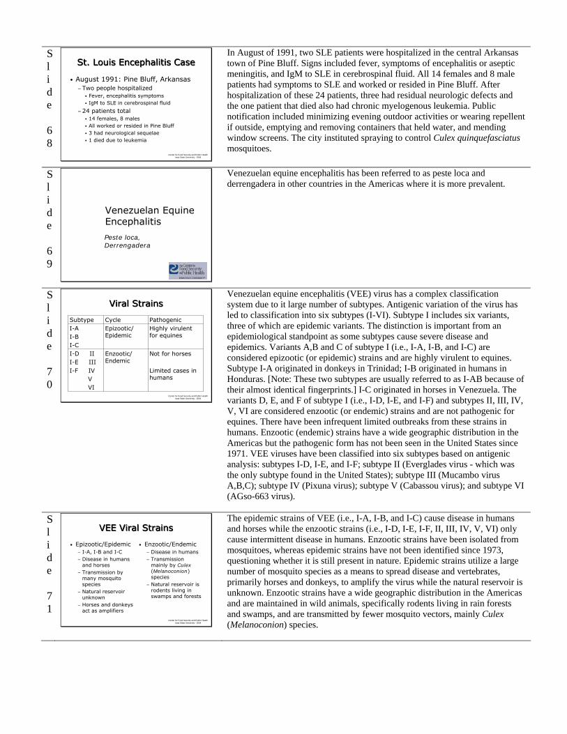
In August of 1991, two SLE patients were hospitalized in the central Arkansas town of Pine Bluff. Signs included fever, symptoms of encephalitis or aseptic meningitis, and IgM to SLE in cerebrospinal fluid. All 14 females and 8 male patients had symptoms to SLE and worked or resided in Pine Bluff. After hospitalization of these 24 patients, three had residual neurologic defects and the one patient that died also had chronic myelogenous leukemia. Public notification included minimizing evening outdoor activities or wearing repellent if outside, emptying and removing containers that held water, and mending window screens. The city instituted spraying to control Culex quinquefasciatus mosquitoes.
Slide 69
Venezuelan Equine Encephalitis
Peste loca, Derrengadera
Venezuelan equine encephalitis has been referred to as peste loca and derrengadera in other countries in the Americas where it is more prevalent.
Slide 70
Center for Food Security and Public Health Iowa State University - 2004
Viral StrainsViral Strains
Not for horses
Limited cases in humans
Enzootic/ Endemic
I-D III-E IIII-F IV
VVI
Highly virulent for equines
Epizootic/ Epidemic
I-AI-BI-C
PathogenicCycleSubtype
Venezuelan equine encephalitis (VEE) virus has a complex classification system due to it large number of subtypes. Antigenic variation of the virus has led to classification into six subtypes (I-VI). Subtype I includes six variants, three of which are epidemic variants. The distinction is important from an epidemiological standpoint as some subtypes cause severe disease and epidemics. Variants A,B and C of subtype I (i.e., I-A, I-B, and I-C) are considered epizootic (or epidemic) strains and are highly virulent to equines. Subtype I-A originated in donkeys in Trinidad; I-B originated in humans in Honduras. [Note: These two subtypes are usually referred to as I-AB because of their almost identical fingerprints.] I-C originated in horses in Venezuela. The variants D, E, and F of subtype I (i.e., I-D, I-E, and I-F) and subtypes II, III, IV, V, VI are considered enzootic (or endemic) strains and are not pathogenic for equines. There have been infrequent limited outbreaks from these strains in humans. Enzootic (endemic) strains have a wide geographic distribution in the Americas but the pathogenic form has not been seen in the United States since 1971. VEE viruses have been classified into six subtypes based on antigenic analysis: subtypes I-D, I-E, and I-F; subtype II (Everglades virus - which was the only subtype found in the United States); subtype III (Mucambo virus A,B,C); subtype IV (Pixuna virus); subtype V (Cabassou virus); and subtype VI (AGso-663 virus).
Slide 71
Center for Food Security and Public Health Iowa State University - 2004
VEE Viral StrainsVEE Viral Strains
• Epizootic/Epidemic− I-A, I-B and I-C− Disease in humans
and horses− Transmission by
many mosquito species
− Natural reservoir unknown
− Horses and donkeys act as amplifiers
• Enzootic/Endemic− Disease in humans− Transmission
mainly by Culex (Melanoconion) species
− Natural reservoir is rodents living in swamps and forests
The epidemic strains of VEE (i.e., I-A, I-B, and I-C) cause disease in humans and horses while the enzootic strains (i.e., I-D, I-E, I-F, II, III, IV, V, VI) only cause intermittent disease in humans. Enzootic strains have been isolated from mosquitoes, whereas epidemic strains have not been identified since 1973, questioning whether it is still present in nature. Epidemic strains utilize a large number of mosquito species as a means to spread disease and vertebrates, primarily horses and donkeys, to amplify the virus while the natural reservoir is unknown. Enzootic strains have a wide geographic distribution in the Americas and are maintained in wild animals, specifically rodents living in rain forests and swamps, and are transmitted by fewer mosquito vectors, mainly Culex (Melanoconion) species.

Slide 72
Center for Food Security and Public Health Iowa State University - 2004
VEE HistoryVEE History
• Western Hemisphere disease−Primarily Central and South America
• 1938: Isolated from a horse brain• 1962-1964−Outbreak in Venezuela
23,000 human cases
• 1967−Outbreak in Colombia
220,000 human cases Over 67,000 horse deaths
Outbreaks of VEE-type diseases have occurred in the Western Hemisphere since the 1920’s, and the virus was finally isolated in 1938 from a horse brain. Annual outbreaks occurred through the 1960’s Colombia and Venezuela. A long standing epidemic, from 1962 to 1964, in Venezuela caused more than 23,000 human cases of VEE with a case-fatality rate of 0.6%. In 1967, a major epidemic hit Colombia, causing 220,000 human cases and over 67,000 horse deaths.
Slide 73
Center for Food Security and Public Health Iowa State University - 2004
VEE HistoryVEE History
• 1969-1971−Largest recorded outbreak in Guatemala−Area from Costa Rica to Rio Grande
Valley in Texas−Thousands of human encephalitis cases−Over 100,000 horses died
• Small outbreaks occur occasionally• 1995−Venezuela and Colombia
Over 90,000 human cases
The largest recorded outbreak of VEE began in 1969 in Guatemala and covered a geographic region from Costa Rica to the Rio Grande Valley of Texas. Human cases of encephalitis were in the thousands, and over 100,000 horses died as a result of VEE. Small, occasional outbreaks have occurred in Peru and Mexico, but most recently in fall of 1995 there were over 90,000 human cases in Venezuela and Colombia.
Slide 74
Center for Food Security and Public Health Iowa State University - 2004
VEE Epizootic TransmissionVEE Epizootic TransmissionPrimary Vector
multiple mosquito species
Dead-end hosts:Humans
Vertebrate Host:Horses
Other species naturally
infected but not amplifiers
Epizootic strains of VEE virus can be transmitted by a large number of mosquito species, such as Mansonia titillans, M. indubitans, Psorophora confinnis, Ps. discolor, Deinocerites pseudes, Aedes thelcter, Ae. sollicitans, Ae. taeniarhynchus, Ae. scapularis and Ae. aegypti. Horses and donkeys are the principal amplifying hosts for VEE and humans are dead-end hosts. There are other domestic species of animals that have been naturally infected with the virus, such as cattle, swine, dogs, and chickens but there is not enough evidence to suggest they are silent amplifiers or reservoirs of disease. Wild species such as cotton rats, opossums, gray foxes, bats and wild birds have been isolated as carriers of the virus in an outbreak, but again their role is unclear as amplifiers or maintainers of VEE.
Slide 75
Center for Food Security and Public Health Iowa State University - 2004
VEE Enzootic TransmissionVEE Enzootic Transmission
Primary VectorCulex
(Melanoconion) species
Dead end hosts:Humans
Vertebrate Host:
Rodents
P. Myers
Enzootic transmission of VEE occurs between mosquitoes (primarily Culex [Melanoconion] species) and rodents who live in or near swamps and rain forests. Humans are dead-end hosts for VEE.
Slide 76
Center for Food Security and Public Health Iowa State University - 2004
VEE EpidemiologyVEE Epidemiology
• 1971−Only U.S. outbreak - in Texas−Enzootic variant Everglades virus in
south Florida2-3 human cases, no horse disease
• Infection in humans less severe than EEE or WEE−Fatalities rare, less than 1%
The only known VEE epidemic in the United States was the outbreak in 1971 in Texas of the subtype I-AB, causing disease in humans and horses; zebras in zoos were also affected. The enzootic variant Everglades virus is limited to the Everglades of south Florida causing only 2 or 3 human cases and no horse cases. Infection in humans with VEE virus is less severe than with EEE and WEE viruses, and fatalities are less than 1%. Adults usually develop an influenza-like illness, while children usually develop overt encephalitis. Effective VEE virus vaccines are available for horses.

Slide 77
Center for Food Security and Public Health Iowa State University - 2004
Human VEEHuman VEE
• Initial signs−Last 24-48 hours −Fever, malaise, dizziness, chills,
headaches, anorexia, severe myalgia, arthralgia, nausea, vomiting
• Lethargy and anorexia can last 2-3 weeks
• 4-15% of cases become neurological• Mortality rates less than 1%−Most often in children with encephalitis
VEE disease in humans varies in its clinical presentation from a mild flu-like illness to neurological involvement. Initial signs lasting 24 to 48 hours include fever, malaise, dizziness, chills, headaches, anorexia, severe myalgia, arthralgia, nausea and vomiting. Lethargy and anorexia can continue for 14 to 21 days. 4-15% of cases become neurological and signs include photophobia, convulsions, stiff neck and altered mental status. Mortality rates are usually less than 1%, with death occurring most often in children with encephalitic signs.
Slide 78
Center for Food Security and Public Health Iowa State University - 2004
Human VEEHuman VEE
• In utero death− Possible in pregnant women who contract the
disease
• Diagnosis− Paired sera with rising titer− ELISA IgG or IgM
• Treatment: Supportive care• No vaccine available• Prognosis
− Variable, often chronic fatigue and headaches
Infection of pregnant women with VEE can result in death of the fetus in utero or a stillbirth. Diagnosis is difficult but involves paired serum samples watching for rising antibody titer or ELISA monitoring IgG or IgM. Treatment involves supportive care and there is no vaccine available. Some patients experience personality changes, fatigue, recurrent headaches and altered senses; prognosis with clinical disease is variable.
Slide 79
Center for Food Security and Public Health Iowa State University - 2004
Animal VEEAnimal VEE
• Incubation period: 1-5 days• Horses most susceptible−Fever, anorexia, depression,
flaccid lips, droopy eyelids and ears, incoordination and blindness
−Death 5-14 days after clinical onset
• Case-fatality rate: 38-83%• In utero transmission results in
abortion, stillbirth
Incubation period in animals is 1 to 5 days, and horses are the most susceptible to epidemic VEE viral infection. Clinical signs include fever, anorexia, depression, flaccid lips, droopy eyelids and ears, head pressing, circling, incoordination and blindness. Death usually occurs 5-14 days after clinical onset, and case-fatality rate ranges from 38-83%. In utero transmission can occur, leading to dead or still born foals.
Slide 80
Center for Food Security and Public Health Iowa State University - 2004
Animal VEEAnimal VEE
• Most domestic animals do not show clinical signs or amplify the virus
• Experimentally− Infected rabbits and dogs die after
inoculation−Laboratory animals susceptible
Act as sentinelsGuinea pigs, mice, hamsters
• Enzootic strains do not cause disease in animals
Most other domestic animals, including swine, cattle and chickens, have not shown clinical signs after natural infection, but rabbits and dogs that were experimentally infected died shortly after inoculation. Laboratory animals vary in their response to the disease, but many are susceptible and act as sentinels for disease prevalence. While guinea pigs experience fever and fatalities without central nervous system signs, mice will experience meningoencephalomyelitis and death. Enzootic strains only cause disease in humans.
Slide 81
Center for Food Security and Public Health Iowa State University - 2004
Animal VEEAnimal VEE
• Diagnosis−Paired sera with rising titer−ELISA IgG or IgM
• Treatment: Supportive care • Vaccine available for horses−Trivalent, formalin inactivated
WEE, EEE, VEE combinationDays 0 and 30
−Annual or biannual booster
Diagnosis is made via serial serum samples monitoring a rise in antibody titer, or by using ELISA for IgG or IgM. Because of the nature of the CNS signs, treatment involves supportive care. Prevention is a better option, and a vaccine is available in the U.S. for horses as a trivalent, formalin-inactivated vaccine (EEE, WEE and VEE). The best protection requires immunization on day 0 followed by a booster 30 days later, and some veterinarians recommend boostering every six months.

Slide 82
Center for Food Security and Public Health Iowa State University - 2004
VEE as a Biological WeaponVEE as a Biological Weapon
• Aerosolized VEE• Human and equine disease occur
simultaneously• Flu-like symptoms in humans• Possible neurological signs in horses• Large number of cases in a given
geographic area
If a bioterrorism attack were to occur using a viral encephalitis agent, experts feel the most likely agent for weaponization would be Venezuelan equine encephalitis. The virus particles would be aerosolized and disseminated, with human disease as the primary event. Equidae would also be susceptible, but disease would most likely occur simultaneously without animals acting as sentinels. Disease symptoms in humans would resemble the flu and be hard to distinguish, so basis of an outbreak would likely be a large number of sick individuals and horses in a given geographic area. Horses may or may not exhibit neurological signs, depending on how the virus was weaponized.
Slide 83
Prevention and Control
Slide 84
Center for Food Security and Public Health Iowa State University - 2004
Management of Mosquito-Borne Diseases
Management of Mosquito-Borne Diseases
• Source reduction• Surveillance• Biological control • Chemical control−Larvicide−Adulticide
• Educating the public−How to protect themselves
Prevention and control of mosquito-borne diseases involve source reduction, surveillance, biological control, chemical control (larvicides and adulticides) and educating the public on how to protect themselves.
Slide 85
Center for Food Security and Public Health Iowa State University - 2004
Source ReductionSource Reduction
• Make habitats unavailable or unsuitable for egg laying and larval development
• Minimize irrigation and lawn watering
• Punch holes in old tires• Fill tree holes with cement• Clean bird baths, outside waterers,
fountains
By trying to eliminate the source of mosquitoes, humans and animals can decrease their risk of exposure. Concentrate efforts on making habitats for egg laying and larval development unsuitable. Utilize less irrigation or manage ditches so that water does not sit undisturbed for more than 2 days, punch holes in old tires to encourage drainage, fill tree holes with cement, clean bird baths and outside animal waterers at least once a week.
Slide 86
Center for Food Security and Public Health Iowa State University - 2004
Source Reduction Cont’dSource Reduction Cont’d
• Drain or fill temporary pools with dirt• Keep swimming pools
treated and circulating−Avoid stagnant water
• Open marsh water management−Connect to deep water
habitats and flood occasionally−Fish access
Drain or fill temporary pools with dirt and keep swimming pools treated and circulating to avoid stagnant water. Eliminate puddles in gutters, around faucets, air conditioners and septic tanks. Also, manage open marshes by connecting mosquito areas by shallow ditches to deep water habitats that allow drainage or fish access.

Slide 87
Center for Food Security and Public Health Iowa State University - 2004
SurveillanceSurveillance
• Record keeping−Weather data, mosquito larval
populations, adult flight patterns
• Sentinel chicken flocks−Blood test and utilize ELISA to monitor
seroconversion of EEE, WEE, SLE
Many states and local governments utilize surveillance programs when there are established risk factors for human disease present. For example, heavy winter snow fall followed by heavy spring rains can lead to flooding and more standing water for mosquitoes to lay eggs upon. Seasonal weather patterns and historical records are kept to predict mosquito larval occurrence and adult flights. Instituting surveillance programs using sentinel chicken flocks and mosquito trapping and testing are ways to monitor disease prevalence in a given area. Blood testing birds, either wild or young, unexposed chickens, and monitoring viral seroconversion or antibody titer allows authorities time to alert the general public if there is concern. These are common practices for EEE, WEE and SLE.
Slide 88
Center for Food Security and Public Health Iowa State University - 2004
SurveillanceSurveillance
Mosquito trapping and testing for viral presence
Other surveillance programs utilize mosquito trapping and testing for viral presence in a given area. When established mosquito larval and adult threshold populations are exceeded, control activities can be initiated.
Slide 89
Center for Food Security and Public Health Iowa State University - 2004
Biological ControlBiological Control
• Predators, natural and introduced, to eat larvae and pupae−Mosquito fish
Gambusia affinis, G. holbrooki most commonFundulus spp., Rivulus spp., killifish
• Other agents have been used but are not readily available
• Copepods
Biological control involves using different predators that eat mosquito larvae and pupae. The mosquito fish, Gambusia affinis and G. holbrooki are the most commonly used supplemental control because they are easily reared. They are indiscriminate feeders and may eat other things, such as tadpoles, zooplankton, aquatic insects and other fish eggs. Some naturally occurring fish such as Fundulus spp., Rivulus spp. and killifish play an important role in controlling mosquitoes in open marsh water and rotational impoundment management. There are other agents such as fungi, protozoa and nematodes that have been tried but are not readily available. A predacious copepod, Mesocyclops longisetus, preys on mosquito larvae and is a candidate for local rearing with Paramecium spp. for food.
Slide 90
Center for Food Security and Public Health Iowa State University - 2004
Chemical ControlChemical Control
• Essential when source reduction is not enough or surveillance shows increased population of virus-carrying mosquitoes
• Requires properly trained personnel• Larvicides, adulticides• Toxic to many birds, fish, wildlife,
aquatic invertebrates, honeybees• Human exposure is uncommon
Chemical control is often warranted when source reduction is not enough and surveillance shows an increased population of virus-carrying mosquitoes. All insecticide use requires proper training by the personnel applying it, and can be targeted at the immature (larvicides) or adult (adulticides) mosquitoes. While it is limited, there is a risk of toxic effects on nontarget organisms, such as birds, fish, wildlife, aquatic vertebrates and honeybees, so low levels of pesticide and proper training of applicators are used. Humans are often concerned with the use of chemicals but low application rates, ultra low volume (ULV) methods, spraying at night while people are indoors, and notifying the public prior to application all decrease exposure risks.
Slide 91
Center for Food Security and Public Health Iowa State University - 2004
Chemical ControlChemical Control
• Federal Food Drug and Cosmetic Act limits the quantity of adulticide used− Due to wind drift onto agricultural crops
• Method used varies− Type of target mosquito− Type of targeted habitat− Aerial spraying covers wide area
• Funding providing by state or local government− Rarely federal
To further protect human exposure, the Federal Food Drug and Cosmetic Act (FFDCA) limits the quantity of poisonous or deleterious substances added to food, specifically adulticides carried by wind drift over agricultural crops. The method selected depends on the type of mosquitoes that need to be controlled and the targeted habitat. Aerial spraying can cover a wide geographic area to control nuisance mosquitoes in emergency situations. Costs for such application is often provided by state or local emergency funds, and rarely by federal funds unless a natural disaster has occurred.

Slide 92
Center for Food Security and Public Health Iowa State University - 2004
LarvicidesLarvicides
• Use when source reduction and biological control not feasible
• More effective and target-specific• Less controversial than adulticides• Applied to smaller geographic areas−Larvae concentrate in specific locations
Larvicides are used when immature mosquito populations become larger than source reduction can manage or biological control can handle. They are often more effective and target-specific than adulticides, making them less controversial. They can be applied to smaller geographic areas than adulticides because larvae are often concentrated in specific locations, such as standing water.
Slide 93
Center for Food Security and Public Health Iowa State University - 2004
Pyrenone, Pyronyl (A, L)Pyrethrins
VectoLex (L)Bacillus sphaericus
Aquabac, Bactimos, LarvX, Teknar, Dunks (L)
Bacillus thuringiensis israelensis (BTI)
Product (Larvae, Pupae, Adult)
Name
Agnique (L, P)Monomolecular film
BVA, Golden Bear (L, P)Oils
Altosid (L)Methoprene
Abate (L)Temephos
LarvicidesLarvicides
This chart depicts the various types of larvicides used in the United States with their chemical or biological name as well as the commercial product name. There may be others on the market that this chart does not cover.
Slide 94
Center for Food Security and Public Health Iowa State University - 2004
AdulticidesAdulticides
• Necessary when other control measures unsuccessful
• Least efficient• Proper type and time of
application helps efficacy− Ultra low volume (ULV)
foggers1 ounce per acre
− Small droplets contact and kill adults
Despite the efforts listed in previous slides, there are times when the environment prevails or humans are unable to prevent large swarms of mosquitoes. Adulticide use then becomes necessary. It is often the least efficient control program but ultra low volume spray either on the ground or aerially can reduce the population when the proper type and time of application is followed. Effective adult mosquito control with adulticides requires small droplets that drift through mosquito areas and come in contact with adults to kill them. Large droplets settle on the ground or vegetation do not contact mosquitoes and may cause undesirable effects on nontargeted organisms. Insecticides are applied in a concentrated form at very low volumes such as 1 oz (29.6 mL) per acre. Excessive wind and updrafts reduce control, but light wind is necessary for drifting spray droplets.
Slide 95
Center for Food Security and Public Health Iowa State University - 2004
AdulticidesAdulticides
AnvilSumithrin
ScourgeResmethrin
ProductChemical Name
Permanone, AquaResilin, Biomist, Mosquito Beater
Permethrin
BatexFenthion
Dibrom, TrumpetNaled
Fyfanon, Atrapa, PrentoxMalathion
This chart displays the various types of chemicals used as adulticides, namely the organophosphates, malathion and naled. Natural pyrethrins, fenthion, and synthetic pyrethroids such as permethrin, resmethrin and sumithrin and their product names are also listed.
Slide 96
Center for Food Security and Public Health Iowa State University - 2004
Personal ProtectionPersonal Protection
• Stay inside during the evening when mosquitoes are most active
• Wear long pants and sleeves• Use mosquito repellent when
necessary−Follow label directions −DEET
Do not use on pets
Humans can protect themselves in two ways: reduce contact with mosquitoes and reduce population of infected mosquitoes in the environment. Personal protection involves reducing time outdoors in the early evening hours outdoors when mosquitoes are most active, wearing long pants and long sleeved shirts and applying mosquito repellent containing DEET to exposed skin areas. DEET can be sprayed on clothing, but this is unnecessary because the underlying skin is protected from insect bites by the clothing. DEET should not be used on pets.

Slide 97
Center for Food Security and Public Health Iowa State University - 2004
Personal ProtectionPersonal Protection
• Make sure window and door screens are "bug tight"
• Replace your outdoor lights with yellow "bug" lights−Bug zappers are not very effective
• ULV foggers for backyard use• Keep vegetation and standing water
in check around the dwelling
It is important to protect yourself by making sure mosquitoes cannot enter your home. Check window screens for holes and make sure they are bug tight so as not to allow entry. Replacing your outdoor lights with yellow bulbs decreases the attractiveness of many bugs to entry ways. Bug zappers are not specific to mosquitoes and are not much help with control. Ultra low volume foggers can be purchased for backyard use to decrease the mosquito population in the event that people will be outdoors during mosquito feeding hours. Keep vegetation and standing water in check around the dwelling to avoid larval habitats.
Slide 98
Center for Food Security and Public Health Iowa State University - 2004
VEE as a Biological WeaponVEE as a Biological Weapon
• 50 kg virulent VEE particles • Aerosolized over city of 5 million
people−150,000 people exposed−30,000 people ill
300 deaths
In 1969, the World Health Organization (WHO) estimated that if 50kg of virulent Venezuelan equine encephalitis particles were aerosolized over a city with 5 million people, 150,000 people would be exposed in a 1 kilometer extent downwind from the release in approximately 5 to 7 minutes. The result could be 30,000 illnesses and 300 deaths from this bioweapon.
Slide 99
Center for Food Security and Public Health Iowa State University - 2004
Internet ResourcesInternet Resources
• CDC Division of Vector Borne Infectious Diseases-Arboviral Encephalitides −www.cdc.gov/ncidod/dvbid/arbor/arbdet
.htm
• American Mosquito Control Association −www.mosquito.org
Slide 100 Center for Food Security and Public Health
Iowa State University - 2004
AcknowledgmentsAcknowledgments
Development of this presentation was funded by a grant from the Centers for Disease Control and Prevention to the Center for Food Security and Public Health at Iowa State University.
Slide 101 Center for Food Security and Public Health
Iowa State University - 2004
AcknowledgmentsAcknowledgments
Author:
Co-author:
Reviewer:
Radford Davis, DVM, MPH
Danelle Bickett-Weddle, DVM, MPH
Jean Gladon, BS