salting out the ionic selectivity of a wide channel: the asymmetry of ompf
TRANSCRIPT

Salting Out the Ionic Selectivity of a Wide Channel: The Asymmetryof OmpF
Antonio Alcaraz,* Ekaterina M. Nestorovich,y Marcel Aguilella-Arzo,* Vicente M. Aguilella,*and Sergey M. Bezrukovy
*Departamento de Ciencias Experimentales, Universidad Jaume I, Castellon, Spain; and yLaboratory of Physical and Structural Biology,National Institute of Child Health and Human Development, National Institutes of Health, Bethesda, Maryland
ABSTRACT Although the crystallographic structure of the bacterial porin OmpF has been known for a decade, the physicalmechanisms of its ionic selectivity are still under investigation. We address this issue in a series of experiments with varied pH,salt concentrations, inverted salt gradient, and charged and uncharged lipids. Measuring reversal potential, we show that OmpFselectivity (traditionally regarded as slightly cationic) depends strongly on pH and salt concentration and is conditionallyasymmetric, that is, the calculated selectivity is sensitive to the direction of salt concentration gradient. At neutral pH andsubdecimolar salt concentrations the channel exhibits nearly ideal cation selectivity (t1G ¼ 0:986 0:01). Substituting neutralDPhPC with DPhPS, we demonstrate that the fixed charge of the host lipid has a small but measurable effect on the channelreversal potential. The available structural information allows for a qualitative explanation of our experimental findings. Thesefindings now lead us to re-examine the ionization state of 102 titratable sites of the OmpF channel. Using standard methods ofcontinuum electrostatics tailored to our particular purpose, we find the charge distribution in the channel as a function of solutionacidity and relate the pH-dependent asymmetry in channel selectivity to the pH-dependent asymmetry in charge distribution. Inan attempt to find a simple phenomenological description of our results, we also discuss different macroscopic models ofelectrodiffusion through large channels.
INTRODUCTION
Large, highly conductive channels from the bacterial outer
membrane are designed by nature to allow exchange of
solutes other than halide, alkaline, or alkaline-earth metal
ions (see recent reviews by Nikaido, 2003, and Delcour,
2003). The major functional role of these large channels in
the cell life-cycle is to support and to regulate the influx of
nutrients and the extrusion of waste products. Yet, their
selectivity for the small ions may play a certain physiological
role too.
However, a much more compelling reason to study the
small-ion selectivity of a large channel is to test our
understanding of electrostatics at the nanoscale. Questions
like the Debye screening in channels that allow one or two
counterions only, electric field effects at distances smaller
that the Bjerrum length, properties of solvents and solutes in
the vicinity of protein surfaces, etc., invariably ignite
a researcher’s interest and imagination.
For our study we choose OmpF (outer membrane protein
F), a channel from the outer bacterial membrane, whose
structure is known with 0.24 nm resolution (Cowan et al.,
1992). Gram-negative bacteria like Escherichia coli are
enclosed in a double membrane cellular envelope. The outer
membrane serves as a selective permeability barrier that
protects bacteria from harmful components and acts as
a passive filter regulating the flow of solutes (Delcour, 2003;
Nikaido, 2003; Jap and Walian, 1990). This function is
accomplished by a number of membrane channel proteins
called porins, which display a wide range of permeability
profiles. Among these channels are general diffusion porins
(with limited substrate selectivity) and solute-specific chan-
nels (with high degree of solute selectivity).
OmpF is one of the general diffusion porins of E. coli. Itallows the passage of ions and small neutral solutes up to
;650 Da. OmpF channels are trimers of identical subunits
(Cowan et al., 1992) where each monomer forms a 16-
stranded b-barrel, which acts as a water-filled pore with an
elliptical cross-section of 2.7 3 3.8 nm2 between the main-
chain atoms in its walls. The shape of this pore is
significantly modified by a polypeptide loop L3 folding into
the barrel. The channel is asymmetric: protein structure on
the extracellular and periplasmic sides differs significantly
not only in the pore dimensions but also in the amount and
ionization state of the charged residues facing the pore
(Karshikoff et al., 1994).
Because of the presence of loop L3 in the b-barrel, theOmpF pore has an hourglass shape, with a narrow
constriction (0.7 3 1.1 nm2) at approximately half the
height of the channel. The solute exclusion limit of the
channel, as well as its ion selectivity, to a great extent is
determined by this constriction. Acidic residues of loop L3
and a cluster of basic residues on the barrel wall opposite the
loop give rise to a transverse electrostatic field strong enough
to orient water molecules (Tieleman and Berendsen, 1998)
and, according to recent molecular dynamics (MD) and
Brownian dynamics (BD) simulations (Schirmer and Phale,
Submitted March 24, 2004, and accepted for publication May 19, 2004.
Address reprint requests to Prof. Vicente M. Aguilella, Universidad Jaume
I, Dep. Ciencias Experimentales, Castellon 12080, Spain. Tel.:
34-964-728045; E-mail: [email protected].
� 2004 by the Biophysical Society
0006-3495/04/08/943/15 $2.00 doi: 10.1529/biophysj.104/043414
Biophysical Journal Volume 87 August 2004 943–957 943

1999; Phale et al., 2001; Im and Roux, 2002a), to produce
well-separated specific ion pathways.
When reconstituted into planar lipid membranes, OmpF
porin forms trimeric channels, whose conductance in 1 M
salt solutions is in a nano-Siemens range (Schindler and
Rosenbusch, 1978). Recordings of ion currents do not reveal
any difference between the monomers of the trimer in regard
to their conductance and blockage by water-soluble
polymers (Rostovtseva et al., 2002) or antibiotics (Nestor-
ovich et al., 2002). Similar observations have been reported
for the specific porin LamB where the time-resolved events
of sugar translocation demonstrate mutual independence of
the monomers in the trimer (Kullman et al., 2002).
Reconstituted OmpF channels display a cation selectivity
at physiological salt concentrations, which decreases at
1-molar concentration (Benz et al., 1979, 1985; Saint et al.,
1996). Presently, there is no quantitative explanation for this
loss of selectivity at high salt concentration, although it is
generally admitted that ions screen both the electrostatic field
created by the protein-charged residues and the reaction field
from the dielectric environment of the pore. However, the
size of the constriction (;0.7 nm3) and the average ion
concentration (;1.2 ions/nm3 for 1 M KCl) make the
application of the Debye screening theory questionable.
Recent studies using BD simulations have shown that
continuum theories fail in narrow pores because they do not
take into account the self-energy contributions (Corry et al.,
2000; Im and Roux, 2002a). Alternatively, this behavior
could be explained by assuming effective binding of small
ions in the vicinity of oppositely charged residues (Miller,
1999).
It has been reported that OmpF selectivity is highly
sensitive to the charge state of the channel lining at the
constriction (Schirmer, 1998; Nestorovich et al., 2003).
OmpF mutants (R42C, R82C, or R132P) after substitution of
any of the three arginines by neutral cysteine or proline
exhibit an increased cationic selectivity (Saint et al., 1996).
Correspondingly, substitution of an acidic group like D113
for a neutral residue reduces considerably the cation
preference of the channel (Saint et al., 1996; Bredin et al.,
2002).
Despite these straightforward correlations, it appears that
channel selectivity cannot be attributed solely to the net
charge and geometry of the narrowest region of the channel.
In fact, net charge in the constriction is positive whereas the
whole channel is cation-selective. Selectivity measurements
in OmpF mutants with no charge at the constriction showed
significantly higher cation selectivity, close to threefold the
selectivity of the wild-type OmpF (Phale et al., 2001).
Furthermore, ion trajectories from MD and BD simulations
suggest that a number of charged residues distributed over
the OmpF aqueous pore also contribute to the channel charge
specificity (Im and Roux, 2002a).
The known crystal structure helps to clarify the role
of each group in the channel selectivity. However, this
information needs to be supplemented by careful estimates
of the pKa values of these groups. This is not an easy task
because of the mutual interference between the neighboring
residues and the difficulties in estimating the electric
potential and the dielectric properties of the solvent in their
vicinities. Several studies aimed at the pKa values de-
termination were using slightly different computational
methods and assumptions (Karshikoff et al., 1994; Varma
and Jakobsson, 2004). As a result, there is no agreement, for
instance, concerning the charge state of some residues at
neutral pH.
The exact orientation of the trimer in the bilayer
reconstitution experiments is also unknown. Some authors
think it is likely that the short periplasmic loops of the porin
first insert into the membrane, thus leaving the long
extracellular loops on the side of the protein addition (cisside) (Im and Roux, 2002a). What is well established,
however, is that under the experimental protocols used in the
present study the OmpF channel inserts predominantly in an
oriented manner with the error rate of ;3% (Nestorovich
et al., 2003).
Here we discuss experiments on OmpF reversal potential
at different lipid membrane composition, salt type, salt
concentration gradient, and pH of the surrounding solution.
The calculated OmpF selectivity is found to be dependent
not only on salt concentration but also on the direction of salt
concentration gradient (first reported by Alcaraz et al., 2003).
Reversal potential measurements show a substantial asym-
metry (of ;25%) upon inverting the gradient. This
asymmetry depends on the bathing solution pH and on the
absolute salt concentration. Experiments with charged
(DPhPS) and neutral (DPhPC) lipid bilayers show that the
fixed charge of the host lipid has a small but measurable
effect on the channel reversal potential.
Taking advantage of the known crystal structure of OmpF,
we complement our experimental study with numerical
calculations of the electric potential all over the protein and
electrodiffusion region. To compute the potential it is
necessary to have the electric charge distribution, which is
not known a priori despite the availability of the three-
dimensional structure of the protein. This charge distribution
has two main contributions: the fixed partial charges which
come from the different electronegativities of the atoms in
the molecule and the charges coming from the ionization of
the groups. The latter depends on the ionization equilibrium
established between the protons of the solution and the
titratable residues of the protein. A grossly simplified
approach consists in using the amino acid pKa values in
free solution (known as ‘‘model pKa,’’ pKma ) and obtaining
the protonation state for a given residue. However, the
ionization state of a given residue in the protein will be
influenced, among other factors, by the charge distribution in
its vicinities and its position in the protein (i.e., whether the
residue is buried or not). For this reason, the effective pKa
values are, in some cases, very different from the model pKa
944 Alcaraz et al.
Biophysical Journal 87(2) 943–957

values. Previous work shows that it is possible to estimate
these pKa values in the protein from the model pKa values by
calculating the work (DG) necessary to carry the set of amino
acids in free solution (unfolded protein) to their actual
position in the protein (Antosiewicz et al., 1996; Ullmann
and Knapp, 1999). Several alternative methods have been
reported for the pKa calculations (Karshikoff et al., 1994;
Varma and Jakobsson, 2004). The common routine is to
divide the pKa shift calculation in two main steps: 1)
‘‘single-site titration’’ ignores the presence of other titratable
residues; 2) ‘‘multiple-site titration’’ accounts for the pre-
sence of all the remaining titratable groups. We use a Monte
Carlo approach for the second step.
Here we use our calculations of the pKa values of the
charged groups in OmpF to obtain the charge distribution in
the channel. We find some differences between our results
and the pKa values reported previously by other authors
(Karshikoff et al., 1994), but in agreement with the pKa shifts
recently computed using a different method for the
‘‘multiple-site titration’’ (Varma and Jakobsson, 2004). We
demonstrate a correlation between the reversal potential
asymmetry and the asymmetry in the total charge near the
OmpF mouths. We also compare several simple macroscopic
models for the reversal potential description identifying
a model that accounts best for the reversal potential changes
with salt concentration and concentration gradient.
MATERIALS AND METHODS
Experimental
Wild-type OmpF (a generous gift of Dr. Mathias Winterhalter) was isolated
and purified from an Escherichia coli culture. Bilayer membranes were
formed from two monolayers made from a 5 mg/ml solution of
diphytanoylphosphatidylcholine (DPhPC) or diphytanoylphosphatidylser-
ine (DPhPS) (Avanti Polar Lipids, Alabaster, AL) in pentane (Burdick and
Jackson, Muskegon, MI) on 70–80-mm diameter orifices in the 15-mm-thick
Teflon partition that separated two chambers (Bezrukov and Vodyanoy,
1993; Montal and Mueller, 1972). The orifices were pretreated with a 1%
solution of hexadecane in pentane. The total capacitance depended on the
actual location of the orifice in the film (and thus area of the film exposed to
salt solution), but membrane capacitance was always ;30–35 pF. Aqueous
solutions of KCl were buffered by 5 mMMES at pH values below pH 6, by 5
mM HEPES at pH values (6 O 8), by 5 mM CHES at pH 9, and by 10 mM
CAPS at pH values above 9. All measurements were performed on single
OmpF channels at room temperature (23.0 6 1.5)�C. Single-channel
insertion was achieved by adding 0.1–0.3 ml of a 1-mg/ml solution of OmpF
in the buffer that contained 1 M KCl and 1% (v/v) of Octyl POE (Alexis,
Switzerland) to 1 ml aqueous phase at the cis side of the membrane only
while stirring.
The membrane potential was applied using Ag/AgCl electrodes in 2 M
KCl, 1.5% agarose bridges assembled within standard 200-ml pipette tips
(Bezrukov and Vodyanoy, 1993). Potential is defined as positive when it is
greater at the side of protein addition (the cis side of the membrane cell). An
Axopatch 200B amplifier (Axon Instruments, Foster City, CA) in the
voltage-clamp mode was used for measuring the current and applying
potential. Data were filtered by a lowpass, eight-pole Butterworth filter
(Model 9002, Frequency Devices, Haverhill, MA) at 15 kHz and recorded
simultaneously by a VCR operated in a digital mode, and directly saved into
the computer memory with a sampling frequency of 50 kHz. The membrane
chamber and the headstage were isolated from external noise sources with
a double metal screen (Amuneal Manufacturing, Philadelphia, PA).
The reversal potential was obtained as follows. First, a lipid membrane
was formed at a given concentration gradient. Second, a single OmpF
channel was inserted at zero potential and the channel conductance was
checked by applying150 mV and then switching potential polarity (Fig. 1).
Third, the ionic current through the channel was manually set to zero by
adjusting the applied potential. The potential needed to achieve zero current
was read as the ‘‘reversal potential.’’ Each point was measured for at least
three different channels in three different experiments to assure re-
producibility and to estimate standard deviation. In some experiments
negative potentials of �(100 O 200) mV were applied to speed up channel
insertion.
Model calculations: numerical procedure
The entire trimer, built using the Protein Data Bank file (Cowan et al., 1992,
1995) was inserted in a membrane that, for simplicity, was represented by
spheres of radius 8 A spaced at 8 A. The membrane dimensions were 2563296 3 40 A3. Overlapping of spheres ensured that the whole space of the
membrane was filled by a low dielectric medium. The University of Houston
Brownian Dynamics Program code (Davis et al., 1991; Madura et al., 1995)
was used for the calculation of the pKa of ionizable residues following the
procedure described by Antosiewicz et al. (1994). This approach involves
the calculation of the interaction energy between the ionizable groups of the
protein and of the differences between the ionization energies of each group
in the neutral protein and in free solution. All these energies were assumed
to be of purely electrostatic nature so that they could be calculated by means
FIGURE 1 A typical recording of transmembrane ion current that
illustrates reversal potential measurements. First, spontaneous insertion of
a single OmpF channel at zero transmembrane voltage was achieved
(arrow). Because of the channel cationic selectivity, the insertion is seen as
a non-zero current that flows from the more concentrated 1.5 M KCl solution
at the cis side of the membrane to the less concentrated 0.1 M KCl on the
trans side. Second, 650 mV potential was applied to make certain that the
number of OmpF channels was equal to 1. Reasonable conductance values
determined from the difference in current readings at positive and negative
potentials as well as the rare flickering to two-thirds of the total channel
conductance at 150 mV provided the evidence for single channel insertion.
Finally, ion current was manually set to zero by adjusting the transmembrane
potential to �27.7 mV. This potential was read as a ‘‘reversal potential.’’
Time resolution was 1 ms. The DPhPC membrane was bathed by solutions
buffered by 5 mM MES at pH 6.
Selectivity and Asymmetry of OmpF 945
Biophysical Journal 87(2) 943–957
of the Poisson-Boltzmann equation. The protein and the membrane were
represented by a single region of low dielectric constant.
Three energy contributions were considered to be involved in the pKa
shift of the charged groups of the protein: 1), the ion self-energy (Born
energy) that takes into account the penalty in free energy for transferring
a charged group from solution with a high dielectric constant to the actual
position in the protein medium of a low dielectric constant; 2), the
background term that includes the electrostatic interaction between residues
with the remaining permanent charges of the protein; and 3), the interaction
of the charged group with other ionizable charged groups. The first two
terms lead to the so-called intrinsic pKa (pKint). All three contributions were
computed using the linear form of the Poisson-Boltzmann equation. We start
from the free energy of ionization of the model compound at each given pH,
DGs, then we add the change in free energy of the ionized residue when
carried from the free solution to the protein environment, DGAs;p. Finally, the
change in free energy of the neutral residue when carried from the free
solution to the protein environment, DGAHs;p , is subtracted. This is sum-
marized in the thermodynamic cycle of Fig. 2.
In practice, the calculation of all these energies requires two finite-
difference calculations per residue: a first calculation with the charge on
the ionizable residue in free solution and another with the charge on the
ionizable residue in the neutral protein (but with the permanent partial
charges present). To avoid problems coming from the mesh discretization
and the self-energy of the charges, all the calculations were carried out with
the residue located at exactly the same position on the mesh, which in our
calculations has initial dimensions (before ‘‘focusing’’) 1303 1303 60 A3
and spacing of 2.5 A. This treatment allows the exact removal of the mesh-
dependent self-energy and the correct finite-difference calculation of
Coulombic energy differences when free energies are computed. The
calculated potential on the large grid is used to provide boundary conditions
for all successive ‘‘focusing’’ steps, where a smaller grid (153 153 15 and
spacing 1.2 A, 0.75 A, and 20 3 20 3 20 and spacing 0.25 A) is built
centered on each titratable residue to reach better accuracy. As for the energy
due to the interaction between titratable groups, we have used a Monte Carlo
method to obtain the statistical distribution of the ionization state for each
residue and, from it, the titration curves for all titratable groups. One
alternative and most direct possibility, namely calculating all possible
ionization states and choosing that of higher Boltzmann probability, is
simply unfeasible in our model due to the large number of ionizable residues
present in the trimer (3 3 102 ¼ 306, which would give 2306 different
states). Nevertheless, this problem can be considerably simplified by
introducing a cutoff distance and considering only the residues within this
distance (Varma and Jakobsson, 2004).
We use the same dielectric constant, em, for the membrane and the
protein. The remaining space, corresponding to the membrane-bathing
solution, is assigned a dielectric constant of 80. The calculations have been
performed both for em ¼ 20 and em ¼ 4 to check the influence of this
parameter on the calculation results. The salt concentration was set in all
cases equal to 150 mM and the ionic radius of the permeating ions was set to
2 A. Typical calculations on a PC (P4, 2.4 GHz processor) took;3 h for the
energies of all charged residues and 45 min for the Monte Carlo procedure.
RESULTS AND DISCUSSION
To assess OmpF small-ion selectivity, we have measured the
channel reversal potential under carefully controlled pH and
salt concentrations on both sides of the membrane formed
from either neutral or charged lipids. One of the typical
measurement protocols is illustrated by Fig. 1. Special care
was used to exclude any exchange of electrolyte solutions
between the cis and trans cell compartments before the
insertion of a single channel. In each new measurement,
including those with the same pairs of cis and transconcentrations, a new membrane was formed on fresh salt
solutions.
‘‘Salting out’’ channel selectivity
In the main series of experiments the OmpF channel was
inserted in a neutral lipid bilayer (DPhPC). The use of
an uncharged lipid discards any side effect of counterion
accumulation on both sides of the membrane that, in the case
of asymmetric lipid compositions or in the case of asym-
metric salt concentrations, could contribute to the voltage
drop across the channel. First, the salt concentration on the
trans side,Ctrans, was kept constant (0.1 M), whereas the con-
centration on the cis side, Ccis, was increased from 0.1 M up
to 3 M. Thus, the concentration ratio, r [ Ccis/Ctrans, was
changed between 1 and 30.
As shown in Fig. 3, for both KCl and NaCl, the main
change in the OmpF reversal potential occurs at small
concentration ratios, mostly up to a fivefold gradient. Only
a weak concentration dependence of Erev is found beyond
this gradient. The channel is not equally cation-selective in
KCl and in NaCl solutions, although the difference is small
(#5 mV in the reversal potential). Measured values of the
reversal potential are similar to those previously reported
in the literature (Benz et al., 1979; Saint et al., 1996;
Nestorovich et al., 2003), although most of the reported
experiments were performed only in a 10-fold salt gradient.
Apparently, the channel reversal potential follows the
well-known Goldman-Hodgkin-Katz (GHK) equation
(Goldman, 1943; Hodgkin and Katz, 1949). For large
concentration gradients, GHK equation predicts a limiting
value defined by the permeability ratio (see Appendix). The
asymptotic behavior seen in the plots for both salts agrees
qualitatively with such prediction. However, according to the
GHK theory, reversal potential for a given salt is only
dependent on the concentration ratio (corrected by activity
coefficients), r, and not on the absolute values of salt
FIGURE 2 Thermodynamic cycle used in calculation of pKa shifts for the
dissociation equilibrium of a model residue (A) in solution (subscript s) andin the protein environment (subscript p).
946 Alcaraz et al.
Biophysical Journal 87(2) 943–957

concentration on both sides of a selective membrane or
a pore.
Fig. 4 demonstrates that this is not the case. The reversal
potential measurements with KCl performed at a constant
concentration ratio, r, but at different concentrations revealthat the reversal potential is also a function of absolute
concentrations. In these experiments both Ctrans and Ccis
were changed to keep their ratio at a particular value
specified in the figure. It is seen that the absolute value of the
reversal potential decreases as salt concentration increases.
Importantly, in the whole concentration range used in these
experiments, the KCl activity coefficient changes only by
36% from 0.87 for 0.02 M to 0.56 for 3 M, and, therefore,
cannot explain the measured trends.
As might be expected, concentrated solutions screen the
channel fixed charges more effectively, and, therefore, the
channel cationic preference gets smaller. At Ccis ¼ 3 M,
the reversal potential is reduced by more than one-half of
its value at the physiological concentration. For a 10-fold
gradient (open circles), the change in Erev in the explored
concentration range is greater than 20 mV. It is seen that in
contrast with the GHK prediction, the reversal potential
changes with salt concentration for all the ratios between cisand trans concentrations (r ¼ 10, 3, and 1.5). Fig. 4 (inset)gives the cation transport numbers calculated using Gold-
man’s constant field approximation (Goldman, 1943;
Hodgkin and Katz, 1949; Markin and Chizmadzhev,
1974). It is seen that as both concentrations increase, the
selectivity of the channel decreases, from nearly ideal
cationic selectivity of 0.98 6 0.01 to slightly cationic of
0.6 O 0.7 for all concentration ratios. At a given cis side
concentration (which is always larger than the trans side
concentration), the calculated selectivity also depends on the
ratio itself: the higher is the ratio, the higher is the transport
number. This observation is in accord with the charge
screening idea. Indeed, at a given cis side concentration,
higher ratios correspond to smaller trans side concentrationsand, therefore, to less pronounced screening.
Reversal potential asymmetry
The structure of the OmpF channel is asymmetric. This
concerns both the geometry of the water-filled pore (Cowan
et al., 1992) and the fixed charge distribution (Karshikoff
et al., 1994). Therefore, because of the difference in salt
concentrations at the cis and trans sides of the membrane, the
fixed charge is screened differently and we should expect
certain a sensitivity of the reversal potential to the direction
of the salt gradient.
It is known that when OmpF is added from one side of the
membrane only, it inserts in a predominantly oriented
manner (Rostovtseva et al., 2002; Nestorovich et al., 2002,
2003; Danelon et al., 2003). We made use of this finding to
study asymmetry in the reversal potential. Fig. 5 shows
results of two series of reversal potential measurements
where not only the absolute gradient but also the gradient
direction was changed. In the first series, the KCl
concentration ratio r is greater than unity (Ccis . Ctrans)
(same data are compared in Fig. 3 with measurements for
NaCl); in the second one, the gradient is inverted, i.e., r , 1
(Ccis , Ctrans). Salt concentration in the less concentrated
solution was kept constant at 0.1 M in all cases. Indeed, the
FIGURE 3 Reversal potential as a function of cis/trans concentration
ratio for KCl (solid circles) and NaCl (open diamonds) at pH 6. Bathing
solution concentration on the trans side was fixed at 0.1 M, whereas the
concentration on the cis side was increased from 0.1 M to 3 M. Membranes
were formed from DPhPC. The channel is more permeable to K1 ions along
the whole range of studied salt gradients. The asterisks represent the
difference between the absolute values of Erev in KCl and NaCl solutions.
FIGURE 4 Reversal potential measured at three constant cis/trans KCl
concentration ratios (circles, 10; squares, 3; pentagons, 1.5) but different
absolute concentrations. Membranes were formed from DPhPC at pH 6. The
channel exhibits cationic selectivity: higher salt concentration at the cis sidegenerates negative reversal potential. The absolute value of this potential
decreases as salt concentration goes up. (Inset) Cationic transport numbers
calculated according to Eq. A5 (Appendix) with salt concentrations
corrected with activity coefficients. Solid lines through the data are drawn
to guide the eye. It is seen that at Ccis ¼ 3 M cationic selectivity of the
channel drops to t1 � 0.6 O 0.7.
Selectivity and Asymmetry of OmpF 947
Biophysical Journal 87(2) 943–957

reversal potential was always negative at the side with the
higher salt concentration. However, to facilitate comparison
of the two datasets, we plotted �Erev for the experiments
where the cis side concentration was smaller. The channel
functional asymmetry manifests itself as a difference reach-
ing ;25% in the measured reversal potential. The absolute
value of Erev is greater when the more concentrated solution
is on the cis side than when the opposite happens. The
asymmetry in Erev (,6 mV) is only slightly concentration-
dependent, as shown in the bottom plot of Fig. 5. These
results demonstrate once again that OmpF trimer inserts in an
oriented manner rather than randomly.
The asymmetry in Erev is pH-dependent. Fig. 6 shows the
change of the reversal potential with pH for a 10-fold
concentration ratio. For the ease of comparison, the values of
–Erev have been plotted for r, 1 (less concentrated solution
in the cis side). Again, different absolute values of Erev are
found for 0.1 M trans/1 M cis (solid circles) and the oppositegradient, 1 M trans/0.1 M cis (open circles) at every pH
except for ;pH 4.
The variation of the reversal potential with pH agrees
qualitatively with previously reported measurements (Benz
et al.,. 1979; Schirmer and Phale, 1999; Nestorovich et al.,
2003). It is seen that the reversal potential changes its sign as
pH is decreased below ;pH 4. Thus, in acidic solutions
the OmpF channel becomes anion-selective. At the other
extreme, in basic solutions, the cationic selectivity increases
beyond its value for neutral pH. Recently, we reported
(Nestorovich et al., 2003) that pH-dependent selectivity
behavior resembles that of the channel conductance. OmpF
conductance in its fully open state displays three character-
istic regimes. Under acidic conditions (corresponding to
most dramatic selectivity change) conductance strongly
depends on solution pH, increasing threefold when pH is
increased from pH 1.0 to pH 5. At neutral pH (where channel
is slightly selective to cations with tK1 � 3tCl
�), it displaysa plateau that extends from pH 5 to pH 9. In basic solutions
from pH 9 to pH 12 (corresponding to a slight increase
in cationic selectivity), channel conductance is again pH-
sensitive and increases by 15–30%, depending on the sign of
the applied voltage. Analysis of open channel noise and
stepwise time-resolved transients in the open state (Nestor-
ovich et al., 2003) demonstrated that at least three different
ionization processes are involved in the pH-dependent
modification of the channel transport properties. Noise is
minimal in the pH range corresponding to the conductance
and selectivity plateaus.
The large amount of titratable residues lining the pore
lumen does not allow a simple explanation of selectivity
versus pH curves in terms of one or a few pKa of charged
groups. However, it is evident that the asymmetry in Erev
does not remain constant but is also pH-dependent.
Moreover, the difference in Erev between the opposite
gradient configurations changes its sign from low to high pH:
DjErevj is;–4 mV at pH 2 and DjErevj is;15 mV at neutral
pH.
Interestingly, at ;pH 3.25 the ‘‘sign’’ of selectivity
measured in 1 M/0.1 M KCl gradient depends on the
gradient direction. The selectivity is cationic for Ccis . Ctrans
and anionic for Ccis , Ctrans. This inversion of measured
selectivity is a consequence of the finite concentration
gradient used in experiment. Indeed, as Ccis approaches Ctrans
the system moves toward equilibrium, so that cis-to-transand trans-to-cis selectivities have to be equal in order not
to violate equilibrium thermodynamics. Once again, the
measured inversion demonstrates that the selectivity ob-
tained for a particular concentration gradient is a function of
FIGURE 5 OmpF reversal potential as a function of the concentration
ratio for two series of measurements with oppositely directed gradients.
(Solid circles) Erev obtained in the series where Ccis . Ctrans ¼ 0.1 M KCl.
(Open circles) Plotted as –Erev for the reversed gradient Ctrans . Ccis ¼ 0.1
M KCl. The channel is asymmetric: the absolute value of the reversal
potential is greater when the more concentrated solution is on the cis side of
the membrane. The asterisks represent the difference. Membranes were
formed from the DPhPC at pH 6.
FIGURE 6 Dependence of the reversal potential on solution pH at Ccis ¼1 M KCl and Ctrans ¼ 0.1 M KCl (solid circles) and at the inverted gradient
of the same concentrations (open circles). To facilitate comparison, the data
for the inverted gradient are plotted as �Erev. Note that the difference in the
reversal potentials (asterisks) changes its sign from high pH to low pH.
Thus, DjErevj ;5 mV at the wide pH range from 5 to 11 and DjErevj ;(�4 mV) at pH 2. Membranes were formed from DPhPC.
948 Alcaraz et al.
Biophysical Journal 87(2) 943–957

this gradient, its direction, and the absolute salt concentration
(Figs. 4 and 5).
Electrostatics versus other interactions
The experimental findings reported above strongly favor
electrostatic interactions as the main cause of small-ion
selectivity of the large channel formed by OmpF. The
decrease in the selectivity with the increasing salt concen-
tration (Fig. 4), selectivity asymmetry (Fig. 5), and its
titration at the changing solution acidity (Fig. 6)—all are in
perfect agreement with intuitive reasoning based on
electrostatic considerations.
Further support has been found in experiments with
charged membranes. The charge of lipid polar groups is able
to modify the surface potential significantly (McLaughlin
et al., 1970; Ninham and Parsegian, 1971). However, taking
into account that the OmpF channel is comprised of three
monomers whose size exceeds the typical Debye length of
the solutions we use (,1 nm), it is reasonable to expect that
the charge on lipid polar groups will have only small
influence on the potential near the channel mouths. We have
checked this conjecture by carrying out a series of measure-
ments at different concentration gradients in a neutral
membrane (DPhPC) and a negatively charged membrane
(DPhPS). The results for pH 6 are shown in Fig. 7. The
concentration of KCl on the trans side was 0.1 M and the
concentration on the cis side was varied from 0.1 M up to
3 M. The reversal potential in the charged lipid is always
higher, although the difference in Erev is small (#5 mV).
This means that the effect of the lipid charge on OmpF
selectivity is quite measurable but does not change the
channel selectivity drastically.
It is seen that the difference between reversal potentials in
DPhPC and DPhPS is constant for the concentration ratio
exceeding 10. This is an indication that the Debye length in
the cis side is very small and the lipid charge is screened out,
so that the major contribution comes from the trans side.Previously it was shown that the effect of the phospha-
tidylserine charge on the channel conductance can be
‘‘titrated out’’ by increasing acidity of the membrane-
bathing solution (Rostovtseva et al., 1998; Aguilella and
Bezrukov, 2001). In a series of experiments we measured the
reversal potential of the OmpF channel reconstituted in
DPhPS and DPhPC membranes as a function of pH (data not
shown). We found that as solution pH was lowered below
pH 4, the difference in the reversal potentials began to level
off reaching virtual zero at pH 2.
Negative charge of DPhPS headgroups in the vicinity of
the channel is expected to accumulate positive ions to an
excess of their bulk concentration. Because the OmpF
channel is cation-selective at neutral pH, this accumulation
may lead to a measurable increase in channel conductance.
Fig. 8 compares single channel conductance in a neutral and
a negatively charged membrane. The effect is an order-
of-magnitude smaller than the effect of charged lipid on
gramicidin channels (Apell et al., 1979; Rostovtseva et al.,
1998) and is comparable (although still smaller) to the effect
on L1 state of alamethicin channels (Aguilella and Bezrukov,
2001). This reduced influence of lipid charge is expected on
the basis of electrostatic considerations that account for the
channel structural features and the increased distance
between the nearest lipid molecules and the water-filled
pore of the channel (Apell et al., 1979; Bell and Miller, 1984;
Moczydlowski et al., 1985; Rostovtseva et al., 1998;
Aguilella and Bezrukov, 2001).
FIGURE 7 Reversal potential is sensitive to the lipid charge. It is larger
(by the absolute value) for the negatively charged DPhPS membranes (open
triangles) than for the neutral DPhPCmembranes (solid circles) in the whole
range of concentration gradients at pH 6. Salt concentration on the trans sidewas kept constant at 0.1 M KCl; the cis side concentration increased from
0.1 MKCl to 3 MKCl. The asterisks represent the difference between Erev in
DPhPC and DPhPS.
FIGURE 8 A single open OmpF channel has larger conductance when
reconstituted into a negatively charged bilayer at pH 6. At 0.1 M KCl
concentration and �100 mV applied voltage the channel conductance in the
DPhPS membranes exceeds that in the DPhPC membranes by ;15%. In
1 M KCl solutions this difference is smaller. In this particular experiment it
was ;4%, which was close to the reproducibility of channel conductance
from experiment to experiment (SD was ;3%). Symmetrical salt solutions
were used in these measurements.
Selectivity and Asymmetry of OmpF 949
Biophysical Journal 87(2) 943–957

Brownian dynamics simulations of the reversal potential
in a model OmpF channel that is totally uncharged yielded
some residual selectivity: the channel would be 40% more
selective to K1 than to Cl� ions (Schirmer and Phale, 1999).
Within purely electrostatic considerations there is no
satisfactory explanation for this residual selectivity. There-
fore, other factors like hydrophobic or hydrophilic effects
and other short-range interactions cannot be ruled out com-
pletely (see also Danelon et al., 2003); however, the results
presented above demonstrate that electrostatic interactions
dominate.
Charge distribution and reversalpotential asymmetry
Table 1 shows the calculated pKa shifts of the ionizable
residues that are presumably most relevant to the channel
selectivity: the residues facing the pore lumen and those
located in its immediate vicinity. The criterion used for the
selection of these 27 residues among the total 102 of one
monomer was to pick those lying within some arbitrary
distance (0.2 nm) of the solvent-accessible surface of the
pore for each axial position in the channel. Also shown
is Glu-296 whose ionization properties deserve a special
comment. In addition, the residues are divided into three
main groups corresponding to those which lie closer to the
periplasmic side (toward the inside of the cell), those in
the constriction zone (approximately in the center of the
channel), and those closer to the extracellular side of the
channel. In particular, residues with all their atoms having
z-coordinate values between 2.6 nm and 4.0 nm are assigned
to the constriction zone, and those with z , 2.6 nm and z .4.0 nm to the periplasmic region and the extracellular re-
gion, respectively. To show the effect of the dielectric con-
stant used to imitate the protein media, pKa calculations have
been performed both for em ¼ 4 and em ¼ 20. The last
column of the table includes the commonly accepted values
of pKma in free solution for each group. It is seen that in
agreement with a recent study (Varma and Jakobsson, 2004),
calculations done with em ¼ 20 yield more realistic pKa
values than those obtained with em ¼ 4.
Most of the residues with large pKa shifts are tyrosines,
with positive changes between six and nine units (both for em¼ 20 and em ¼ 4), so that tyrosines appear to be stabilized in
their protonated state. These tyrosines (only six are shown in
the table) are mostly buried between the other residues of the
trimer. The significant shift for the tyrosines is not rare since
the predicted charge of the protein is huge at high pH
(�100e at pH 14.0). This leads to high electrostatic
interaction energies, unless dramatic rearrangements in the
structure of the protein occur in such an extremely basic
environment. The large shifts for tyrosines have been
previously reported by Karshikoff et al. (1994), although
in our model with em ¼ 20 the shifts are generally smaller.
The constriction contains the so-called ‘‘cluster of
arginines,’’ which includes Arg-42, Arg-82, and Arg-132,
all of them positively charged at neutral pH, and the acidic
residues Asp-113 and Glu-117, negatively charged at neutral
pH. As already mentioned, the position of these residues, with
the arginines at the side close to the axis of the trimer and the
aspartic and glutamic residues at the other side, creates a strong
transverse field. We also include in the constriction zone three
additional groups, which may have some influence on the
channel selectivity: Lys-16, Tyr-106, and Tyr-102.
In the physiological pH range most of the residues in our
model OmpF channel are in the ionization state correspond-
ing to the so-called ‘‘null model,’’ which assigns the same
pKa to the charged groups in the protein as in free solution.
The only exception is Glu-296. According to our calcu-
lations, Glu-296 in the protein has an effective pKa of 8.9,
whereas its pKa in free solution is ;4.4. This suggests that
this residue should be protonated at pH 7. It should be noted
that, according to our calculations, Arg-82 should be charged
at neutral pH. This is in agreement with previously reported
experimental results (Schirmer and Phale, 1999), and in
contradiction with calculations by Karshikoff et al. (1994),
who used em ¼ 4.
Recently Varma and Jakobsson (2004) have reported the
protonation states of titratable residues lining the lumen of
the wild-type OmpF channel at neutral bulk pH. Their
extensive calculations performed under different conditions
of protein dielectric constant, solute shielding, and pro-
tonation sites yield pKa shifts that are in close agreement
with our results. Particularly, for em ¼ 20 we find that at
neutral pH the ionization state of the residues lining the pore
is the same as in their prediction. The use of a rather unusual
dielectric constant of 20 is also supported by an extensive
comparison of measured pKa values of ionizable groups in
proteins with the computed values carried out by Antosie-
wicz et al. (1994).
For this reason hereafter we use the results obtained at
em ¼ 20. We first calculate fields in the pore lumen that are
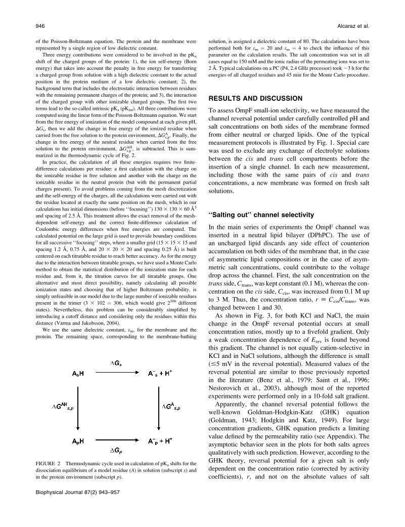
produced by all charges in a monomer. Fig. 9 shows the
electrostatic potential (dashed line) sensed by an ion crossingan OmpF monomer at pH 7, following a path approximately
along the geometric center of the pore lumen. It also gives
the modulus of the transverse electric field (solid line) alongthe same path. Parameter z is the axial coordinate from the
OmpF PDB file; z ¼ 0 refers to the periplasmic side. The
shaded region approximately represents the constriction
zone. The pore lumen is not exactly perpendicular to the
membrane surface and has a rather complex geometry, so
that ion trajectories differ considerably from a straight line
parallel to z axis.It is seen that the electric potential is slightly asymmetric
and exhibits a minimum in the constriction zone near z¼ 3.5
nm. The depth of the potential well at this point exceeds
3.5 kT. Qualitatively, this explains nearly perfect cationic
selectivity of the channel measured for the lowest KCl
950 Alcaraz et al.
Biophysical Journal 87(2) 943–957

concentrations used in this study (Fig. 4, inset). However,quantitative analysis of the selectivity data based on the
electric potential profile illustrated in Fig. 9 is difficult
because of the strong transverse field. This field introduces
a complex distribution of potential in the channel pore.
Indeed, recent MD and BD simulations have shown that
cation and anion pathways through the pore are somewhat
separated (Im and Roux, 2002a; Schirmer and Phale, 1999).
It was also shown (Robertson and Tieleman, 2002) that the
transversal field is able to orient dipolar molecules; experi-
ments with ampicillin translocation through OmpF con-
firmed this prediction (Nestorovich et al., 2002).
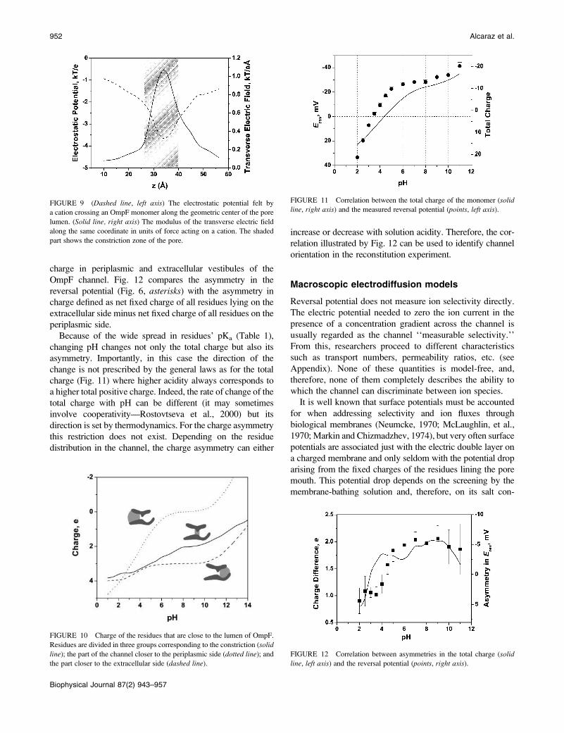
The charge of the ionizable residues located near the lumen
of the channel as a function of pH is shown in Fig. 10.
Separate plots are presented for the fixed charge of the
residues corresponding to the periplasmic region, the con-
striction zone, and the extracellular region of the channel.
It is seen that, except for the charge in the periplasmic region
of the pore at pH . 10, fixed charges lining the pore are
positive, which would imply anionic selectivity. However, this
conjecture contradicts experimental observations: the results
plotted in Fig. 6 show that the channel has cationic selectivity
everywhere except for acidic solutions of pH , 3.5.
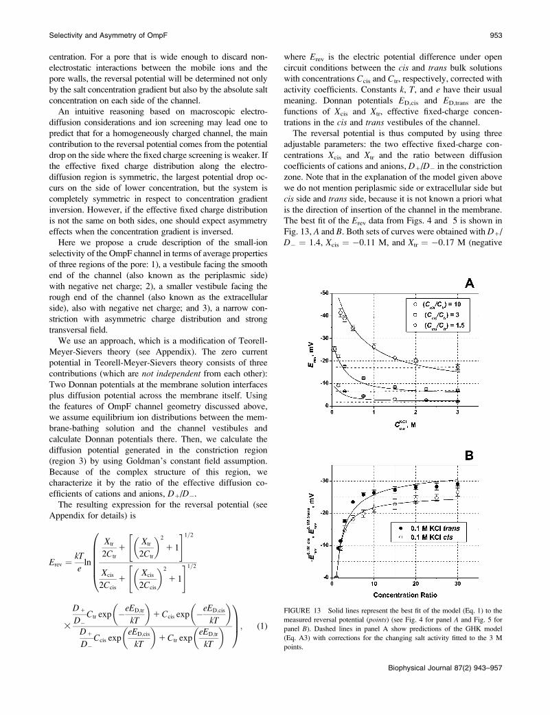
It turns out that the correlation between the total charge of
the monomer and the reversal potential is much better. Fig.
11 shows that as pH is decreased, the total charge goes from
negative to positive, implying the selectivity transition from
cationic to anionic. The solid line represents the sum of all
charges in a monomer (i.e., the total charge of the 102
ionizable groups). The discrepancy in the exact pH of the
sign change between the calculated total charge and the
measured reversal potential can be explained by different
influence of different titratable residues on channel selectiv-
ity. Residues which are farther from the permeating ion paths
influence channel transport properties to a smaller degree.
Therefore, such a discrepancy is expected even in the case of
purely electrostatic interactions.
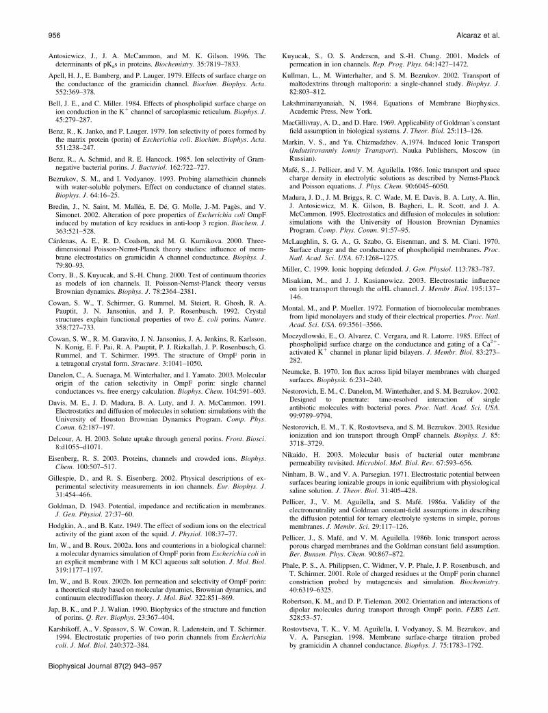
It is interesting to note that not only the pH-dependent total
charge correlates with the pH-dependent reversal potential.
It is also true for the pH-dependent asymmetry in charge.
Indeed, if the asymmetry in Erev is mainly of electrostatic
origin, we expect it to correlate with the difference in the fixed
TABLE 1 Comparison of pKa values of selected residues at different protein dielectric constants
e ¼ 20 e ¼ 4
Residue Residue number pKint pKa pKshift pKint pKa pKshift pKma
Closer to the periplasmic side
GLU 2 4,9 3,7 �0,7 7,5 7,0 2,6 4,4
LYS 10 10,0 12,5 2,1 8,9 10,2 �0,2 10,4
ASP 12 5,4 3,0 �1,0 12,0 11,2 7,2 4
LYS 46 10,0 13,8 3,4 8,4 9,6 �0,8 10,4
GLU 48 5,5 3,6 �0,8 10,0 9,1 4,7 4,4
LYS 89 9,7 12,5 2,1 7,8 8,9 �1,5 10,4
GLU 181 5,0 4,2 �0,2 7,0 6,0 1,6 4,4
LYS 219 10,0 14,5 4,1 8,5 12,3 1,9 10,4
ASP 221 4,6 3,0 �1,0 5,8 3,8 �0,2 4
LYS 305 10,1 12,6 2,2 9,3 10,6 0,2 10,4
TYR 310 11,6 18,8 9,2 19,3 20,0 10,4 9,6
Constriction zone
LYS 16 10,1 14,2 3,8 8,5 9,2 �1,2 10,4
ARG 42 11,8 16,2 4,2 9,1 9,2 �2,8 12
ARG 82 11,6 15,7 3,7 7,6 5,8 �6,2 12
ASP 113 5,0 3,2 �0,8 6,8 5,6 1,6 4
GLU 117 5,9 6,2 1,8 10,1 15,9 11,5 4,4
ARG 132 10,8 15,5 3,5 3,4 3,7 �8,3 12
TYR 40 11,2 14,3 4,7 15,6 15,1 5,5 9,6
TYR 102 10,7 13,3 3,7 13,7 15,9 6,3 9,6
TYR 106 10,6 16,4 6,8 14,2 19,0 9,4 9,6
Closer to the extracellular side
GLU 29 4,7 4,3 �0,1 6,1 6,2 1,8 4,4
LYS 80 10,3 13,1 2,7 9,3 11,4 1,0 10,4
ARG 167 10,6 14,4 2,4 3,9 4,0 �8,0 12
ARG 168 10,7 14,4 2,4 5,8 4,7 �7,3 12
LYS 243 9,9 13,1 2,7 7,7 9,7 �0,7 10,4
TYR 22 11,0 17,7 8,1 16,0 20,0 10,4 9,6
TYR 32 10,0 17,2 7,6 12,5 20,0 10,4 9,6
Other
GLU 296 5.5 8.9 4.5 11.2 19.2 14.8 4.4
Selectivity and Asymmetry of OmpF 951
Biophysical Journal 87(2) 943–957
charge in periplasmic and extracellular vestibules of the
OmpF channel. Fig. 12 compares the asymmetry in the
reversal potential (Fig. 6, asterisks) with the asymmetry in
charge defined as net fixed charge of all residues lying on the
extracellular side minus net fixed charge of all residues on the
periplasmic side.
Because of the wide spread in residues’ pKa (Table 1),
changing pH changes not only the total charge but also its
asymmetry. Importantly, in this case the direction of the
change is not prescribed by the general laws as for the total
charge (Fig. 11) where higher acidity always corresponds to
a higher total positive charge. Indeed, the rate of change of the
total charge with pH can be different (it may sometimes
involve cooperativity—Rostovtseva et al., 2000) but its
direction is set by thermodynamics. For the charge asymmetry
this restriction does not exist. Depending on the residue
distribution in the channel, the charge asymmetry can either
increase or decrease with solution acidity. Therefore, the cor-
relation illustrated by Fig. 12 can be used to identify channel
orientation in the reconstitution experiment.
Macroscopic electrodiffusion models
Reversal potential does not measure ion selectivity directly.
The electric potential needed to zero the ion current in the
presence of a concentration gradient across the channel is
usually regarded as the channel ‘‘measurable selectivity.’’
From this, researchers proceed to different characteristics
such as transport numbers, permeability ratios, etc. (see
Appendix). None of these quantities is model-free, and,
therefore, none of them completely describes the ability to
which the channel can discriminate between ion species.
It is well known that surface potentials must be accounted
for when addressing selectivity and ion fluxes through
biological membranes (Neumcke, 1970; McLaughlin, et al.,
1970; Markin and Chizmadzhev, 1974), but very often surface
potentials are associated just with the electric double layer on
a charged membrane and only seldom with the potential drop
arising from the fixed charges of the residues lining the pore
mouth. This potential drop depends on the screening by the
membrane-bathing solution and, therefore, on its salt con-
FIGURE 10 Charge of the residues that are close to the lumen of OmpF.
Residues are divided in three groups corresponding to the constriction (solid
line); the part of the channel closer to the periplasmic side (dotted line); andthe part closer to the extracellular side (dashed line).
FIGURE 11 Correlation between the total charge of the monomer (solidline, right axis) and the measured reversal potential (points, left axis).
FIGURE 12 Correlation between asymmetries in the total charge (solidline, left axis) and the reversal potential (points, right axis).
FIGURE 9 (Dashed line, left axis) The electrostatic potential felt by
a cation crossing an OmpF monomer along the geometric center of the pore
lumen. (Solid line, right axis) The modulus of the transverse electric field
along the same coordinate in units of force acting on a cation. The shaded
part shows the constriction zone of the pore.
952 Alcaraz et al.
Biophysical Journal 87(2) 943–957
centration. For a pore that is wide enough to discard non-
electrostatic interactions between the mobile ions and the
pore walls, the reversal potential will be determined not only
by the salt concentration gradient but also by the absolute salt
concentration on each side of the channel.
An intuitive reasoning based on macroscopic electro-
diffusion considerations and ion screening may lead one to
predict that for a homogeneously charged channel, the main
contribution to the reversal potential comes from the potential
drop on the side where the fixed charge screening is weaker. If
the effective fixed charge distribution along the electro-
diffusion region is symmetric, the largest potential drop oc-
curs on the side of lower concentration, but the system is
completely symmetric in respect to concentration gradient
inversion. However, if the effective fixed charge distribution
is not the same on both sides, one should expect asymmetry
effects when the concentration gradient is inversed.
Here we propose a crude description of the small-ion
selectivity of the OmpF channel in terms of average properties
of three regions of the pore: 1), a vestibule facing the smooth
end of the channel (also known as the periplasmic side)
with negative net charge; 2), a smaller vestibule facing the
rough end of the channel (also known as the extracellular
side), also with negative net charge; and 3), a narrow con-
striction with asymmetric charge distribution and strong
transversal field.
We use an approach, which is a modification of Teorell-
Meyer-Sievers theory (see Appendix). The zero current
potential in Teorell-Meyer-Sievers theory consists of three
contributions (which are not independent from each other):
Two Donnan potentials at the membrane solution interfaces
plus diffusion potential across the membrane itself. Using
the features of OmpF channel geometry discussed above,
we assume equilibrium ion distributions between the mem-
brane-bathing solution and the channel vestibules and
calculate Donnan potentials there. Then, we calculate the
diffusion potential generated in the constriction region
(region 3) by using Goldman’s constant field assumption.
Because of the complex structure of this region, we
characterize it by the ratio of the effective diffusion co-
efficients of cations and anions, D1/D�.The resulting expression for the reversal potential (see
Appendix for details) is
Erev ¼ kT
eln
Xtr
2Ctr
1Xtr
2Ctr
� �2
1 1
" #1=2
Xcis
2Ccis
1Xcis
2Ccis
� �2
1 1
" #1=2
0BBBBB@
3
D1
D�Ctr exp �eED;tr
kT
� �1Ccis exp �eED;cis
kT
� �D1
D�Ccis exp
eED;cis
kT
� �1Ctr exp
eED;tr
kT
� �1CCA; (1)
where Erev is the electric potential difference under open
circuit conditions between the cis and trans bulk solutions
with concentrations Ccis and Ctr, respectively, corrected with
activity coefficients. Constants k, T, and e have their usual
meaning. Donnan potentials ED,cis and ED,trans are the
functions of Xcis and Xtr, effective fixed-charge concen-
trations in the cis and trans vestibules of the channel.The reversal potential is thus computed by using three
adjustable parameters: the two effective fixed-charge con-
centrations Xcis and Xtr and the ratio between diffusion
coefficients of cations and anions, D1/D� in the constriction
zone. Note that in the explanation of the model given above
we do not mention periplasmic side or extracellular side but
cis side and trans side, because it is not known a priori what
is the direction of insertion of the channel in the membrane.
The best fit of the Erev data from Figs. 4 and 5 is shown in
Fig. 13, A and B. Both sets of curves were obtained with D1/
D� ¼ 1.4, Xcis ¼ �0.11 M, and Xtr ¼ �0.17 M (negative
FIGURE 13 Solid lines represent the best fit of the model (Eq. 1) to the
measured reversal potential (points) (see Fig. 4 for panel A and Fig. 5 for
panel B). Dashed lines in panel A show predictions of the GHK model
(Eq. A3) with corrections for the changing salt activity fitted to the 3 M
points.
Selectivity and Asymmetry of OmpF 953
Biophysical Journal 87(2) 943–957

fixed-charge concentration in both vestibules). It is seen that
such a simple model for the reversal potential seems to reflect
the main trend of its dependence on the concentration
gradient as well as on the absolute bulk salt concentration.
For comparison, Fig. 13 A gives predictions of Goldman’s
approximation (dashed lines) that does not take into account
the complex structure of the channel. Corrections by activity
coefficients introduce certain dependence on salt concentra-
tion, but deviations from straight lines are hardly seen.
Earlier, the selectivity of a wide uniform channel was
addressed by Zambrowicz and Colombini (1993). Their
model is able to explain the reduction in the channel
selectivity with the increase in salt concentration. However,
because it is devised to deal with regular cylindrical pores
and homogeneous charge distributions only, it cannot be
used to approach the asymmetry in the reversal potential.
Thus, the asymmetry in the reversal potential measure-
ments shown in Fig. 5 permits a simple qualitative
explanation in terms of the structural inhomogeneity of
OmpF. Indeed, the ‘‘real’’ concentration gradient over the
constriction zone of the channel does not simply invert its
sign when bulk solutions are interchanged; it also changes its
absolute value. Together with the change in the Donnan
potentials on both sides it leads to the observed asymmetry.
Our simple model describes this asymmetry, but only
qualitatively. The best fit of experimental data is obtained for
both series by using the same values of Xcis; Xtr, and the ratio
of ionic diffusivities. Therefore, both sets of measurements
(Figs. 4 and 5) can be described using the same effective
fixed charge densities. Together with the correlation of the
pH dependences for the asymmetries in the reversal potential
and the charge distribution (Fig. 12), this may suggest that in
our experiments the OmpF channel inserts with its
periplasmic end on the trans side of the membrane.
One of the shortcomings of the model is that values ofD1/
D� could be different for the opposite gradients as an
expected consequence of the limitations of the model.
Indeed, the central part of the channel, the constriction,
exhibits an asymmetric charge distribution; therefore, guided
by electrostatic arguments presented above, one could expect
a dependence of selectivity on the central part of the channel
on the direction of the concentration gradient.
Clearly, any rigorous description of channel transport
properties requires detailed and sophisticated accounting for
the fine structural features of the channel-forming protein.
The exact residue positions from channel atomic structure
and their ionization equilibria are now routinely taken as
input information in both continuum treatments and MD and
BD simulations (Cardenas et al., 2000; Corry et al., 2000;
Kuyucak et al., 2001; Im and Roux, 2002a,b; Gillespie and
Eisenberg, 2002; Eisenberg, 2003; Misakian and Kasiano-
wicz, 2003). Nevertheless, our simple electrodiffusion model
seems to be a minimal one that still captures the main
features of the large-channel reversal potential and, there-
fore, sheds light on the physics of small-ion selectivity.
CONCLUSIONS
We have studied the small-ion selectivity of a large
membrane channel, OmpF. Neutral and charged lipids were
used for channel reconstitution. The reversal potential of
ionic current was measured as a function of pH, salt gradient,
and absolute salt concentrations. Water solutions of KCl
were used because of the close mobilities of potassium and
chloride ions in the bulk. Our main findings are:
1. The reversal potential and, therefore, channel selectivity
are strong functions of absolute salt concentrations. At
neutral pH the channel properties change from those of
a nearly ideal cation-selective channel at small salt
concentration (for example, t1G ¼ 0:986 0:01 at salt
gradient of 0.06 M KCl on the cis side versus 0.02 M
KCl on the trans side) to those of a poorly cation-
selective channel at high salt concentrations.
2. Measured channel selectivity is asymmetric. The reversal
potential depends on the direction of the applied salt
gradient and, for a 10-fold gradient can be different by
;5 mV for the opposite directions.
3. Membrane lipid charge has a moderate, but statistically
significant effect on channel selectivity. At neutral pH
and salt gradients .10, a negatively charged lipid is able
to increase the absolute value of the reversal potential by
;5 mV.
4. Channel selectivity and its asymmetry are functions of
the membrane-bathing solution pH. In acidic solutions
the channel becomes anion-selective; at a pH value
corresponding to the transition between anionic and
cationic selectivity the measured selectivity can be either
cationic or anionic, depending on the direction of the salt
gradient.
We attribute our findings to electrostatic interactions
between the charges of ionizable residues and permeating
ions. Comparing the pH-dependent selectivity of the channel
with the calculated pH-dependent ionization state of the
channel residues, we find that selectivity correlates with the
total charge of the protein rather than with the charge of
the residues only in the vicinity of the pore.
APPENDIX
Macroscopic electrodiffusion models ofchannel selectivity
Description of membrane ion selectivity usually involves the Nernst-Planck
flux equations combined with the Poisson equation, a system of coupled
nonlinear differential equations that cannot be solved in closed form even in
the case of a homogeneous membrane. Therefore, to obtain analytical
solutions for the zero current potential, two main approximations are used
(Markin and Chizmadzhev, 1974; Sten-Knudsen, 1978).
First approximation is based on the assumption of net charge neutrality
all over the diffusion region, which leads to the classical Planck’s expression
(Markin and Chizmadzhev, 1974; Sten-Knudsen, 1978; Lakshminaraya-
naiah, 1984). Straightforward integration of the Nernst-Planck equations
954 Alcaraz et al.
Biophysical Journal 87(2) 943–957

with the condition of equal cation and anion concentration for a 1:1
electrolyte and an uncharged membrane gives the following zero-current
potential (potential is defined as positive if it is greater at the cis side),
Ediff;P ¼ kT
e
� �D� � D1
D� 1D1
lnCcis
Ctr
; (A1)
where k, T, and e have their usual meanings, Ccis and Ctr are salt
concentrations corrected with activity coefficients, and D� and D1 are
coordinate-independent diffusion coefficients for anions and cations,
correspondingly.
Second approximation is based on Goldman’s assumption of constant
electric field along the diffusion zone (Goldman, 1943), which leads to the
well-known Goldman-Hodgkin-Katz equation (Hodgkin and Katz, 1949). In
the simplest case of uncharged pores and membranes the integration of
Nernst-Planck equation gives
Ediff;G ¼ kT
elnD1Ctr 1D�Ccis
D1Ccis 1D�Ctr
: (A2)
For a charged membrane or pore, it takes a slightly different form when
written in terms of boundary ion concentrations instead of bulk salt
concentration,
Ediff;G ¼ kT
elnD1C1;tr 1D�C�;cis
D1C1;cis 1D�C�;tr
; (A3)
where overbars denote concentrations at the membrane surfaces to
distinguish them from the bulk values.
Ion selectivity is often described in terms of the cation or anion transport
numbers, which, in the case of neutral membranes or pores, are represented
by t1 ¼ D1/(D� 1 D1) and t� ¼ 1–t1, correspondingly. Planck’s
approximation (Eq. A1) gives
t1P ¼ 1
21� eEdiff;P
kT lnðCcis=CtrÞ� �
; (A4)
whereas Goldman’s approximation (Eq. A2) leads to
t1G ¼ Ccis � ½Ctr expðeEdiff;G=kTÞ�
ðCcis � CtrÞ ½11 expðeEdiff;G=kTÞ�: (A5)
Thus, at arbitrary concentration gradients the two approximations give different
values for the transport numbers even for a homogeneous neutral membrane. A
recent discussion concerning applicability of Goldman’s approximation to
complex molecular pores can be found in Im and Roux (2002a).
In the limit of small gradients, Ccis � Ctr, and, therefore, small diffusion
potentials, Ediff � e/kT, both approximations give the same result:
t1P ¼ t
1G ¼ 1
21� eEdiff
kT
� �Ctr
Ccis � Ctr
� �: (A6)
Both approximations also give identical answers, the Nernst equation,
Ediff ¼ 6(kT/e)ln(Ccis/Ctr), for an ideally selective channel with the plus
sign corresponding to the anion-selective channel (D1 ¼ 0).
In an attempt to account for the charge distribution and nonregular
geometry of OmpF channel, in this article we use a more elaborate
electrodiffusion model (Mafe et al., 1986) by dividing the channel into three
domains (see main text). We assume that ionic fluxes are small enough to
ensure quasiequilibrium conditions at both channel solution interfaces
(similarly to the description of charged surfactant transport through bilayer
membranes—Neumcke, 1970; Markin and Chizmadzhev, 1974) and within
its relatively large vestibules. Then, to find Donnan potential at the
trans interface, ED,tr, we use the equations (equilibrium and electro-
neutrality)
C1;tr ¼ Ctr exp �eED;tr
kT
� �;
C�;tr ¼ Ctr expeED;tr
kT
� �;
Xtr 1C1;tr � C�;tr ¼ 0; (A7)
where Xtr is the effective fixed charge concentration in the trans vestibule of
the channel, related to the actual density of charged ionizable groups.
Solving these equations we have
ED;tr ¼ kT
eln
Xtr
2Ctr
1Xtr
2Ctr
� �2
1 1
" #1=20@
1A: (A8)
The corresponding expression for the Donnan potential on the cis side
differs only by subscript substitution (cis for trans) and a minus sign at the
first term under the logarithm sign.
We calculate the reversal potential as a sum of the diffusion potential of
the central constriction (Eq. A3) and the two Donnan potentials,
Er ¼ ED;tr 1ED;cis 1Ediff;G; (A9)
arriving to Eq. 1 of the main text. This simple approach permits us to
describe several important features found in reversal potential experiments
including salt-concentration dependence and asymmetry.
The constant field approximation for calculating the diffusion potential in
the constriction seems to be a better choice than Planck’s approximation.
Two reasons can be given. First, the constant field approximation gives
better results than the other when the electrodiffusion region is shorter or
comparable to Debye length (Syganow and von Kitzing, 1999; Pellicer et al.,
1986a; MacGillivray and Hare, 1969). Second, the requirement of constant
electric field at the constriction seems not so strict as that of electroneutrality:
independence of cation and anion currents (with separate pathways) found in
MD and BD simulations (Im and Roux, 2002a) support the constant field
assumption, whereas charge neutrality looks a bit unrealistic as a hypothesis
for the narrow constriction.
It is interesting to note that both approximations make use of additional
assumptions to replace Poisson’s equation and to decouple the Nernst-
Planck equations. Because the assumptions are different, the outcomes are
different too. Although Goldman’s approximation leads to a linear profile
of the electric potential, the electroneutrality condition results in a nonlinear
profile (Sten-Knudsen, 1978). Furthermore, it can be proved that Planck’s
approximation invariably overestimates the magnitude of the diffusion
potential (that is, underestimates transport numbers calculated from the
measured reversal potential), whereas the constant field assumption can
be ‘‘exact’’ under particular experimental situations (Pellicer et al.,
1986b).
We are grateful to V. Adrian Parsegian for fruitful discussions. V.A. thanks
financial support from Fundacio Caixa-Castello (P1-1B2001-20) and from
Ministry of Science and Technology of Spain (project BFM2001-3293).
REFERENCES
Aguilella, V. M., and S. M. Bezrukov. 2001. Alamethicin channelconductance modified by lipid charge. Eur. Biophys. J. 30:233–241.
Alcaraz, A., E. M. Nestorovich, V. M. Aguilella, and S. M. Bezrukov.2003. Salting out large pore ion selectivity: studies with OmpF. Biophys.J. 84:533a.
Antosiewicz, J., J. A. McCammon, and M. K. Gilson. 1994. Prediction ofpH-dependent properties of proteins. J. Mol. Biol. 238:415–436.
Selectivity and Asymmetry of OmpF 955
Biophysical Journal 87(2) 943–957
Antosiewicz, J., J. A. McCammon, and M. K. Gilson. 1996. Thedeterminants of pKas in proteins. Biochemistry. 35:7819–7833.
Apell, H. J., E. Bamberg, and P. Lauger. 1979. Effects of surface charge onthe conductance of the gramicidin channel. Biochim. Biophys. Acta.552:369–378.
Bell, J. E., and C. Miller. 1984. Effects of phospholipid surface charge onion conduction in the K1 channel of sarcoplasmic reticulum. Biophys. J.45:279–287.
Benz, R., K. Janko, and P. Lauger. 1979. Ion selectivity of pores formed bythe matrix protein (porin) of Escherichia coli. Biochim. Biophys. Acta.551:238–247.
Benz, R., A. Schmid, and R. E. Hancock. 1985. Ion selectivity of Gram-negative bacterial porins. J. Bacteriol. 162:722–727.
Bezrukov, S. M., and I. Vodyanoy. 1993. Probing alamethicin channelswith water-soluble polymers. Effect on conductance of channel states.Biophys. J. 64:16–25.
Bredin, J., N. Saint, M. Mallea, E. De, G. Molle, J.-M. Pages, and V.Simonet. 2002. Alteration of pore properties of Escherichia coli OmpFinduced by mutation of key residues in anti-loop 3 region. Biochem. J.363:521–528.
Cardenas, A. E., R. D. Coalson, and M. G. Kurnikova. 2000. Three-dimensional Poisson-Nernst-Planck theory studies: influence of mem-brane electrostatics on gramicidin A channel conductance. Biophys. J.79:80–93.
Corry, B., S. Kuyucak, and S.-H. Chung. 2000. Test of continuum theoriesas models of ion channels. II. Poisson-Nernst-Planck theory versusBrownian dynamics. Biophys. J. 78:2364–2381.
Cowan, S. W., T. Schirmer, G. Rummel, M. Steiert, R. Ghosh, R. A.Pauptit, J. N. Jansonius, and J. P. Rosenbusch. 1992. Crystalstructures explain functional properties of two E. coli porins. Nature.358:727–733.
Cowan, S. W., R. M. Garavito, J. N. Jansonius, J. A. Jenkins, R. Karlsson,N. Konig, E. F. Pai, R. A. Pauptit, P. J. Rizkallah, J. P. Rosenbusch, G.Rummel, and T. Schirmer. 1995. The structure of OmpF porin ina tetragonal crystal form. Structure. 3:1041–1050.
Danelon, C., A. Suenaga, M. Winterhalter, and I. Yamato. 2003. Molecularorigin of the cation selectivity in OmpF porin: single channelconductances vs. free energy calculation. Biophys. Chem. 104:591–603.
Davis, M. E., J. D. Madura, B. A. Luty, and J. A. McCammon. 1991.Electrostatics and diffusion of molecules in solution: simulations with theUniversity of Houston Brownian Dynamics Program. Comp. Phys.Comm. 62:187–197.
Delcour, A. H. 2003. Solute uptake through general porins. Front. Biosci.8:d1055–d1071.
Eisenberg, R. S. 2003. Proteins, channels and crowded ions. Biophys.Chem. 100:507–517.
Gillespie, D., and R. S. Eisenberg. 2002. Physical descriptions of ex-perimental selectivity measurements in ion channels. Eur. Biophys. J.31:454–466.
Goldman, D. 1943. Potential, impedance and rectification in membranes.J. Gen. Physiol. 27:37–60.
Hodgkin, A., and B. Katz. 1949. The effect of sodium ions on the electricalactivity of the giant axon of the squid. J. Physiol. 108:37–77.
Im, W., and B. Roux. 2002a. Ions and counterions in a biological channel:a molecular dynamics simulation of OmpF porin from Escherichia coli inan explicit membrane with 1 M KCl aqueous salt solution. J. Mol. Biol.319:1177–1197.
Im, W., and B. Roux. 2002b. Ion permeation and selectivity of OmpF porin:a theoretical study based on molecular dynamics, Brownian dynamics, andcontinuum electrodiffusion theory. J. Mol. Biol. 322:851–869.
Jap, B. K., and P. J. Walian. 1990. Biophysics of the structure and functionof porins. Q. Rev. Biophys. 23:367–404.
Karshikoff, A., V. Spassov, S. W. Cowan, R. Ladenstein, and T. Schirmer.1994. Electrostatic properties of two porin channels from Escherichiacoli. J. Mol. Biol. 240:372–384.
Kuyucak, S., O. S. Andersen, and S.-H. Chung. 2001. Models ofpermeation in ion channels. Rep. Prog. Phys. 64:1427–1472.
Kullman, L., M. Winterhalter, and S. M. Bezrukov. 2002. Transport ofmaltodextrins through maltoporin: a single-channel study. Biophys. J.82:803–812.
Lakshminarayanaiah, N. 1984. Equations of Membrane Biophysics.Academic Press, New York.
MacGillivray, A. D., and D. Hare. 1969. Applicability of Goldman’s constantfield assumption in biological systems. J. Theor. Biol. 25:113–126.
Markin, V. S., and Yu. Chizmadzhev. A.1974. Induced Ionic Transport(Indutsirovanniy Ionniy Transport). Nauka Publishers, Moscow (inRussian).
Mafe, S., J. Pellicer, and V. M. Aguilella. 1986. Ionic transport and spacecharge density in electrolytic solutions as described by Nernst-Planckand Poisson equations. J. Phys. Chem. 90:6045–6050.
Madura, J. D., J. M. Briggs, R. C. Wade, M. E. Davis, B. A. Luty, A. Ilin,J. Antosiewicz, M. K. Gilson, B. Bagheri, L. R. Scott, and J. A.McCammon. 1995. Electrostatics and diffusion of molecules in solution:simulations with the University of Houston Brownian DynamicsProgram. Comp. Phys. Comm. 91:57–95.
McLaughlin, S. G. A., G. Szabo, G. Eisenman, and S. M. Ciani. 1970.Surface charge and the conductance of phospholipid membranes. Proc.Natl. Acad. Sci. USA. 67:1268–1275.
Miller, C. 1999. Ionic hopping defended. J. Gen. Physiol. 113:783–787.
Misakian, M., and J. J. Kasianowicz. 2003. Electrostatic influenceon ion transport through the aHL channel. J. Membr. Biol. 195:137–146.
Montal, M., and P. Mueller. 1972. Formation of biomolecular membranesfrom lipid monolayers and study of their electrical properties. Proc. Natl.Acad. Sci. USA. 69:3561–3566.
Moczydlowski, E., O. Alvarez, C. Vergara, and R. Latorre. 1985. Effect ofphospholipid surface charge on the conductance and gating of a Ca21-activated K1 channel in planar lipid bilayers. J. Membr. Biol. 83:273–282.
Neumcke, B. 1970. Ion flux across lipid bilayer membranes with chargedsurfaces. Biophysik. 6:231–240.
Nestorovich, E. M., C. Danelon, M. Winterhalter, and S. M. Bezrukov. 2002.Designed to penetrate: time-resolved interaction of singleantibiotic molecules with bacterial pores. Proc. Natl. Acad. Sci. USA.99:9789–9794.
Nestorovich, E. M., T. K. Rostovtseva, and S. M. Bezrukov. 2003. Residueionization and ion transport through OmpF channels. Biophys. J. 85:3718–3729.
Nikaido, H. 2003. Molecular basis of bacterial outer membranepermeability revisited. Microbiol. Mol. Biol. Rev. 67:593–656.
Ninham, B. W., and V. A. Parsegian. 1971. Electrostatic potential betweensurfaces bearing ionizable groups in ionic equilibrium with physiologicalsaline solution. J. Theor. Biol. 31:405–428.
Pellicer, J., V. M. Aguilella, and S. Mafe. 1986a. Validity of theelectroneutrality and Goldman constant-field assumptions in describingthe diffusion potential for ternary electrolyte systems in simple, porousmembranes. J. Membr. Sci. 29:117–126.
Pellicer, J., S. Mafe, and V. M. Aguilella. 1986b. Ionic transport acrossporous charged membranes and the Goldman constant field assumption.Ber. Bunsen. Phys. Chem. 90:867–872.
Phale, P. S., A. Philippsen, C. Widmer, V. P. Phale, J. P. Rosenbusch, andT. Schirmer. 2001. Role of charged residues at the OmpF porin channelconstriction probed by mutagenesis and simulation. Biochemistry.40:6319–6325.
Robertson, K. M., and D. P. Tieleman. 2002. Orientation and interactions ofdipolar molecules during transport through OmpF porin. FEBS Lett.528:53–57.
Rostovtseva, T. K., V. M. Aguilella, I. Vodyanoy, S. M. Bezrukov, andV. A. Parsegian. 1998. Membrane surface-charge titration probedby gramicidin A channel conductance. Biophys. J. 75:1783–1792.
956 Alcaraz et al.
Biophysical Journal 87(2) 943–957

Rostovtseva, T. K., T.-T. Liu, M. Colombini, V. A. Parsegian, and S. M.Bezrukov. 2000. Positive cooperativity without domains or subunitsin a monomeric membrane channel. Proc. Natl. Acad. Sci. USA. 97:7819–7822.
Rostovtseva, T. K., E. M. Nestorovich, and S. M. Bezrukov. 2002.Partitioning of differently sized poly(ethylene glycol)s into OmpF porin.Biophys. J. 82:160–169.
Saint, N., K.-L. Lou, C. Widmer, M. Luckey, T. Schirmer, and J. P.Rosenbusch. 1996. Structural and functional characterization of OmpFporin mutants selected for larger pore size. II. Functional characteriza-tion. J. Biol. Chem. 271:20676–20680.
Schindler, H., and J. P. Rosenbusch. 1978. Matrix protein from E. coli outermembranes forms voltage-controlled channels in lipid bilayers. Proc.Natl. Acad. Sci. USA. 75:3751–3755.
Schirmer, T. 1998. General and specific porins from bacterial outermembranes. J. Struct. Biol. 121:101–109.
Schirmer, T., and P. S. Phale. 1999. Brownian dynamics simulation of ionflow through porin channels. J. Mol. Biol. 294:1159–1167.
Sten-Knudsen, O. 1978. Passive transport processes. In MembraneTransport in Biology. I. Concepts and Models. G. Giebisch, D. C.Tosteson, and H. H. Ussing, editors. Springer-Verlag, New York.
Syganow, A., and E. von Kitzing. 1999. (In)validity of the constant fieldand constant currents assumptions in theories of ion transport. Biophys. J.76:768–781.
Tieleman, D. P., and H. J. C. Berendsen. 1998. A molecular dynamicsstudy of the pores formed by Escherichia coli OmpF porin in a fullyhydrated palmitoyloleoylphosphatidylcholine bilayer. Biophys. J. 74:2786–2801.
Ullmann, G. M., and E.-W. Knapp. 1999. Electrostatic models forcomputing protonation and redox balances in proteins. Eur. Biophys. J.28:533–551.
Varma, S., and E. Jakobsson. 2004. Ionization states of residues in OmpFand mutants: effects of dielectric constant and interactions betweenresidues. Biophys. J. 86:690–704.
Zambrowicz, E. B., and M. Colombini. 1993. Zero-current potentials ina large membrane channel: a simple theory accounts for complexbehavior. Biophys. J. 65:1093–1100.
Selectivity and Asymmetry of OmpF 957
Biophysical Journal 87(2) 943–957