sccp opinion on hc violet n° 1 - european...
TRANSCRIPT

SCCP/1025/06
EUROPEAN COMMISSION HEALTH & CONSUMER PROTECTION DIRECTORATE-GENERAL Directorate C - Public Health and Risk Assessment C7 - Risk assessment
SCIENTIFIC COMMITTEE ON CONSUMER PRODUCTS
SCCP
Opinion on
HC Violet n° 1
COLIPA N° B66
Adopted by the SCCP during the 8th plenary meeting of 20 June 2006

SCCP/1025/06
Opinion on HC Violet n° 1 ____________________________________________________________________________________________
2
TABLE OF CONTENTS
1. BACKGROUND ……………………………………………….... 3 2. TERMS OF REFERENCE ………………………………………………… 3 3. OPINION ………………………………………………… 3 4. CONCLUSION ………………………………………………… 27 5. MINORITY OPINION ………………………………………………… 27 6. REFERENCES ……………………………………………….... 28 7. ACKNOWLEDGEMENTS ………………………………………………… 29

SCCP/1025/06
Opinion on HC Violet n° 1 ____________________________________________________________________________________________
3
1. BACKGROUND Submission I on 1-Amino-3-methyl-4(2-hydroxyethyl)-amino-6-nitrobenzene was submitted by COLIPA (European Cosmetics Toiletry and Perfumery Association) in July 1995. On 20 May 1998, the Scientific Committee on Cosmetic Products and Non-food Products intended for Consumers (SCCNFP) adopted its first opinion on that substance. A second opinion was adopted in 18 March 2003 (SCCNFP/0635/03, final). The above mentioned substance is regulated under reference number 27 of Annex III, Part 2 (List of substances provisionally allowed) of the Cosmetics Directive (76/768/EEC). Submission II presents updated scientific data on the above mentioned substance in line with the second step of the strategy for the evaluation of hair dyes (http://pharmacos.eudra.org/F3/cosmetic/doc/HairDyeStrategyInternet.pdf) within the framework of the Cosmetics Directive 76/768/EEC. 2. TERMS OF REFERENCE 1. Is 1-Amino-3-methyl-4(2-hydroxyethyl)-amino-6-nitrobenzene safe for use in hair dye
formulations taken into account the data provided? 2. Does the SCCP recommend any further restrictions with regard to the use 1-Amino-3-
methyl-4(2-hydroxyethyl)-amino-6-nitrobenzene in hair dye formulations? 3. OPINION 3.1. Chemical and Physical Specifications 3.1.1. Chemical identity 3.1.1.1. Primary name and/or INCI name HC Violet n° 1 (INCI name) 3.1.1.2. Chemical names Ethanol, 2-[(4-amino-2-methyl-5-nitrophenyl)amino]- (CAS name) 1-Amino-3-methyl-4(2-hydroxyethyl)-amino-6-nitrobenzene 1-Amino-3-methyl-4(2-hydroxyethylamino)-6-nitrobenzene

SCCP/1025/06
Opinion on HC Violet n° 1 ____________________________________________________________________________________________
4
3.1.1.3. Trade names and abbreviations Trade name: Imexine FAA COLIPA: B66 3.1.1.4. CAS / EINECS number CAS: 82576-75-8 ELINCS: 417-600-7 (Imexine FAA) 3.1.1.5. Structural formula
NH2
CH3
NHOH
O2N
3.1.1.6. Empirical formula Formula: C9H13N3O3 3.1.2. Physical form Bright anthracitic crystalline powder 3.1.3. Molecular weight Molecular weight: 211.2 g/mol 3.1.4. Purity, composition and substance codes 0507912 Op.2 Op.3 Op.T8 Appearance A bright anthracitic crystalline powder Titre Potentiometry (g/l00g) 99.2 99.6 98.2 99.9 Evaluation * (g/l00g) #96 #98.5 #98.5 Vis. Spectrum Comparable Water content (g/100g) 0.19 0.27 Melting point (°C) 143.4 140 139 139 Impurities (g/l00g), HPLC Impurity A 0.16 0.70 0.11 Impurity B < 0.1 D 0.06 0.11 Impurity C 3.20 0.72 0.90 Impurity D 0.18 Residual solvents (µg/g), GC

SCCP/1025/06
Opinion on HC Violet n° 1 ____________________________________________________________________________________________
5
0507912 Op.2 Op.3 Op.T8 Ethanol 1000 1200 1120 Acetone 300 310 120 Isopropanol < 100 ND < 100 ND IR Spectrum In accordance with
the proposed structure
In accordance with the proposed structure
Mass spectrum Compatible with the proposed structure
lH and l3C NMR Spectra In accordance with the proposed structure
D: Detected ND: Not detected * Evaluation of Titre (g/l00g): 100 - (impurities A, B, C, D + Water content + Residual
solvents) 3.1.5. Impurities / accompanying contaminants Total impurities content: < 4 % Heavy metals: < 10 µg/g Loss on drying: < 0.5 g/100 g Impurity A: 2-Methyl-5-nitro-benzene-1,4-diamine Impurity B: 2-[4-(2-hydroxy-ethylamino)-2-methyl-5-nitro-phenylamino]-ethanol Impurity C: 4-(2-hydroxy-ethylamino)-5-methyl-2-nitro-phenol Impurity D: l-Amino-3-methyl-4-(N-carboxyethylamino)-6-nitrobenzene 3.1.6. Solubility In g/100 ml at 22 °C after 24 h: Water: < 1 (according to EC method A6) Ethanol: < 1 DMSO: 20 Water solubility (according to supplier MSDS): 338 mg/l at 20 °C 3.1.7. Partition coefficient (Log Pow) Log Po/w: 0.92 at 24 °C (experimental, according to EC method A8 and supplier MSDS) 3.1.8. Additional physical and chemical specifications - Melting point: 139 – 145 °C - vapour pressure: / - boiling point: / - density at 20 °C: /

SCCP/1025/06
Opinion on HC Violet n° 1 ____________________________________________________________________________________________
6
- viscosity: / - pKa: / - UV/VIS absorption spectrum: maximum at 248.4 nm and 489 nm - Refractive index at 20 °C: / 3.1.9. Stability The content of a suspension of HC Violet n° 1 in 0.5% aqueous carboxymethylcellulose was within ± 5 % of theoretical over a 24 h period. No data are available on the stability in hair dye formulations. General Comments on Physico-chemical characterisation - HC Violet n° 1 is a secondary amine, and thus is prone to nitrosation. The nitrosamine
content in HC Violet n° 1 is not reported. - No data are available on the stability in hair dye formulations. 3.2. Function and uses HC Violet n° 1 is used in semi-permanent hair dye formulations at a maximum concentration of 0.28%. HC Violet n° 1 is also used in oxidative hair dye formulations at a maximum concentration of 0.5% which corresponds to a 0.25% concentration upon application after 1:1 mixing with hydrogen peroxide just prior to use. 3.3. Toxicological Evaluation 3.3.1. Acute toxicity 3.3.1.1. Acute oral toxicity Study 1 Guideline: OECD 401 (1981) Species/strain: Sprague-Dawley rat Group size: 10 animals (5 males and 5 females) Observation: 14 days Test substance: Imexine FAA Batch: Op 2 Purity: 98.5% Dose level: 2000 mg/kg bw (administered by gavage suspended in 0.5%
carboxymethylcellulose) GLP: in compliance

SCCP/1025/06
Opinion on HC Violet n° 1 ____________________________________________________________________________________________
7
A single oral dose of the test substance was administered to the animals by gavage. The dose was 2000 mg/kg bw and the dose volume was 10 ml/kg bw. The animals were observed daily for 14 days and deaths and evidence for overt toxicity were recorded. Body weights were recorded and all animals were subjected to gross necropsy at the end of the observation period. Results There were no deaths during the observation period and no evidence of systemic toxicity was noted. All animals showed dark brown staining of the fur which was attributed to administration of the test material. According to the author, body weight gains were as expected. No abnormalities were noted at necropsy. Conclusion Under the conditions of the study the acute oral LD50 in the Sprague-Dawley strain of rats was >2000 mg/kg bw.
Ref.: 1 Study 2 Guideline: OECD 420 (2001) Species/strain: Sprague-Dawley Rj: SD (IOPS Han) rat Group size: 5 female animals Observation: 14 days Test substance: HC Violet n° 1 Batch: 0507912 Purity: 96% Dose level: 2000 mg/kg bw (administered by gavage in a 0.5% suspension of
carboxymethylcellulose in water) GLP: in compliance A single oral dose of 2000 mg/kg bw was administered by gavage (dose volume 10 ml/kg bw) to one (sighting test) and four female rats (main test). Clinical signs and mortality were checked for a period of 14 days. The animals were checked for body weight gain and subjected to necropsy at the end of the observation period. Results There was no mortality. In the sighting test, no clinical signs were observed whereas in the main test, hypoactivity, piloerection and dyspnea were observed in all animals on day 1. Body weight gain was not affected and at necropsy no apparent abnormalities were observed. Conclusion Under the conditions of the study (fixed dose method) the maximum non lethal dose of the test item was >2000 mg/kg bw.
Ref.: 2 3.3.1.2. Acute dermal toxicity No data submitted

SCCP/1025/06
Opinion on HC Violet n° 1 ____________________________________________________________________________________________
8
3.3.1.3. Acute inhalation toxicity No data submitted 3.3.2. Irritation and corrosivity 3.3.2.1. Skin irritation Study 1 Guideline: OECD 404 (1981) Species/strain: New Zealand White rabbits Group size: 3 male animals Test substance: Imexine FAA Batch: Op 3 Purity: 98.5% Application: one application of 500 mg to an approx. 6 cm2 area of clipped skin Exposure: 4 hours GLP: in compliance A study on acute dermal irritation was conducted in New Zealand White rabbits. A quantity of 500 mg of the test item was placed on an approximately 6 cm2 gauze pad moistened with 0.5 ml water which was then applied to the skin (clipped flank) of 3 male rabbits. The test item was held in contact with the skin for 4 hours by means of an adhesive aerated semi-occlusive dressing and a restraining bandage. The dressing was removed and any residual test item was wiped off. Cutaneous reactions were assessed 1, 24, 48 and 72 hours after removal of the dressing. Results One hour and 24 hours after removal of the dressing, the evaluation of erythema was obscured by a red colouration of the skin caused by the test item. A slight oedema (score of 2) in 2 animals or severe oedema (maximal score of 4) in one animal was recorded after one hour but not after 24 hours. After 48 and 72 hours, the colouration of the skin diminished and no cutaneous reactions were noted. The author of the study report concluded that under the conditions of the study and according to the classification criteria laid down in Directive 83/467/EEC, the test item was non-irritant in rabbits in the absence of reactions after 48 and 72 hours.
Ref.: 5 Comment According to OECD Guideline 404, a substance or a preparation is considered to be irritating to the skin if significant inflammation is caused which persists for 24 hours or more. In the present study, however, scoring of erythema was not possible after one and 24 hours. Therefore, the SCCP cannot evaluate the irritating potential of the substance.

SCCP/1025/06
Opinion on HC Violet n° 1 ____________________________________________________________________________________________
9
Study 2 Guideline: / Species/strain: New Zealand White rabbits Group size: 3 animals (sex not indicated) Test substance: Imexine FAA Batch: Op 3 Purity: 98.5% Application: one application of 500 mg to an approx. 6 cm2 area of abraded and intact
skin Exposure: 24 hours GLP: in compliance A study on primary skin irritation in rabbits was provided. According to the author, the method used followed that described in the Journal Officiel de la Republique Francaise (1982). A quantity of 500 mg of the test material moistened with 0.5 ml water was placed on an approximately 6 cm2 gauze pad which was then applied under a patch to the abraded and intact skin of 3 rabbits. The patches and any residual test material were removed 24 hours after the application. Approximately 1 and 48 hours after removal of the patches (corresponding to the 25- and 72-hour observation, respectively), the test sites were examined for evidence of primary irritation and scored according to the scale devised by Draize (1959). Results Purple staining of the skin, caused by the test material, was noted at all treated skin sites. Very slight erythema and haemorrhage of the dermal capillaries was noted at all treated skin sites at the 25-hour observation. Very slight oedema at three intact and two abraded sites was also noted at this time. Very slight erythema and desquamation was noted at one intact and one abraded skin site at the 72-hour observation. Desquamation was also noted at one other intact skin site. Conclusion Under the conditions of the test, Imexine FAA showed some irritant potential for rabbit skin.
Ref.: 6 3.3.2.2. Mucous membrane irritation Study 1 Guideline: / Species/strain: New Zealand White rabbits Group size: 3 animals (sex not indicated) Test substance: Imexine FAA Batch: Op 3 Purity: 98.5% Application: instillation of 0.1 ml (corresponding to 73 mg, concentration not indicated)
into the conjunctival sac of the eye GLP: in compliance

SCCP/1025/06
Opinion on HC Violet n° 1 ____________________________________________________________________________________________
10
A study on primary eye irritation was conducted in rabbits. According to the author, the method used followed that described in the Journal Officiel de la Republique Francaise (1984) “Official Method for Evaluation of Eye Irritation”. A volume of 0.1 ml of the test material (corresponding to 73 mg, concentration not indicated) was placed into the conjunctival sac of one eye. Ocular damage/irritation was assessed approximately 1 hour and 1, 2, 3, 4 and 7 days following treatment, according to the numerical evaluation described by Draize (1959). Results No corneal effects or iridial inflammation was noted in any treated eye. Pale violet staining of the iris was noted in one treated eye 1 hour after treatment. Moderate conjunctival irritation, identified as redness (grade 2), chemosis (grade 2) and discharge (grade 2) was noted in one treated eye 1 hour after treatment. Minimal conjunctival irritation, identified as redness (grade 1) and chemosis (grade 1), with or without discharge (grade 1) was noted in the other two treated eyes at that time. Minimal conjunctival redness was noted in two treated eyes on Day 1. The other treated eye appeared normal at this time. All treated eyes appeared normal on days 2, 3, 4 and 7. Conclusion Under the conditions of the test, Imexine FAA showed transient irritation to rabbit eyes.
Ref.: 3 Study 2 Guideline: OECD 405 (2002) Species/strain: New Zealand White rabbits Group size: 3 male animals Test substance: HC Violet n° 1 Batch: 0507912 Purity: 96% Application: instillation of 0.1 ml at the concentration of 1% (in a 0.5% suspension of
methylcellulose) into the conjunctival sac of the eye GLP: in compliance In a study on acute eye irritation, a single dose of 0.1 ml of the test item (1% in a 0.5% suspension of methylcellulose) was instilled into the left conjunctival sac of the eyes of three male rabbits. Ocular reactions, i.e. conjunctival reaction, iritis and corneal opacification, were evaluated approximately 1 hour, 24, 48 and 72 hours after the administrations. Results A very slight redness (grade 1) of the conjunctiva was observed in 2 out of 3 animals on days 1 and 2. No other ocular reactions were noted during the study. Mean scores calculated for each animal over 24, 48 and 72 hours were 0.0, 0.0 and 0.0 for chemosis, 0.0, 0.3 and 0.3 for redness of the conjunctiva, 0.0, 0.0 and 0.0 for iris lesions and 0.0, 0.0 and 0.0 for corneal opacity.

SCCP/1025/06
Opinion on HC Violet n° 1 ____________________________________________________________________________________________
11
Conclusion The test item was well tolerated when administered by the ocular route to rabbits under the conditions of the study.
Ref.: 4 3.3.3. Skin sensitisation Maximisation test Guideline: OECD 406 Species/strain: Albino Dunkin-Hartley guinea pigs Group size: 20 test and 10 control female animals (in the main study) Test substance: Imexine FAA Batch: Op 2 Purity: 98.5% Concentrations: Intradermal induction: 25% (w/v) in arachidis oil 25% (w/v) in a Freund’s Complete Adjuvant
(FCA)/arachidis oil (1:1) Topical induction: 50% (w/w) in arachidis oil Topical challenge: 25% (w/w) in arachidis oil GLP: in compliance The dermal sensitisation potential was evaluated in Albino Dunkin-Hartley guinea pigs. ‘Sighting tests’ were carried out in order to select concentrations for the main study: Individual animals were intradermally injected with a 1%, 5%, 10% or 25% (w/v) preparation of the test material in arachidis oil. The highest concentration that did not cause local necrosis, ulceration or systemic toxicity (25%), was selected for the intradermal induction stage in the main study. Two animals per group (intradermally injected with Freund’s Complete Adjuvant (FCA) eight days earlier) were treated with 50%, 25%, 10% and 5% (w/w) preparations of the test material in arachidis oil. The highest concentration producing only mild to moderate dermal irritation after a 48-hour occlusive exposure (50%), was selected for the topical induction stage of the main study. On day 14 of the main study 50% and 25% (w/w) preparations of the test material in arachidis oil were applied occlusively to the flanks of two guinea pigs for a period of 24 hours. These animals had been treated identically to the control animals of the main study up to day 14. The maximum non-irritant concentration of the test material (25%) was selected for the topical challenge stage of the main study. In the main study, 20 test animals were intradermally induced on day 1 with: - FCA plus distilled water (1:1), - a 25% (w/v) dilution of the test material in arachidis oil, and - a 25% (w/v) dilution of the test material in FCA plus arachidis oil (1:1)

SCCP/1025/06
Opinion on HC Violet n° 1 ____________________________________________________________________________________________
12
One week later the test material (50% w/w in arachidis oil) was topically applied to the same skin areas under occlusive conditions for 48 hours. Erythematous reactions were quantified one and 24 hours following removal of the occlusive patches using a 0-3 scale. Control animals (10) received intradermal injections of: - FCA plus distilled water (1:1), - arachidis oil and - FCA plus arachidis oil (1:1). One week later the vehicle was accordingly applied and skin reactions were quantified as for the test animals. On day 21 the animals were challenged by applying 0.1-0.2 ml of the test material formulation (25% w/w in arachidis oil) topically to the shorn right flank of each animal. The vehicle alone was similarly applied to the left shorn flank. After 24 hours of occlusive exposure the dressing was removed and 24 and 48 hours later erythematous reactions were quantified using a four-point scale. Results After topical induction brown/red-coloured staining, which was noted at all sites treated with the test material, prevented accurate evaluation of erythema. No adverse skin reactions were noted at the skin sites of the control animals treated with vehicle alone. After topical challenge red/pink-coloured staining was noted at the sites treated with the test material. No adverse reactions were noted at the test material and vehicle control sites of the test or control animals at the 24 and 48-hour observations. Bodyweight gains in the test group were comparable to those observed in the control group. According to the authors under the conditions of the study, the test material did not cause a skin sensitising effect in guinea pigs and was regarded as “non-sensitising”.
Ref.: 7 Comment Evaluation of erythema may have been disturbed by red/pink staining of the skin. Local Lymph Node Assay (LLNA) Guideline: OECD 429 (2002) Species/strain: CBA/J mice Group size: 4 female animals / dose group Test substance: HC Violet n° 1 Batch: 0507912 Purity: 96% Concentrations: 1st experiment: 1, 2.5, 5, 10, 25 % (w/v) in DMF 2nd experiment: 0.1, 0.25, 0.5, 1, 2.5 % (w/v) in DMF GLP: in compliance The skin sensitising potential was investigated in CBA/J mice by measuring the cell proliferation in the draining lymph nodes after topical application of the test item on the ear. In a preliminary

SCCP/1025/06
Opinion on HC Violet n° 1 ____________________________________________________________________________________________
13
test to assess the irritant potential (measurement of ear thickness), the test item was found to be non-irritant up to the highest concentration tested (25%). In the first experiment 25 µl of 0 (control), 1, 2.5, 5, 10 or 25% (w/v) of the test item in dimethylformamide (DMF) were applied to the dorsal surface of both ears of each of five female CBA/J mice per group for three consecutive days. Alpha-hexylcinnamaldehyde (HCA) at a concentration of 25% (v/v) in DMF was used as the positive control. In the second experiment, 25 µl of 0 (control), 0.1, 0.25, 0.5, 1 or 2.5% (w/v) of the test item in dimethylformamide (DMF) were applied. Animals were checked for clinical signs, signs of morbidity or mortality at least once daily. Body weights were determined on the first day of the study (day 1) and on the day of sacrifice (day 6). On day 6, the animals received a single intravenous injection of 250 µl of 0.9% NaCl containing 20 µCi of 3H-TdR (specific activity 25 Ci/mmol). Approximately five hours later, the mice were killed by cervical dislocation and the auricular lymph nodes were removed. The lymph nodes were pooled for each experimental group and a single cell suspension was prepared by mechanical disaggregation. The cells were precipitated with trichloroacetic acid (TCA) and the radioactivity was determined by means of liquid scintillation counting as disintegrations per minute (dpm). The mean dpm per treated group and per node were determined and the stimulation indices (SI) were calculated (test item in relation to the vehicle control). Results In both experiments, no clinical signs and no mortality were observed. The body weight changes were similar for control and treated animals. A red colouration of the skin of the ears which could have masked a possible erythema (grade 1 to 5) was noted in all treated animals given the test item at the concentrations > 0.5%. There was no noteworthy increase in ear thickness. In the first experiment, positive lymphoproliferative responses were noted at all tested concentrations together with consideration of a dose-response relationship between 1 and 5% (mean stimulation indices of 4.08, 6.11, 9.55, 9.11, 5.64 and 9.79 were obtained for the test concentrations of 1, 2.5, 5, 10 and 25 %, respectively). In the absence of local irritation, these positive responses were attributed to delayed contact hypersensitivity. In the second experiment, a dose-related increase in the SI was noted. Mean stimulation indices of 1.20, 1.67, 1.30, 3.40, 3.83 and 11.05 were obtained for the test concentrations of 0.1, 0.25, 0.5, 1 and 2.5%, respectively. On the basis of these findings, an EC3 value (equal to the concentration inducing an SI of 3) of 0.9% was calculated. This response was considered positive and indicative of a strong sensitiser (EC3 ≥ 0.1% - ≤ 1%). In both experiments, the positive control alpha-hexylcinnamaldehyde (HCA) caused a stimulation index of > 9 which demonstrated the sensitivity of the test system. Experiment 1 Experiment 2
Treatment Concentration (%)
Stimulation Index (SI)
Concentration (%)
Stimulation Index (SI)
HC Violet No.1 1 4.08 0.1 1.20 HC Violet No.1 2.5 6.11 0.25 1.67 HC Violet No.1 5 9.55 0.5 1.30 HC Violet No.1 10 9.11 1 3.40 HC Violet No.1 25 5.64 2.5 3.83
HCA 25 9.79 25 11.05

SCCP/1025/06
Opinion on HC Violet n° 1 ____________________________________________________________________________________________
14
Ref.: 8
Comment Under the conditions of the study, the test item induced delayed contact hypersensitivity in the murine Local Lymph Node Assay. On the basis of the EC3 value in the second experiment, HC Violet n° 1 should be considered as a strong sensitiser. 3.3.4. Dermal / percutaneous absorption Percutaneous absorption in vitro Guideline: OECD draft 428 (2002) Tissue: human breast and abdominal skin Method: flow-through diffusion cell Test substance: HC Violet n° 1 Batch: 0507912 CFQ13621 (radio-labelled test material) Purity: 96% 99.3% (radio-labelled test material) Applied amount: ca. 20 mg/cm2 formulation Concentration: semi-permanent formulation: 0.28 % oxidative formulation: 0.25 % No. of cells: semi-permanent formulation: 11 (2 breast and 2 abdominal skin donors) oxidative formulation: 12 (2 breast and 3 abdominal skin donors) GLP: in compliance Skin absorption of HC Violet n° 1 under in-use conditions (oxidative and semi-permanent conditions) was examined in vitro using human breast and abdominal split-thickness skin membranes mounted in flow-through diffusion cells. The integrity of the skin was assessed at the beginning of the experiment by determination of the permeability coefficient for tritiated water which was <2.5 x 10⁻³ cm/h for all selected membranes. [14C]-HC Violet n° 1 was applied to the skin in semi-permanent and oxidative hair dye formulations containing 0.28% (w/w) and 0.25% (w/w) HC Violet n° 1, respectively. According to the study report, these concentrations correspond to typical in-use conditions. The solubility of HC Violet n° 1 in water is 338 mg/L. The target formulation application rate of the formulations was ca. 20 mg/cm2. The target application rate of [14C]-HC Violet n° 1 in the semi-permanent and oxidative hair dye formulations was 56 µg/cm2 and 50 µg/cm2, respectively. Thirty minutes after application, the test item was removed by washing the skin with water, then sodium dodecyl sulphate (SDS) solution (2% w/v), water again and dried. Absorption was assessed by collecting receptor fluid (phosphate buffered saline - PBS) samples hourly from 0-24 h after application of the test materials. The surface area of exposed skin within the cells was 0.64 cm2 and the flow rate was 1.5 ml/h. At the end of the experiment, the underside of the skin was rinsed with receptor fluid. The skin was then removed from the cells, dried and the stratum corneum removed by tape stripping. The remaining skin was divided into exposed and unexposed skin. All liquid samples were analysed by liquid scintillation counting (LSC) and the remaining samples by combustion and LSC. Results and Conclusions
SCCP/1025/06
Opinion on HC Violet n° 1 ____________________________________________________________________________________________
15
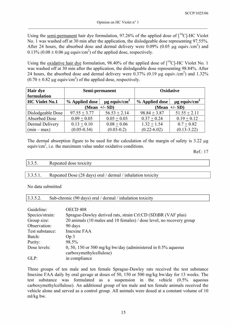
Using the semi-permanent hair dye formulation, 97.26% of the applied dose of [14C]-HC Violet No. 1 was washed off at 30 min after the application, the dislodgeable dose representing 97.55%. After 24 hours, the absorbed dose and dermal delivery were 0.09% (0.05 µg equiv./cm2) and 0.13% (0.08 ± 0.06 µg equiv/cm2) of the applied dose, respectively. Using the oxidative hair dye formulation, 98.40% of the applied dose of [14C]-HC Violet No. 1 was washed off at 30 min after the application, the dislodgeable dose representing 98.84%. After 24 hours, the absorbed dose and dermal delivery were 0.37% (0.19 µg equiv./cm2) and 1.32% (0.70 ± 0.82 µg equiv/cm2) of the applied dose, respectively. Hair dye formulation
Semi-permanent Oxidative
HC Violet No.1 % Applied dose µg equiv/cm2 % Applied dose µg equiv/cm2 (Mean +/- SD) (Mean +/- SD) Dislodgeable Dose 97.55 + 3.77 56.53 + 2.14 98.84 + 3.87 51.55 + 2.11 Absorbed Dose 0.09 + 0.05 0.05 + 0.03 0.37 + 0.24 0.19 + 0.12 Dermal Delivery (min – max)
0.13 + 0.10 (0.05-0.34)
0.08 + 0.06 (0.03-0.2)
1.32 + 1.54 (0.22-6.02)
0.7 + 0.82 (0.13-3.22)
The dermal absorption figure to be used for the calculation of the margin of safety is 3.22 µg equiv/cm2, i.e. the maximum value under oxidative conditions.
Ref.: 17 3.3.5. Repeated dose toxicity 3.3.5.1. Repeated Dose (28 days) oral / dermal / inhalation toxicity No data submitted 3.3.5.2. Sub-chronic (90 days) oral / dermal / inhalation toxicity Guideline: OECD 408 Species/strain: Sprague-Dawley derived rats, strain Crl:CD (SD)BR (VAF plus) Group size: 20 animals (10 males and 10 females) / dose level, no recovery group Observation: 90 days Test substance: Imexine FAA Batch: Op 3 Purity: 98.5% Dose levels: 0, 50, 150 or 500 mg/kg bw/day (administered in 0.5% aqueous
carboxymethylcellulose) GLP: in compliance Three groups of ten male and ten female Sprague-Dawley rats received the test substance Imexine FAA daily by oral gavage at doses of 50, 150 or 500 mg/kg bw/day for 13 weeks. The test substance was formulated as a suspension in the vehicle (0.5% aqueous carboxymethylcellulose). An additional group of ten male and ten female animals received the vehicle alone and served as a control group. All animals were dosed at a constant volume of 10 ml/kg bw.

SCCP/1025/06
Opinion on HC Violet n° 1 ____________________________________________________________________________________________
16
The animals were observed daily for clinical signs and mortality. Food consumption and individual bodyweights were recorded weekly. Both eyes of all animals were examined before the start of the treatment and of control and high-dose animals during week 13 (ophthalmoscopy). Blood and urine samples were taken from all animals during weeks 4 and 13 and examined for haematology, clinical-chemistry and urinalysis parameters. At the end of the treatment period, all animals were killed and subjected to necropsy (external examination and macroscopic examination of the cranial, thoracic and visceral cavities). The weights of selected organs were recorded, and histopathological examinations were carried out on organs and tissues from control and high dose animals, all gross lesions from all animals and specified tissues from animals that had died during the study. Results Two animals (one male of the high dose group and one female of the mid dose group) died during the first week of the study. The cause of death could not be established. Purple fur and tail staining were noted in animals of the treatment groups from the first week onward. Hair loss and scab were noted in both control and treated animals from day 48 onward (in males only in the treatment groups, in females more frequently in the control group). There were no relevant ocular changes or abnormalities. Females of the low-, mid- and high- dose groups showed slight reductions in bodyweight gain compared with the controls (9%, 8% and 12%, respectively). Since the terminal mean bodyweights were within 10% of the control value the author of the study report did not consider this as a treatment-related effect. Food consumption was comparable in all groups (within 97.4% and 105.8% of the controls). In the high-dose females food consumption was slightly (3.6%) reduced. There were several statistically significant (p < 0.05) differences in haematology parameters compared with the controls (week 4: PCV in low- and high-dose males, MCHC in males from all treatment groups and mid-dose females, WBC in high-dose females; week 13: MCHC in low- and high-dose females, lymphocytes in low-dose males and monocytes in high-dose males). These differences were not considered toxicologically relevant since they showed no consistent pattern (not dose-related, observed at one time point only) and all values were within the historical control ranges. There were several significant differences (p<0.05) in clinical-chemistry parameters compared with the controls. In weeks 4 and 13 females of the high-dose group showed an increase in ALP activity (there was a trend in the low- and mid-dose groups also). In males of all treatment groups ALP activities were decreased in week 4 (there was a trend also in week 13). In males of the mid- and high-dose groups there were dose related increases in ALT activities in week 13 (trends are notable also regarding AST activities in males and ALT and AST activities in females of the mid- and high-dose groups). Apart from ALT and AST activities in male animals of the mid- and high-dose groups in week 13, which were caused by one animal in each of these groups, all values were within the historical control ranges. Other statistically significant differences (BUN, glucose, total protein, albumin, bilirubin, Ca, Phosphate, K, Na, Cl) were not considered toxicologically relevant since they showed no consistent pattern (not dose-related, observed at one time point only) and all values were within the historical control ranges. Most of the urine analyses could not be carried out in the higher dose groups due to the dark purple discolouration of the urine caused by the test compound. According to the author, the analyses performed showed no relevant effects. A summarising table showing the statistically analysed data, however, was not presented (solely individual data were provided). Compared with the controls, there was a dose-related increase in mean absolute and relative liver weights in male animals of the treatment groups, which was significant (p<0.05, <0.01 or

SCCP/1025/06
Opinion on HC Violet n° 1 ____________________________________________________________________________________________
17
<0.001) in the mid- and high-dose groups (absolute 4%, 15% and 29% and relative 4%, 12% and 32%, respectively). In females there was a slight but significant (4%) decrease in absolute liver weights in the low-dose group and a dose-related increase in the mid- and high-dose groups (not significant, 9% and 7%, respectively). Relative liver weights were slightly decreased in the low-dose group (5%) and showed a dose-related and significant increase in the mid- and high-dose groups (12% and 16%, respectively). According to the author, the values were at the upper end of the normal ranges for animals of this strain and age, the respective historical control data, however, were not provided. High-dosed males showed a slight increase (6%) in relative kidney weights which was not statistically significant. Absolute kidney weights were slightly but not significantly increased in males of the mid- and high-dose groups. Female animals of the treatment groups showed a significant and dose-related increase in relative kidney weights (7%, 11% and 18%, respectively). Absolute kidney weights in the mid- and high-dose groups were also increased but without statistical significance (9%). Females of all treatment groups showed dose-related but not significant increases in ovary weights (absolute 10%, 16%, 19% and relative 19%, 21% and 29%). According to the author, relative ovary weights were normal for rats of this age and strain. Relative thyroid weights were significantly increased in high-dose females (22%). An increase in low-dose females (12%) was not significant. Increases in absolute thyroid weights in the low- and high-dose groups (6% and 12%, respectively) were not significant. In the macroscopic examinations purple staining of the skin and tail was noted in animals from all treated groups. Histopathological findings in organs and tissues including the kidney, pituitary gland and ovaries were comparable in the control and treatment groups. According to the author, the findings were within the normal ranges of background alterations seen in untreated rats of this age and strain. The only notable effect was that livers of male animals in the high-dose group showed moderate congestion (8 versus 0 animals out of 10). Conclusion The author concluded that the no-observed toxic effect level in this study was 50 mg/kg/day.
Ref.: 9 Comment The SCCP considered the dose of 50 mg/kg bw/d as LOAEL due to the change in the kidney weight in this group. 3.3.5.3. Chronic (> 12 months) toxicity No data submitted
SCCP/1025/06
Opinion on HC Violet n° 1 ____________________________________________________________________________________________
18
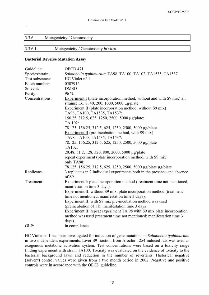
3.3.6. Mutagenicity / Genotoxicity 3.3.6.1 Mutagenicity / Genotoxicity in vitro Bacterial Reverse Mutation Assay Guideline: OECD 471 Species/strain: Salmonella typhimurium TA98, TA100, TA102, TA1535, TA1537 Test substance: HC Violet n° 1 Batch number: 0507912 Solvent: DMSO Purity: 96 % Concentrations: Experiment I (plate incorporation method, without and with S9 mix) all
strains: 1.6, 8, 40, 200, 1000, 5000 µg/plate Experiment II (plate incorporation method, without S9 mix) TA98, TA100, TA1535, TA1537: 156.25, 312.5, 625, 1250, 2500, 5000 µg/plate; TA 102: 78.125, 156.25, 312.5, 625, 1250, 2500, 5000 µg/plate Experiment II (pre-incubation method, with S9 mix) TA98, TA100, TA1535, TA1537: 78.125, 156.25, 312.5, 625, 1250, 2500, 5000 µg/plate TA102: 20.48, 51.2, 128, 320, 800, 2000, 5000 µg/plate repeat experiment (plate incorporation method, with S9 mix): only TA98: 78.125, 156.25, 312.5, 625, 1250, 2500, 5000 µg/plate µg/plate Replicates: 3 replicates in 2 individual experiments both in the presence and absence
of S9. Treatment: Experiment I: plate incorporation method (treatment time not mentioned;
manifestation time 3 days). Experiment II: without S9 mix, plate incorporation method (treatment time not mentioned; manifestation time 3 days).
Experiment II: with S9 mix pre-incubation method was used (preincubation of 1 h; manifestation time 3 days).
Experiment II: repeat experiment TA 98 with S9 mix plate incorporation method was used (treatment time not mentioned; manifestation time 3 days).
GLP: in compliance HC Violet n° 1 has been investigated for induction of gene mutations in Salmonella typhimurium in two independent experiments. Liver S9 fraction from Aroclor 1254-induced rats was used as exogenous metabolic activation system. Test concentrations were based on a toxicity range finding experiment with strain TA100. Toxicity was evaluated on the evidence of toxicity to the bacterial background lawn and reduction in the number of revertants. Historical negative (solvent) control values were given from a two month period in 2002. Negative and positive controls were in accordance with the OECD guideline.

SCCP/1025/06
Opinion on HC Violet n° 1 ____________________________________________________________________________________________
19
Results Evidence of toxicity was not demonstrated in the toxicity range finding study. In the main experiment toxic effects occurred at concentrations of 1000 µg/plate and above in TA102 (without and with S9 mix, both methods), in TA1537 at 5000 µg/plate (plate incorporation method) and in TA100 and TA1537 (with S9 mix, pre-incubation method). Slight thinning of the background bacterial lawn also was observed in TA98 and TA100 at the highest tested concentration of 5000 µg/plate especially in Experiment II with S9 mix. In TA98 a twofold increase of the revertant numbers occurred at 1000 and 625 µg/plate, respectively in two independent experiments at the plate incorporation method with S9 mix. In a third repeat experiment with S9 mix using the pre-incubation method a slight increase at 1250 µg/plate and higher was observed. Since this latter increase showed at least some evidence of a dose relationship and although these increases were weak, they may be sufficient to be considered as biologically relevant. Additional statistical significant findings with TA100 and TA 102 which were reached at intermediate doses, showed no dose response relationship and were not reproducible, were consequently considered as not biologically relevant. Conclusion The study had some deficiencies (positive controls of TA102, choice of the test concentrations). Under the experimental conditions HC Violet n° 1 may be slightly mutagenic in this gene mutation tests in strain TA98.
Ref.: 10 In vitro Mammalian Cell Gene Mutation Test (hprt locus) Guideline: OECD 476 Cells: L5178Y tk+/- mouse lymphoma cells Test substance: HC Violet n° 1 Batch number: 0507912 Purity: 96 % Solvent: DMSO Concentrations: Experiment I: 0 - 800 µg/ml without S9 mix
0 - 1000 µg/ml with S9 mix Experiment II: 0 - 900 µg/ml without S9 mix 0 - 1000 µg/ml with S9 mix
Treatment Both experiments: 3 h both without and with S9 mix; expression period 7 days.
Replicates: duplicate cultures in two independent experiments GLP: in compliance HC Violet n° 1 has been investigated for induction of gene mutations at the hprt locus (6-thioguanine resistance) in L5178Y mouse lymphoma cells using a fluctuation protocol. Test concentrations for treatment were based on the level of toxicity measured as relative survival in a range-finding experiment. The highest doses for evaluating, that means for survival plating and 6-thioguanine resistance were the lowest precipitating doses at the end of the treatment period (800 and 900 µg/ml respectively, without S9 mix and 1000 µg/ml with S9 mix). Liver S9 fraction from Aroclor 1254-induced rats was used as exogenous metabolic activation system. In the main test, cells were treated for 3 h followed by an expression period of 7 days to fix the DNA damage into a stable hprt mutation. Toxicity was measured as relative survival of the

SCCP/1025/06
Opinion on HC Violet n° 1 ____________________________________________________________________________________________
20
treated cultures relative to the relative survival of the solvent control cultures after treatment. Negative and positive controls were in accordance with the OECD guideline. Results In both experiments the required relative survival of 10 - 20% was not reached. After 3 h treatment precipitation was observed at the higher doses. The highest doses evaluated were the lowest precipitating doses at the end of the treatment period. No increases in the mutant frequencies were observed up to the evaluated concentrations in both experiments without and with metabolic activation. Conclusion Under the experimental conditions used, HC Violet n° 1 did not lead to an increase in the mutation frequency at the hprt locus of mouse lymphoma cells.
Ref.: 12 In vitro Micronucleus Test Guideline: In accordance with the recommendations of the "International Workshop
on Genotoxicity Testing (IWGT)" Cells: Peripheral blood lymphocytes from 2 healthy non smoking female donors Test substance: HC Violet n° 1 Batch number: 0507912 Purity: 96 % Solvent: DMSO Concentrations: Experiment 1: 283.5, 354.3, 442.9 and 692.1 µg/ml without S9 mix
354.3, 442.9, 865.1 and 1690 µg/ml with S9 mix Experiment 2: 442.9, 553.6, 865.0 and 1352 µg/ml without S9 mix
1475, 1634 and 1720 µg/ml with S9 mix Treatment: Experiment 1: 24 h PHA, 20 h treatment and 28 h recovery without S9 mix 24 h PHA, 3 h treatment and 45 h recovery with S9 mix Experiment 2: 48 h PHA, 20 h treatment and 28 h recovery without S9 mix 48 h PHA, 3 h treatment and 45 h recovery with S9 mix Replicates: duplicate cultures in two independent experiments GLP: in compliance HC Violet n° 1 has been investigated for induction of micronuclei in PHA stimulated human lymphocytes. Test concentrations for treatment were based on the level of toxicity measured as decreased replication index in each experiment. The highest concentrations chosen for analysis induced approximately 60 % reduction in replication index. Treatment periods were 20 h without S9 and 3 h with S9. Harvest times were 48 h after the beginning of treatment. The final 28 h of incubation was in the presence of cytochalasin B (at a final concentration of 6 µg/ml). Liver S9 fraction from Aroclor 1254-induced rats was used as exogenous metabolic activation system. Negative and positive controls were in accordance with the IWGT recommendations.
SCCP/1025/06
Opinion on HC Violet n° 1 ____________________________________________________________________________________________
21
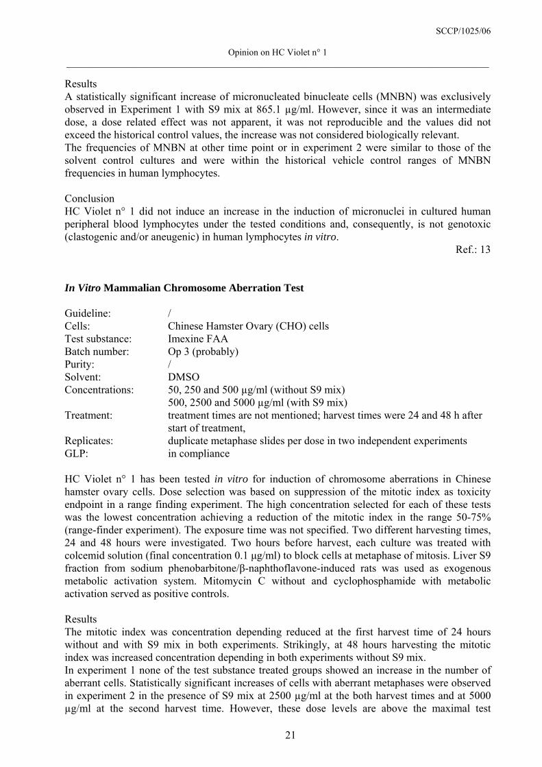
Results A statistically significant increase of micronucleated binucleate cells (MNBN) was exclusively observed in Experiment 1 with S9 mix at 865.1 µg/ml. However, since it was an intermediate dose, a dose related effect was not apparent, it was not reproducible and the values did not exceed the historical control values, the increase was not considered biologically relevant. The frequencies of MNBN at other time point or in experiment 2 were similar to those of the solvent control cultures and were within the historical vehicle control ranges of MNBN frequencies in human lymphocytes. Conclusion HC Violet n° 1 did not induce an increase in the induction of micronuclei in cultured human peripheral blood lymphocytes under the tested conditions and, consequently, is not genotoxic (clastogenic and/or aneugenic) in human lymphocytes in vitro.
Ref.: 13 In Vitro Mammalian Chromosome Aberration Test Guideline: / Cells: Chinese Hamster Ovary (CHO) cells Test substance: Imexine FAA Batch number: Op 3 (probably) Purity: / Solvent: DMSO Concentrations: 50, 250 and 500 µg/ml (without S9 mix) 500, 2500 and 5000 µg/ml (with S9 mix) Treatment: treatment times are not mentioned; harvest times were 24 and 48 h after
start of treatment, Replicates: duplicate metaphase slides per dose in two independent experiments GLP: in compliance HC Violet n° 1 has been tested in vitro for induction of chromosome aberrations in Chinese hamster ovary cells. Dose selection was based on suppression of the mitotic index as toxicity endpoint in a range finding experiment. The high concentration selected for each of these tests was the lowest concentration achieving a reduction of the mitotic index in the range 50-75% (range-finder experiment). The exposure time was not specified. Two different harvesting times, 24 and 48 hours were investigated. Two hours before harvest, each culture was treated with colcemid solution (final concentration 0.1 µg/ml) to block cells at metaphase of mitosis. Liver S9 fraction from sodium phenobarbitone/β-naphthoflavone-induced rats was used as exogenous metabolic activation system. Mitomycin C without and cyclophosphamide with metabolic activation served as positive controls. Results The mitotic index was concentration depending reduced at the first harvest time of 24 hours without and with S9 mix in both experiments. Strikingly, at 48 hours harvesting the mitotic index was increased concentration depending in both experiments without S9 mix. In experiment 1 none of the test substance treated groups showed an increase in the number of aberrant cells. Statistically significant increases of cells with aberrant metaphases were observed in experiment 2 in the presence of S9 mix at 2500 µg/ml at the both harvest times and at 5000 µg/ml at the second harvest time. However, these dose levels are above the maximal test

SCCP/1025/06
Opinion on HC Violet n° 1 ____________________________________________________________________________________________
22
concentrations of the current guidelines (10 mM = 2110 µg/ml). The maximal test concentration was chosen considering that at higher concentrations toxicity may cause the genotoxic effect. Yet, in the present experiments, high toxicity, measured as a reduction in MI, at the positive dose levels was not observed. Not statistically significant increases were found in experiment 2 without S9 mix at dose levels below the maximal test dose. Strikingly, in experiment 1 the background level of polyploid cells is rather high whereas occasionally it is even increased after treatment. The same was observed in experiment 2 at the second harvest time. Conclusion Although the test was performed with GLP compliance, it has several deficiencies. The test is not performed according an OECD guideline, the purity of the test compound is not given; exposure times are not mentioned, the CHO cells suffer from a high level of polyploidy cells, historical background levels are not reported. These short comings reduce the value of this test and, therefore, it can only be used as supportive. Under the conditions of this study it can not be excluded that HC Violet n° 1 may induce chromosomal aberrations or aneuploidy in CHO cells.
Ref.: 11 3.3.6.2 Mutagenicity / Genotoxicity in vivo Mammalian Erythrocyte Micronucleus Test, mice Guideline: EU B12 Species/strain: CD-1 mice Group size: 5 mice/sex/dose Test substance: Imexine FAA Batch number: OPT 8 Purity: 99.9 % Route: gavage, single application, as suspension Vehicle: aqueous carboxymethylcellulose 0.5% Dose levels: 500, 1000, 2000 mg/kg Sacrifice time: 24 hours (all doses) and 48 hours (2000 mg/kg only) after treatment GLP: in compliance HC Violet n° 1 has been investigated for induction of micronuclei in the bone marrow cells of mice. Test concentrations were based on the results of a preliminary study of the maximum non lethal dose; 2000 mg/kg bw was selected as the highest dose-level which showed analysable bone marrow slides and no toxicity signs or mortality. In the main experiment mice were exposed to a single oral administration (gavage) of 0, 500, 1000 and 2000 mg/kg bw. 24 h or 48 h (highest dose only) after dosing bone marrow cells were collected. The study did not include a vehicle control group for the treatment of 48 hours. 1000 polychromatic erythrocytes (PCE) per animal were analysed for the frequency of micronuclei. Cytotoxicity was assessed by scoring the ratio between normochromatic (NCE) and polychromatic erythrocytes (NCE/PCE). Negative and positive controls were in accordance with the EU guideline.
SCCP/1025/06
Opinion on HC Violet n° 1 ____________________________________________________________________________________________
23
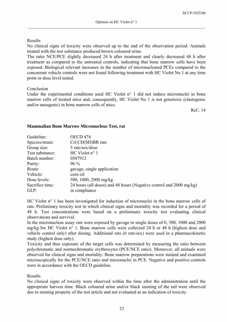
Results No clinical signs of toxicity were observed up to the end of the observation period. Animals treated with the test substance produced brown coloured urine. The ratio NCE/PCE slightly decreased 24 h after treatment and clearly decreased 48 h after treatment as compared to the untreated controls, indicating that bone marrow cells have been exposed. Biological relevant increases in the number of micronucleated PCEs compared to the concurrent vehicle controls were not found following treatment with HC Violet No 1 at any time point or dose level tested. Conclusion Under the experimental conditions used HC Violet n° 1 did not induce micronuclei in bone marrow cells of treated mice and, consequently, HC Violet No 1 is not genotoxic (clastogenic and/or aneugenic) in bone marrow cells of mice.
Ref.: 14 Mammalian Bone Marrow Micronucleus Test, rat Guideline: OECD 474 Species/strain: Crl:CD(SD)BR rats Group size: 5 rats/sex/dose Test substance: HC Violet n° 1 Batch number: 0507912 Purity: 96 % Route: gavage, single application Vehicle: corn oil Dose levels: 500, 1000, 2000 mg/kg Sacrifice time: 24 hours (all doses) and 48 hours (Negative control and 2000 mg/kg) GLP: in compliance HC Violet n° 1 has been investigated for induction of micronuclei in the bone marrow cells of rats. Preliminary toxicity test in which clinical signs and mortality was recorded for a period of 48 h. Test concentrations were based on a preliminary toxicity test evaluating clinical observations and survival. In the micronucleus assay rats were exposed by gavage to single doses of 0, 500, 1000 and 2000 mg/kg bw HC Violet n° 1. Bone marrow cells were collected 24 h or 48 h (highest dose and vehicle control only) after dosing. Additional rats (6 rats/sex) were used in a pharmacokinetic study (highest dose only). Toxicity and thus exposure of the target cells was determined by measuring the ratio between polychromatic and normochromatic erythrocytes (PCE/NCE ratio). Moreover, all animals were observed for clinical signs and mortality. Bone marrow preparations were stained and examined microscopically for the PCE/NCE ratio and micronuclei in PCE. Negative and positive controls were in accordance with the OECD guideline. Results No clinical signs of toxicity were observed within the time after the administration until the appropriate harvest time. Black coloured urine and/or black staining of the tail were observed due to staining property of the test article and not evaluated as an indication of toxicity.

SCCP/1025/06
Opinion on HC Violet n° 1 ____________________________________________________________________________________________
24
The ratio of PCE/NCE was statistical significantly decreased in females treated with 1000 mg/kg bw at 24 h and with 2000 mg/kg bw at 48 hours. The ratio was not changed in males. The results of the toxicokinetics study demonstrated elevated plasma levels of HC Violet No 1 (B 066) in treated rats indicating to systemic exposure with HC Violet No 1 (B 066) after oral administration. The mean micronucleated PCE frequencies in the bone marrow were not significantly increased in any of the groups treated with the test substance. Conclusion HC Violet n° 1 did not induce micronuclei in bone marrow cells of treated rats after oral treatment under the test conditions used and, consequently, was not genotoxic (clastogenic and/or aneugenic) in bone marrow cells of rats.
Ref.: 15 3.3.7. Carcinogenicity No data submitted 3.3.8. Reproductive toxicity 3.3.8.1. Two generation reproduction toxicity No data submitted 3.3.8.2. Teratogenicity Guideline: / Species/strain: Sprague-Dawley derived rats, strain Crl:CD(SD)BR (VAF plus) Group size: 24 mated female animals / dose level Observation: 20 days Test substance: Imexine FAA Batch: Op 3 Purity: 98.5% Dose levels: 0, 50, 200 or 800 mg/kg bw/day (administered by gavage in 0.5% aqueous
carboxymethylcellulose) from days 6 to 15 of pregnancy GLP: in compliance Although it was not mentioned in the study report, the study was to a large extent carried out in accordance with OECD Guideline 414. Three groups of 24 mated female Sprague-Dawley rats received the test substance by gavage at doses of 50, 200 or 800 mg/kg bw/day from day 6 through day 15 of gestation. The test substance was formulated as a suspension in the vehicle (0.5% aqueous carboxymethylcellulose). An additional group of 24 mated female animals received the vehicle only and served as a control group. A standard dose volume of 10 ml/kg bw was given. The day of observation of sperm positive smear was designated as day 0 of pregnancy. The animals were checked daily for clinical signs. Bodyweights and food consumption were recorded at designated intervals during pregnancy. On day 20 of pregnancy, the females were killed and major maternal organs were examined macroscopically. Foetuses were removed by Caesarean section. The following litter parameters

SCCP/1025/06
Opinion on HC Violet n° 1 ____________________________________________________________________________________________
25
were recorded: number of corpora lutea and implantation sites, number and distribution of early and late resorptions and number and distribution of dead and live foetuses. Live foetuses were weighed, sexed and subjected to external, soft tissue and skeletal examinations. Results There were no premature deaths during the study. Treatment-related clinical observations were limited to a dose-related red colouring of the urine, tail, fur, limbs and body in the treated groups. At 800 mg/kg bw/day food consumption was reduced in comparison with the control group throughout the dosing period, the difference being statistically significant over days 6 to 12 of pregnancy. Bodyweight gains were also reduced during the dosing period, with statistical significance over days 6 to 12. There were no statistically significant differences in bodyweights although these were also generally lower than in the controls. Maternal necropsy revealed dose-related pink/purple staining of tail, fur, limbs and body but no other treatment-related internal abnormalities. There were no statistically significant differences between the treatment groups and the control group regarding pregnancy data, i.e. numbers of corporae luteae, implantations and live foetuses, pre- and post-implantation losses and sex ratio. The visceral and skeletal examinations of the foetuses revealed no toxicologically relevant differences in the incidences of malformations or variations for any of the treatment groups compared with the control group. Conclusion Oral administration of Imexine FAA to pregnant rats during organogenesis induced maternal toxicity at 800 mg/kg bw/day, characterised by a reduction of bodyweight gain and food consumption throughout the dosing period. At 200 and 50 mg/kg bw/day, there was no evidence of maternal toxicity. Red coloured urine and red staining of the tail and fur was observed in all treated groups. There was no evidence of embryolethality, embryonic growth retardation or teratogenicity at any of the dose levels administered. The NOAEL of embryo/foetotoxicity is 800 mg/kg bw/day. The NOAEL of maternal toxicity is 200 mg/kg bw/day.
Ref.: 16 3.3.9. Toxicokinetics No data submitted 3.3.10. Photo-induced toxicity 3.3.10.1. Phototoxicity / photoirritation and photosensitisation No data submitted 3.3.10.2. Phototoxicity / photomutagenicity / photoclastogenicity No data submitted

SCCP/1025/06
Opinion on HC Violet n° 1 ____________________________________________________________________________________________
26
3.3.11. Human data No data submitted 3.3.12. Special investigations No data submitted 3.3.13. Safety evaluation (including calculation of the MoS)
CALCULATION OF THE MARGIN OF SAFETY
(HC Violet n° 1)
(Oxidative/semi-permanent) Maximum absorption through the skin A (µg/cm2) = 3.22 µg/cm² Skin Area surface SAS (cm2) = 700 cm2 Dermal absorption per treatment SAS x A x 0.001 = 2.25 mg Typical body weight of human = 60 kg Systemic exposure dose (SED) SAS x A x 0.001/60 = 0.04 mg/kg Lowest observed adverse effect level (mg/kg), oral, rat, sub-chronic LOAEL = 50 mg/kg Margin of Safety LOAEL / SED = 1331 3.3.14. Discussion Physico-chemical properties HC Violet n° 1 is a secondary amine, and thus it is prone to nitrosation. The nitrosamine content in HC Violet n° 1 is not reported. No data are available on the stability in hair dye formulations. General toxicity In an acute oral toxicity study in rats the maximum non lethal dose of the test item was >2000 mg/kg bw. In an oral subchronic toxicity in rats, the LOAEL is considered being at 50 mg/kg bw/d due to the change in kidney weight in this group. Oral administration of HC Violet n° 1 to pregnant rats during organogenesis induced maternal toxicity at 800 mg/kg bw/day. The NOAEL of maternal toxicity is 200 mg/kg bw/day. There was no evidence of embryolethality, embryonic growth retardation or teratogenicity at any of the dose levels administered, the NOAEL of embryotoxicity is 800 mg/kg bw/d.

SCCP/1025/06
Opinion on HC Violet n° 1 ____________________________________________________________________________________________
27
Irritation / sensitisation The substance has some irritant potential to the rabbit skin and showed transient irritation to the rabbit eyes. On the basis of the EC3 value in the second experiment of the local lymph node assay HC Violet n° 1 should be considered as a strong sensitiser. Percutaneous absorption The dermal absorption figure to be used for the calculation of the margin of safety is 3.22 µg equiv./cm2, i.e. the maximum value under oxidative conditions. The margin of safety was calculated on the basis of LOAEL of 50 mg/kg bw/d of subchronic toxicity as being 1331. Mutagenicity HC Violet n° 1 was slightly mutagenic in a single bacterial strain of the Ames test, but induced no mutations at the hprt locus of L5178Y mouse lymphoma cells. It was non-clastogenic in cultured CHO cells, in an in vitro micronucleus test and in two in vivo micronucleus tests up to doses of 2000 mg/kg/day by oral route. Although HC Violet n° 1 showed some mutagenic activity in vitro in the Ames test, this activity was observed only in a single experiment and strain (TA98 strain with S9) and was relatively weak. However, this activity was not confirmed in another in vitro test on the gene mutation endpoint (hprt test with mouse lymphoma cells). All other tests yielded negative results. 4. CONCLUSION Based on the information provided, the SCCP is of the opinion that the use of HC Violet n° 1 itself as a semipermanent hair dye at a maximum concentration of 0.28 % and as an oxidative hair dye at a maximum concentration of 0.25% in the finished cosmetic product (after mixing with hydrogen peroxide) does not pose a risk to the health of the consumer, apart from its sensitising potential. HC Violet n° 1 is a secondary amine, and thus is prone to nitrosation. It should not be used in combination with nitrosating substances. The nitrosamine content should be < 50 ppb. Studies on genotoxicity/mutagenicity in finished hair dye formulations should be undertaken following the relevant SCCNFP/SCCP opinions and in accordance with the Notes of Guidance 5. MINORITY OPINION Not applicable

SCCP/1025/06
Opinion on HC Violet n° 1 ____________________________________________________________________________________________
28
6. REFERENCES References in italics were not submitted as full reports in the present dossier. They consist of report for stability and homogeneity of dosage forms study (16), reports of preliminary toxicity studies (17, 18), reports for studies considered inadequate (19-24), and can be provided upon request. 1. R.L. Guest. Imexine FAA Lot OP2: Acute Oral Toxicity (Limit Test) in the Rat.
Safepharm Laboratories Limited, Project n° 109/407, 1990. 2. B. Griffon. Acute Oral Toxicity in Rats (“Fixed Dose Method”). CIT; Study No.
26920TAR, 2004 3. R.L. Guest. Imexine FAA Op 3: Primary eye irritation study in the rabbit. Safepharm;
Project N° 109/268R, 1994 4. B. Griffon. Acute Eye Irritation in Rabbits. CIT; Study No. 26921TAL, 2004 5. J. Clouzeau. Acute skin irritation study in the rabbit. CIT, Study Number: 7996TAL, 1991 6. R.L. Guest. Imexine FAA Op 3: Primary skin irritation study in the rabbit. Project
Number: 109/272R. Safepharm Laboratories Limited, 1994 7. R.L. Guest. Imexine FAA lot Op2. Magnusson & Kligman maximisation study in the
guinea pig. Safepharm Laboratories; Project Number 109/406; 1990 8. B. Griffon. Evaluation of Skin Sensitization Potential in Mice Using the Local Lymph
Node Assay (LLNA). CIT Study No. 26922 TSS, 2004 9. S.A. Brownlie. IMEXINE FAA. 13 week oral toxicity study in the rat. Toxicol
Laboratories Limited, Report N° LRL/21/93, 1994 10. M. Ballantyne. HC Violet N°1 (B066): Reverse Mutation in five Histidine-requiring
strains of Salmonella typhimurium. Covance Laboratories Ltd. Study Number: 413/64; 2004
11. M.D. Kelly. Imexine FAA-Metaphase Analysis of Chinese Hamster Ovary (CHO) Cells. Toxicol Laboratories Limited. Report N° M/CCA/38504; 1994
12. M. Lloyd. HC Violet N°1 (B066): Mutation at the hprt Locus of L5178Y Mouse Lymphoma Cells using the MicrotitreR Fluctuation Technique. Covance Laboratories Ltd ; Study N° 413/65, 2004
13. J. Whitwell. HC Violet N°1 (B066): Induction of micronuclei in cultured human peripheral blood lymphocytes. Covance Laboratories Ltd; Study N° 413/66, 2004
14. R. Mollà. Micronucleus Test. Test substance: Imexine FAA. Centro de Investigacion y Desarrollo Aplicado; Report N° CD-96/4902T, 1996
15. G. L. Erexson. HC Violet N°1 (B066): In vivo Rat Micronucleus Assay. Covance Laboratories Inc. Study N° 6182-108, 2004
16. L. Chandler. Imexine FAA: Oral (Gavage) Rat Developmental Toxicity Study. Toxicol Laboratories Limited; Report Ref. LRL/22/93, 1994
17. C.S. Roper. The In vitro Percutaneous Absorption of Radiolabelled HC Violet N°1 Through Human Skin. INVERESK Project N° 205348 – INVERESK Report N° 23871, 2004
18. R. Groult. Validation of the Analytical Method and Determination of Homogeneity and Stability of Dosage Forms. CIT Study No. 26923 AHS, 2004
19. S.A. Brownlie. IMEXINE FAA. 14 day oral (gavage) toxicity study in the rat. Toxicol Laboratories Limited, Report N° LRL/19/93, 1993
20. L. Chandler and L. Irvine. Imexine FAA: Oral (Gavage) Rat Developmental Toxicity Dose Ranging Study. Toxicol Laboratories Limited; Report Ref. LRL/20/93, 1993

SCCP/1025/06
Opinion on HC Violet n° 1 ____________________________________________________________________________________________
29
21. R. Forster. Reverse Mutation in Salmonella Typhimurium. Test Substance: C-5185-P. LSR-RTC Report N° 088038-M-04484, 1984
22. K.G. Dossou. Test du Micronoyau sur Moelle Osseuse de Souris traitée in vivo par voie Intrapéritonéale. Produit testé : B66 (1-amino-3-methyl-4-(2-hydroxyethyl)-amino-6-nitro-benzene). Département des Contrôles Biologiques de la Recherche Fondamentale de L’OREAL. Report N° CS/JC/93/004; 1993
23. "Pénétration du Colorant Imexine FAA à travers l'Epiderme Humain monté sur Cellules de Diffusion type Franz". L’OREAL - Chimie Analytique -Contrôles Biologiques et Méthodes Alternatives. Reports N°92/09/28A, 92/09/28B, 92/10/7A and 92/10/07B; 1992
24. "Pénétration in vitro du colorant Imexine FAA, étudié en présence d'eau oxygénée, à travers l'épiderme humain isolé monté sur cellules de diffusion type Franz". L'OREAL - Laboratoires Absorption Transcutanée - Reports N° 94/05/30 - 94/05/31 and 94/06/96 ; 1994
7. ACKNOWLEDGEMENTS Members of the working group are acknowledged for their valuable contribution to this opinion. The members of the working group are: Dr. C. Chambers Prof. R. Dubakiene Prof. V. Kapoulas Prof. C. Lidén Prof. J.-P. Marty Prof. T. Platzek (Chairman) Dr. S.C. Rastogi Prof. T. Sanner Dr. J. van Engelen Dr. I.R. White External experts Dr. A. Pöting, BfR, Germany (Rapporteur) Dr. H. Norppa, Institute for Occupational Health, Finland Dr. J. van Benthem, RIVM, the Netherlands