science china life sciences - springer · sispa, random pcr amplification, displacement...
TRANSCRIPT

SCIENCE CHINA Life Sciences
© The Author(s) 2013. This article is published with open access at Springerlink.com life.scichina.com www.springer.com/scp
*Corresponding author (email: [email protected])
SPECIAL TOPIC: Haunted with and hunting for viruses August 2013 Vol.56 No.8: 688–696
• REVIEW • doi: 10.1007/s11427-013-4516-y
New strategy for virus discovery: viruses identified in human feces in the last decade
XIE GuangCheng, YU JieMei & DUAN ZhaoJun*
National Institute of Viral Disease Control and Prevention, Chinese Center for Disease Control and Prevention, Beijing 102206, China
Received May 27, 2013; accepted June 16, 2013
Emerging and re-emerging viruses continue to surface all over the world. Some of these viruses have the potential for rapid and global spread with high morbidity and mortality, such as the SARS coronavirus outbreak. It is extremely urgent and im-portant to identify a novel virus near-instantaneously to develop an active preventive and/or control strategy. As a culture- independent approach, viral metagenomics has been widely used to investigate highly divergent and completely new viruses in humans, animals, and even environmental samples in the past decade. A new model of Koch’s postulates, named the meta-genomic Koch’s postulates, has provided guidance for the study of the pathogenicity of novel viruses. This review explains the viral metagenomics strategy for virus discovery and describes viruses discovered in human feces in the past 10 years using this approach. This review also addresses issues related to the metagenomic Koch’s postulates and the challenges for virus discov-ery in the future.
next-generation sequencing, novel viruses, metagenomic Koch’s postulates, viral metagenomics
Citation: Xie G C, Yu J M, Duan Z J. New strategy for virus discovery: viruses identified in human feces in the last decade. Sci China Life Sci, 2013, 56: 688–696, doi: 10.1007/s11427-013-4516-y
In the past decade, several serious emerging viral infectious diseases have left deep impression on many people. The world witnessed the first pandemic of the new millennium in 2003. Instead of the influenza virus, severe acute respira-tory syndrome coronavirus (SARS-CoV) caused the first serious and widespread zoonotic disease, having a huge global impact on health, travel, and the economy [1–5]. The great global impact of the SARS outbreak was in some ways intensified by the delay in identifying the causative agent of the disease. Within a few months, SARS-CoV caused approximately 916 deaths and affected around 30 countries. When the novel SARS-CoV was isolated by a joint effort of the World Health Organization (WHO) SARS Collaborative Network, six months had passed. Hence, identifying the causative agent of a new epidemic is one of the most important steps for active control and prevention
of viral disease outbreaks. With the development of novel modern molecular bio-
logy technology, especially sequence-independent single- primer amplification (SISPA) [6], next-generation sequenc-ing (NGS) such as 454-pyrosequencing [7,8], and viral metagenomics [9–11], multiple viruses can be detected simultaneously, and novel and highly divergent viruses can be discovered and genetically characterized quickly. A nov-el phlebovirus of the Bunyaviridae family, known as severe fever with thrombocytopenia syndrome bunyavirus, was discovered using SISPA technology [12,13]. Human bo-cavirus was also identified using the above-mentioned methods in 2005 [14]. Recently, a novel coronavirus, HCoV-EMC, was isolated in Jeddah, Saudi Arabia on June 13, 2012 [15]. Subsequently, the complete genome of MERS-hCoV was determined using an unbiased virus dis-covery approach involving NGS techniques, which deter-

Xie G C, et al. Sci China Life Sci August (2013) Vol.56 No.8 689
mined that it was closely related to bat coronavirus but was distant from SARS-CoV [16]. This novel coronavirus, MERS-hCoV, reminds us to pay attention to animal coro-naviruses, which may be the cause of severe disease in hu-mans, and to develop strategies to rapidly determine un-known viral agents [17]. In fact, only limited data are available regarding the diversity of viruses present in hu-mans and animals. A great number of human and animal viruses are still unknown.
1 Traditional methods of virus discovery
Before the advent of modern molecular methods, traditional virus discovery included filtration, tissue culture, electron microscopy, serology, and vaccination; cell culture for virus propagation was the virus discovery gold standard. Howev-er, many viruses cannot be easily propagated in cell culture, thus limiting our understanding of virology. Two milestone findings solved these difficulties, which allowed those in-terested in understanding new viruses first to amplify and then to sequence viral nucleic acids, namely DNA amplifi-cation by polymerase chain reaction (PCR) [18] and DNA sequencing with chain-terminating inhibitors (Sanger se-quencing or first-generation sequencing) [19]. The number of sequences deposited on GenBank between August 2009 and August 2010 has totaled 970 million and 43 million bases of viral and phage origin, respectively, representing an annual growth of 20%–24% [20]. After that, several se-rious emerging viruses, such as Hendravirus [21], Nipah virus [22], Menangle virus [23], Melaka virus [24], and Reston Ebola virus [25], were discovered in different coun-tries. The trend in virology research has shown gradual sub-stitution of the traditional virus discovery methods with novel modern molecular biology technology. Nevertheless, tradi-tional methods to isolate, identify, and characterize viruses play complementary roles in the virus discovery effort.
2 Novel modern molecular techniques of virus discovery
There are two major types of molecular methods for virus discovery, including sequence-dependent and sequence- independent methods. Sequence-dependent methods require knowledge of the nucleic acid sequence. Such methods in-clude consensus primer PCR and microarrays. Several vi-ruses, such as human immunodeficiency virus [26], simian retroviruses [27–29], and hepatitis E virus [30], have been identified using consensus sequences of known viruses; however, these methods have little or no value for charac-terizing completely novel viruses. Microarrays use probes that can hybridize to known viral sequences and potentially novel viruses with sufficient sequence similarity [31]. Mi-croarrays have been applied in the detection of swine res-
piratory viruses in clinical samples [32] and for dengue vi-rus [33]. Unlike sequence-dependent methods, the sequence- independent approaches do not rely on any knowledge of the virus. SISPA circumvents the viral load limitation of suppression subtractive hybridization (SSH). By now, there are several variations to the original protocol created by Reyes et al. [6]. The main strategy of SISPA is to exploit the sensitivity and the specificity of PCR amplification us-ing primers that bind oligonucleotide fragments ligated to any putative viral DNA material in the sample. SISPA has been modified to allow the detection of both DNA and RNA viruses after the removal of genomic and contaminating nucleic acids [34]. Human astrovirus [35] and parvoviruses 2 and 3 [36] were discovered using SISPA methods. Another sequence-independent and culture-independent approach, viral metagenomics, provides superior capability for detect-ing known and unknown viruses and will be described in detail below.
3 Viral metagenomics
The term ‘metagenome’ was proposed earlier by Handels-man in PubMed, in 1998, in relation to classifying uncul-turable bacteria from soil samples [37]. Metagenomics can be defined as the characterization of genetic information directly from samples. It is a culture- and sequence-inde- pendent approach that does not rely on the presence of any particular gene in all subject entities [7,10]. This approach was originally developed as a tool for ‘functional and se-quence-based analysis of collective microbial genomes contained in environmental samples’ [38,39]. The first ap-plication of metagenomics to the field of virology was in the analysis of viral communities sampled at two near-shore marine locations in San Diego, California [40]. Since then, viruses in numerous environments, including freshwater, marine sediment, soil, and the human gut, were surveyed. More information on published environmental viral meta-genomes, DNA viral families, and RNA viral families in environmental samples and organisms found using the viral metagenomic approach can be found in Rosario’s review [9]. The availability of NGS has forced viral metagenomics to be evaluated by unbiased means at previously unforeseen resolution and is providing a wealth of new opportunities in two major areas: viral candidate pathogen discovery and viral ecology [7].
Currently, several commercially available high-throughput sequencing platforms exist that vary by way of their se-quencing principle, sequencing speed, expense, and read length. Figure 1 shows a schematic flow diagram summa-rizing the viral metagenomic studies that used sequence- independent amplification and high-throughput sequencing for virus discovery in 2012. A metagenomic analysis essen-tially entails the following steps: sample preparation, se-quence-independent amplification, high-throughput sequenc-

690 Xie G C, et al. Sci China Life Sci August (2013) Vol.56 No.8
Figure 1 Overview of viral metagenomic studies in 2012. Viruses discovered through viral metagenomic studies from 2002 to 2011 have been described in detail by Mokili et al. [10].
Figure 2 Schematic flow diagram of a typical viral metagenomic study.
ing, and bioinformatics analysis (Figure 2). Below, we briefly discuss each step of this viral metagenomic process. More detailed descriptions can be obtained in a previously published paper [41].
(i) Sample preparation. Theoretically, any type of sam-ple can be analyzed using the viral metagenomic approach; however, viral genomes are relatively short and of low con-centration in many samples, and bacterial and eukaryotic nucleic acids can interfere with the isolation and detection of viral DNA or RNA. Thus, one of the most important, yet difficult, tasks of viral metagenomics is removal of non-viral nucleic acid while preserving viral nucleic acids [11,41,42]. Toward this aim, several measures have been taken, such as SYBR-gold staining, filtration, sucrose or cesium chloride density centrifugation [41], DNase diges-tion [43], and chloroform treatment [44]. Total RNA con-tent is usually estimated by measuring ribosomal RNA (rRNA), because the abundance of rRNA and its association with ribosomes makes the detection of contaminating RNA viruses difficult. However, RNases are ineffective for deg-radation of rRNA. Therefore, other strategies must be em-ployed. One rRNA depletion strategy is to use random pri-
mers during cDNA synthesis that do not target rRNA se-quences [45,46]. Another strategy is to use biotin-labeled probes that target ribosomal sequences for depletion [47–49].
(ii) Sequence-independent amplification. Sequence- independent amplification is the important step in viral metagenomics that shows the true genetic composition of the sample and simultaneously amplifies several viral ge-nomes and makes them available, including highly diver-gent and completely novel viruses, for characterization [11,34]. SISPA, random PCR amplification, displacement amplification, arbitrarily primed PCR, and rolling circle amplification are typically used for sequence-independent amplification. Detailed descriptions of these methods have been made available in previously published papers [11,50].
(iii) High-throughput sequencing. To identify known, highly divergent, or new viruses, sequencing is often uti-lized. The Sanger sequencing method can create high-quality sequence reads up to nearly 1000 nt; however, this highly laborious process limits its usage [43]. High-throughput sequencing, or NGS, is used more and more often in viral metagenomics. The 454 sequencing platform (Roche diag-

Xie G C, et al. Sci China Life Sci August (2013) Vol.56 No.8 691
nostics) [8,51], SOLiD sequencing (Life Technologies) [7,52], Illumina sequencing [7,52], Helicos sequencing (Helicos Biosciences), PacBio sequencing (Pacific Biosci-ences), and Ion Torrent (Life Technologies) [7,52] are all commercial instruments in the marketplace, and all use slightly different methodologies to achieve clonal amplifi-cation and sequencing. The advantages and disadvantages of each of these instruments have been detailed in other papers [7,5254].
(iv) Bioinformatics. Post-sequencing, the analysis of the vast amount of sequencing data produced, is the chal-lenging part of viral metagenomics. The datasets from met-agenomic studies are complicated, and they typically con-tain a mixture of different species. The genomes in the da-tasets are usually incomplete, with some cases having only a small number of short fragments belonging to each genome. There are two approaches that can be used for analysis of read data, de novo assembly using software such as Velvet [55] and mapping strategies using a mapper such as BWA [56]. Other programs and platforms also have been devel-oped, the details of which can be found in the NGS review by Blomstrom et al. [50].
4 Viruses discovered in human feces
Feces comprise a community of many different microor-ganisms, including bacteria and viruses. Many viral diseases are transmitted by the oral-fecal route, and the viral agents are excreted in the feces. With the development of modern molecular methods and viral metagenomics, several new viruses have been discovered in feces in the past decade.
(i) Picornaviruses. Enteroviruses constitute the largest genus within the family Picornaviridae, which have non- enveloped, positive-sense, single-stranded RNA (+ssRNA) genomes surrounded by four structural proteins, VP1-VP4. A majority of these viruses infect humans and cause diseas-es ranging from minor respiratory illness to severe neuro-logical disorders like meningitis, encephalitis, and poliomy-elitis [57]. Metagenomic analyses of viruses in stool sam-ples from children with acute flaccid paralysis (AFP) found numerous viruses, particularly HEV-C, including one from a potentially novel enterovirus genotype [58].
Human cosavirus (HCoSV) is a proposed new genus in the family Picornaviridae originally identified in 2008 in feces [59]. Since then, HCoSVs have been detected in feces from Australian [60] and Chinese [61] children. HCoSV infection with accompanying diarrhea in Thailand [62], the prevalence of a new species (F) of HCoSV, and another 26 new HCoSV genotypes in human feces of healthy children and children with AFP [63] were also reported. HCoSV pathogenicity in humans has remained unknown because detection rates in patients and healthy controls were similar in the only available cohort studies of patients with AFP in Asia [59] and with gastroenteritis in China [61]. In Brazil,
the 3.6% detection rate in children with gastroenteritis [64] was comparable to the 1.8% rate in a cohort study of gas-troenteritis in China [61]. The large number of HCoSV genotypes/serotypes reported indicates that HCoSV infec-tion is associated with a range of diseases, such as unex-plained diarrhea, AFP, or others. Future work will focus on defining whether HCoSV is a true human pathogen.
Two other novel picornaviruses have also been described since 2008 due to the advent of metagenomics: klassevirus [65] and salivirus [66]. The reported association between salivirus shedding and diarrhea indicates that such infec-tions may account for a significant fraction of the unex-plained cases of diarrhea occurring worldwide. Future stud-ies to determine a possible link to disease in humans and any unique characteristics of the viral life cycle will be re-quired. Viral culture in human cell lines, especially those from the gastrointestinal tract, could be suggestive that the virus is competent to replicate in human cells and that hu-mans could be a bona fide host of klassevirus [65]. Further epidemiological screening and serological assays will be necessary to understand the diversity within this possible genus, the prevalence of these novel viruses, and the age range of those susceptible to infection.
(ii) Parvoviruses. Parvoviruses are, as their name sug-gests, small viruses, with a single-stranded DNA genome and are widespread pathogens that cause a wide range of diseases in humans and animals. Allander et al. [14] first reported the discovery of a previously unidentified human parvovirus in 2005. From 2009 to 2010, the bocavirus genus was expanded to include three additional species of human bocaviruses, HBoV2 [67], HBoV3 [68], and HBoV4 [69]. HBoV2-4 seems to be found primarily in human stool [67–71]. HBoV1 is predominantly a respiratory pathogen, whereas HBoV2 and possibly HBoV3 are associated with gastroenteritis [67,71]. A variety of signs and symptoms have been described in patients with HBoV infection, in-cluding rhinitis, pharyngitis, cough, dyspnea, and diarrhea, among others [72].
Human parvovirus 4 (PARV4) was identified in 2005 in a plasma sample [73]. Another two novel parvoviruses closely related to PARV4, porcine hokovirus and bovine hokovirus, were discovered in feces specimens from Hong Kong [74]. Furthermore, a proposed new Parvoviridae genus associated with acute diarrhea has been identified; however, wider geographic sampling of human and animal fecal samples will provide a better understanding of the genetic diversity of this proposed genus. Serological assays and case- control studies will also help determine whether members of this viral clade are associated with diarrhea or other symp-toms [75].
(iii) Astroviruses. The family Astroviridae consists of small, non-lipid enveloped, single-stranded, positive-sense RNA viruses with genomes that contain three open reading frames (ORFs), designated ORF1a, ORF1b, and ORF2. Astroviruses are known to infect a variety of human and

692 Xie G C, et al. Sci China Life Sci August (2013) Vol.56 No.8
animal hosts. Recently, two highly divergent members of the astrovirus family, MLB1 [76–78] and VA1 [79], were identified and associated with human viral diarrhea. Astro-viruses VA2, MLB2, and VA3 were also reported as novel astroviruses in human stool [80], as was HMOAstV A-C [81]. With the rising number of human astrovirus species detected in infections associated with unexplained AFP and/or diarrhea, increased understanding of the genetic di-versity within viral families infecting humans will assist in future studies of their pathogenicity and allow the design of specific diagnostic assays [81].
(iv) Polyomaviruses. Viruses in the Polyomaviridae family typically possess ~5000 bp of circular, double- stranded DNA. The genome can be divided into three parts, including the regulatory region, the early region, and the later region. Over the past five years, seven novel polyoma-viruses have been discovered in humans, namely KI poly-omavirus (KIPyV) [82], WU polyomavirus (WUPyV) [83], Merkel cell polyomavirus (MCPyV) [84], human polyoma-virus 6 (HPyV-6), human polyomavirus 7 (HPyV-7) [85], human polyomavirus 9 (HPyV-9) [86], and trichodysplasia spinulosa-associated polyomavirus (TSPyV) [87]. In 2012, another two novel polyomaviruses were discovered in hu-man stool and provisionally named MW polyomavirus (MWPyV) [88] and MX polyomavirus (MXPyV) [89]. The number of polyomaviruses found in the human body con-tinues to grow, raising the question of how many more spe-cies have yet to be identified and what roles they play in humans with and without manifest disease. Taking MWPyV as an example, one critical question is whether MWPyV is a bona fide infectious agent of humans and, if so, which dis-ease(s), if any, might be associated with MWPyV infection. The detection of MWPyV in feces of children with diarrhea, many of which are unexplained, raises the possibility that MWPyV might play a role in human diarrhea. It is also pos-sible that MWPyV is a dietary contaminant and does not actively infect humans. Serological studies and antibody- based immune responses to MWPyV and wider sampling and screening are approaches to determine whether MWPyV is a true infectious agent [88].
(v) Circoviruses. Members of the family Circoviridae are non-enveloped, spherical viruses with a single-stranded circular DNA genome of approximately 2 kb––the smallest known autonomously replicating viral genome [90]––and include the genera circovirus, gyrovirus, and gyclovirus (proposed) [91,92]. Until recently, chicken anemia virus (CAV) was the only known representative of the gyrovirus genus. In the last two years, three novel gyroviruses have been reported: avian gyrovirus 2 (AGV2) in chickens [93], a human gyrovirus (HGV) detected on human skin [94], and gyrovirus 3 (GyV3) in human feces and chicken meat [95]. Recently, a novel gyrovirus 4 (GyV4) was also identified in human stool and in chicken meat prepared for human con-sumption [96]. In addition, a new strain of CAV, designated GD-1-12, was isolated from fecal samples of a 12-day-old
commercial broiler in Guangdong province, China [97]. Multiple diverse circoviruses were found in human and chimpanzee feces, such as CyCV-PK5006 and Chimp 17 [92]. Other novel single-stranded, circular DNA viruses also detected were found in porcine [98] and bovine feces [99], respectively.
Diarrhea is the third leading infectious cause of death worldwide, and an estimated 1.4 billion nonfatal episodes occur yearly [100,101]. Importantly, it is estimated that 40% of diarrhea cases are of unknown etiology [60,102]. Viral metagenomics has largely propelled the progress in virus discovery; however, the association of these newly- discovered viruses with specific diseases is largely un-known. An understanding of these newly-discovered viruses’ disease associations and causations is extremely urgent.
5 Metagenomic Koch’s postulates
Viral metagenomics has provided a powerful tool for dis-covering new viruses. However, virus discovery is only the first step to determining the etiology behind a disease. The detection of nucleic acid is not sufficient to prove causality. For that, more comprehensive studies have to be performed [103]. Evidence for causality is not always conclusive, even when the suspect virus is found “at the scene of the crime”. This means that finding a virus in a sample from a patient with an illness of unknown etiology, and even demonstrat-ing an association, does not always prove causation. There-fore, strict guidelines proposed by Robert Koch and later modified by Rivers [104] have been used to assign causality to infectious agents [10]. One of Koch’s postulates requires that the candidate etiological agent be isolated from a dis-eased organism and grown in pure culture. However, not all viruses can be propagated by current culture methods [103].
In many cases, Koch’s postulates will not be satisfied if current culture standards are used to prove causality. Koch’s postulates have since been modified to acknowledge molec-ular methods used to monitor the role played by genes in bacterial virulence [105]. The revised so-called molecular Koch’s postulates can be applied to pathogenic members of a genus or pathogenic strains of a species as well as non-pathogenic strains. Because genes can be expressed at dif-ferent time points during infection, however, new molecular methods do not always distinctively characterize virulence genes and make a clear association with the disease of un-known etiology. Mokili et al. [10] proposed the so-called metagenomic Koch’s postulates, which focus on the identi-fication of metagenomic traits in disease subjects (Figure 3). Molecular markers such as sequence reads, assembled con-tigs, genes, or full genomes that can uniquely distinguish disease-associated metagenomes from those obtained from matched healthy control subjects can all be used to define metagenomic traits. Thus, satisfying metagenomic Koch’s

Xie G C, et al. Sci China Life Sci August (2013) Vol.56 No.8 693
Figure 3 Flow chart of virus discovery, pathogenicity determination, intervention, and metagenomic Koch’s postulates. Traits A, D, E, and J found in the diseased animal (e.g., a mouse) are not present in the healthy control (represented in blue). The acquisition or increase of new metagenomic traits, A, P, and E, are represented in orange. Inoculation of the suspected purified traits into a healthy mouse will induce disease if the traits encompass the etiology of the disease (represented in purple); pathogenicity as a trait is represented in red. The figure is modified from models proposed by Mokili et al. [10] and Delwart [106].
Figure 4 The global aviation network. Lines show direct links between airports, and the color of the line indicates passenger capacity in people per day (thousands (red), hundreds (yellow), and tens (blue)) [110].
postulates is possible when one or multiple viral agents are involved in disease causation.
6 Challenges in virus discovery
Novel viruses are still emerging. In November 2011, a novel orthobunyavirus named Schmallenberg virus (SBV) was detected in plasma samples from a farm near the German town of Schmallenberg by using a metagenomic approach
with NGS [107]; this novel virus is on the rise in Europe [108,109]. The SBV epidemic showed once again that the novel technology of metagenomics is very useful for early detection of novel pathogens in livestock. In fact, veterinary diagnostics in Europe has proved to be a very effective network of institutions studying epizootic diseases. Before any strategy is decided for prediction, precaution, and pre-vention of EIDs in humans, three questions should always be asked and answered: what to expect, what to be prepared for, and what to do.
694 Xie G C, et al. Sci China Life Sci August (2013) Vol.56 No.8
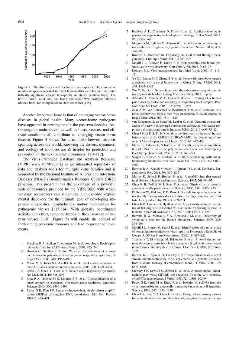
Figure 5 The discovery curve for human virus species. The cumulative number of species reported to infect humans (black circles and line). Sta-tistically significant upward breakpoints are shown (vertical lines). The best-fit curve (solid line) and lower and upper 95% posterior intervals (dashed lines) for extrapolation to 2020 are shown [115].
Another important issue is that of emerging vector-borne diseases in global health. Many vector-borne pathogens have appeared in new regions in the past two decades. An-thropogenic trade, travel, as well as hosts, vectors, and cli-mate conditions all contribute to emerging vector-borne disease. Figure 4 shows the direct links between airports spanning across the world. Knowing the drivers, dynamics, and ecology of zoonoses are all helpful for prediction and prevention of the next pandemic zoonosis [110–112].
The Virus Pathogen Database and Analysis Resource (ViPR; www.ViPRbrc.org) is an integrated repository of data and analysis tools for multiple virus families and is supported by the National Institute of Allergy and Infectious Diseases (NIAID) Bioinformatics Resource Centers (BRC) program. This program has the advantage of a powerful suite of resources provided by the ViPR BRC with which virology researchers can streamline and expedite experi-mental discovery for the ultimate goal of developing im-proved diagnostics, prophylactics, and/or therapeutics for pathogenic viruses [113,114]. With globally coordinated activity and effort, temporal trends in the discovery of hu-man viruses [115] (Figure 5) will enable the control of forthcoming pandemic zoonoses and lead to greater achieve-ments.
1 Fouchier R A, Kuiken T, Schutten M, et al. Aetiology: Koch’s pos-tulates fulfilled for SARS virus. Nature, 2003, 423: 240
2 Drosten C, Gunther S, Preiser W, et al. Identification of a novel coronavirus in patients with severe acute respiratory syndrome. N Engl J Med, 2003, 348: 1967–1976
3 Marra M A, Jones S J, Astell C R, et al. The Genome sequence of the SARS-associated coronavirus. Science, 2003, 300: 1399–1404
4 Peiris J S, Guan Y, Yuen K Y. Severe acute respiratory syndrome. Nat Med, 2004, 10: S88–S97
5 Rota P A, Oberste M S, Monroe S S, et al. Characterization of a novel coronavirus associated with severe acute respiratory syndrome. Science, 2003, 300: 1394–1399
6 Reyes G R, Kim J P. Sequence-independent, single-primer amplifi-cation (SISPA) of complex DNA populations. Mol Cell Probes, 1991, 5: 473–481
7 Radford A D, Chapman D, Dixon L, et al. Application of next- generation sequencing technologies in virology. J Gen Virol, 2012, 93: 1853–1868
8 Margulies M, Egholm M, Altman W E, et al. Genome sequencing in microfabricated high-density picolitre reactors. Nature, 2005, 437: 376–380
9 Rosario K, Breitbart M. Exploring the viral world through meta-genomics. Curr Opin Virol, 2011, 1: 289–297
10 Mokili J L, Rohwer F, Dutilh B E. Metagenomics and future per-spectives in virus discovery. Curr Opin Virol, 2012, 2: 63–77
11 Delwart E L. Viral metagenomics. Rev Med Virol, 2007, 17: 115– 131
12 Yu X J, Liang M F, Zhang S Y, et al. Fever with thrombocytopenia associated with a novel bunyavirus in China. N Engl J Med, 2011, 364: 1523–1532
13 Wu Y, Gao G F. Severe fever with thrombocytopenia syndrome vi-rus expands its borders. Emerg Microbes Infect, 2013, in press
14 Allander T, Tammi M T, Eriksson M, et al. Cloning of a human parvovirus by molecular screening of respiratory tract samples. Proc Natl Acad Sci USA, 2005, 102: 12891–12896
15 Zaki A M, van Boheemen S, Bestebroer T M, et al. Isolation of a novel coronavirus from a man with pneumonia in Saudi Arabia. N Engl J Med, 2012, 367: 1814–1820
16 van Boheemen S, de Graaf M, Lauber C, et al. Genomic characteri-zation of a newly discovered coronavirus associated with acute res-piratory distress syndrome in humans. MBio, 2012, 3: e00473–12
17 Chan J F, Li K S, To K K, et al. Is the discovery of the novel human betacoronavirus 2c EMC/2012 (HCoV-EMC) the beginning of an-other SARS-like pandemic? J Infect, 2012, 65: 477–489
18 Mullis K, Faloona F, Scharf S, et al. Specific enzymatic amplifica-tion of DNA in vitro: the polymerase chain reaction. Cold Spring Harb Symp Quant Biol, 1986, 51(Pt 1): 263–273
19 Sanger F, Nicklen S, Coulson A R. DNA sequencing with chain- terminating inhibitors. Proc Natl Acad Sci USA, 1977, 74: 5463– 5467
20 Benson D A, Karsch-Mizrachi I, Lipman D J, et al. GenBank. Nu-cleic Acids Res, 2011, 39: D32–D37
21 Murray K, Selleck P, Hooper P, et al. A morbillivirus that caused fatal disease in horses and humans. Science, 1995, 268: 94–97
22 Chua K B, Bellini W J, Rota P A, et al. Nipah virus: a recently emergent deadly paramyxovirus. Science, 2000, 288: 1432–1435
23 Philbey A W, Kirkland P D, Ross A D, et al. An apparently new vi-rus (family Paramyxoviridae) infectious for pigs, humans, and fruit bats. Emerg Infect Dis, 1998, 4: 269–271
24 Chua K B, Crameri G, Hyatt A, et al. A previously unknown reovi-rus of bat origin is associated with an acute respiratory disease in humans. Proc Natl Acad Sci USA, 2007, 104: 11424–11429
25 Barrette R W, Metwally S A, Rowland J M, et al. Discovery of swine as a host for the Reston ebolavirus. Science, 2009, 325: 204–206
26 Mokili J L, Rogers M, Carr J K, et al. Identification of a novel clade of human immunodeficiency virus type 1 in Democratic Republic of Congo. AIDS Res Hum Retroviruses, 2002, 18: 817–823
27 Takemura T, Ekwalanga M, Bikandou B, et al. A novel simian im-munodeficiency virus from black mangabey (Lophocebus aterrimus) in the Democratic Republic of Congo. J Gen Virol, 2005, 86: 1967– 1971
28 Barlow K L, Ajao A O, Clewley J P. Characterization of a novel simian immunodeficiency virus (SIVmonNG1) genome sequence from a mona monkey (Cercopithecus mona). J Virol, 2003, 77: 6879–6888
29 Clewley J P, Lewis J C, Brown D W, et al. A novel simian immu-nodeficiency virus (SIVdrl) pol sequence from the drill monkey, Mandrillus leucophaeus. J Virol, 1998, 72: 10305–10309
30 Reyes G R, Purdy M A, Kim J P, et al. Isolation of a cDNA from the virus responsible for enterically transmitted non-A, non-B hepatitis. Science, 1990, 247: 1335–1339
31 Chou C C, Lee T T, Chen C H, et al. Design of microarray probes for virus identification and detection of emerging viruses at the ge-

Xie G C, et al. Sci China Life Sci August (2013) Vol.56 No.8 695
nus level. BMC Bioinformatics, 2006, 7: 232 32 Nicholson T L, Kukielka D, Vincent A L, et al. Utility of a panviral
microarray for detection of swine respiratory viruses in clinical sam-ples. J Clin Microbiol, 2011, 49: 1542–1548
33 Yozwiak N L, Skewes-Cox P, Stenglein M D, et al. Virus identifica-tion in unknown tropical febrile illness cases using deep sequencing. PLoS Negl Trop Dis, 2012, 6: e1485
34 Bexfield N, Kellam P. Metagenomics and the molecular identifica-tion of novel viruses. Vet J, 2011, 190: 191–198
35 Finkbeiner S R, Li Y, Ruone S, et al. Identification of a novel astro-virus (astrovirus VA1) associated with an outbreak of acute gastro-enteritis. J Virol, 2009, 83: 10836–10839
36 Allander T, Emerson S U, Engle R E, et al. A virus discovery meth-od incorporating DNase treatment and its application to the identifi-cation of two bovine parvovirus species. Proc Natl Acad Sci USA, 2001, 98: 11609–11614
37 Handelsman J, Rondon M R, Brady S F, et al. Molecular biological access to the chemistry of unknown soil microbes: a new frontier for natural products. Chem Biol, 1998, 5: R245–R249
38 Riesenfeld C S, Schloss P D, Handelsman J. Metagenomics: ge-nomic analysis of microbial communities. Annu Rev Genet, 2004, 38: 525–552
39 Schloss P D, Handelsman J. Biotechnological prospects from meta-genomics. Curr Opin Biotechnol, 2003, 14: 303–310
40 Breitbart M, Salamon P, Andresen B, et al. Genomic analysis of un-cultured marine viral communities. Proc Natl Acad Sci USA, 2002, 99: 14250–14255
41 Thurber R V, Haynes M, Breitbart M, et al. Laboratory procedures to generate viral metagenomes. Nat Protoc, 2009, 4: 470–483
42 Edwards R A, Rodriguez-Brito B, Wegley L, et al. Using pyrose-quencing to shed light on deep mine microbial ecology. BMC Ge-nomics, 2006, 7: 57
43 Breitbart M, Rohwer F. Method for discovering novel DNA viruses in blood using viral particle selection and shotgun sequencing. Bio-techniques, 2005, 39: 729–736
44 Willner D, Furlan M, Schmieder R, et al. Metagenomic detection of phage-encoded platelet-binding factors in the human oral cavity. Proc Natl Acad Sci USA, 2011, 108(Suppl 1): 4547–4553
45 Endoh D, Mizutani T, Kirisawa R, et al. Species-independent detec-tion of RNA virus by representational difference analysis using non-ribosomal hexanucleotides for reverse transcription. Nucleic Acids Res, 2005, 33: e65
46 Armour C D, Castle J C, Chen R, et al. Digital transcriptome profil-ing using selective hexamer priming for cDNA synthesis. Nat Methods, 2009, 6: 647–649
47 He S, Wurtzel O, Singh K, et al. Validation of two ribosomal RNA removal methods for microbial metatranscriptomics. Nat Methods, 2010, 7: 807–812
48 Stewart F J, Ottesen E A, DeLong E F. Development and quantita-tive analyses of a universal rRNA-subtraction protocol for microbial metatranscriptomics. ISME J, 2010, 4: 896–907
49 Chen Z, Duan X. Ribosomal RNA depletion for massively parallel bacterial RNA-sequencing applications. Methods Mol Biol, 2011, 733: 93–103
50 Blomstrom A L. Viral metagenomics as an emerging and powerful tool in veterinary medicine. Vet Q, 2011, 31: 107–114
51 Gilles A, Meglecz E, Pech N, et al. Accuracy and quality assessment of 454 GS-FLX Titanium pyrosequencing. BMC Genomics, 2011, 12: 245
52 Loman N J, Misra R V, Dallman T J, et al. Performance comparison of benchtop high-throughput sequencing platforms. Nat Biotechnol, 2012, 30: 434–439
53 Quail M A, Smith M, Coupland P, et al. A tale of three next genera-tion sequencing platforms: comparison of Ion Torrent, Pacific Bio-sciences and Illumina MiSeq sequencers. BMC Genomics, 2012, 13: 341
54 Liu L, Li Y, Li S, et al. Comparison of next-generation sequencing systems. J Biomed Biotechnol, 2012, 2012: 251364
55 Zerbino D R, Birney E. Velvet: algorithms for de novo short read
assembly using de Bruijn graphs. Genome Res, 2008, 18: 821–829 56 Li H, Durbin R. Fast and accurate short read alignment with Bur-
rows-Wheeler transform. Bioinformatics, 2009, 25: 1754–1760 57 Berlin L E, Rorabaugh M L, Heldrich F, et al. Aseptic meningitis in
infants <2 years of age: diagnosis and etiology. J Infect Dis, 1993, 168: 888–892
58 Victoria J G, Kapoor A, Li L, et al. Metagenomic analyses of viruses in stool samples from children with acute flaccid paralysis. J Virol, 2009, 83: 4642–4651
59 Kapoor A, Victoria J, Simmonds P, et al. A highly prevalent and genetically diversified Picornaviridae genus in South Asian children. Proc Natl Acad Sci USA, 2008, 105: 20482–20487
60 Holtz L R, Finkbeiner S R, Kirkwood C D, et al. Identification of a novel picornavirus related to cosaviruses in a child with acute diar-rhea. Virol J, 2008, 5: 159
61 Dai X Q, Hua X G, Shan T L, et al. Human cosavirus infections in children in China. J Clin Virol, 2010, 48: 228–229
62 Khamrin P, Chaimongkol N, Malasao R, et al. Detection and molec-ular characterization of cosavirus in adults with diarrhea, Thailand. Virus Genes, 2012, 44: 244–246
63 Kapusinszky B, Phan T G, Kapoor A, et al. Genetic diversity of the genus Cosavirus in the family Picornaviridae: a new species, recom-bination, and 26 new genotypes. PLoS ONE, 2012, 7: e36685
64 Stocker A, Souza B F, Ribeiro T C, et al. Cosavirus infection in persons with and without gastroenteritis, Brazil. Emerg Infect Dis, 2012, 18: 656–659
65 Greninger A L, Runckel C, Chiu C Y, et al. The complete genome of klassevirus––a novel picornavirus in pediatric stool. Virol J, 2009, 6: 82
66 Li L, Victoria J, Kapoor A, et al. A novel picornavirus associated with gastroenteritis. J Virol, 2009, 83: 12002–12006
67 Arthur J L, Higgins G D, Davidson G P, et al. A novel bocavirus associated with acute gastroenteritis in Australian children. PLoS Pathog, 2009, 5: e1000391
68 Kapoor A, Slikas E, Simmonds P, et al. A newly identified bo-cavirus species in human stool. J Infect Dis, 2009, 199: 196–200
69 Kapoor A, Simmonds P, Slikas E, et al. Human bocaviruses are highly diverse, dispersed, recombination prone, and prevalent in en-teric infections. J Infect Dis, 2010, 201: 1633–1643
70 Chieochansin T, Kapoor A, Delwart E, et al. Absence of detectable replication of human bocavirus species 2 in respiratory tract. Emerg Infect Dis, 2009, 15: 1503–1505
71 Chow B D, Ou Z, Esper F P. Newly recognized bocaviruses (HBoV, HBoV2) in children and adults with gastrointestinal illness in the United States. J Clin Virol, 2010, 47: 143–147
72 Jartti T, Hedman K, Jartti L, et al. Human bocavirus––the first 5 years. Rev Med Virol, 2012, 22: 46–64
73 Jones M S, Kapoor A, Lukashov V V, et al. New DNA viruses iden-tified in patients with acute viral infection syndrome. J Virol, 2005, 79: 8230–8236
74 Lau S K, Woo P C, Tse H, et al. Identification of novel porcine and bovine parvoviruses closely related to human parvovirus 4. J Gen Virol, 2008, 89: 1840–1848
75 Phan T G, Vo N P, Bonkoungou I J, et al. Acute diarrhea in West African children: diverse enteric viruses and a novel parvovirus ge-nus. J Virol, 2012, 86: 11024–11030
76 Finkbeiner S R, Allred A F, Tarr P I, et al. Metagenomic analysis of human diarrhea: viral detection and discovery. PLoS Pathog, 2008, 4: e1000011
77 Finkbeiner S R, Kirkwood C D, Wang D. Complete genome se-quence of a highly divergent astrovirus isolated from a child with acute diarrhea. Virol J, 2008, 5: 117
78 Finkbeiner S R, Le B M, Holtz L R, et al. Detection of newly de-scribed astrovirus MLB1 in stool samples from children. Emerg In-fect Dis, 2009, 15: 441–444
79 Finkbeiner S R, Li Y, Ruone S, et al. Identification of a novel astro-virus (astrovirus VA1) associated with an outbreak of acute gastro-enteritis. J Virol, 2009, 83: 10836–10839
80 Finkbeiner S R, Holtz L R, Jiang Y, et al. Human stool contains a

696 Xie G C, et al. Sci China Life Sci August (2013) Vol.56 No.8
previously unrecognized diversity of novel astroviruses. Virol J, 2009, 6: 161
81 Kapoor A, Li L, Victoria J, et al. Multiple novel astrovirus species in human stool. J Gen Virol, 2009, 90: 2965–2972
82 Allander T, Andreasson K, Gupta S, et al. Identification of a third human polyomavirus. J Virol, 2007, 81: 4130–4136
83 Gaynor A M, Nissen M D, Whiley D M, et al. Identification of a novel polyomavirus from patients with acute respiratory tract infec-tions. PLoS Pathog, 2007, 3: e64
84 Feng H, Shuda M, Chang Y, et al. Clonal integration of a polyoma-virus in human Merkel cell carcinoma. Science, 2008, 319: 1096–1100
85 Schowalter R M, Pastrana D V, Pumphrey K A, et al. Merkel cell polyomavirus and two previously unknown polyomaviruses are chronically shed from human skin. Cell Host Microbe, 2010, 7: 509–515
86 Scuda N, Hofmann J, Calvignac-Spencer S, et al. A novel human polyomavirus closely related to the african green monkey-derived lymphotropic polyomavirus. J Virol, 2011, 85: 4586–4590
87 van der Meijden E, Janssens R W, Lauber C, et al. Discovery of a new human polyomavirus associated with trichodysplasia spinulosa in an immunocompromized patient. PLoS Pathog, 2010, 6: e1001024
88 Siebrasse E A, Reyes A, Lim E S, et al. Identification of MW poly-omavirus, a novel polyomavirus in human stool. J Virol, 2012, 86: 10321–10326
89 Yu G, Greninger A L, Isa P, et al. Discovery of a novel polyoma-virus in acute diarrheal samples from children. PLoS ONE, 2012, 7: e49449
90 Fringuelli E, Scott A N, Beckett A, et al. Diagnosis of duck circovirus infections by conventional and real-time polymerase chain reaction tests. Avian Pathol, 2005, 34: 495–500
91 Blinkova O, Victoria J, Li Y, et al. Novel circular DNA viruses in stool samples of wild-living chimpanzees. J Gen Virol, 2010, 91: 74–86
92 Li L, Kapoor A, Slikas B, et al. Multiple diverse circoviruses infect farm animals and are commonly found in human and chimpanzee feces. J Virol, 2010, 84: 1674–1682
93 Rijsewijk F A, Dos S H, Teixeira T F, et al. Discovery of a genome of a distant relative of chicken anemia virus reveals a new member of the genus Gyrovirus. Arch Virol, 2011, 156: 1097–1100
94 Sauvage V, Cheval J, Foulongne V, et al. Identification of the first human gyrovirus, a virus related to chicken anemia virus. J Virol, 2011, 85: 7948–7950
95 Phan T G, Li L, O'Ryan M G, et al. A third gyrovirus species in hu-man faeces. J Gen Virol, 2012, 93: 1356–1361
96 Chu D K, Poon L L, Chiu S S, et al. Characterization of a novel gy-rovirus in human stool and chicken meat. J Clin Virol, 2012, 55: 209–213
97 Zhang X, Xie Q, Ji J, et al. Complete genome sequence analysis of a recent chicken anemia virus isolate and comparison with a chicken
anemia virus isolate from human fecal samples in China. J Virol, 2012, 86: 10896–10897
98 Sikorski A, Arguello-Astorga G R, Dayaram A, et al. Discovery of a novel circular single-stranded DNA virus from porcine faeces. Arch Virol, 2013, 158: 283–289
99 Kim H K, Park S J, Nguyen V G, et al. Identification of a novel sin-gle-stranded, circular DNA virus from bovine stool. J Gen Virol, 2012, 93: 635–639
100 Kosek M, Bern C, Guerrant R L. The global burden of diarrhoeal disease, as estimated from studies published between 1992 and 2000. Bull World Health Organ, 2003, 81: 197–204
101 O’'Ryan M, Prado V, Pickering L K. A millennium update on pedi-atric diarrheal illness in the developing world. Semin Pediatr Infect Dis, 2005, 16: 125–136
102 Denno D M, Stapp J R, Boster D R, et al. Etiology of diarrhea in pe-diatric outpatient settings. Pediatr Infect Dis J, 2005, 24: 142–148
103 Fredericks D N, Relman D A. Sequence-based identification of mi-crobial pathogens: a reconsideration of Koch's postulates. Clin Mi-crobiol Rev, 1996, 9: 18–33
104 Rivers T M. Viruses and Koch’s Postulates. J Bacteriol, 1937, 33: 1–12
105 Falkow S. Molecular Koch’s postulates applied to microbial patho-genicity. Rev Infect Dis, 1988, 10(Suppl 2): S274–S276
106 Delwart E. Animal virus discovery: improving animal health, under-standing zoonoses, and opportunities for vaccine development. Curr Opin Virol, 2012, 2: 344–352
107 Hoffmann B, Scheuch M, Hoper D, et al. Novel orthobunyavirus in Cattle, Europe, 2011. Emerg Infect Dis, 2012, 18: 469–472
108 Garigliany M M, Bayrou C, Kleijnen D, et al. Schmallenberg virus: a new Shamonda/Sathuperi-like virus on the rise in Europe. Antivi-ral Res, 2012, 95: 82–87
109 Beer M, Conraths F J, van der Poel W H. ‘Schmallenberg virus’––a novel orthobunyavirus emerging in Europe. Epidemiol Infect, 2013, 141: 1–8
110 Kilpatrick A M, Randolph S E. Drivers, dynamics, and control of emerging vector-borne zoonotic diseases. Lancet, 2012, 380: 1946– 1955
111 Morse S S, Mazet J A, Woolhouse M, et al. Prediction and preven-tion of the next pandemic zoonosis. Lancet, 2012, 380: 1956–1965
112 Karesh W B, Dobson A, Lloyd-Smith J O, et al. Ecology of zoono-ses: natural and unnatural histories. Lancet, 2012, 380: 1936–1945
113 Pickett B E, Greer D S, Zhang Y, et al. Virus pathogen database and analysis resource (ViPR): a comprehensive bioinformatics database and analysis resource for the coronavirus research community. Vi-ruses, 2012, 4: 3209–3226
114 Pickett B E, Sadat E L, Zhang Y, et al. ViPR: an open bioinformatics database and analysis resource for virology research. Nucleic Acids Res, 2012, 40: D593–D598
115 Woolhouse M E, Howey R, Gaunt E, et al. Temporal trends in the discovery of human viruses. Proc Biol Sci, 2008, 275: 2111–2115
Open Access This article is distributed under the terms of the Creative Commons Attribution License which permits any use, distribution, and reproduction
in any medium, provided the original author(s) and source are credited.