sensory conditioning and sensory stimulation do not affect gabaa
TRANSCRIPT

Sensory conditioning and sensory stimulation do not affect GABAA receptor binding in the barrel field of mice
Beata Jablonska and Jolanta Skangiel-Kramska
Department of Neurophysiology, Nencki Institute of Experimental Biology, 3 Pasteur St., 02-093 Warsaw, Poland
Abstract. The whisker-to-barrel system of adult mice was used in a study on the effects of short-lasting tactile stimulation and sensory conditioning training on GABAA receptor binding in the barrel field of somatosensory cortex. In vitro receptor binding autoradiography was used to examine the pattern and intensity of [3~]muscimol binding to GABAA receptors. A well-defined pattern of GABAA receptors in the barrel field remained unaffected after both procedures used. Also, no differences in intensity of GABAA receptor binding were observed. These results suggest that GABAA receptors are not involved in the plastic changes developing during sensory conditioning training.
-
Key words: GABAA receptors, barrel field, conditioning mice

290 B. Jablohska and J. Skangiel-Kramska
Cortical representations in adult animals can undergo considerable remodelling following sen- sory deprivation as well as after behavioural train- ing - classical conditioning and tactil estimulation (Warren et al. 1989, Siucihska and Kossut 1994). This strongly suggests that alteration of synaptic connectivity may be involved. Gamma-aminobu- tyric acid (GABA) is the major inhibitory neuro- transmitter in the CNS. The components of the GABAergic system are sensitive to various manipula- tions of the sensory periphery. The occlusion of one eye in the adult monkey resulted in a reduction of GABA and GAD levels within deprived ocular dominance columns in primary visual cortex (Hendry and Jones 1986). Similarly, a decrease of irnmunoreactivity of GABA~receptors was found in the visual cortex of the monkey after monocular deprivation (Henry et al. 1990). The down-regulation ofthe GABAergic system following the elimination of afferent activity was also observed in the somatosensory system. Krohn et al. (1992) found a decrease in GAD activity in cortical layers II-IV of the hindlimb representation in SI area of the rat after sciatic nerve injury.
An excellent model for studying use-disuse changes is the whisker-to-barrel system because the manipulation of peripheral input in adult mice leads to plastic changes in the barrel field - a homotopic representation of all facial whiskers in layer IV of SI cortex, where a special arrangement of neurones is observed. Each barrel is related to one sensory whisker (Van der Loos and Woolsey 1973). The components of the GABAergic system also appear to be regulated in an activity-dependent way in the barrel cortex. Welker et al. (1989b) found that in- jury of follicular nerves caused a permanent loss of GAD-immunoreactivity in somatosensory cortex of adult mice. In contrast, continuous tactile stimu- lation of whiskers resulted in an increase of GAD immunoreactivity within the corresponding barrel field. Transient down-regulation of GABAA re- ceptors was observed in the barrel field after removal of vibrissal follicles of adult mice (Skangiel- -Kramska et al. 1994).
A previous study (Kossut 1992) showed that conditioned training, during which tactile stimula-
tion of a selected row of whiskers is accompanied by an electric shock applied to the tail, increased the cortical representation of this row by about 40% as revealed using 2-deoxyglucose autoradiography. Tactile stimulation alone did not alter the cortical representation of this row of whiskers.
The aim of the present study was to examine whether the tactile stimulation procedure and con- ditioned training results in alteration of the level of GABAA binding sites in the stimulated row of bar- rels in somatosensory cortex. For this purpose after a period of training or tactile stimulation quantita- tive receptor autoradiography was performed using [3~]muscimol as a ligand.
Six adult Swiss-Webster mice were used for the experiment. These were divided into two groups of three. The tactile stimulated group of mice had row B of whiskers stroked with an artist's paintbrush for 9 s unilaterally. The overall duration of the session was 10 min per day. The stimulation was repeated during 4 successive days. The conditioned group consisted of animals in which a stimulated row of whiskers was paired with mild electric irritation (0.5 mA, 0.5 s) applied to the tail. One hour after the last experimental session the mice were killed by cervical dislocation. The brains were removed rapidly from the skull, and the cerebral cortex was dissected and flattened as described by Strominger and Woolsey 1987, and frozen in isopentane at -
7 0 ' ~ . Sections (10 pm) were cut tangentially to the barrel field on a cryostat at - 2 0 ' ~ . In both groups the hemisphere ipsilateral to the stimulated row of whiskers served as a control.
To label GABAA receptors ['~]muscimol(18.6 Cilmmol, Amersham) was used. Glass-mounted sections were preincubated for 20 min at 4 ' ~ in 50 mM Tris-citrate buffer (pH 7.1). After preincuba- tion the dried slices were incubated for 40 min at4'C in 50 mM Tris-citrate (pH 7.1) containing 50 nM ['~]muscimol. Then the slices were rinsed twice in cold buffer and distilled water and blown dry under cold air. Non-specific binding was estimated in neighbouring sections by adding 100 pM GABA to the medium solution (Skangiel-Kramska et al. 1994). The labelled tissue sections were apposed to
['HI muscirnol binding in the barrel field 291
tritium-sensitive film ( 3 ~ Hyperfilm, Amersham) together with radioactive plastic standards ( 3 ~ - microscales, Amersham). After exposure for 4 weeks the films were developed with a Kodak 19 developer. Quantative analysis of the autoradio- grams was done on image-analysing system (Im- aging Research Inc.).
To identify the barrel field the autoradiogram was superimposed over a neighbouring section which was counterstained with succinyl dehydroge- nase (SDH). Three sections with a clearly visible pattern of barrel fields from each hemisphere were analysed and 2 readings were made per individual rows of barrels. The density of the receptor binding sites was calculated from the mean grey level measured in an individual row of barrels using a calibration curve plotted from the radioactivity of the tissue standards (kBq1mg tissue equivalent) and the densitometrically determined optical density values of the respective autoradiograms. The bind- ing density was expressed in pmoles of specific li- gand binding per milligrams of protein.
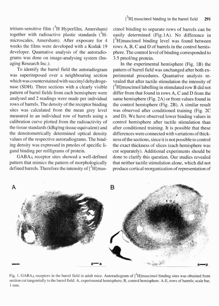
GABAA receptor sites showed a well-defined pattern that mimics the pattern of morphologically defined barrels. Therefore the intensity of [3~]mus-
cimol binding to separate rows of barrels can be easily determined (Fig.lA). No difference in [3~]muscimol binding level was found between rows A, B, C and D of barrels in the control hemis- phere. The control level of binding corresponded to 3.5 pmollmg protein.
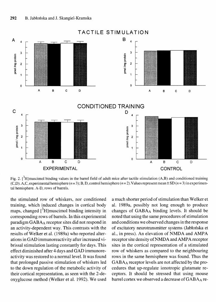
In the experimental hemisphere (Fig. 1B) the pattern of barrel field was unchanged after both ex- perimental procedures. Quantative analysis re- vealed that after tactile stimulation the intensity of [3~]muscimol labelling in stimulated row B did not differ from that found in rows A, C and D from the same hemisphere (Fig. 2A) or from values found in the control hemisphere (Fig. 2B). A similar result was observed after conditioned training (Fig. 2C and D). We have observed lower binding values in control hemisphere after tactile stimulation than after conditioned training. It is possible that these differences were connected with variations of thick- ness of the sections, since it is not possible to control the exact thickness of slices (each hemisphere was cut separately). Additional experiments should be done to clarify this question. Our studies revealed that neither tactile stimulation alone, which did not produce cortical reorganization of representation of
Fig. 1. GABAA receptors in the barrel field in adult mice. Autoradiogram of [3~]muscimol binding sites was obtained from section cut tangentially to the barrel field. A, experimental hemisphere; B, control hemisphere. A-E, rows of barrels; scale bar, 1 mm.
292 B. Jablonska and J. Skangiel-Kramska
T A C T I L E S T I M U L A T I O N
CONDITIONED TRAINING C D
EXPERIMENTAL CONTROL
Fig. 2. [3~]muscimol binding values in the barrel field of adult mice after tactile stimulation (A,B) and conditioned training (C,D). A,C, experimental hemisphere (n= 3); B, D, control hemisphere (n = 2).Values represent mean + SD (n = 3) in experimen- tal hemisphere. A-D, rows of barrels.
the stimulated row of whiskers, nor conditioned training, which induced changes in cortical body maps, changed [3~]muscimol binding intensity in corresponding rows of barrels. In this experimental paradigm GABAA receptor sites did not respond in an activity-dependent way. This contrasts with the results of Welker et al. (1 989a) who reported alter- ations in GAD immunoreactivity after increased vi- brissal stimulation lasting constantly for days. This effect diminished after 4 days and GAD immunore- activity was restored to a normal level. It was found that prolonged passive stimulation of whiskers led to the down regulation of the metabolic activity of their cortical representation, as seen with the 2-de- oxyglucose method (Welker et al. 1992). We used
a much shorter period of stimulation than Welker et al. 1989a, possibly not long enough to produce changes of GABAA binding levels. It should be noted that using the same procedures of stimulation and conditions we observed changes in the response of excitatory neurotransmitter systems (Jablofiska et al., in press). An elevation of NMDA and AMPA receptor site density of NMDA and AMPA receptor sites in the cortical representation of a stimulated row of whiskers as compared to the neighbouring rows in the same hemisphere was found. Thus the GABAA receptor levels are not affected by the pro- cedures that up-regulate ionotropic glutamate re- ceptors. It should be stressed that using mouse barrel cortex we observed a decrease of GABAA re-

[ 3 ~ ] muscimol binding in the barrel field 293
ceptor binding after elimination of sensory input but tactile stimulation and conditioning training did not result in GABAA receptor site change.
This work was supported by grant 6 P203 014 06 from the State Committee for Scientific Re- search.
Henry S.H.C., Jones E.G. (1986) Reduction in number of im- munostained GABAergic neurones in deprived-eye do- minance columns of monkey area 17. Nature 320: 750-753.
Henry S.H.C., Fuchs J., deBlas A.L., Jones E.G. (1990). Dis- tribution and plasticity of immunocytochemically lo- calized GABAA receptors in adult monkeys visual cortex. J. Neurosci. 10: 2438-2450.
Jablonska B., Kossut M., Skangiel-Kramska J. (1995). Tran- sient increase of AMPA and NMDA receptor binding in the barrel cortex of mice after tactile stimulation. Neuro- biol. Learn. Memory (in press).
Kossut M. (1992). Plasticity of the barrel cortex neurons. Prog. Neurobiol. 39: 389-422.
Krohn K., Rothe T., Biesold D. (1992) High-affinity uptake of GABA and decarboxylase activity in rat primary soma- tosensory cortex after sciatic nerve injury. Mol. Chem. Neuropathol. 16: 159-69.
SiucinskaE., Kossut M. (1994) Short term changes of cortical body maps following partial vibrissectomy in adult mice. Acta Neurobiol. Exp. 54: 345-352.
Skangiel-Kramska J., Glazewski S., Jablonska B., Siucinska E., Kossut M. (1994) Reduction of GABAA receptor bind- ing of [3~]muscimol in the barrel field of mice after de- nervation: transient and long-lasting effects. Exp. Brain Res. 100: 39-46.
Strominger R.N., Woolsey T.A. (1987) Templates for locating the whisker area in fresh flattened mouse and rat cortex. J. Neurosci. Methods 22: 1 13- 1 18.
Van der Loos H., Woolsey T. (1973) Somatosensory cortex: structural alternations following early injury to sense or- gans. Science 1796: 395-397.
Warren R., Tremblay N., Dykes R.W. (1989) Quantitative study of glutamic acid decarboxylase-immunoreactive neurons and cytochrome oxidase activity in normal and partially deafferented rat hindlimb somatosensory cortex. J. Comp. Neurol. 288: 583-592.
Welker E. Rao S.B., Dorfl J., Malzer P., Van der Loos H. (1992) Plasticity in the barrel cortex of the adult mice: ef- fects of chronic stimulation upon deoxyglucose uptake in the behaving animal. J. Neurosci. 12: 153- 170.
Welker E., Soriano E., Dorfl J., Van der Loos H. (1989a) Plas- ticity in the barrel cortex of the adult mouse: transient in- crease of GAD-immunoreactivity following sensory stimulation. Exp. Brain Res. 78: 659-664.
Welker E., Soriano E., Van der Loos H. (1989b) Plasticity in the barrel cortex of the adult mouse: effects of peripheral deprivation on GAD-immunoreactivity. Exp. Brain Res. 74: 441-452.
Received 27 April 1995, accepted 1 October 1995