sensory neural hearing loss in children
DESCRIPTION
SensoryNeuralHearingLossinChildrenTRANSCRIPT

1
Chapter 176: Sensorineural Hearing Loss in Children
Patrick E. Brookhouser
Early identification of educationally significant sensorineural hearing impairment inyoung children, which varies in incidence from 1:1000 to 1:2000 depending on the populationstudied, should be a major public health priority. Early (re)habilitative intervention, asprovided under Public Law 94-457, is a prerequisite for the development of age-appropriatespeech and language skills by school age, to provide a basis for attaining literacy skills.Credible research supports the existence of a critical period in the first years of life foroptimal acquisition of speech and language. The absence of adequate auditory stimulation inthe young infant may also impair the full development and maturation of central auditorypathways. Universal hearing screening for all newborns would be a laudable goal, butbehavioral measures (both automated and direct observation) have proven too labor intensive,as well as unreliable, and auditory brain stem response testing is too technologicallysophisticated and costly for general acceptance by clinicians. Evoked otoacoustic emissiontesting may prove to be the long-awaited, cost-effective objective evaluation tool, but presentclinical practice focuses on evaluation of neonates and infants who are identified as being atrisk for hearing impairment. The major argument against limiting early identification effortsto such targeted testing of high-risk populations is data indicating that as many as 50% ofinfants with significant hearing loss may be missed.
In 1982 the Joint Committee on Infant Hearing recommended seven criteria foridentifying infants at risk for hearing impairment. Revised recommendations were publishedin 1991 (Bess, 1991) that expand the risk criteria to include not only neonates (birth to 28days) but also infants (29 days to 2 years). As many as 2% to 5% of neonates manifestinghigh risk factors can be confirmed as having moderate to severe sensorineural hearing loss.Additionally, recommendations were made regarding optimal audiologic screening protocolsand early intervention strategies for hearing-impaired infants and their families.
The high risk factors identified for neonates can be broadly classified in fourcategories: family history, physical findings, events during birth and postnatal hospital stay,and infections. A family history of congenital or delayed-onset childhood sensorineuralhearing loss (SNHL) should serve as a red flag for referral of the infant for early audiologicevaluation. High-risk physical findings include birth weight less than 1500 g (3.3 lb);craniofacial anomalies involving the ear, skull, and mouth region; and stigmata associatedwith a hearing loss syndrome, such as white forelock Waardenburg's syndrome). Severedepression of vital body systems at birth is reflected by a low Apgar score (0 to 3 at 5minutes), delayed spontaneous respirations (beyond 10 minutes), and persistent hypotoniaduring the first 2 hours of life. In addition to these indicators, significant occurrences duringthe neonatal period include hyperbilirubinemia requiring exchange transfusion, prolongedmechanical ventilation, and treatment with ototoxic medications (for example, gentamicin,tobramycin, kanamycin, streptomycin). Infections with the potential to damage hearing maybe acquired prenatally (for example, toxoplasmosis, syphilis, rubella, cytomegalovirus, herpes)or postnatally (for example, bacterial meningitis).

2
In addition to risk factors for neonates, infants (29 days to 2 years) may sustainskull/temporal bone trauma or contract other infectious diseases with potential for causingSNHL (for example, measles or mumps). Neurodegenetative disorders in infancy (forexample, Tay-Sachs disease, myoclonic epilepsy) should also prompt a referral for hearingevaluation. Finally, concern expressed by a parent or surrogate care giver about an infant'shearing, speech and language acquisition, or general development should alert the clinicianto the need for audiologic evaluation. The axiom "moter is always right" is worthremembering when deciding whether to refer an infant for testing. Because behavioralaudiologic testing of hearing in neonates and some infants is fraught with methodologicproblems, it is essential to refer the high-risk neonate for auditory brain stem response (ABR)evaluation, optimally before hospital discharge. If this is not feasible, evaluation by 3 monthsof age is acceptable, but a delay beyond 6 months is unwaranted. It is important to continuefollow-up of those infants with absent or abnormal ABRs or a family history of progressivehearing loss. The conventional "click" stimulus used to elicit an ABR in most centers isaccurate in assessing auditory acuity for higher frequencies (that is, above 2000 Hz), so thata valid and repeatable behavioral audiogram should be obtained as soon as practicable.Appropriate (re)habilitative intervention should be initiated immediately after a sensorineuralhearing loss has been confirmed. Local school districts are obligated to provide and coordinatesuch intervention programs for hearing impaired children living within their boundaries.
The risk factors that identify those neonates, birth to 28 days, who are at risk forsensorineural hearing impairment include the following (ASHA, 1991):
1. Family history of congenital or delayed-onset childhood sensorineural impairment.
2. Congenital infection known or suspected to be associated with sensorineural hearingimpairment such as toxoplasmosis, syphilis, rubella, cytomegalovirus, or herpes.
3. Craniofacial anomalies, including morphologic abnormalities of the pinna and earcanal, absent philtrum, and low hairline.
4. Birth weight less than 1500 g (3.3 lb).
5. Hyperbilirubinemia at a level exceeding indication for exchange transfusion.
6. Ototoxic medications including but not limited to the aminoglycosides used formore than 5 days (for example, gentamicin, tobramycin, kanamycin, streptomycin) and loopdiuretics used in combination with aminoglycosides.
7. Bacterial meningitis.
8. Severe depression at birth, which may include infants with Apgar scores of 0 to 3at 5 minutes or those who fail to initiate spontaneous respiration by 10 minutes or those withhypotonia persisting to 2 hours of age.
9. Prolonged mechanical ventilation for a duration equal to or greater than 10 days (forexample, persistent pulmonary hypertension).

3
10. Stigmata or other findings associated with a syndrome known to includesensorineural hearing loss (for example, Waardenburg or Usher syndrome).
The factors that identify those infants, 29 days to 2 years, who are at risk forsensorineural hearing impairment include the following:
1. Parent or care giver's concern regarding hearing, speech, language, or developmentaldelay.
2. Bacterial meningitis.
3. Neonatal risk factors that may be associated with progressive sensorineural hearingloss (for example, cytomegalovirus, prolonged mechanical ventilation, and inheriteddisorders).
4. Head trauma, especially with either longitudinal or transverse fracture of thetemporal bone.
5. Stigmata or other findings associated with syndromes known to includesensorineural hearing loss (for example, Waardenburg or Usher syndrome).
6. Ototoxic medications including but not limited to the aminoglycosides used formore than 5 days (for example, gentamicin, tobramycin, kanamycin, streptomycin) and loopdiuretics used in combination with aminoglycosides.
7. Children with neurodegenerative disorders such as neurofibromatosis, myoclonicepilepsy, Werdnig-Hoffmann disease, Tay-Sachs disease, infantile Gaucher's disease,Niemann-Pick disease, any metachromatic leukodystrophy, or any infantile demyelinatingneuropathy.
8. Childhood infectious diseases known to be associated with sensorineural hearingloss (for example, mumps, measles).
Neonates, birth to 28 days, who manifest one or more items on the risk criteria shouldbe screened, preferably under the supervision of an audiologist. Optimally, screening shouldbe completed before discharge from the newborn nursery but no later than 3 months of age.The initial screening should include measurement of the auditory brain stem response (ABR)(ASHA 1989). Behavioral testing of newborn infants' hearing has high false-positive andfalse-negative rates and is not universally recommended. Because some false-positive resultscan occur with ABR screening, ongoing assessment and observation of the infant's auditorybehavior are recommended during the early stages of intervention. If the infant is dischargedbefore screening or if ABR screening under audiologic supervision is not available, the childshould be referred for ABR testing by 3 months of age but never later than 6 months of age.
The acoustic stimulus for ABR screening should contain energy in the frequencyregion important for speech recognition. Clicks are the most commonly used signal foreliciting the ABR and contain energy in the speech frequency region (ASHA, 1989). Passcriterion for ABR screening is a response from each ear at intensity levels 40 dB nHL or less.

4
Transducers designed to reduce the probability of ear-canal collapse are recommended.
If consistent electrophysiologic responses are detected at appropriate sound levels, thenthe screening process will be considered complete except in those cases where there is aprobability of progressive hearing loss (for example, family history of delayed onset,degenerative disease, meningitis, intrauterine infections, or infants who had chronic lungdisease, pulmonary hypertension, or received medications in doses likely to be ototoxic). Ifthe results of an initial screening of an infant manifesting any risk criteria are equivocal, thenthe infant should be referred for general medical, otologic, and audiologic follow-up.
Infants, 28 days to 2 years, who exhibit one or more items on the risk criteria shouldbe screened as soon as possible but no later than 3 months after the child has been identifiedas at risk. For infants less than 6 months of age, ABR screening is recommended. For infantsolder than 6 months, behavioral testing using a conditioned response or ABR testing isappropriate. Infants who fail the screen should be referred for a comprehensive audiologicevaluation. This evaluation may include ABR, behavioral testing (6 months), and acousticimmitance measures (see ASHA 1989 Guidelines for recommended protocols bydevelopmental age).
Evaluation of Young Hearing Impaired Child
The comprehensive evaluation of a child with educationally significant hearingimpairment requires the coordinated efforts of a specialty team that should include anotolaryngologist experienced with children, a pediatric audiologic team competent with bothbehavioral and electrophysiologic testing, a pediatrician skilled in detecting subtle findingsassociated with deafness syndromes, a genetic counselor, a pediatric ophthalmologist withcompetence in electroretinography, a psychologist to assess the child's cognitive abilities, aneducator of the deaf with expertise in early intervention, and a speech/language pathologistto assess oral motor function and linguistic development. Consultation in pediatric neurologyand neuroradiology should also be easily available.
A thorough case history provides the foundation for the remainder of the evaluation.In addition to a detailed prenatal, birth, and postnatal history for the child, a complete familyhistory should be obtained that includes hearing loss; speech/language disorders; ear, nose,and throat disorders; and craniofacial deformities; as well as features that accompanysyndromes, such as kidney disorders, sudden death at a young age, thyroid disease,intracranial tumors, progressive blindness, and café-au-lait spots. Because the majority ofgenetic hearing loss is inherited in a recessive manner, it is very important to rule out maritalconsanguinity in the family history. If a family history of hearing loss is identified, anexhaustive pedigree should be constructed, including at least three generations. It is importantto realize that many families with genetic deafness fail to recognize the heritable nature ofthe disorder, because family oral tradition ascribes each case to an extrinsic cause such asmastoid disease, head trauma, or noise damage.
A careful physical examination must involve not only an assessment of the ears butalso a search for subtle findings such as preauricular or branchial pits, heterochromia iridis,blue sclerae, dystopia canthorum, facial asymmetry, and café-au-lait spots. Specific physicalfindings associated with deafness syndromes are detailed in the subsequent text, but no

5
finding should be disregarded until genetic consultation has been obtained. Ophthalmologicevaluation is critical because the visual system assumes an even more important neurosensoryrole in a child with impaired auditory acuity. Additionally, early detection of coexistingretinitis pigmentosa, with electroretinography, can influence (re)habilitative decisions.
Basic laboratory studies should include complete blood count with differential andsickle cell preparations, if indicated; basic blood studies: chemistries, lipids, blood sugar,creatinine, BUN, and thyroid studies; and urinalysis. If hematuria is present, additional renalstudies (such as ultrasound) should be undertaken. Serologic tests aimed at detectingcongenital syphilis and toxoplasmosis, both of which are potentially treatable, are advisable.Knowledge of the rubella vaccination status of the mother should determine the necessity forpursuing that diagnosis. Cytomegalovirus infection presents a diagnostic quandary. It is likelythat CMV accounts for a significant percentage of childhood deafness, but confirmation oftrue congenital infection must be based on positive serology obtained very early in life,probably within the first month or two. After that, antibodies to perinatally or postnatallyacquired CMV can obscure the diagnosis. The role that autoimmune disorders play in theetiology of SNHL in children is presently unclear, and judgments regarding the costeffectiveness of studies aimed at detecting such disease processes must await evidenceregarding their prevalence.
The pediatric audiologic evaluation must determine the type of hearing loss (that is,conductive, sensorineural, or mixed); the degree of loss (that is, mild, moderate, severe,profound, or anacusic); the audiometric configuration and symmetry of the impairment; andfinally, with serial assessment, the stability or progression of the loss. Vestibular dysfunctionmay coexist with hearing loss in some children with genetic deafness (for example, Usher'ssyndrome, type 1) and with hearing loss attributable to such nongenetic etiologcies asbacterial meningitis. The availability of computerized rotational testing has facilitatedvestibular system evaluation in young children, which may also be important in determiningthe cause of delayed gross motor skill development (such as sitting unsupported, walking,standing on one foot). Progressive and fluctuating SNHL, with or without vertigo, in childrenraises the specter of possible perilymphatic fistula (PLF). Although preoperative confirmationof PLF remains elusive, a monitored fistula test seems a reasonable course in these cases. Apositive fistula test may be helpful, but a negative result must be considered inconclusive onthe basis of reported series.
Temporal bone imaging studies are helpful in identifying middle ear anomalies inchildren with conductive or mixed losses, as well as inner ear or internal auditory canalanomalies or lesions in cases of SNHL. The relatively low yield and high cost of such studiesmake their uniform application problematic. The utility of CT scans for assessing profoundlydeaf children for cochlear implant candidacy has resulted in increased use of this diagnosticmodality in recent years.
A diligent search for etiology of SNHL in a child, utilizing state of the art techniques,will prove inconclusive in 30% to 40% of cases. With the declining incidence of non-geneticetiologies such as prenatal rubella, measles, mumps andHaemophilus influenzaemeningitis,genetic hearing loss will affect an increasing percentage of newly identified cases. Manyparents are anxious to determine the etiology of their child's loss for prognostic and familyplanning considerations. Clinicians serving the needs of these families must be prepared to

6
address their concerns regarding the lack of a definitive diagnosis.
Several detailed etiologic classifications of SNHL in children have been suggested.The use of terminology such as genetic versus acquired and congenital versus acquired canbe confusing. It would seem most helpful to classify SNHL cases broadly in a matrix asgenetic versus nongenetic and congenital versus delayed (that is, postnatal) onset. Clearly,many genetic hearing losses are not congenital, whereas certain nongenetic hearingimpairments (for example, rubella deafness) are present at birth. Rapidly expandingknowledge regarding genetic hearing loss should lead to techniques for the prenatal or earlypostnatal diagnosis of these disorders even if clinical manifestations are delayed for monthsor years.
Genetic Hearing Loss
It has been estimated that 50% of childhood sensorineural hearing impairment is dueto genetic factors; 20% to 25% of cases are attributable to identifiable environmental causes,occurring prenatally, perinatally, or later in life; and 25% to 30% are sporadic cases ofuncertain etiology.
Genetic forms of hearing loss may be congenital or delayed in onset, progressive ornonprogressive, and unilateral or bilateral; they may be part of a syndrome (that is, involvingother identifiable physical characteristics in other systems), or there may beno associatedsyndrome, (that is, there is only hearing loss). At least 100 genetic syndromes that involvehearing loss have been identified, and they may be classified on the basis of other organsystems involved; craniofacial/cervical, skeletal, integumentary, ocular, neurologic, renal,metabolic, and "other". Classifications of hearing impairments not associated with syndromes(that is, hearing loss only) are generally based on audiologic characteristics, presence orabsence of progression, age of onset, and mode of inheritance. Approximately 75% to 80%of genetic hearing loss is generally attributed to autosomal recessive genes and 18% to 20%to autosomal dominant genes, with the remainder classified as X-linked, or chromosomal,disorders.
Patterns of Dysmorphology
Based on the classification by Spranger et al (1982), a number of terms can be usedto describe patterns of dysmorphology.
Individual alterations of forms or structure
A malformationis a defect in the morphology of an organ or body region occurringbecause the underlying developmental potential of the involved organ was intrinsicallyabnormal.
If an organ's developmental potential was normal and extrinsic factors, such as trauma,infections, radiation, or pharmaceuticals, cause the defect, the resulting morphologic anomalyis called adisruption.By definition, disruptions cannot be inherited because they are sporadicoccurrences.
7
Intrinsic or extrinsic mechanical forces can produce adeformationin which a part ofthe body assumes an abnormal position, shape, or form. Intrauterine contrainst is an exampleof such a deforming force.
Abnormal cellular organization within tissues is classified as adysplasia, asexemplified by osteogenesis imperfecta resulting from a defect in connective tissue. Adysplasia may involve multiple organs, may be sporadic or multifactorial in etiology, and mayresult from a genetic mutation. Solitary acoustic neuromas and multiple neurofibromatosesare examples of dysplasias.
Patterns of morphologic defects
A polytopic field defectresults from perturbation of a single developmental field,whereas the termsequencedescribes a pattern of several anomalies resulting from a singlecause such as a preexisting anomaly or a mechanical force. A malformation or disruption maybe the underlying cause of a sequence. The Robin sequence (also known as Pierre Robinsyndrome), consisting of micrognathia, cleft palate, and glossoptosis, can result from variousintrinsic and extrinsic forces operative in several different developmental environments.
A syndromeis a pattern of multiple anomalies presumed to be pathogenetically relatedbut not meeting the qualifications for classification as a single sequence or a polytopic fielddefect. Malformation syndromes usually have an identifiable cause, which could includedisorders resulting from chromosomal abnormalities or genetically determined morphogeneticdeviances.
Associationdescribes a nonrandom occurrence of multiple anomalies, not meetingstrict criteria for classification as a polytopic field defect, sequence, or syndrome, in two ormore individuals. In reality, association is a statistical instead of an etiopathogenetic constructthat serves to alert clinicians to possible occult associated anomalies after one defect has beenidentified. CHARGE association consists of the following: C - coloboma, H - heart defects,A - choanal atresia, R - retarded growth and development and/or CNS anomalies, G - genitalhypoplasia, and E - ear anomalies and/or deafness.
Basic Genetic Principles
Human genes are arranged linearly on 22 pairs of autosomes and 1 pair of sexchromosomes, which taken together constitute what might be called a design manual for thehuman body. Each pair of chromosomes carries a distinctive set of gene loci for which theremay be several alternative codes or alleles. For any particular trait, thegenotypemay becomprised of two identical alleles (homozygous) or two different alleles (heterozygous). Thephysical expression of the trait in an individual is called thephenotype,which is determinedby the nature and interaction of the two alleles. An autosomal dominant allele isphenotypically expressed in either the homozygous or heterozygous state, whereas anautosomal recessive allele is expressed only in the homozygous state. An X-linked (that is,sex chromosome-linked) recessive gene will also be expressed in the hemizygous conditionin the male because the Y chromosome does not carry a complementary allele. A parent whois heterozygous for an autosomal dominant gene has a 50% chance of transmitting it to each

8
child (Fig. 176-1). Offspring of heterozygotic carriers of an autosomal recessive gene havea 25% recurrence risk (Fig. 176-2). An X-linked recessive trait is not usually expressed in aheterozygous female, but each male child has a 50% chance of inheriting the gene, whichwould be phenotypically expressed. There is a 50% chance that each of a female carrier'sdaughters would also inherit the gene, but 100% of the daughters of an affected male willinherit his only X chromosome and thus carry the abnormal gene. None of an affected male'ssons inherit his X chromosome with the abnormal gene.
Dominant genes may not be phenotypically expressed in all individuals who areheterozygous for the gene, a phenomenon called decreased penetrance. Variable expressivityis also characteristic of dominant genes in that different family members may show diversemanifestations of the gene. Phenotypic expression may be modified by environmentalinfluences or interaction with other genes.
Chromosomal abnormalities involving autosomes result from extra or deficientchromosomal material being present in each cell, and the phenotypic expression may be quitesevere or even fatal. Phenotypes associated with abnormalities of sex chromosomes aregenerally less severe. In patients with trisomy, three copies of a given chromosome arepresent.Trisomy 21or Down's syndromeis the least severe autosomal trisomy, whereastrisomy 13 (Patau's syndrome)and trisomy 18 (Edward's syndrome)are less common andmore severe. Other autosomal trisomies are almost always lethal, as is the presence of a singlechromosome of a pair, monosomy. Chromosomal deletions (absent segments) or duplications(extra material) may be present, with the nature and severity of each phenotype dependingon the amount and origin of the material involved.
Genetic Linkage Analysis
Genetic linkage analysis is the process of determining the precise chromosomallocation of a specific gene. Genetic material may be randomly exchanged (that is, crossover)between two members of a chromosome pair during cell mitoses, and two genetic loci aresaid to be linked when their alleles are transmitted together more frequency than expected bychance. If the chromosomal location of one gene is known (a marker gene), the linked focuscan be determined. Restriction fragment length polymorphisms are markers based onvariations in DNA sequence rather than on gene product. These DNA probes have alreadyplayed an important role in linkage studies successfully identifying specific chromosomal lociof multiple genes responsible for Usher syndrome (USH 1 and USH 2). Waardenburgsyndrome (WS 1), and branchio-oto-renal syndrome (BOR).
Inner Ear Structural Malformations
By the beginning of the ninth week of gestation, the cochlea reaches full growth (2.75turns). Arrest in normal development (agenesis) or aberrant development (dysgenesis) of innerear structures may result in hearing impairment. Studies utilizing modern temporal boneimaging reveal subtle or severe anomalies of the inner ear in about 20% of children withcongenital sensorineural hearing loss, with about 65% being bilateral and the remainderunilateral.

9
Complete agenesis of the petrous portion of the temporal bone occurs inMichel'saplasia,which is inherited as an autosomal dominant trait. Temporal bone imaging plays anessential role in confirming the diagnosis, although labyrinthitis ossificans, as occursfollowing meningitis, can present a similar appearance. Affected ears are anacusic becauseof the absence of sensory and neural inner ear structures. Vibrotactile devices have been ofsome benefit with these patients, but conventional amplification and cochlear implantation arenot efficacious for obvious reasons.
In Mondini's aplasiathe basal coil of the cochlea can be clearly identified, but theinterscalar septum is absent in the upper coils, which assume a cloacal form. It is postulatedthat the deformity results from a development arrest at approximately the sixth week ofgestation. The anomaly is inherited in an autosomal dominant fashion, may be unilateral orbilateral, and has been described in a number of disorders, including Pendred's, Waardenburg,Treacher Collins, and Wildervaank's syndromes. An aggressive program of early interventionwith conventional amplification is advisable in cases with residual neurosensory structures.
The bony labyrinth and superior portion of the membranous labyrinth, including theutricle and semicircular canals, are normally differentiated in patients withScheibe's aplasia.The organ of Corti is generally poorly differentiated with a deformed tectorial membrane andcollapsed Reissner's membrane. Inherited as an autosomal recessive trait, Scheibe's is the mostcommon aplasia and has been observed in temporal bones from patients with Jervell andLange-Nielsen, Refsum's, Usher's, and Waardenburg's syndromes.
In Alexander's aplasia,cochlear duct differentiation at the level of the basal coil islimited, with resultant effects on the organ of Corti and ganglion cells. High-frequencyhearing loss is the rule in these patients, with enough residual low-frequency hearing tobenefit from amplification.
An enlarged vestibular aqueduct, observed radiographically, has been associated withearly-onset SNHL, which is usually bilateral and may be accompanied by vertigo. Initialhopes that endolymphatic sac surgery in these patients might be beneficial proved incorrect,and such intervention is contraindicated.
Autosomal Dominant Disorders
An autosomal dominant syndrome might be expected to be easily identified by apositive family history of classical dominant inheritance and a recognizable phenotype. Infact, variation in expressivity may be reflected in different phenotypic characteristics inaffected family members. An obligate carrier may not have any detectable phenotypicexpression because of decreased penetrance. If a new mutation has occurred, the familyhistory might be negative, but the disorder is still transmitted in a dominant fashion.
Variable expressivity is observed withWaardenburg's syndrome,which includesunilateral or bilateral sensorineural hearing loss in 20% of cases, as well as pigmentaryanomalies: white forelock in 20% to 30% of cases, heterochromia iridis, premature graying,and vitiligo; and craniofacial features such as dystopia canthorum, broad nasal root, andsynophyrs. Clinically, two distinct types of Waardenburg's syndrome are distinguishable,based on the presence (WS-1) or absence (WS-2) of dystopia canthorum, with hearing loss
10
being more frequent in WS-2. Initial linkage analysis results assign a locus for WS-1 to the2q37 region of the number 2 chromosome (Foy et al, 1990).
Features in individuals withStickler's syndromemay include a small jaw, often witha cleft palate (Robin sequence), myopia that may be accompanied by retinal detachment orcataracts, hypermobility and enlargement of joints with early adult-onset arthritis, andoccasional spondyloepiphyseal dysplasia. Sensorineural or mixed hearing loss is present inabout 15% of cases.
In branchiootorenal (BOR)or Melnick-Fraser syndrome,ear pits (possibly tags) orcervical fistulas are present as are renal findings ranging from agenesis with renal failure tominor asymptomatic renal dysplasia, detectable by ultrasound or intravenous pyelography. Thehearing loss may be sensorineural, conductive, or mixed. Linkage analysis studies haverecently identified the responsible gene to be on chromosome 8q (Kimberling, 1992).
The craniofacial features ofTreacher Collins syndrome(mandibulofacial dysostosis)may include microtia, aural meatal atresia, and conductive hearing impairment about 30% ofthe time. Sensorineural loss and vestibular dysfunction may also be present. Malar hypoplasia,with underdeveloped zygomatic arches, downward slanting palpebral fissures, coloboma ofthe lower eyelids, and a hypoplastic mandible, characterizes the face of affected individuals.Symmetric facies and bilateral eyelid coloboma distinguish Treacher Collins fromGoldenhar'ssyndromeand other oculoauricular vertebral syndromes that involve similar, but unilateral,microtia and craniofacial abnormalities. The oculoauriculovertebral (OAV) spectrum issporadic and thought to be multifactorial, whereas Treacher Collins syndrome is transmittedas an autosomal dominant trait.
Persons affected byneurofibromatosismay present with café-au-lait spots (light brown,variable-sized pigmented spots) and multiple fibromatous tumors. Cutaneous tumors are mostcommon, but the central nervous system, peripheral nerves, and viscera may also be involved.Mental retardation, blindness, and SNHL may result from CNS lesions. Two distinct formshave been identified clinically, the more common beign classic neurofibromatosis(vonRecklinghausen's disease),with an incidence of about 1:3000 persons, which generallyincludes many café-au-lait spots and cutaneous neurofibromas but acoustic neruomas(typically unilateral) in only 5% of cases. Bilateral acoustic neuromas are present in 95% ofcases with central neurofibromatosis (NF-2), which is genetically distinct from the classicalform. Presenting signs and symptoms of the bilateral lesions may not be noted until earlyadulthood. Café-au-lait spots and cutaneous neurofibromatosis in NF-2 patients are fewer thanobserved in von Recklinghausen's disease. Both types of neurofibromatosis are inherited asautosomal dominant traits with high penetrance but variable expressivity. High mutation ratescharacterize both disorders. The gene for at least one form of NF-2 has been assigned tochromosome 22 by linkage analysis.
Otosclerosis appears to be transmitted by an autosomal dominant pattern withdecreased penetrance, so that only 40% of gene carriers will actually demonstrate thephenotype. Delayed-onset conductive or mixed hearing impairment may begin in childhoodbut usually presents in adulthood.

11
Osteogenesis imperfectais characterized by bone fragility, blue (clear) sclerae, hearingloss (conductive, mixed, or sensorineural), and hyperelasticity of joints and ligaments. Thisdisorder is transmitted as an autosomal dominant trait with variable expressivity andincomplete penetrance. A severe congenital type may present with intrauterine fractures severeenough to threaten fetal viability. The age at which the more common "tarda" variety becomesclinically apparent is variable.Van der Hoeve's syndromeis a subtype of osteogenesisimperfecta in which progressive hearing loss begins in early childhood. Although someinvestigators have postulated a relationship between osteogenesis imperfecta and otosclerosis,histopathologic studies of stapedial foot plates in each disorder demonstrate significantdifferences. Minimal peripheral fixation of a markedly thickened stapedial foot plate is presentin a typical case of osteogenesis imperfecta hearing loss.
Nonsyndromic Autosomal Dominant Hearing Loss
Dominant progressive hearing loss (DPHL)is a type of nonsyndromic, noncongenitalsensorineural hearing loss, variable in age of onset and rate of progression, transmitted as anautosomal dominant trait. It is idiopathic and differs from otosclerosis by the absence ofossicular and otic capsule involvement and from presbyacusis by the earlier age of onset. Alltypes of DPHL eventually progress to severe or profound hearing loss, but initial frequencyinvolvement and the rate of progression vary among families. Konigsmark and Gorlin (1976)defined four types of DPHL: early onset, high frequency, mid frequency, and low frequency.
About 80% of genetic deafness in childhood is inherited in an autosomal recessivefashion, with approximately one half of cases involving recognizable syndromes. Variationin penetrance and expressivity is less pervasive than with dominant disorders, but carriers areasymptomatic, making definitive diagnosis in small kindreds difficult unless other syndromiccomponents are identified.
Sensorineural hearing loss and retinitis pigmentosa characterizeUsher syndrome,which has an estimated frequency of 3.0/100.000 in Scandinavia and 4.4/100.000 in the USA.This disorder affects about one half of the 16.000 deaf/blind persons in the USA. Clinically,at least two subtypes, USH 1 and USH2, are distinguishable, based on severity of the hearingloss and the extent of vestibular system involvement. Patients with USH 1 present withcongenital, bilateral profound hearing loss and absent vestibular function, whereas USH 2patients have moderate hearing losses and normal vestibular function (Fig. 176-3). Linkageanalysis studies have localized the gene for USH 2 to the 1q32 region of chromosome 1(Kimberling, 1990). More recently, Kimberling and coworkers have identified chromosome11q as the probable site of the gene for USH 1 that occurs in patients who are not of Acadianbackground and do not reside in Louisiana (Kimberling, 1992). Ophthalmologic evaluationis an essential part of the diagnostic workup, and subnormal electroreginographic (ERG)patterns have been observed in affected children as young as 2 to 3 years of age, beforeretinal changes are evident fundoscopically.
In the autosomal recessive disorderPendred's syndrome,SNHL is associated withabnormal iodine metabolism resulting in a euthyroid goiter. The perchlorate discharge testdemonstrates abnormal organification of nonorganic iodine in these patients, and exogenousthyroid hormone is the therapy of choice.

12
In patients withJervellandLange-Nielsen syndrome,a severe, congenital sensorineuralhearing impairment is inherited in a recessive pattern together with a cardiac conductiondefect that can produce syncopal episodes early in life and even sudden death.Electrocardiographically, large T waves and prolongation of the QT interval are observed, sothat an electrocardiogram should be performed on all children with early-onset hearing lossof uncertain etiology. Beta-adrenergic blockers have proven effective in treating the disorder.
Konigsmark and Gorlin (1976) described three subtypes ofnonsyndromic recessiveSNHL: congenital severe; congenital moderate;and early-onset,which usually progressesrapidly from onset at age 1.5 years to profound loss by 6 years of age.
Sex-Linked Disorders
Approximately 6% of nonsyndromic profound losses in males may be attributable tosex-linked disorders. InNorrie's syndromecongenital or rapidly progressive blindness,development of pseudoglioma, opacification, and ocular degeneration resulting inmicrophthalmia are observed. Progressive sensorineural hearing loss, with onset in the secondor third decade, affects approximately one third of individuals with this sex-linked disorder.
Characteristics associated withotopalatodigital syndromemay include hypertelorism,craniofacial deformity involving the supraorbital area, flat midface, small nose, and cleftpalate. Affected individuals are also short in stature with broad fingers and toes that vary inlength, with an excessively wide space between the first and second toe. An associatedconductive hearing loss usually results from an ossicular malformation.
Wildervaank's syndromeis comprised of the Klippel-Feil malformation involving fusedcervical vertebrae, sensorineural or mixed hearing impairment, and cranial nerve VI paralysiscausing retraction of the eye on lateral gaze. Wildervaank's syndrome is almost alwaysobserved in females because of the high degree of lethality associated with the X-linkeddominant form in males.
In Alport's syndromeSNHL is associated with renal impairment of varying severity.The progressive hearing impairment may not become clinically evident until the seconddecade of life. The renal disease may cause hematuria in infancy but generally remainsasymptomatic for several years before onset of renal insufficiency. Renal involvement isespecially severe in males, and death from uremia before 30 years of age was the rule beforethe availability of renal dialysis and kidney transplantation. Although genetic heterogeneitymay be involved in some cases, the gene for the X-linked form of Alport's codes for acollagen gene (COL4A5) on the X chromosome.
Two types ofnonsyndromic congenital severe sensorineural hearing losshave beendescribed:early-onset, rapidly progressiveandmoderately slowly progressive.
13
Multifactorial Genetic Disorders
Some genetic disorders appear to result from a combination of genetic factorsinteracting with environmental influences. Such multifactorial disorders associated withhearing loss include clefting (that is, cleft lip/palate) syndromes, involving conductive hearingloss, and themicrotia/hemifacial microsomia/Goldenhar's spectrum.
Findings may include preauricular tags/pits, vertebral anomalies such as hypoplasticvertebrae or hemivertebrae in the cervical region, epibulbar dermoids, and coloboma of theupper lip.Goldenhar's syndrome(oculoauriculovertebral dysplasia) has also been describedas being inherited in an autosomal dominant pattern in some families.
Autosomal Chromosomal Syndromes
Middle ear and mastoid disease is often observed in Down's syndrome children.Turner's syndromepatients, monosomic for all or part of one X chromosome (X0), generallypresent as females with gonadal dysgenesis, short stature, and often a webbed neck or shieldchest. Sensorineural, conductive, or mixed hearing loss may be seen with Turner's syndrome.
Genetic Evaluation and Counseling
Family histories of persons with genetic hearing impairment often involve a numberof marriages between deaf individuals, with hearing losses of uncertain etiology. Reasonablecriteria for obtaining cytogenetic studies would be the presence of two major malformationsor a single major malformation with two or more minor malformations. The specific etiologyof a hearing loss may still remain uncertain after an intensive, and often expensive,evaluation. A complete genetic evaluation involves diagnosis, including careful delineationof the phenotype, prognosis, and estimation of recurrence risk. Consideration of the usualpattern of penetrance of a gene must be considered in counseling regarding recurrence risksfor autosomal dominant disorders. Recurrence risk for children of an individual withrecessively inherited deafness depends on the genetic status of his or her mate. The recurrencerisk in deaf x deaf marriages entirely depends on the etiology of the deafness. If both parentshave identical recessively inherited deafness, for example, the recurrence rate would be 100%,but if parents have disparate etiologies, the risk may be quite small.
Bieber and Nance (1978) developed empiric risk tables based on an "averaged" risk,taking into account various possibilities, including the number of affected an unaffectedchildren in the family. For instance, the range of recurrence risk for future offspring in afamily whose only child has an unexplained hearing impairment is 10% to 16%. Eachadditional normal hearing child born to such a family reduces the probability of a geneticetiology with a consequet decrease in estimated recurrence risk.
If a normal hearing individual marries a person with sensorineural hearing loss ofuncertain etiology, their risk of having a hearing-impaired child could range from negligible(nongenetic or uncommon recessive) to 50% if the gene is a fully penetrant dominant one.The averaged empiric risk cited for such a couple is 6%, which would decrease with eachunaffected child born to them.

14
When both husband and wife have hearing losses of uncertain etiology, mostcounselors assign an empiric risk for a deaf child of about 10%. The risk declines with thebirth of each hearing child but increases to 62% if their firstborn is hearing impaired.
Infectious Diseases
Congenital and early-onset infectious disease
Congenital and neonatal infections
Infections acquired during the prenatal or perinatal/neonatal period occur in about 10%of all live births. In most instances these infections are asymptomatic or present non-specificfindings. New diagnostic techniques, including fetal ultrasound, sampling of peripheralumbilical blood, and polymerase chain reaction technology, hold promise for more specificdiagnostic capability in the future.
Viral infections
A number of viruses have been implicated as etiopathogenic agents for congenital andacquired hearing loss. Cytomegalovirus has been isolated from human labyrinthine fluids.Seroconversion studies and virus isolation from urine and nasopharyngeal secretions haveconfirmed an association of labyrinthitis with rubella, rubeola, mumps, influenza, varicella-zoster, Epstein-Barr, poliomyelitis, variola, adenoviruses, and parainfluenza viruses. Beforethe advent of an effective vaccine, mumps was a common etiology of acquired profoundunilateral sensorineural hearing loss in childhood. Effective immunization programs forrubella, rubeola, and mumps in developed countries have decreased their importance in theetiology of childhood hearing loss, but these infections continue to play a significant role inthe Third World.
Histopathologic studies of temporal bones from patients who were affected by prenatalrubella, mumps, rubeola, or cytomegalovirus (CMV) reveal evidence of an endolymphaticlabyrinthitis with pathologic changes limited to the cochlear duct, saccule, and utricle. Thesefindings are consistent with a blood-borne spread of infection most likely via vessels of thestria vascularis. Measles, mumps, and CMV may also present with a meningoencephalitis,permitting direct spread along meningeal and neural structures into the perilymphatic spaces,where inflammatory changes may later progress to fibrosis. Bordley and Kapur (1972) studiedtemporal bones of patients who suffered acute smallpox, varicella (chickenpox), or measlesand found the most severe pathologic changes in the middle ears, although two measlespatients also revealed histopathologic evidence of endolabyrinthitis.
Davis and Johnson (1976) demonstrated selective vulnerability of inner ear structuresof experimental animals to specific viruses. In newborn hamsters, influenza virus infectedmesenchymal cells in the perilymphatic system, whereas mumps virus infected principallyendolymphatic structures. Rubeola and vaccinia viruses infected both perilymphatic andendolymphatic cells, whereas herpes simplex involvement was essentially limited to thesensory cells of the labyrinth.

15
Cytomegalovirus infection
Cytomegaloviruses (CMVs) are members of the herpes family that are widelyprevalent in human populations in both developed and developing countries. Human CMVappears to be species specific with no known external reservoirs or animal vectors involvedin transmission. CMVs may be transmitted either vertically (that is, from mother to child) orhorizontally from person to person, and primary infection is accompanied by viral sheddingthat may persist for months to years. Congenital CMV infection differs from rubella andtoxoplasmosis in that the virus can be transmitted in utero in the course of both primarymaternal infection and infection resulting from reactivation of latent virus in immune women.The probability that her baby will have a harmful congenital CMV infection is decreased inthe mother with recurrent infection, being approximately one ninth as great as after primaryinfection.
In populations of higher socioeconomic status (SES) approximately 55% of womenof childbearing age are CMV immune, with the remaining 45% susceptible to primary CMVinfection. In lower SES groups, fully 85% of fertile women demonstrate immunity.
About 1% to 4% of susceptible women will experience primary CMV infection duringpregnancy, with a resultant fetal infection rate of 40%. Higher SES women with evidence ofCMV immunity at conception will deliver infants with congenital infection in 0.15% of cases,whereas the congenital infection rate among offspring of immune lower SES mothers variesfrom 0.5% to 1.0% (Fig. 176-4)/. Although Hardy (1973) found that the gestational age ofthe fetus is the most important factor determining the extent and long-range consequences ofcongenital viral infections, the time of greatest susceptibility for CMV is not known.
CMV infection is endemic in child day-care centers, and transmission of the virusfrom young children who acquire CMV at such centers to their pregnant mothers is importantepidemiologically.
Congenital CMV infection, which is definitely diagnosed by isolation of the virus fromneonates in the first few weeks of life, is currently the most common cause of intrauterineinfection in humans, occurring in approximately 1% of all live births. An additional 4% to10% of infants acquire the infection during and after birth through such sources as cervicalvirus shedding, virus in breast milk, and blood transfusions. Perinatally infected infantsusually begin to excrete virus between 3 and 12 weeks of age, making early viral isolationstudies essential to the diagnosis of a true congenital infection. Of congenitally infectedinfants, 10% to 15% are symptomatic, and 90% of those demonstrate typical cytomegalicinclusion disease (CID), characterized by involvement of the central nervous system andreticuloendothelial system, with hepatosplenomegaly, petechiae, and jaundice as commonpresenting findings. Microcephaly, intrauterine growth retardation, and prematurity alsocharacterize the CID population, which may experience a mortality rate as high as 30%. Asmany as 90% of children with true CID will develop severe mental and perceptual deficitsby 2 years of age, including severe to profound sensorineural hearing impairment and suchocular abnormalities as chorioretinitis and optic atrophy in 25% to 30% of cases.

16
Before current viral isolation techniques were available, speculation regarding the direoutcome of congenital CMV infection was based solely on data from the 10% of congenitallyinfected infants who are symptomatic. Investigators were unaware of the large percentage ofasymptomatic infected neonates. Such subclinical infections, which account for 90% ofcongenitally infected infants, may become apparent months to years after birth. Althoughprognosis for life with normal neurologic development among these infants is better,approximately 10% to 15% will develop significant SNHL, ranging from mild to profound.SNHL is the most common irreversible sequela of congenital cytomegalovirus infection, withan estimated 30% to 50% incidence in symptomatic cases and 10% to 15% in asymptomaticinfants with congenital infection. It is bilateral in about 50% of cases, varying in magnitudefrom 50 to 100 dB. Stagno and coworkers (1982) found that nearly 25% of SNHL eitherdevelops or increases in severity after the first year of life, reiterating the need forlongitudinal screening programs during the preschool years. With the vaccine-related declinein congenital rubella, congenital CMV infection is likely the most common cause ofnongenetic congenital sensorineural deafness in developed countries. Whereas rubella occursin epidemic cycles, CMV infection extracts an anually recurring toll on the newbornpopulation. Thus far, research directed toward developing a clinically effective vaccine, aswell as a safe and effective treatment regimen, has yet to bear fruit.
Histopathologic study of temporal bones of infants dying of CID has revealedcharacteristic inclusion bodies in the superficial cells of the stria vascularis, Reissner'smembrane, the limbus spiralis, saccule, utricle, and semicircular canals. Although noinclusion-bearing cells were present in the organ of Corti, cristae, or ganglia, endolymphatichydrops was noted to be present in at least a portion of each cochlear duct.
Congenital rubella
During the Australian rubella epidemic of 1941, the ophthalmologist Gregg (1941) firstrecognized the teratogenic potential of prenatal maternal infection; Swan and coworkers(1943) were first to describe deafness as a component of the rubella triad, along withcongenital cataracts and heart defects. Initial estimates of fetal damage following first-trimester maternal rubella ranged from 16% to 59%. Following isolation of the rubellamyxovirus in 1962, clinically applicable methods for confirmation of infection by serologicand virus isolation techniques became feasible. Laboratory-based case identification permitteddocumentation of subclinical rubella infection in expectant mothers, as well as confirmationof congenital rubella infection in infants who did not manifest all components of the classicalrubella syndrome. During the 1963-1965 rubella epidemic in the USA, these laboratorymethods for documenting the presence and timing of rubella infection led to the descriptionof rubella-related disorders in addition to the classical triad. Included in the expanded rubellasyndrome are deafness, eye defects, congenital heart defects, microcephaly, mental or motorretardation, newborn hepatosplenomegaly, thrombocytopenia, radiolucencies in long bones,interstitial pneumonitis, encephalitis, and low birth weight. Prospective studies were carriedout by obtaining paired sera sampes from pregnant women, thus pinpointing the exact timingof maternal rubella infection, clinical or subclinical. Of 165 laboratory-documented cases ofmaternal rubella in one study, 49% were clinically apparent and 51% were subclinical.Maternal rubella during the first trimester of pregnancy exacted the greatest toll on the fetus,but some offspring of mothers with second-trimester infection were also found to havedeafness, microcephaly, cataracts, and mental-motor retardation. Infants with only one or two

17
components of the rubella triad were clearly demonstrated to be victims of congenital rubellainfection.
Hearing loss was the single most common deficit in the laboratory-documentedcongenital rubella population, being the only defect found in 22% of cases. Among thoseinfants from whom rubella virus was isolated, 57% had hearing impairment, whereas 41.5%of infants in whom only serologic confirmation could be obtained were hearing impaired.Approximately 30% of babies born of mothers with subclinical but laboratory-confirmedinfection had hearing loss. The hearing loss was typically sensorineural in character, rangingin severity from patient to patient and to a lesser extent between the ears of the same patient.Some patients showed no response at any frequency, but the most frequently observedaudiometric curves were of the "cookie-bite" type with greatest loss in the middle frequenciesbetween 500 and 2000 Hz. Serial audiograms demonstrated a progressive decrease of auditoryacuity in 25% of cases.
Prolonged virus shedding was observed in many infants who failed to thrive, most ofwhom were progeny of mothers with first-trimester rubella. Viral excretion can pose a hazardto rubella-susceptible health care and child care personnel interacting with infected babies.A general miniaturization of organs from rubella-infected fetuses and infants was noted atautopsy, and histopathologic examination revealed hypocellularity. Highly specialized cells,such as fiber cells in the lens of the eye, appear to become more susceptible to the effects ofintracellular virus with increasing cellular specialization. As a consequence, gradualprogression in cataract formation was noted in the postnatal period.
Temporal bone histopathologic findings in rubella infants include Scheibe-typecochleosaccular changes, but the utricle, semicircular canals, and spiral ganglion wereunaffected. Partial collapse of Reissner's membrane with adherence to the stria vascularis andorgan of Corti was observed in a number of cases. In some sections the tectorial membranewas found to be rolled and lying in the internal sulcus. Saccular collapse with histologicevidence of recent acute inflammation was documented in a number of temporal bones. Theorgan of Corti per se was relatively unaffected but the stria vascularis was noted to be smallerthan normal and areas of cystic dilatation were observed at the junction of Reissner'smembrane and the spiral ligament.
Before the availability of an effective rubella vaccine, epidemics in most developedcountires occurred at 6- to 9-year intervals. Up to 60% of cases of newly identified deafnessin infants and young children were attributed to prenatal rubella in epidemic years, falling toless than 1% in non-epidemic years. Widespread and, in some instances, mandatoryadministration of the rubella vaccine to children and nonpregnant women of childbearing agein the USA has markedly decreased the incidence of rubella. In some parts of the world,however, prenatal rubella still poses a threat to hearing, and it must be considered as anetiologic possibility in immigrant children with sensorineural hearing impairment.

18
Herpes simplex encephalitis
Herpes simplex encephalitis (HSE) is uncommon, but 25% to 30% of cases involvethe pediatric age group. As anticipated in light of the rapidity rising incidence of genitalherpes infection in women of childbearing age, recent studies reveal a tenfold increase in theincidence of neonatal HSV (herpes simplex virus) infection. A large percentage of HSV-infected neonates are born prematurely of young, often nulliparous mothers. Neonatal HSVpatients may present with mucocutaneous involvement or disseminated infection, with onefourth to one third of the infants also affected by meningoencephalitis. On the other hand, upto 20% of newborns with HSV infection never manifest cutaneous involvement. The route bywhich the HSV reaches the brain during primary infection is not clear, althoughhematogenous dissemination, direct spread from the nasopharynx via the cribriform plate, andretrograde spread from infected ganglia have all been postulated. The infection hasdemonstrated a predilection for the temporal and frontal areas of the brain.
Herpes simplex meningoencephalitis, which has an incubation period ranging up to 4weeks, is most likely to occur in the second or third postpartum week. Epidemiologically,only 50% of neonatal HSV infections can be related by history to a definite maternal orpaternal infection, so that the absence of a positive history does not exclude the disease.Nonspecific clinical findings, including fever and an altered mental state, are accompaniedby abnormal CSF findings in over 90% of patients. Electroencephalography and imagingstudies, computerized tomographic scanning, and magnetic resonance imaging are of benefitin detecting focal meningoencephalitis. Brain biopsy, which is positive in 33% to 55% ofHSE cases, represents the only definitive means of diagnosing HSE, as well as excludingother disease processes that may mimic the condition. Chemotherapy for HSE involves theantiviral drug acyclovir for a 10- to 14-day period. Clinicians caring for a child withundiagnosed focal encephalitis are advised to introduce broad-spectrum antimicrobial therapy,in addition to acyclovir, until definitive diagnostic tests have been completed. Effective teammanagement of a child with HSE requires the involvement of neurologists, neurosurgeons,pulmonologists, intensivists, and infectious disease specialists.
Congenital toxoplasmosis
Toxoplasmosis is spread by the ingestion of oocysts on food contaminated by cat fecesor consumption of tissue cysts in undercooked meat products, as well as by congenitaltransmission. The cat is the definitive natural host for the protozoan organismToxoplasmagondii, and ingested feline fecal material containing oocysts leads to the liberation oforganisms that invade human intestinal mucosa and are widely disseminated. Cysts may beformed in all tissues of the body including the human placenta, through which fetal infectionoccurs. Except in immunocompromised mothers, fetal infection is usually the result ofprimary maternal infection during gestation. Although the incidence of fetal infection isgreater if maternal infection occurs during the third trimester, greater fetal damage followsfirst-trimester infection. Two clinical forms are recognized, the more common neurologicallydominant type and a disseminated variant with multiple organ system involvement. Clinicalpresentation of congenitalToxoplasmainfection may include chorioretinitis, hydrocephalus,and generalized intracranial calcifications. Such infants have a very poor prognosis for normaldevelopment. In 90% of cases, congenital toxoplasmosis is subclinical and must be confirmedby laboratory studies. If subclinical infection is unrecognized and consequently untreated,

19
there is a high likelihood of subsequent chorioretinitis with decreasing visual acuity,progressive CNS involvement with decreased intellectual function, deafness, and precociouspuberty.
French investigators conducted extensive studies of pregnancies at risk for congenitaltoxoplasmosis, and compulsory programs for the identification of seronegative women earlyin pregnancy have been implemented in that country (Daffos, 1988; Decoster et al, 1988;Desmonts and Couvreur, 1974; Stepick-Biek, 1990). Maternal infection is confirmed by theappearance of specific IgG in a previously seronegative patient or a rising IgG titer after a3-week or greater interval. Prompt and intensive maternal treatment with a combination ofpyrimethamine plus a sulfonamide of the sulfapyrimidine type is advocated as a means ofdecreasing the likelihood and mitigating the effects of fetal infection. Prenatal diagnosis offetal infection is accomplished by acquisition of amniotic fluid through amniocentesis andfetal blood samples from the umbilical cord under ultrasound control. Inoculation of fetalblood and amniotic fluid into mice, IgM immunosorbent assays, and quantitative maternal andfetal IgG studies are utilized to document the presence of fetal infection. In the Frenchstudies, which included prenatal treatment of maternal infection, congenital infection occurredin 0.6% of cases with documented maternal infection during the preconceptual/earlypregnancy perion, in 3.7% of fetuses carried by mothers who were infected from the sixth tothe sixteenth week of gestation, and in 20% of infants born after maternal infection duringthe sixteenth through the twenty-fifth week of pregnancy. The relatively low incidence of fetalinfection among the 16- to 25-week group was 70% less than reported in previous studies,demonstrating that adequate treatment of primary maternal infection during pregnancy reducesthe likelihood of transmission to the fetus. Extended treatment is also advocated forcongenitally infected infants, in the form of alternating courses of pyrimethamine andsulfonamide. Infected infants should receive very careful follow-up, including CT scans toassess central nervous system status and ophthalmologic examination for chorioretinitis.French investigators found that treatment of congenitally infected infants, begun at birth,reduced the frequency of chorioretinitis from 60% to 10%.
Accurate incidence figures for congenital toxoplasmosis in the USA are elusive, withestimates ranging from 1:1000 to 1:8000. It is estimated that as many as 85% to 90% ofAmerican women of childbearing age may be previously uninfected and consequently at riskfor primary infection. The most effective strategy for control of fetal damage is preventionof maternal infection by careful attention to hygienic measures, including cleaning of fruitsand vegetables and thorough cooking of meat products. Expectant mothers should beespecially careful when cleaning cat litter boxes. Careful serologic testing of women at thetime of diagnosis of pregnancy and during prenatal visits will facilitate accurate identificationof seroconverters and prompt institution of maternal treatment during pregnancy. Prenatal andneonatal evaluation of the fetus and infant can document congenital infection so that postnataltreatment can be instituted as early as possible. Longitudinal follow-up studies in Franceshould further demonstrate the effectiveness of recommended treatment regimens inpreventing sequelae, including deafness.

20
Syphilis
Congenital syphilis, resulting from the transplacental transmission of the causativeorganismTreponema pallidumto the fetus after the fourth month of gestation, may bemanifest at birth or may be inapparent until as late as the fifth decade of life. Recent reportsindicate a rapid increase in the number of acquired and congenital syphilis cases. There isevidence that concurrent human immunodeficiency virus (HIV) infection in patients withacquired syphilis may increase the likelihood of early progression to neurosyphilis and beassociated with failure to respond to penicllin therapy. Although SNHL has been reported inpatients with primary, secondary, and later acquired syphilis, the primary concern in childrenis congenital infection.
Classic stigmata of congenital syphilis include deafness, interstitial keratitis,Hutchinson's teeth (notched incisors), and nasal septal perforation. Prevalence estimates ofhearing loss among patients with congenital syphilis range from 3% to 38%, with about 37%of cases presenting before 10 years of age, 51% between 25 and 35 years of age, and 12%becoming apparent even later in life. In some instances, sensorineural hearing loss may be theonly presenting symptom. A commonly reported audiometric configuration is a bilateral, flatsensorineural hearing loss, which may present in children as a sudden, bilateral profoundimpairment, usually without vertigo. In late congenital syphilis the hearing loss may besudden, asymmetric, fluctuating, or progressive, accompanied in many cases by episodictinnitus and vertigo. Speech discrimination scores tend to be poorer than expected from theaudiometric configuration, loudness recruitment severe, and caloric responses weak to absent.A positive labyrinthine fistula test may be present (Hennebert's sign), and Tullio'sphenomenon (dysequilibrium in response to loud sounds) may be observed.
Because any attempt to carry out direct dark-field examination of perilymph forTreponema pallidumwould present an unacceptable risk of additional hearing loss, serologictests present the best alternative means for diagnosing otosyphilis.Treponema palliduminfection gives rise to both nonspecific reagin antibody and specific antitreponemal antibodies.Nonspecific tests are utilized for screening large numbers of patients from low-incidencepopulations, such as state-required tests of cord blood from all newborns. On the other hand,reports from large otologic centers indicate a prevalence of 570/100.000 of presumedotosyphilis among otologic referrals, justifying the use of more expensive treponema-specificprocedures. The most widely utilized treponema-specific test is the fluorescent treponemaantibody absorpion test (FTA-ABS) with a high sensitivity rate to all stages of syphilis anda low rate of false-positive results; the specificity rate approaches 98% in large studies. Mostinstances of false-positive results involve autoimmune or drug-induced collagen vasculardiseases, such as systemic lupus erythematosus. Other treponema-specific tests that may beused to confirm FTA-ABS results are the micro-hemagglutination assay forT. pallidum(MHA-TP) and theT. pallidum inhibition test,a complex and costly but highly specific (99%)procedure. Because the treponema-specific tests may remain positive following adequatetreatment, Birdsall and coworkers (1990) introduced a western blot assay to identifyantitreponeaml antibody isotype and determine whether or not the infection remains active.In the presence of active infection, both antitreponemal IgM and IgG antibodies are present.After successful treatment, only IgG antibodies persist.

21
High-dose parenteral penicillin is the treatment of choice in all nonallergic patientswith a presumptive diagnosis of otosyphilis, and consideration must be given, in dosagedetermination, to the potential limits on penicillin diffusion posed by the blood-CSF andblood-perilymph barriers. The discovery that treponemes may lie dormant for as long as 90days between replications in late congenital syphilis cases dictates the necessity for longer-term treatment schedules than formerly recommended. Current recommendations for dosageand treatment course may be found in publications from the Centers for Disease Control,which should be consulted before initiating therapy.
As an adjunct to adequate antimicrobial therapy, systemically administeredcorticosteroids (generally oral prednisone) have demonstrated effectiveness in stabilizing orimproving hearing in approximately 50% of patients with syphilitic deafness. Greaterimprovement is usually observed in speech discrimination scores than in pure-tone thresholds.The mechanisms of steroid action in these cases are uncertain but may involve nonspecificreduction of vasculitis, suppression of immune reaction to spirochetal antigens, orenhancement of penicillin diffusion into the perilymph. Relative contraindications to steroidtherapy include lack of immunity to varicella, recent vaccination, hypertension, diabetesmellitus, glaucoma, pregnancy, and peptic ulcer disease. Fatal cases of varicella have beenreported in young children receiving steroid therapy, and careful case selection is essential.Steroids, initially administered in a gradually tapered dosage regimen, are usually discontinuedafter 4 to 8 weeks if hearing does not improve. On the other hand, long-term maintenancetherapy may be required to sustain hearing improvement. Alternate-day dosage appears tocarry less risk of sequelae, such as cataract formation, bone growth disturbance, and adrenalsuppression.
Temporal bone histopathologic features in cases of congenital otosyphilis includeobliterative endarteritis, as well as mononuclear cell infiltrates, osteitis of the otic capsule, andvarying degrees of tissue necrosis. Early congenital syphilis may involve the labyrinth, as wellas the eighth nerve, with round cell infiltration but may also present as a meningolabyrinthitis.Osteitis of the temporal bone with secondary involvement of the membranous labyrinth canalso be found in late congenital otosyphilis. Atrophy of the organ of Corti, as well asinvolvement of the stria vascularis, spiral ganglion, and eighth nerve fibers, has been reported.Gummatous changes may be found in all bones of the ear including the ossicles, which mayadd a conductive component to a preexisting sensorineural hearing loss.
Neonatal sepsis
Neonatal sepsis, characterized by signs of infection and septicemia in the first monthof life, carries a significant mortality, ranging from 10% to 50%, with an inverse relationshipto the age and weight of the child. The bacterial pathovens involved may vary from centerto center, but group B streptococci and gram-negative enteric bacilli are most commonlyobserved. Meningitis is a common outcome of bacteremia in these infants, and uncommonorganisms such asListeria monocytogenesmay play a significant role in such cases.Postmeningitic deafness should be suspected in all survivors of neonatal meningitis, andappropriate evaluation with techniques such as auditory brain stem response testing shouldbe undertaken.

22
Later-onset infectious disease
Mumps and measles
Measles and mumps immunization as part of standard well-baby care has effected a90% to 95% decrease in the incidence of these diseases in the USA. Before the availabilityof an effective vaccine for mumps, the most common otologic sequela attributed to this viruswas unilateral profound SNHL or unilateral anacusis without vertigo or vestibular impairment.Data from adults suggest that the time of onset of mumps hearing loss is usually within a fewdays after the disease has reached the acute stage.
Two distinct pathogenetic mechanisms have been implicated in SNHL as a sequela ofmeasles or mumps. In cases of SNHL without signs of meningoencephalitis, the virus appearsto access the inner ear via the stria vascularis during viremia. Sluggish strial circulation,coupled with an intraepithelial capillary network, predisposes to the onset of inflammation inthis structure, which is followed by degeneration and scarring. The resultant change in thevolume and constitution of endolymph has been invoked to explain subsequent degenerationof the stria vascularis, the organ of Corti, the tectorial membrane, and peripheral cochlearneurons, proceeding from the cochlear base to the apex. Collapse of Reissner's membranewith adherence to underlying structures has also been described. The perilymphatic system,the vestibular sensory organs, and the internal auditory canal contents are generallyuninvolved. These histopathologic changes are also compatible with findings in cases ofSNHL caused by prenatal rubella.
Temporal bone findings in patients who suffered meningoencephalitis in conjunctionwith measles or mumps are substantially similar to those associated with meningogenicbacterial labyrinthitis. It appears that direct extension of the viral inflammatory process occursalong the nerves and vessels in the internal auditory meatus into the inner ear. Findingscompatible with such a transmeatal route of infection include severe degeneration of neuralelements inthe modiolus, contrasted with lesser degrees of involvement of neural structuresin the cochlear duct. Temporal bones from infants who died during the acute stage ofmeningoencephalitis demonstrate lymphocytic infiltration along nerves and vessels in theinternal auditory canal without concurrent involvement of the stria vascularis. Intralabyrinthinefibrosis and osteoneogenesis in perilymphatic spaces were found in patients who had survivedthe acute disease process.
Bacterial meningitis
Bacterial meningitis in young children was associated with a 90% to 100% mortalityduring the preantibiotic era. By 1986, clinical investigators reported a mortality of 2% to 3%among children over the age of 1 month with bacterial meningitis that had been adequatelytreated. In cases of neonatal meningitis,Escherichia coli and group B beta-hemolyticstreptococci are the most common causative organisms. The highest percentage of childhoodmeningitis cases in the USA occur in children 6 to 9 months of age, withHaemophilusinfluenzae, Neisseria meningitidis,andStreptococcus pneumoniaecollectively accounting for84% of cases. Among this group of infants,H. influenzaeis the most commonly isolatedpathogen.

23
Unfortunately, the dramatic decline in mortality was not accompanied by aconcomitant decrease in the incidence of immediate and long-term sequelae of the disease.Seizures are experienced by 28% to 40% of meningitis patients before or during their hospitalstay, and at least one handicap was detected during a 1-year follow-up in 57% ofpneumococcal meningitis survivors and 14.5% of children in whichH. influenzaewas theetiologic agent. Apart from sensorineural hearing impairment, meningitis sequelae mayinclude mental retardation, hydrocephalus, seizure disorders, motor abnormalities (hemiparesis,diplegia, quadriplegia, cerebral palsy), vestibular deficits, speech defects, language disorders,hyperactivity, poor impulse control, visual impairment, and learning disabilities. Computerizedtomographic imaging studies of the CNS in children with severe sequelae reveal evidence ofbrain infarction, arterial occlusion, and brain/spinal cord necrosis.
The reported incidence of SNHL following meningitis varies from3% to 40%, withmost reports clustered in the 15% to 20% range. In my series of 280 meningitis survivors,89% of those who experienced postmeningitic hearing loss contracted the disease before theirthird birthday (Brookhauser et al, 1988). The majority of patients with post-meningitic hearingloss sustain permanent, bilateral, severe-to-profound sensorineural impairment, but in a seriesof 64 carefully documented cases, coworkers and I found 38% to have bilateral, asymmetricSNHL and 11% to have unilateral SNHL. Some reports have documented improvement inhearing acuity in a small number of cases observed longitudinally, whereas in other patientshearing losses fluctuated or progressed. These reports can be broadly classified into thoseusing behavioral audiologic tests and those using primarily ABR (Tables 176-1 and 176-2).
A number of observations can be drawn from these studies. The onset of hearing lossassociated with bacterial meningitis usually occurs early in the course of the disease, and noparticular regimen of antibiotic therapy appears to offer any degree of protection against thissequela. These findings are consistent with recent observations regarding the role that thehosts's initial inflammatory response may have in damage sustained by neural tissue. Directspread of the infection to the labyrinth from the middle ear during a bout of otitis media ismuch less likely than (1) the penetration of bacteria and toxins along the cochlear aqueductor internal auditory canal leading to a suppurative labyrinthitis, perineuritis, or neuritis of theeighth nerve; (2) serous or toxic labyrinthitis that constitutes a sterile inflammatory response;(3) septic thrombophlebitis or embolization of small labyrinthine vessels; and (4) hypoxicinsult to the eighth nerve or central auditory pathways.
Meningitis patients who exhibit normal ABR after the first few days of hospitalizationand antibiotic therapy are unlikely to develop later SNHL. Late-onset SNHL, after dischargefrom the hospital, is also distinctly uncommon. Some patients whose ABR results areabnormal early in their hospital course may demonstrate a normal ABR by the time ofdischarge or in follow-up evaluation. These observations may, in some cases, reflect theresolution of a co-existing conductive loss caused by middle ear effusion. Most patients withpostmeningitic losses who show improvement of auditory thresholds over time initiallydemonstrate mild or moderate, rather than severe or profound, SNHL. Thresholds in patientswho exhibit improvement, deterioration, or fluctuation of auditory acuity often require a yearor more to stabilize following meningitis. Some patients with improving thresholds continueto exhibit absent vestibular responses. The likely pathophysiologic explanation for changingbone conduction thresholds during and after an episode of bacterial meningitis is theoccurence of serous labyrinthitis. All meningitis survivors should have careful evaluation of

24
auditory function, initially by ABR in younger children, to be followed by behavioralaudiologic testing when the child's condition permits. Because postmeningitis hearing lossesmay assume a wide range of audiometric configurations, click-evoked ABR, which assesseshearing sensitivity primarily in the high-frequency region (2000 to 4000 Hz) may lead toerroneous conclusions regarding auditory acuity at lower frequencies. Careful longitudinalfollow-up of postmeningitis children will avoid diagnostic errors.
Neurologic sequelae of bacterial meningitis, including sensorineural hearing loss, maybe attributable to direct bacterial action on nervous tissue, the patient's inflammatory response,or a combination of both factors. Studies of the progression of events at the molecular levelduring an episode of bacterial meningitis suggest that significant injury to the central nervoussystem occurs during the first hours of antibiotic therapy. Experimental animal modelsdemonstrate that bacterial surface components, including endotoxin and cell wall structures,are sufficient to produce the symptom complex of meningitis even in the absence of viablebacteria. Bacterial disintegration resulting from antibiotic action produces a veritable floodof cell wall antigens and endotoxins, stimulating an intense immunologic response by thehost. The damage attributable to such an inflammatory reaction was first demonstrated inanimal studies of pneumococcal meningitis and later with gram-negative pathogens. Reportsinvolving neonates and young children with meningitis have also documented a rapid hostinflammatory response following antibiotic-induced release of bacterial components. It ishypothesized that inflammatory exudate that forms as a result of meningeal invasion bybacteria is potentially harmful and is not critical for containment of the infection. By studyingpaired CSF samples drawn at admission and 18 to 30 hours later, clinical investigators havefound a correlation between the intensity of inflammation as reflected in CSF findings andsubsequent clinical outcome. Poorer outcome observed in neonates receiving intraventriculargentamicin rather than just intravenous antibiotics may also be attributable to the patient'sresponse to an additional load of bacterial degradation products in the CSF.
The local production of interleukin-1B (IL-1B) and tumor necrosis factor (TNF) inresponse to CNS tissue interacting with bacterial breakdown products is thought to be the firststep in a complex series of molecular events that result in inflammation and tissue destruction.Based on the hypothesis that down-modulation of the host inflammatory response couldameliorate the severity of postmeningitic sequelae, corticosteroids have been used asadjunctive therapy to the usual course of bactericidal antibiotics. Corticosteroids act to inhibitthe activity of phospholipase with a consequent decrease in prostaglandin E2, thromboxane,and leukotriene formation. Dexamethasone has also been shown to decrease the productionof IL-1B and TNF. In double-blind, placebo-controlled trials with bacterial meningitispatients, CNS indices of infection severity, such as decreased glucose levels, elevated protein,and increased lactate levels, were significantly less pronounced after 24 hours of combinedtherapy with antibiotics and dexamethasone than with antibiotics alone (Odio et al, 1991;Laebel et al, 1988). In a population of 200 infants and older children, patients receivingdexamethasone became afebrile earlier and were less likely to suffer moderate to severesensorineural hearing loss than controls. Fourteen percent of patients in the placebo group hadsevere or profound hearing loss, whereas only 1% of the steroid-treated group were affected(Laebel et al, 1988). Because of a natural reluctance by some clinicians to administer steroidsin the face of a potentially life-threatening infection, other strategies for downmodulating hostresponse to the infection are being explored in current studies.

25
Ototoxic Drugs and Chemicals
As a result of more accurate reporting of adverse effects of pharmaceutical agentsmandated by federal regulations, a steadily increasing number of potentially ototoxic drugsand chemicals are being identified. Some medications, such as thalidomide, taken early inpregnancy produce severe embryopathic effects that may include deformities of the external,middle, or inner ear with associated sensorineural hearing loss. Other ototoxic drugs andchemicals produce more profound effects after the sensory end-organ of the inner ear hasreached a greater degree of differentiation later in pregnancy.
A partial listing of drugs and chemicals with ototoxic potential follows:
1. Antibioticsa. Streptomycinb. Dihydrostreptomycinc. Neomycind. Gentamicine. Kanamycinf. Vancomycing. Polymyxin Bh. Erythromycin
2. Chemicalsa. Carbon monoxideb. Mercuryc. Goldd. Leade. Arsenicf. Aniline dyes
3. Loop diureticsa. Ethacrynic acidb. Furosemide
4. Other drugsa. Cisplatinb. Quininec. Chloroquined. Salicylatese. Polybrenef. Nitrogen mustardg. Thalidomide.

26
Aminoglycosides
Aminoglycoside antibiotics are administered parenterally because they are poorlyabsorbed from the gastrointestinal tract, with only 3% actually entering the bloodstream.Normal serum half-life is about 2 hours in patients with unimpaired renal function. This classof antimicrobials is not metabolized in the body and is excreted almost entirely by glomerularfiltration so that urinary concentrations can approach 100 times serum levels if renal functionis impaired. Hemodialysis and peritoneal dialysis are effective in lowering serum levels inrenal-compromised patients. It has been documented that aminoglycosides may alsoaccumulate in the perilymph, particularly in patients with suboptimal renal function. Schacht(1986) demonstrated that the drugs are transmitted into hair cells by an energy-dependentprocess involving binding to the phospholipid phosphatidyl-inositol biophosphate.Histopathologic evidence of injury has been observed in the stria vascularis, suprastrial spiralligament, pericapillary tissues in the spiral prominence, the outer sulcus, and Reissner'smembrane. Cochlear damage usually proceeds from the basilar turn toward the apex withinitial destruction being observed in the inner row of outer hair cells. Although the remainingrows of outer hair cells may also be destroyed, inner hair cells are usually spared except incases of overwhelming toxicity. In the vestibular system, type I hair cells of the cristaampullaris appear to be more sensitive to damage by streptomycin, kanamycin, andgentamicin than type II.
Streptomycin, gentamicin, and tobramycin are primarily vestibulotoxic, whereaskanamycin and amikacin are principally chochleotoxic. Fee (1980) observed that gentamicinwas significantly more vestibulotoxic than tobramycin, the toxicity being further potentiatedby high hematocrit, elevated creatinine clearance, criticality of illness, and duration of therapybeyond 10 days. The margin between therapeutically effective blood levels and potentiallytoxic concentrations for aminoglycosides is relatively small, so that careful monitoring of peakserum levels (30 minutes after intravenous administration) and tough levels (immediatelybefore the next IV dose) is essential. Dosage levels must be adjusted downward appropriatedin patients with renal dysfunction. In addition to compromised renal function, other factorsthought to predispose to aminoglycoside ototoxicity include preexisting hearing loss,temporally related noise exposure, age, duration of therapy, prior use of aminoglycosides, orconcomitant use of additional ototoxic drugs such as other aminoglycosides and loop diuretics(for example, ethacrynic acid and furosemide).
Aminoglycosides are utilized with some regularity to treat serious infections inneonates and young infants, but controversy exists as to the inherent risk posed to hearing andbalance function. On the basis of extensive studies, Finitzo-Hieber and colleagues (1985)concluded that aminoglycosides at appropriate dosage levels pose little risk of ototoxicity inchildren. Eviatar and Eviatar (1982), however, conducted controlled prospective studies ofinfants receiving aminoglycosides for neonatal sepsis, evaluating such parameters asacquisition of head control and nystagmus induced by position change, perrotatory stimulationwith a torsion swing, and caloric irrigations. Nearly 10% of the infants who had receivedaminoglycosides demonstrated vestibular abnormalities and delay in acquisition of headcontrol as would be compatible with vestibulotoxicity. Prolonged administration of the drugs(that is, 25 to 45 days) increases the risk of damage as does concomitant administration ofother ototoxic drugs. Serial audiometric screening, including frequencies above 12.000 Hz,and vestibular evaluation may be of benefit for providing early warning of impending damage.

27
Some investigators report partial return of vestibular function after an initial deficit has beendemonstrated during extended gentamicin therapy.
Loop diuretics
Powerful "loop" diuretics, such as ethacrynic acid and furosemide, promote the rapidexcretion of large amounts of isoosmotic urine. Early clinical observations of a transientototoxic effect associated with the use of these agents were followed by reports of permanentototoxicity, particularly when these diuretics are used concurrently with apparently safe levelsof aminoglycosides. Estimates of the incidence of ototoxic effects with this class ofpharmaceuticals reported by Matz (1990) range from 0.7% with ethacrynic acid to 6.4% withfurosemide. Temporal bone histopathology in animals treated with toxic levels of these drugsreveals changes in the stria vascularis and significant hair cell loss, Rybak and coworkers(1990) postulate that furosemide and ethacrynic acid have different mechanisms of action onthe cochlea. Because of the repeatedly documented synergy between the ototoxic effects ofloop diuretics and aminoglycosides, the respective dosages of these medications should becarefully adjusted when they are administered concurrently.
Erythromycin
The ototoxic potential of erythromycin became evident with the relatively largeintravenous dosage levels utilized to treat pneumonia caused byLegionella pneumophilla.Bilateral SNHL, often accompanied by tinnitus and vertigo, was noted in adults witherythromycin doses of 2 g/day, particularly in patients with concomitant renal or hepaticfailure. Blood levels at which ototoxic effects were observed ranged upward from 63 mg/L.
Salicylates
High therapeutic doses of salicylates (levels exceeding 20 mg/dL) are known to beassociated with reversible hearing loss and tinnitus in humans. Hawkins' work (1973)demonstrated that salicylates in the perilymph can lead to a reduction in transaminases,cochlear adenosine triphosphate levels, and cochlear blood flow. Changes in cochlear functionhave been attributed to a salicylate-mediated increase in the membrane conductance of outerhair cells. The association of the ingestion of therapeutic doses of salicylate in young febrilechildren with the occurrence of Reye's syndrome has led to a marked decrease in theutilization of this class of drugs in the pediatric and adolescent age groups without strictindications.
Vancomycin
Nephrotoxicity and ototoxicity have been reported in patients receiving vancomycin,particularly if blood levels exceed 45 mg/L. Adults have described tinnitus at slightly lowerserum levels, which could serve as early warning of impending damage. Matz (1990) reportsthat premature infants are more susceptible to the ototoxic effects than full-term babiesbecause they have larger volumes of distribution of the drug, which has a longer half-life inthese neonates.

28
Cisplatin
Cisplatin (cis-diaminoedichloroplatinum), which is utilized for treating a range ofneoplasms, has been implicated as an ototoxic agent, with the potential for permanentlyaffecting both cochlear and vestibular function. Studies employing serial high-frequencyaudiometry demonstrate that the hearing loss usually begins in the 10.000 to 18.000 Hz range,gradually involving frequencies below 8.000 Hz if treatment continues. Audiometric screeningat 12.000 Hz and 14.000 Hz has proven helpful in early identification of ototoxicity. Thepathophysiologic mechanism by which damage occurs involves drug-mediated blockade ofouter hair cell transduction channels, coupled with a decrease in adenylate cyclase.Histopathologically, outer hair cell loss is observed, particularly in the basal turn of thecochlea. Estimates of the risk of ototoxicity posed by cisplatin vary with the audiometricfrequencies that are evaluated. Early estimates placed the risk at 20% or less. In a studyevaluating thresholds from 8 to 20 kHz, Dreschler and colleagues (1985) utilized conservativecriteria for damage, including deterioration of 20 dB or more at one frequency, 15 dB at twofrequencies, or 10 dB at four frequencies. They found that 46% to 83% of patients in thevarious study groups demonstrated some degree of loss, principally at higher frequencies.Eighty percent of these losses were bilateral, with the remainder having unilateralinvolvement. Most investigators agree that dosage levels exceeding 3 to 4 mg/kg of bodyweight carry a definite risk of ototoxicity. In one report of 54 cases, no ototoxic changes werenoted in patients where the peak plasma concentration of cisplatin did not exceed 1 microg/L.Davidson and coworkers (1989) reported that permanent, bilateral, high-frequencysensorineural hearing loss was detected in 88% of children receiving cisplatin doses over 450mg/m2. Cochlear damage, which was related to cumulative doses exceeding 279 mg/m2, wasinversely proportional to the age of the child. Animal studies have confirmed that the ototoxiceffects of noise exposure (85 dB SPL or greater) and cisplatin are synergistic, producinggreater hair cell loss and hearing impairment at high frequencies in combination than isobserved with either agent alone. Although conclusive human studies addressing this issue arenot available, prudence would dictate avoidance of exposure to potentially damaging soundlevels during cisplatin treatment.
Quinine and chloroquine
The antimalarial agents quinine and chloroquine phosphate have both demonstratedototoxic potential. Mothers receiving quinine during pregnancy may give birth to infants withvarying degrees of severe to profound hearing impairment. Both retinopathy and hearing losshave been observed in offspring of pregnant mothers treated with chloroquine. Thepathophysiologic mechanism involved with both drugs appears to be vasculitis and ischemiain the inner ear, with subsequent degenerative changes of the stria vascularis, organ of Corti,and neuronal elements.
Anoxia/Hypoxia
Anoxia and hypoxia during the perinatal period, resulting from such factors as cordcompression and neonatal seizures, are strongly correlated, statistically, with subsequentSNHL in young adults. Objective evidence of anoxia/hypoxia in the neonatal history caninclude meconium staining, primary apnea, a history of resuscitation at birth, low Apgarscores at 1 and 5 minutes, postnatal apneic episodes, and the need for prolonged postnatal

29
ventilatory assistance. Histopathologic evidence demonstrates that anoxic damage occurs inthe brain stem reticular formation and cochlear nuclei, which show decreased cell numbersand volume in direct proportion to the length and severity of oxygen deprivation. In somecases of perinatal anoxia, infants may have multiple high risk factors, making the precise roleof oxygen deprivation in the etiology of hearing loss unclear. On the other hand, recentstudies among neonates with chronic hypoxemia resulting from persistent fetal circulationrevealed a 20% incidence of SNHL, three fourths of which was moderate to severe and onefourth of which was profound.
Hyperbilirubinemia
A causal relatinship between elevated bilirubin levels in neonates, with associatedkernicterus, and SNHL has been appreciated for decades. Whether resulting from inadequateconjugation, impaired albumin binding, or increased unconjugated bilirubin production, thebilirubin that crosses the blood-brain barrier may be deposited in the basal ganglia,particularly the ventrocochlear nucleus, resulting in neurologic sequelae including SNHL.Histopathologically, the middle and inner ears of these patients are generally free ofabnormality. Improved prenatal care including the introduction of Rh immunoglobulin,exchange transfusion, and phototherapy has markedly decreased the importance ofhyperbilirubinemia as a cause of early acquired SNHL. Recent studies suggest that therapeuticintervention should be instituted at lower serum bilirubin levels in premature neonates thanwas previously thought necessary.
Prematurity and Full-Term Low Birth Weight
Although a number of high risk factors, such as perinatal hypoxia, sepsis, andkernicterus, may be present in premature and dysmature neonates, prematurity itselfconstitutes a reliable risk indicator. Admission to a neonatal intensive care unit (NICU) isstandard care for these small infants, and NICU outcome studies reveal that premature infantsmay be as much as 20 times more likely to be severely hearing impaired than infants ofnormal weight and gestational age. As many as 2% of all infants with birth weight of 3pounds or less manifest significant SNHL. Audiologic evaluation and habilitation of thesehearing impaired infants may be further complicated by the presence of additional handicaps.
Recurrent Otitis Media and Mastoid Disease
Conductive hearing loss is the usual sequela of recurrent otitis media and mastoiditis,but studies by Paparella et al (1970, 1984) and others indicate that otitis-prone childrenshowed an increased likelihood to have a coexisting sensorineural loss than would bepredicted by incidence figures for the general population. Mechanisms postulated to accountfor this observation include penetration of the labyrinthine windows (particularly the roundwindow membrane) by infectious by-products or toxins, topical medications with ototoxicpotential, or a combination thereof. Studies of round window permeability in experimentalanimals suggest a greater risk for penetration by middle ear contents into the inner ear thanhas been observed empirically in humans. It has been postulated that edema and granulationtissue that accompany middle ear infection may actually help prevent transmission ofunwanted substances through the round window membrane.

30
Ear and Head Trauma
Middle ear and inner ear truma can result from diverse etiologies, including a slap inthe ear, a foreign body (for example, cotton swab) inserted in the external canal, impactagainst water while diving or water skiing, and skull trauma, which may include temporalbone fracture. An infant's foreshortened ear canal can predispose to transcanal injury, whereashead trauma is a more common etiology in older children and adolescents. Youngsters maybe able to report symptoms such as acute pain, bloody otorrhea, hearing loss, tinnitus, andpossibly vertigo. Mild to moderate conductive hearing loss is the most common audiometricfinding, but intralabyrinthine injury may produce a concurrent, usually high-frequency,sensorineural component. More severe conductive impairment suggests possible ossiculardamage, and labyrinthine membrane disruption, including perilymphatic fistula, should besuspected in patients with fluctuating or progressive sensorineural losses. Large traumatictympanic membrane perforations, particularly if accompanied by ossicular injury, may requiresurgical intervention, but most heal with surprisingly little residual scarring.
Motor vehicle accidents, bicycle or skateboard mishaps, and falls are among thecommon etiologies for temporal bone fractures in children and adolescents. Such fractures arebroadly classified as longitudinal or transverse with respect to the long axis of the temporalbone, with the longitudinal variety accounting for 70% to 90% of these injuries. Longitudinalfractures are associated with middle ear and ossicular damage, usually sparing inner ear andinternal canal structures. Transverse fractures often disrupt labyrinthine integrity or transectneural structures in the internal auditory canal, leading to permanent sensorineural deafnessor facial nerve paralysis, as well as vertigo.
Temporal bone fractures have been reported to occur in 7% of children who wereadmitted to the hospital following head injury, and 13% of these patients sustained SNHL.In a group of 324 children and adolescents with unilateral SNHL, 10.8% of losses wereattributed to head trauma, which in 35% of cases had been sustained at or before 6 years ofage.
Noise-Induced Hearing Loss
Sounds of sufficient loudness and duration will produce ear damage resulting intemporary or permanent hearing loss, often accompanied by tinnitus. Irreversible inner eardamage from loud sounds is cumulative over time, can occur at any age, and is not presentlyamenable to medical or surgical treatment. An important consequence of a typical noise-induced hearing loss (NIHL) is difficulty in understanding speech sounds, which could impairclassroom performance. Utilizing current knowledge about ear protection, NIHL is entirelypreventable except in rare cases of accidental exposure.
About 10.000.000 Americans have hearing losses that are at least partly attributableto damage from loud sounds. More than 20 million Americans, including children andadolescents, are exposed on a regular basis to hazardous sound levels that could result inhearing loss. Much of this noise exposure occurs in the workplace, but the proliferation ofpotentially harmful noise sources at home, on the farm, and in recreational environments hasplaced increasing numbers of unsuspecting children and adolescents at risk (Table 176-3).
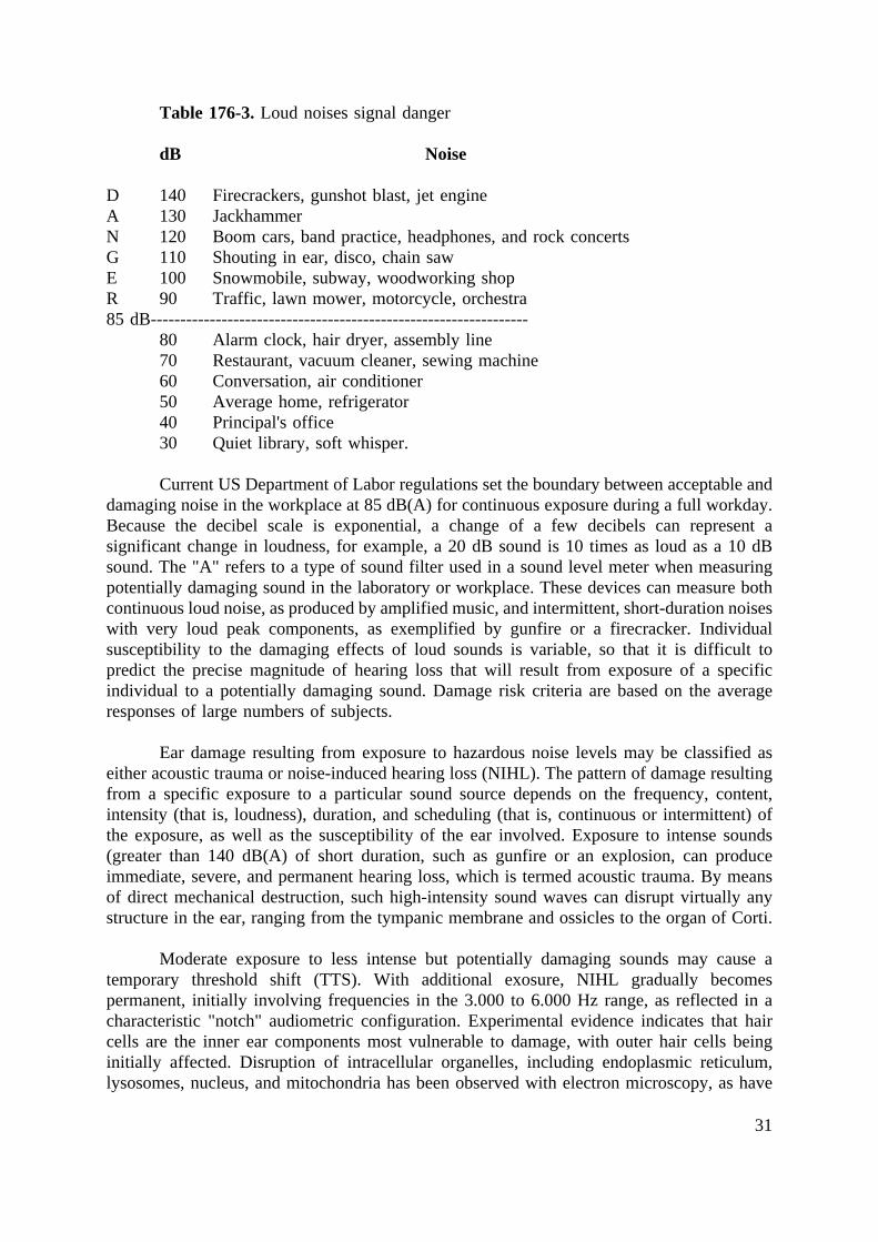
31
Table 176-3.Loud noises signal danger
dB Noise
D 140 Firecrackers, gunshot blast, jet engineA 130 JackhammerN 120 Boom cars, band practice, headphones, and rock concertsG 110 Shouting in ear, disco, chain sawE 100 Snowmobile, subway, woodworking shopR 90 Traffic, lawn mower, motorcycle, orchestra85 dB----------------------------------------------------------------
80 Alarm clock, hair dryer, assembly line70 Restaurant, vacuum cleaner, sewing machine60 Conversation, air conditioner50 Average home, refrigerator40 Principal's office30 Quiet library, soft whisper.
Current US Department of Labor regulations set the boundary between acceptable anddamaging noise in the workplace at 85 dB(A) for continuous exposure during a full workday.Because the decibel scale is exponential, a change of a few decibels can represent asignificant change in loudness, for example, a 20 dB sound is 10 times as loud as a 10 dBsound. The "A" refers to a type of sound filter used in a sound level meter when measuringpotentially damaging sound in the laboratory or workplace. These devices can measure bothcontinuous loud noise, as produced by amplified music, and intermittent, short-duration noiseswith very loud peak components, as exemplified by gunfire or a firecracker. Individualsusceptibility to the damaging effects of loud sounds is variable, so that it is difficult topredict the precise magnitude of hearing loss that will result from exposure of a specificindividual to a potentially damaging sound. Damage risk criteria are based on the averageresponses of large numbers of subjects.
Ear damage resulting from exposure to hazardous noise levels may be classified aseither acoustic trauma or noise-induced hearing loss (NIHL). The pattern of damage resultingfrom a specific exposure to a particular sound source depends on the frequency, content,intensity (that is, loudness), duration, and scheduling (that is, continuous or intermittent) ofthe exposure, as well as the susceptibility of the ear involved. Exposure to intense sounds(greater than 140 dB(A) of short duration, such as gunfire or an explosion, can produceimmediate, severe, and permanent hearing loss, which is termed acoustic trauma. By meansof direct mechanical destruction, such high-intensity sound waves can disrupt virtually anystructure in the ear, ranging from the tympanic membrane and ossicles to the organ of Corti.
Moderate exposure to less intense but potentially damaging sounds may cause atemporary threshold shift (TTS). With additional exosure, NIHL gradually becomespermanent, initially involving frequencies in the 3.000 to 6.000 Hz range, as reflected in acharacteristic "notch" audiometric configuration. Experimental evidence indicates that haircells are the inner ear components most vulnerable to damage, with outer hair cells beinginitially affected. Disruption of intracellular organelles, including endoplasmic reticulum,lysosomes, nucleus, and mitochondria has been observed with electron microscopy, as have

32
disorganization, fusion, and loss of stereocilia. The stereocilia rootlet structure, which anchorsit into the top of the hair cell, appears to be affected first. Degeneration of damaged hair cellsmay lead to eventual loss of auditory nerve fibers and changes in auditory areas of the centralnervous system. With repeated exposure, hair cell loss will continue to increase, and there isno evidence that degenerated hair cells are ever replaced in mammals. Recent studies in birds,however, have documented some hair cell regeneration after initial loss following noiseexposure. The anatomic and physiologic changes that occur in the inner ear during a TTS arequite subtle and may be limited to swelling of hair cells and underlying afferent nerve fiberterminals that resolve during a rest period away from loud sounds. Acute changes instereocilia rootlets might alter cochlear micromechanics, whereas vascular spasm andmetabolic exhaustion could also play a role in TTS.
Impulse noises, as generated by gunfire or an exploding firecracker, are characterizedby short duration and very high sound intensity levels, 132 to 170 dB(A) during the initialacoustic pulse of a typical gun discharge. Large-caliber rifles and shotguns are particularlyhazardous, having the potential to produce immediate and permanent injury to sensitive innerear structures. Sound levels produced by toy weapons measured at a distance of 50 cmshowed mean peak values from143 to 153 dB(A), while firecrackers produced peak levelsmeasured at 3 m of 125 to 156 dB.Adolescents employed in noisy environments, who alsoengage in shooting or setting off firecrackers, may add to whatever hearing loss they sustainas a result of occupational exposure.
Whereas an explosion poses an obvious hearing risk to an unprotected ear, otherpotentially damaging environmental sounds may be less readily apparent. Parents and publicpolicy makers have expressed concern about hearing risks posed by exposure to rock music,"boom" cars, motorcycles, and personal cassette players (for example, "Walkman").
Average sound levels in a typical discotheque approximate 95 dB(A), but at rockconcerts it is common to experience amplified sound as loud as 105 to 115 dB(A), well abovethe discomfort level for most people. Not only the audience but also the rock musicians,stagehands, ushers, and concessionaires are at risk for acquiring NIHL after prolongedexposure to these very high sound levels. Short periods of exposure during a typical concertcan be experienced by most children and youth without suffering a permanent hearing loss,but practically everyone exposed to such sound levels will experience a temporary loss (TTS)often accompanied by tinnitus.
Personal cassette players are capable of producing sound levels in excess of 110 to115 dB(A) at the ear. Because they are portable and often used by listeners to drown out loudbackground noises, exposure time can be quite long. A child may unwittingly compensate fora temporary threshold shift by simply turning up the volume to even more hazardous levels.Surveys reveal that about 80% of children in the middle elementary school age group ownor use personal cassette players and 5% to 10% listen for extended periods at potentiallydangerous volume settings. A child who experiences a full feeling in the ear, muffled hearing,or tinnitus following use of a personal cassette player should be cautioned to modify listeninghabits to avoid NIHL. Sound levels attained in many "boom" cars containing very powerfulstereo amplifiers and speakers may exceed 120 dB(A). All individuals, including children,exposed to such high ambient sound levels for prolonged periods risk permanent NIHL.

33
In a recent paper my coworkers and I (Brookhauser et al, 1992) reported results of astudy of 114 children and adolescents (19 years old and younger) who were diagnosed ashaving NIHL on the basis of history and audiometric configuration. In 42 children the losswas unilateral, and bilateral losses were present in 72. The gender distribution of the samplewas consistent with the findings of other investigators, in that 90.3% of the affected childrenwere males. The mean age of referral for evaluation was 12.7 years (range of 1.2 to 19.8years; standard deviation of 4.21 years), but 26% of these losses were diagnosed in childrenaged 10 years or younger. Parents or guardians were asked to identify the potentiallydamaging noise sources to which their children was exposed. Among 70 children for whomvery reliable information was available, fireworks or firearms were identified as the sole noisesource in 21 of 58 (36%) children with bilateral losses and 8 of 12 (67%) youngsters withunilateral losses. Histories of 14 of 19 children (74%) with exposure to multiple noise sourcesalso mentioned fireworks or firearms. Consequently, noise exposure histories on 43 of 94children (46%) included exposure to impulse sounds produced by fireworks or firearms.Although cap pistols and noisy toys have been identified in the literature as possiblehazardous noise sources, none of the histories mentioned either. Such an omission may reflecta lack of care giver awareness of the noise levels produced by such devices. Live or amplifiedmusic was identified in 11 of 94 (12%) cases as the principal source of noise exposure.
The age of identification of the hearing loss in relation to the type of noise sourceimplicated in the history was also examined (Table 176-4). Although the "loud music"category contained only youth age 10 years or greater, all other categories included youngstersin the preschool and early elementary age groups. The noise exposure histories were repletewith descriptions of children riding with a parent on a recreational vehicle (for example,motorcycle, snowmobile), assisting a parent in a home workshop, or accompanying a parenton a hunting trip or a visit to a target range. Eight of forty-seven children (17%) for