serbian biochemical society eighth conference · transferrin/igfbp-3 complex: crossroad for the igf...
TRANSCRIPT
Serbian Biochemical Society Eighth Conference
with international participation
University of Novi Sad – Rectorate Hall16.11.2018. Novi Sad, Serbia
“Coordination in Biochemistry and Life”
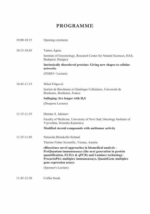
PROGRAMME
10:00-10:15 Opening ceremony
10:15-10:45 Tantos Ágnes
Institute of Enzymology, Research Center for Natural Sciences, HAS, Budapest, Hungary
Intrinsically disordered proteins: Giving new shapes to cellular networks
(FEBS3+ Lecture)
10:45-11:15 �������������
Institut de Biochimie et Génétique Cellulaires, Université de Bordeaux, Bordeaux, France
Sulfaging: live longer with H2S
(Diaspora Lecture)
11:15-11:35 Dimitar S. Jakimov
Faculty of Medicine, University of Novi Sad; Oncology Institute of Vojvodina, Sremska Kamenica
Modified steroid compounds with antitumor activity
11:35-11:45 Natascha Brinskelle-Schmal
Thermo Fisher Scientific, Vienna, Austria
eBioscience novel approaches in biomedical analysis –ProQuantum immunoassays (the next generation in protein quantification, ELISA & qPCR) and Luminex technology: ProcartaPlex multiplex immunoassays, QuantiGene multiplex gene expression assays
(Sponsor's Lecture)
11:45-12:30 Coffee break
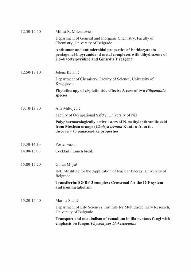
12:30-12:50 ����� �������������
Department of General and Inorganic Chemistry, Faculty of Chemistry, University of Belgrade
Antitumor and antimicrobial properties of isothiocyanato pentagonal-bipyramidal d metal complexes with dihydrazone of 2,6-diacetylpyridine and Girard's T reagent
12:50-13:10 ����� �� � ���
Department of Chemistry, Faculty of Science, University of Kragujevac
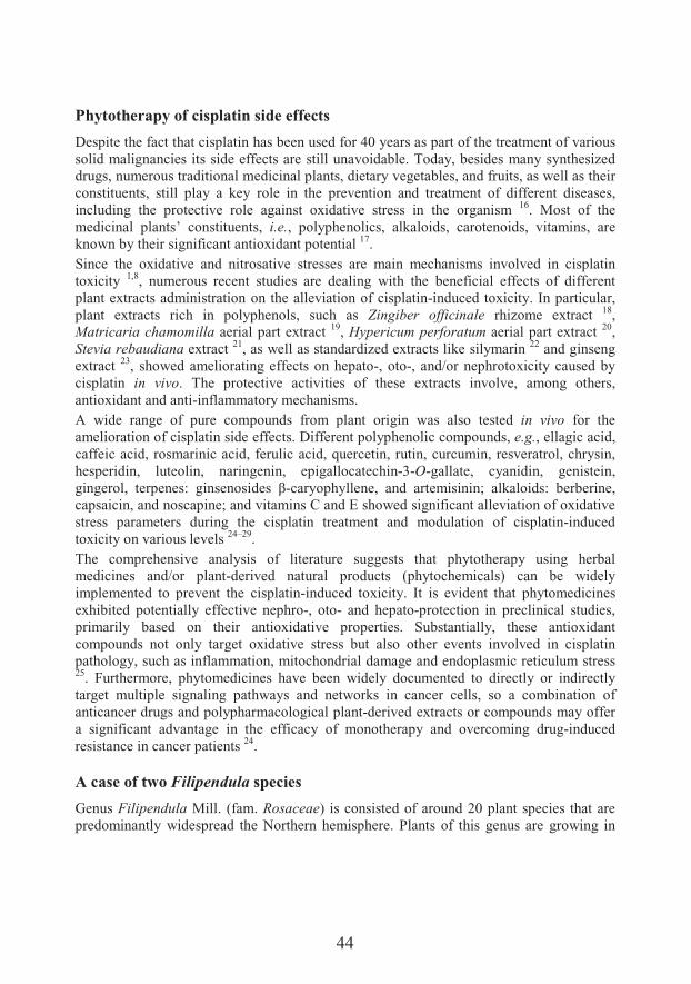
Phytotherapy of cisplatin side effects: A case of two Filipendulaspecies
13:10-13:30 Ana ���������
Faculty of Occupational Safety, University of Niš
Polypharmacologically active esters of N-methylanthranilic acid from Mexican orange (Choisya ternata Kunth): from the discovery to panacea-like properties
13.30-14.30 Poster session
14.00-15.00 Cocktail / Lunch break
15:00-15:20 Goran Miljuš
INEP-Institute for the Application of Nuclear Energy, University of Belgrade
Transferrin/IGFBP-3 complex: Crossroad for the IGF system and iron metabolism
15:20-15:40 � ��� ��� ����
Department of Life Sciences, Institute for Multidisciplinary Research, University of Belgrade
Transport and metabolism of vanadium in filamentous fungi with emphasis on fungus Phycomyces blakesleeanus
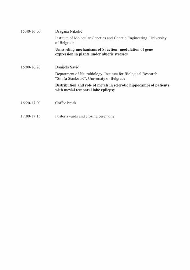
15:40-16:00 �� � � ��������
Institute of Molecular Genetics and Genetic Engineering, University of Belgrade
Unraveling mechanisms of Si action: modulation of gene expression in plants under abiotic stresses
16:00-16:20 � ����� �� ��
Department of Neurobiology, Institute for Biological Research“Siniš ��� �����”, University of Belgrade
Distribution and role of metals in sclerotic hippocampi of patients with mesial temporal lobe epilepsy
16:20-17:00 Coffee break
17:00-17:15 Poster awards and closing ceremony
Poster Session
Miloš Avramov Department of Biology and Ecology, Faculty of Sciences, University of Novi SadIntrinsic disorder and insect diapause – A first look
Sanja Berežni Department of Chemistry, Biochemistry and Environmental Protection, Faculty of Sciences, University of Novi SadClassification and elucidation of Anthriscus sylvestris lignans using spectral information
���� ���� ������Department of Biology and Ecology, Faculty of Science, University of KragujevacCytotoxic and proapoptotic effects of extracts from Vitis vinifera L. petioleon colon cancer cell lines
Luka BreberinaDepartment of Biochemistry, Faculty of Chemistry, University of BelgradeComputational analysis of non-covalent interactions in phycocyanin subunit interfaces
����� �� ���Department of Pharmacy, Faculty of Medicine, University of Novi SadConsumption of monoclonal antibodies in Serbia in period 2006-2016
��� � �����Department of Pharmacy, Faculty of Medicine, University of Novi SadExcipients in iron medications as potential causes of side effects in pediatric population
����� ������������Life Sciences Department, Institute for Multidisciplinary Research, University of BelgradeBiliverdin-copper complex at the physiological pH
Yaraslau U. DzichenkaInstitute of Bioorganic Chemistry of National Academy of Sciences, Minsk, BelarusModified androstane and estrane steroids as novel ligands of cytochromes P450
�� �!� ����� ������� ��� "��#�����$%� "�&���#��� ���� �������� �� ��&� ��'� (����� � �� �����)%�University of BelgradeEthyl pyruvate has tolerogenic effects on dendritic cells
Maja HitlDepartment of Pharmacy, Faculty of Medicine, University of Novi SadHuman pharmacokinetics of rosmarinic acid
���� � �"����Department of Biology and Ecology, Faculty of Sciences, University of Novi SadEffects of Acetamiprid on gene-specific DNA methylation in zebrafish (Danio rerio)embryos
� � ��� ��������Department of Pharmacy, Faculty of Medicine Novi Sad, University of Novi SadDoes peppermint’s post distilation waste can reduce lipid peroxidation?
Rebeka JójártDepartment of Organic Chemistry, University of Szeged, Szeged, Hungary�������� ��� �������� ��������� �� ����� ���-estrone derivatives for targeted intracrine modulation of estrogen biosynthesis and transport
Dunja KokaiDepartment of Biology and Ecology, Faculty of Sciences, University of Novi SadBiological effects of long-term exposure of human vascular endothelial cells to bisphenol A
����� ���� �Life Sciences Department, Institute for Multidisciplinary Research, University of BelgradeRedox interactions of epinephrine with iron at physiological pH
Gordana KošaninDepartment of Biology and Ecology, Faculty of Sciences, University of Novi SadT-2 toxin inhibits ovulatory genes expression and steroidogenesis through cAMP signaling pathway in human granulosa cells
Ahmed LatifInstitute of Pharmacognosy, University of Szeged, Szeged, HungaryAnticancer and antioxidant effects of naringenin and its semi-synthetic oxime ethers
* �� � �� ����Department of Chemistry, Biochemistry and Environmental Protection, Faculty of Sciences, University of Novi SadPlantago species as modulators of thromboxane A2 and prostaglandin E2 production in inflammation
+� �� �������Faculty of Agriculture, University of Novi SadMitigating biotic stress in soybean (Glycine max L.) by plant-growth-promoting fungi Trichoderma asperellum
���� ������ �����Department of Biology and Ecology, Faculty of Science, University of KragujevacExpression of protein synthesis elongation factor 1A in different physiological stages of winter wheat varieties
�� � � ���������Department of Laboratory Diagnostics, Health Care ,������-�������� ���� �. ����","�/ijaRelationship between hematological parameters and glycemic control in type 2 diabetes mellitus patients
����� ���������Department of Biochemistry, Faculty of Chemistry, University of BelgradeAmyloid fibrillation of ovalbumin
Nikolett NagyDepartment of Pharmacodynamics and Biopharmacy, University of Szeged, Szeged,HungaryAntitumor effects of herbal sesquiterpenes
Ivana NemešDepartment of Chemistry, Biochemistry and Environmental Protection, Faculty of Sciences, University of Novi SadPhenolic content and antioxidant potential of extracts of parsley (Petroselinum crispum) cultivated in the Province of Vojvodina
+� ��� � ������Department of Biochemistry, Faculty of Chemistry, University of BelgradeThe pro-inflammatory effect of Act d 1, cysteine protease from kiwifruit (Actinidia deliciosa), on intestinal epithelial cells in vitro
" � ��������Laboratory for Plant Molecular Biology, Institute of Molecular Genetics and Genetic Engineering, University of BelgradeAnalysis of Arabidopsis intrinsically disordered DSS1(V) protein mutants exposed tooxidative stress
���0 � �1�2��Department of Biology and Ecology, Faculty of Sciences, University of Novi SadSeasonal variation in fatty acid composition of fat body lipids in worker honey bee (Apis mellifera L.)
Marijana .����*#�#�� �Department of Field and Vegetable Crops, Faculty of Agriculture, University of Novi SadThe influence of extraction solvent and maturity stage on antioxidant capacity of fruits of sweet cherry
�� � � �.��� �Department of Chemistry, Biochemistry and Environmental Protection, Faculty of Sciences, University of Novi SadNatural vs. commercial products: comparisson of antioxidant activity

Jovana J. Plavša-PužDepartment of Biology and Ecology, Faculty of Sciences, University of Novi SadCrystal structures of AKR1C3 protein with new potent steroid inhibitors
�#��� ����.����Department of Histology and Embryology, Faculty of Medicine, University of Novi SadAntitumor interaction and safety of metformin and itraconazole low doses in hamster fibrosarcoma
��&� ����.����Department of Pharmacy, Faculty of Medicine, University of Novi SadPhysicochemical and immunohistochemical changes of experimental fibrosarcomas in hamsters treated with low doses of metformin and caffeine
"&� �� �.�����-��&���Department of Biochemistry, Faculty of Chemistry, University of BelgradeProduction, purification and structural characterisation of recombinant BanLec-Bet v 1
+� �������Department of Biochemistry, Faculty of Chemistry, University of Belgrade Isolation, characterization and biological activity of R-phycoerythrin from red macroalgae Porphyra spp.
+���& � ������& �����Department of Analytical Chemistry, Faculty of Chemistry, University of BelgradeCadmium as the main endocrine disrupter in papillary thyroid carcinoma
Seyyed Ashkan Senobar TahaeiDepartment of Pharmacodynamics and Biopharmacy, University of Szeged, Szeged,HungarySynthesis and antiproliferative activities of 16-triazolyl-methyl-17-estradiol derivatives
� ��� �* &���Department of Biochemistry, Institute f��� �������� �� ��&� ��'� (����� � �� �����)%�University of BelgradeThe effects of fructose-rich diet and/or chronic unpredictable stress on antioxidant enzymes function in the rat kidney
�� ��&� �*���������Department of Pharamacy, Faculty of Medicine, University of Novi SadEffects of different formulation of basil extracts (Ocimum basilicum L.) on normoglycemic and diabetic rats
��� �� �*� �����Department of Pharmacy, Faculty of Medicine Novi Sad, University of Novi SadEnzymes as active pharmaceutical ingredients in registered drugs in Serbia
Iva UzelacDepartment of Biology and Ecology, Faculty of Sciences, University of Novi SadThe effect of spermidine supplementation on catalase and Cu, Zn superoxide dismutase gene expression in honey bees
S��0 � �3��� ��Life Sciences Department, Institute for Multidisciplinary Research, University of BelgradeHormetic effects of low-dose radiation on lipid production in Chlorella sorokiniana
����� �3#���Department of Chemistry, Faculty of Science, University of KragujevacProoxidative effects of shikonin derivatives in human breast cancer cell line MDA-MB-231
Milorad Živanov Laboratory for Soil and Agroecology, Institute of Field and Vegetable Crops, Novi SadThe potential of biochar in improving microbial activity of soils in Vojvodina Province
11
Foreword
Dear Colleagues
Welcome to the 8th Conference of the Serbian Biochemical Society, entitled "Coordination in Biochemistry and Life".
The title of this year’s Conference refers to an important place of coordinationchemistry in biochemistry and biomedicine, but also to a need to coordinate the efforts towards new knowledge with fellow scientists from other fields in order to reach more. The collaboration within FEBS3+ (Croatia, Hungary, Slovenia, and Serbia) Meeting Programme continues with the invited lecture of our dear colleague Tantos Ágnes from Research Center for Natural Sciences, Budapest, Hungary. For the first time we have 4�� &�� �5���#��6� �' ��7���� 8�� ������ � 8$������� �������%� � ��� 4�� #��6� ��� ���8� ��biochemistry who is now affiliated at the Université de Bordeaux. We have more than forty PhD students from Serbia, Hungary, and Belarus with poster presentations, and for the first time the Conference is held outside the capital. It believe that we are getting better each year, and that we are prepared for future challenges.
I would like to express my gratitude to the members of the Scientific Board who suggested lecturers, to all respected colleagues who accepted the invitation, and to our dear hosts from the University of Novi Sad.
Editor of the Proceedings����������� �

13
Proceedings
Agnes Tantos
Intrinsically disordered proteins: Giving new shapes to cellular networks (19)
� ���� � �� �
Sulfaging: live longer with H2S (23)
Dimitar S. Jakimov
Modified steroid compounds with antitumor activity (25)
� � ������� ����� �
Antitumor and antimicrobial properties of isothiocyanato pentagonal-bipyramidal d metal complexes with dihydrazone of 2,6-diacetylpyridine and Girard's T reagent (33)
������������ �
Phytotherapy of cisplatin side effects: A case of two Filipendula species (41)
����� ����� �
Polypharmacologically active esters of N-methylanthranilic acid from Mexican orange (Choisya ternata Kunth): from the discovery to panacea-like properties (53)
Goran Miljuš
Transferrin/IGFBP-3 complex: Crossroad for the IGF system and iron metabolism (67)
��� ������� ��
Transport and metabolism of vanadium in filamentous fungi with emphasis on fungus Phycomyces blakesleeanus (81)
��������� �� �
Unraveling mechanisms of Si action: modulation of gene expression in plants under abiotic stresses (93)
14
��� �������� �
Distribution and role of metals in sclerotic hippocampi of patients with mesial temporal lobe epilepsy (105)
Miloš Avramov Intrinsic disorder and insect diapause – A first look (113)
Sanja Berežni Classification and elucidation of Anthriscus sylvestris lignans using spectral information(115)
�������!������ �Cytotoxic and proapoptotic effects of extracts from Vitis vinifera L. petioleon colon cancer cell lines (117)
Luka BreberinaComputational analysis of non-covalent interactions in phycocyanin subunit interfaces (119)
�������"��� Consumption of monoclonal antibodies in Serbia in period 2006-2016 (121)
!�����"� �Excipients in iron medications as potential causes of side effects in pediatric population(123)
� ������ # �� ��� �Biliverdin-copper complex at the physiological pH (125)
Yaraslau U. DzichenkaModified androstane and estrane steroids as novel ligands of cytochromes P450 (127)
��$��%�$� �Ethyl pyruvate has tolerogenic effects on dendritic cells (129)

15
Maja HitlHuman pharmacokinetics of rosmarinic acid (131)
! ���������� �Effects of Acetamiprid on gene-specific DNA methylation in zebrafish (Danio rerio)embryos (133)
����� �������# �Does peppermint’s post distilation waste can reduce lipid peroxidation? (135)
Rebeka Jójárt�$��'�&�&� � � 8���'���� �� � �# ����� ��� ����� 9:;-estrone derivatives for targeted intracrine modulation of estrogen biosynthesis and transport (137)
Dunja KokaiBiological effects of long-term exposure of human vascular endothelial cells to bisphenol A (139)
�����������Redox interactions of epinephrine with iron at physiological pH (141)
Gordana KošaninT-2 toxin inhibits ovulatory genes expression and steroidogenesis through cAMP signaling pathway in human granulosa cells (143)
Ahmed LatifAnticancer and antioxidant effects of naringenin and its semi-synthetic oxime ethers (145)
&����������� �Plantago species as modulators of thromboxane A2 and prostaglandin E2 production in inflammation (147)
���������� �Mitigating biotic stress in soybean (Glycine max L.) by plant-growth-promoting fungi Trichoderma asperellum (149)
16
��������������� �Expression of protein synthesis elongation factor 1A in different physiological stages of winter wheat varieties (151)
��������� ���� �Relationship between hematological parameters and glycemic control in type 2 diabetes mellitus patients (153)
��� ���� ���� �Amyloid fibrillation of ovalbumin (155)
Nikolett NagyAntitumor effects of herbal sesquiterpenes (157)
Ivana NemešPhenolic content and antioxidant potential of extracts of parsley (Petroselinum crispum)cultivated in the Province of Vojvodina (159)
��$� �������� �The pro-inflammatory effect of Act d 1, cysteine protease from kiwifruit (Actinidia deliciosa), on intestinal epithelial cells in vitro (161)
������� �� �Analysis of Arabidopsis intrinsically disordered DSS1(V) protein mutants exposed to oxidative stress (163)
���'����(�) �Seasonal variation in fatty acid composition of fat body lipids in worker honey bee (Apis mellifera L.) (165)
��� �����*� ��&+�+����The influence of extraction solvent and maturity stage on antioxidant capacity of fruits of sweet cherry (167)
� ��$���* ����Natural vs. commercial products: comparisson of antioxidant activity (169)
17
Jovana J. Plavša-PužCrystal structures of AKR1C3 protein with new potent steroid inhibitors (171)
�+� ������*�� �Antitumor interaction and safety of metformin and itraconazole low doses in hamster fibrosarcoma (173)
�������*�� �Physicochemical and immunohistochemical changes of experimental fibrosarcomas in hamsters treated with low doses of metformin and caffeine (175)
� $���*�� �-� ��Production, purification and structural characterisation of recombinant BanLec-Bet v 1(177)
����� #� �Isolation, characterization and biological activity of R-phycoerythrin from red macroalgae Porphyra spp. (179)
������$������������ �Cadmium as the main endocrine disrupter in papillary thyroid carcinoma (181)
Seyyed Ashkan Senobar TahaeiSynthesis and antiproliferative activities of 16-triazolyl-methyl-17-estradiol derivatives(183)
��� ���&� ��The effects of fructose-rich diet and/or chronic unpredictable stress on antioxidant enzymes function in the rat kidney (185)
!��� �����&�� �� ��Effects of different formulation of basil extracts (Ocimum basilicum L.) on normoglycemic and diabetic rats (187)
��#�����&$�� �Enzymes as active pharmaceutical ingredients in registered drugs in Serbia (189)
18
Iva UzelacThe effect of spermidine supplementation on catalase and Cu, Zn superoxide dismutase gene expression in honey bees (191)
���'����,��$ �Hormetic effects of low-dose radiation on lipid production in Chlorella sorokiniana (193)
Mil����,+� �Prooxidative effects of shikonin derivatives in human breast cancer cell line MDA-MB-231 (195)
Milorad Živanov The potential of biochar in improving microbial activity of soils in Vojvodina Province (197)
19
Intrinsically disordered proteins: Giving new shapes to cellular networks
Agnes Tantos1*, Peter Tompa1,2
1Institute of Enzymology, Research Centre for Natural Sciences, Hungarian Academy ofSciences, Budapest, Hungary2VIB, Center for Structural Biology (CSB), Vrije Universiteit Brussel (VUB), Brussels,Belgium
*e-mail: [email protected]
With the deeper understanding of the molecular details of the cellular processes, it has become increasingly clear that protein function is inseparable from the context of its encompassing interaction network. The application of high-throughput techniques enabled the mapping of complete protein-protein interaction (PPI) network of certain organisms 1–3.These PPI networks appear to display scale free topology, consisting of many nodes with small number interaction partners and a few hubs with a high number of partners 4.Cellular PPI networks are also highly dynamic 5, consisting of elements that can be modified, shifting interaction preferences in reaction to external signals.Hub proteins need to posses specific properties that enable them to interact with many structurally and sequentially different partners. It has been shown that intrinsically disordered proteins (IDPs) and proteins with intrinsically disordered regions (IDRs) are significantly overrepresented among hub proteins 6. IDPs and IDRs, lacking a stable three-dimensional fold, exist in a state described as conformational ensembles, sampling a multitude of different conformations under physiological conditions 7. The particular adaptability resulting of this conformational variability enables IDPs/IDRs to recognize and bind their various partners, making them central players in PPI networks.During the past few years, many specific features of IDP-partner recognition and binding have been uncovered, starting from the use of linear motifs 8, through binding without folding, known as fuzzy complexes 9, to the driving of liquid-liquid phase separation 10.The most intriguing new feature of IDP interaction is the recognition of their role in phase transitions inside the cells. The presence of phase separated cellular compartments, also known as membraneless organelles, has been known for many decades, but the molecular details of their formation and exact composition have been only recently started to be uncovered. These organelles take part in the regulation of many physiological processes like stress response 11, signal transduction 12, and the control of gene expression 13. Not separated from their environment by lipid bilayers, for long it has remained unclear how membraneless organelles are able to maintain and regulate their structures and compositions. A major step towards understanding this phenomenon was when it became
20
clear that phase separation is mostly driven by the intermolecular interactions formed by low complexity proteins sequences that contain repetitive sequences of single amino acids (such as polyQ or polyN) or amino acid motifs. These regions reside in disordered segments of proteins, shedding new light on the importance and function of IDPs. Although the ability to phase separate is not exclusive to disordered protein regions, as folded proteins are also known to go through phase separation under specific conditions, IDR containing proteins do so faster and easier either by themselves or by binding to nucleic acids 14. Another important feature of this process is that it can be regulated by the post-translational modifications that often occur in the disordered segments of proteins. Given that phase transitions are generally triggered by the interaction of alternately charged residues, subtle changes in charge arising from phosphorylation, methylation and acetylation could easily initiate the switch between different states 14.Indeed, strict regulation of the process of phase separation is necessary, since mounting evidence shows that dysregulated phase transitions are largely involved in pathological processes such as neurodegenerative disorders 15, and even cancers 16. Mutations in the region of proteins responsible for phase separation might accelerate the process 17,18, or cause aberrant protein-RNA interactions 19, resulting in loss of function of the components.The realization that IDPs/IDRs are directly involved in the process of phase separation has significantly broadened our understanding of the role they play in rewiring functional networks within the cells and may also provide new strategies in fighting against several diseases.
References1. Uetz P, et al. A comprehensive analysis of protein-protein interactions in Saccharomyces
cerevisiae. Nature 2000;403:623–7.2. Guruharsha KG, et al. A protein complex network of Drosophila melanogaster. Cell
2011;147:690–703.3. Rual JF, et al. Towards a proteome-scale map of the human protein-protein interaction network.
Nature 2005;437:1173–8.4. Cumberworth A, Lamour G, Babu MM, Gsponer J. Promiscuity as a functional trait:
intrinsically disordered regions as central players of interactomes. Biochem J 2013;454:361–9.5. de Lichtenberg U. Dynamic complex formation during the yeast cell cycle. Science
2005;307:724–7.6. Haynes C, et al. Intrinsic disorder is a common feature of hub proteins from four eukaryotic
interactomes. PLoS Comput Biol 2006;2:e100.7. Varadi M, et al. pE-DB: a database of structural ensembles of intrinsically disordered and of
unfolded proteins. Nucleic Acids Res 2014;42:D326–35.8. Davey NE, et al. Attributes of short linear motifs. Mol Biosyst 2012;8:268–81.9. Miskei M, et al. Fuzziness enables context dependence of protein interactions. FEBS Lett
2017;591:2682–95.10. Uversky VN, Kuznetsova IM, Turoverov KK, Zaslavsky B. Intrinsically disordered proteins as
crucial constituents of cellular aqueous two phase systems and coacervates. FEBS Lett2015;589:15–22.
11. Riback JA, et al. Stress-triggered phase separation is an adaptive, evolutionarily tuned response. Cell 2017;168:1028–40.
12. Li P, et al. Phase transitions in the assembly of multivalent signalling proteins. Nature 2012;483:336–40.
21
13. Sawyer IA, Bartek J, Dundr M. Phase separated microenvironments inside the cell nucleus are linked to disease and regulate epigenetic state, transcription and RNA processing. Semin Cell Dev Biol 2018;10.1016/j.semcdb.2018.07.001.
14. Bergeron-Sandoval LP, Safaee N, Michnick SW. Mechanisms and consequences of macromolecular phase separation. Cell 2016;165:1067–79.
15. Boeynaems S, et al. Protein phase separation: A new phase in cell biology. Trends Cell Biol2018;28:420–35.
16. Boeynaems S, Tompa P, Van Den Bosch L. Phasing in on the cell cycle. Cell Div 2018;13:1.17. Conicella AE, Zerze GH, Mittal J, Fawzi NL. ALS mutations disrupt phase separation mediated
8$� ;-helical structure in the TDP-43 low-complexity C-terminal domain. Structure 2016;24: 1537–49.
18. Patel A, et al. A liquid-to-solid phase transition of the ALS protein FUS accelerated by disease mutation. Cell 2015;162:1066–77.
19. Wojciechowska M, Krzyzosiak WJ. Cellular toxicity of expanded RNA repeats: focus on RNA foci. Hum Mol Genet 2011;20:3811–21.

23
Sulfaging: live longer with H2S
�������������������������������������-Roux, Bikash Adhikari, Jan �� !�������"#$%�&���*
Institut de Biochimie et Génétique Cellulaires, CNR, Université de Bordeaux, Bordeaux, France
*e-mail: [email protected]
Although considered a toxic gas for more than a century, hydrogen sulfide (H2S) was one of the essential ingredients required for life to emerge on Earth. During the past decade numerous studies have revealed important physiological/pathophysiological roles of H2S as a signaling molecule. Oxidative posttranslational modification of cysteine residues, called protein persulfidation, has been proposed as a unifying mechanism behind this numerous effects. Persulfides are difficult to label and study due to their reactivity and similarity with cysteine. We now report a facile strategy for chemoselective persulfide bioconjugation to achieve highly selective, rapid, and robust persulfide labeling in biological samples with broad utility. Using this new method we show that persulfidation is evolutionary conserved posttranslational modifications. Waves of persulfidation are employed by cells to resolve sulfenylation and prevent irreversible cysteine overoxidation to preserve protein function. This is used for both signaling purposes (like in the case of receptor tyrosine kinase signaling) and general protection. We report an age-associated decline in persulfidation which is conserved across evolutionary boundaries. Accordingly, pharmacological or dietary interventions (such as calorie restriction) to increase persulfidation, result in increased longevity and improved capacity to cope with stress stimuli.

25
Modified steroid compounds with antitumor activity
Dimitar Jakimov1*�'������ �1�(� �)��!��1�*������+�,������1, Jovana ) ��!���2�)�����-!���2���,�� �.������2, Katarina Penov Gaši2, Marija ��!�/2����������0���-Ušaj1���1�����������-Šanta2
1Oncology Institute of Vojvodina, Faculty of Medicine, University of Novi Sad, SremskaKamenica, Serbia2Department of Chemistry, Biochemistry and Environmental Protection, Faculty ofSciences, University of Novi Sad, Novi Sad, Serbia
*e-mail: [email protected]
�������� ������ �������� ��� �2�-picolyl and 17(E)-picolinylidene androstane derivatives and five 16,17-seco-steroid dinitriles were examined for their in vitro antiproliferative effect and proapoptotic potential. The antiproliferative impact was examined on six human tumor cell lines, including two types of breast (MCF-7 and MDA-MB-231), prostate (PC3), cervical (HeLa), colon (HT 29) and lung cancer (A549), one leukemia (K562), as well as one normal fetal lung fibroblasts cell line (MRC-5). All of the tested steroidal compounds showed antiproliferative activity against the MDA-MB-231 triple-negative breast adenocarcinoma and against most of the treated human solid tumors cell lines. We used MDA-MB-231 cell line, as the most affected by steroidal compounds, to investigate their apoptotic potential. Apoptosis of the treated MDA-MB-231 cells was confirmed by three different methods, including apoptosis detection by flow cytometry, expression of proteins of the apoptotic signaling pathway and apoptotic morphology screening. Tested androstane derivatives modulated the cell cycle distribution and induced apoptosis and necrosis. Compounds had different and specific mode of action, depending on derivative type and exposure time. Structure–activity relationship analysis was performed to find correlations between the structure variations of investigated derivatives and observed biological effects. Results of presented study show that some of the investigated androstane derivatives have good biomedical potential and could be candidates for the development of effective and selective anticancer drugs.
Introduction
The discovery of new effective and selective antitumor drugs is especially important today, when there is a large number of malignant diseases in the world, and therapy is often not adequate, acting also on healthy tissues, undermining the health of patients.Steroids are endogenous molecules in human organism. By modifying the structure of steroidal molecules, their physical and chemical properties, and thus the influence on
26
biological systems, also change. Even small changes in the structure can lead to significant changes in the biological response. Therefore, the correlation of the structure of tested compounds and their pharmacological activity must be investigated.The present study aimed at investigating the biological properties of 14 selected steroidal derivatives through the examination of in vitro antiproliferative effect and apoptotic potential against human cancer cell lines. The results presented in this study are published in several scientific publications 1-5.
Materials and methods
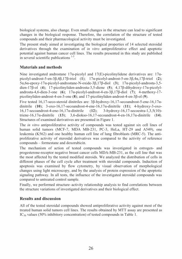
����� ���&��� �� � � ��&� ��� 9<;-picolyl and 17(E)-������$�� ���� ��� ���&� ��=� 9<;-picolyl-androst-5-en-:>%?>%9<>-triol (1@A� 9<;-picolyl-androst-5-en-:>%?;%9<>-triol (2); B;%C;-epoxy-9<;-picolyl-androstane-N-oxide-:>%9<>-diol (3@A� 9<;-picolyl-androsta-3,5-dien-9<>-ol (4); 17-picolinyliden-androsta-3,5-diene (5@A� ?%9<>-dihydroxy-9<;-picolyl-androsta-4,6-dien-3-one (6@A� 9<;-picolyl-androst-4-en-:>%9<>-diol (7); 4-methoxy-17-picolinyliden-androst-4-en-3-one (8); and 17-picolinyliden-androst-4-en-:>-ol (9).Five tested 16,17-seco-&����� � ��������&� ��=�:>-hydroxy-16,17-secoandrost-5-ene-9C%9<;-dinitrile (10); 3-oxo-16,17-secoandrost-4-ene-9C%9<;-dinitrile (11); 4-hydroxy-3-oxo-16,17-secoandrost-4-ene-9C%9<;-dinitrile (12); 3-hydroxy-16,17-secoestra-1,3,5(10)-triene-9C%9<;-dinitrile (13); 3,6-diokso-16,17-secoandrost-4-en-9C%9<;-dinitrile (14). Structures of examined derivatives are presented in Figure 1.The in vitro antiproliferative activity of compounds was tested against six cell lines of human solid tumors (MCF-7, MDA MB-231, PC-3, HeLa, HT-29 and A549), one leukemia (K562) and one healthy human cell line of lung fibroblasts (MRC-5). The anti-proliferative activity of steroidal derivatives was compared to the activity of reference compounds – formestane and doxorubicin.The mechanism of action of tested compounds was investigated in estrogen- and progesterone-receptor negative breast cancer cells MDA-MB-231, as the cell line that was the most affected by the tested modified steroids. We analyzed the distribution of cells in different phases of the cell cycle after treatment with steroidal compounds. Induction of apoptosis was examined by flow cytometry, by visual observation of morphological changes using light microscopy, and by the analysis of protein expression of the apoptotic signaling pathway. In all tests, the influence of the investigated steroidal compounds was compared to untreated control sample.Finally, we performed structure–activity relationship analysis to find correlations between the structure variations of investigated derivatives and their biological effect.
Results and discussion
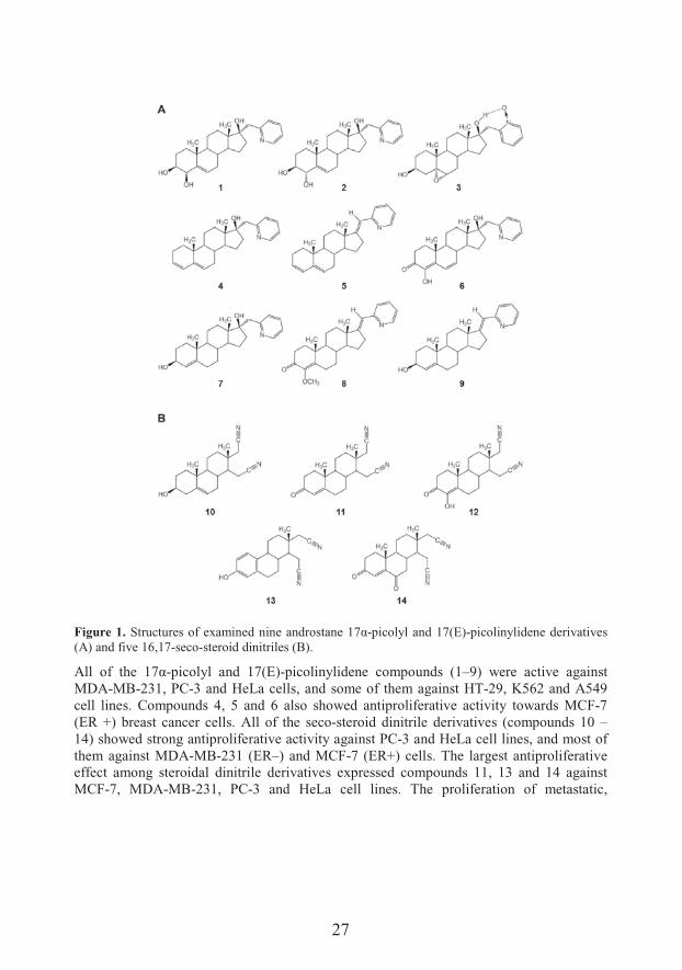
All of the tested steroidal compounds showed antiproliferative activity against most of the treated human solid tumors cell lines. The results obtained by MTT assay are presented as IC50 values (50% inhibitory concentration) of tested compounds in Table 1.
27
Figure 1. Structures of examined ����� � ��&� ���9<;-picolyl and 17(E)-picolinylidene derivatives (A) and five 16,17-seco-steroid dinitriles (B).
+��� ��� �'�� 9<;-picolyl and 17(E)-picolinylidene compounds (1–9) were active against MDA-MB-231, PC-3 and HeLa cells, and some of them against HT-29, K562 and A549 cell lines. Compounds 4, 5 and 6 also showed antiproliferative activity towards MCF-7(ER +) breast cancer cells. All of the seco-steroid dinitrile derivatives (compounds 10 –14) showed strong antiproliferative activity against PC-3 and HeLa cell lines, and most of them against MDA-MB-231 (ER–) and MCF-7 (ER+) cells. The largest antiproliferative effect among steroidal dinitrile derivatives expressed compounds 11, 13 and 14 against MCF-7, MDA-MB-231, PC-3 and HeLa cell lines. The proliferation of metastatic,
28
hormone-independent breast cancer cell line MDA-MB-231 was strongly inhibited by all of the investigated steroid derivatives.
Table 1. The IC50 values [μM] of tested derivatives 1–14 and reference compounds formestane (For) and doxorubicin (Dox) after 48 h treatment. Bolded are values below 10 μM. The IC50 values greaterthen 100 μM are marked with „dash“ symbol (–).
Cell lines
Compounds MCF-7MDA-
MB-231PC-3 HeLa HT-29 A549 K562 MRC-5
Pic
olyl
and
pic
olin
ylid
ene
deri
vati
ves
1 – 6.43 41.29 23.97 5.29 – – –
2 – 4.56 22.38 21.07 2.46 – – –
3 N/A 5.18 28.21 32.73 17.41 – N/A N/A
4 12.32 2.17 19.37 22.38 10.46 – 13.72 –
5 52.70 3.82 20.62 18.69 1.04 16.87 52.44 N/A
6 4.11 1.03 21.37 29.74 – – 30.74 –
7 – 2.50 22.53 24.25 N/A 12.74 27.31 –
8 – 3.46 33.16 10.25 45.83 – – –
9 – 5.18 11.50 7.82 16.43 – 75.57 –
Din
itri
le
deri
vati
ves
10 30.04 34.82 19.12 2.15 – – – N/A
11 15.02 15.51 8.27 5.73 – – – –
12 56.31 39.59 8.69 14.71 – – – –
13 16.96 3.96 11.18 7.32 – – – –
14 – 6.66 9.65 4.02 – – – –
Ref. com-pounds
For – 53.29 45.65 1.90 – – – –
Dox 0.62 0.17 89.90 1.68 0.10 7.52 0.42 0.11
N/A - IC50 value was not available due to nonlinear dose dependence or hormetic effect.
None of the tested steroidal compounds (1–14) was toxic against healthy MRC-5 cells, unlike Doxorubicin, a widely used cytostatic in the therapy of malignant diseases. When compared to the reference compounds, the majority of the investigated steroid derivatives was more effective than Formestane, except in HeLa cell line, and all of them were more toxic than Doxorubicin only to the PC-3 cells.Cell cycle modulation and apoptosis induction. All tested steroidal compounds, more or less, induced apoptosis in MDA-MB-231 cells. Picolyl and picolinylidene derivatives influenced the cell cycle phase distribution, primarily by shifting the cell population toward subG1 or G2/M phase, and reducing the G0/G1 and S phase. The changes were specific for the compound and the treatment period. Steroidal seco-dinitriles also affected the cell cycle phase distribution of MDA-MB-231 cells. Changes depended on the treatment time. Derivatives 11, 12 and 14 lowered the subG1 phase of the cell cycle after

29
48 h, while after 72 h all dinitriles caused an increase of this apoptotic cell fraction, as well as the S phase, with a reduction in G0/G1 and G2/M phases. The largest increase in subG1 phase induced compounds 13 and 14.The Annexin V apoptosis induction assay shown that the tested compounds act via different mechanisms. The apoptotic/necrotic response of cells depended on length of the treatment. According to the results of the flow-cytometric analysis, the largest percentage of specific apoptosis was induced by picolyl and picolinylidene derivatives 1, 4, 6 and 7, after 72 h treatment. All dinitrile derivatives induced low percentage of specific apoptosis during 48 h. However, after 72 h, compounds 10, 11, 13 and 14 achieved a value of specific apoptosis more than 25%, and all of them were more effective than Formestane. Compound 13 induced the highest specific apoptosis (over 50%). The results of apoptosis induced by dinitrile derivatives were very similar to those of the subG1 cell cycle phase modulation, especially after 72 hours of treatment. Among all tested steroidal derivatives, compounds 11 (after 48 h) and 14 (after 72 h) caused the highest necrosis of the treated MDA-MB-231 cells.Expression of apoptotic proteins. All picolyl and picolinylidene derivatives (except compound 1) after 48 h induced a high expression of proapoptotic BAX protein in treated MDA-MB-231 cells and reduced the expression of the antiapoptotic Bcl-2 proteins (with the exception of compounds 1 and 7). Detection of the proteolytic cleavage of the PARP protein in samples treated with all picolyl and picolinylidene compounds confirmed the signaling activity and the completion of the apoptotic process. The lack of caspase-3activation by compounds 3, 4, 5 and 8 after 48 h, as well as by all the picolyl and picolinylidene compounds after 72 h (except compound 3 and formestane), suggests that these derivatives induced apoptosis in caspase-independent manner.The apoptotic protein expression differed significantly among dinitrile derivatives, depending on treatment duration. Thus, the expression of Bcl-2 was increased after 48 h only for compound 14, and the most pronounced decrease was observed after 72 h for compounds 10 and 11. The expression of BAX protein was increased in all samples treated 48 h with dinitrile derivatives, which was even more evident after 72 h treatment. The highest BAX expression was induced by compounds 10, 11, 13 and 14. According to the expression of precursors and active subunit of caspase-3, it was lower than control one only for derivative 14 and Formestane after 48 h, with a nearly control level for all dinitrile compounds after 72 h. Compounds 10, 13 and 14 (as well as Formestane) increased the expression of PARP protein after 48 h, which was smaller and more uniform after 72 h. Evident PARP expression, induced by all the tested steroidal dinitriles regardless their structure, tells about the activity of the signaling pathway in the final stages of apoptosis.Modulation of the apoptotic protein expression showed that some of the investigated steroidal derivatives (4, 5, 6, 7, 8, 13, and 14) have proapoptotic effect and that dinitrile derivatives act differently on the expression of apoptotic protein in comparison to picolyl and picolinylidene compounds.Apoptotic morphology. Among tested picolyl and picolinylidene derivatives, the most of apoptotic morphological changes in MDA-MB-231 cells induced compounds 1, 4–6 and 8,
30
where 1, 5 and 8 more than Formestane. Among dinitrile derivatives, compounds 10 and 11 were the most efficient in the induction of apoptotic morphology after 48 h, and derivatives 12–14 after 72 h treatment. The most apoptotic morphological changes sparked estrane compound 13 after 72 h of treatment.
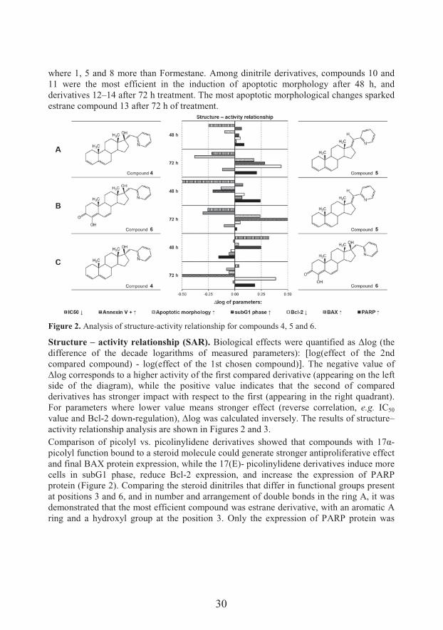
Figure 2. Analysis of structure-activity relationship for compounds 4, 5 and 6.
Structure – activity relationship (SAR). �������� ��������&�7����D# ������ � &�E����F�'��difference of the decade logarithms of measured parameters): [log(effect of the 2nd compared compound) - log(effect of the 1st chosen compound)]. The negative value of E���������&�� &���� �'��'���activity of the first compared derivative (appearing on the left side of the diagram), while the positive value indicates that the second of compared derivatives has stronger impact with respect to the first (appearing in the right quadrant). For parameters where lower value means stronger effect (reverse correlation, e.g. IC50
value and Bcl-2 down-���#� ����@%�E����7 &�� ��#� �� �����&��$��*'����&#��&����&��#��#��–activity relationship analysis are shown in Figures 2 and 3.,�� ��&��� ��� ����$�� &�� ������$�� ���� ��� ���&� &'�7� � �' �� ����#� &� 7��'� 9<;-picolyl function bound to a steroid molecule could generate stronger antiproliferative effect and final BAX protein expression, while the 17(E)- picolinylidene derivatives induce more cells in subG1 phase, reduce Bcl-2 expression, and increase the expression of PARP protein (Figure 2). Comparing the steroid dinitriles that differ in functional groups present at positions 3 and 6, and in number and arrangement of double bonds in the ring A, it was demonstrated that the most efficient compound was estrane derivative, with an aromatic A ring and a hydroxyl group at the position 3. Only the expression of PARP protein was
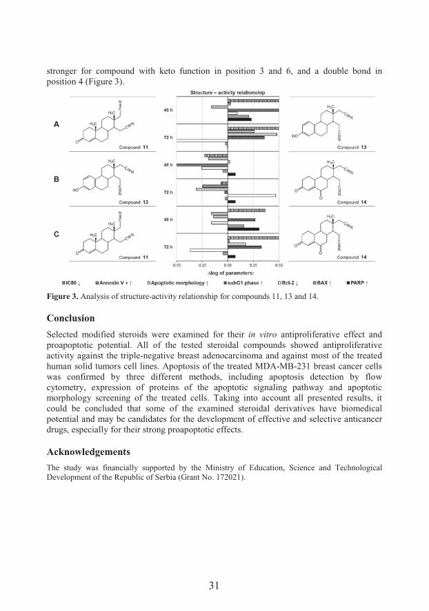
31
stronger for compound with keto function in position 3 and 6, and a double bond in position 4 (Figure 3).
Figure 3. Analysis of structure-activity relationship for compounds 11, 13 and 14.
Conclusion
Selected modified steroids were examined for their in vitro antiproliferative effect and proapoptotic potential. All of the tested steroidal compounds showed antiproliferative activity against the triple-negative breast adenocarcinoma and against most of the treated human solid tumors cell lines. Apoptosis of the treated MDA-MB-231 breast cancer cells was confirmed by three different methods, including apoptosis detection by flowcytometry, expression of proteins of the apoptotic signaling pathway and apoptotic morphology screening of the treated cells. Taking into account all presented results, it could be concluded that some of the examined steroidal derivatives have biomedical potential and may be candidates for the development of effective and selective anticancer drugs, especially for their strong proapoptotic effects.
Acknowledgements
The study was financially supported by the Ministry of Education, Science and Technological Development of the Republic of Serbia (Grant No. 172021).
32
References
1. ��#��� ���G%� ��� ����$��'�&�&� � � �$����H��� �����$���� &����9<-picolyl and 17-picolinylidene androstane derivatives. Eur J Med Chem 2012;54:784-92.
2. +� #������%���� ����$��'�&�&%�&��#��#� �� � �$&�&� � � ����#���� �����$���������9< ����$�� � �17(E)-picolinylidene A-modified androstane derivatives. Bioorgan Med Chem 2015;23:1557-68.
3. ��������+%���� ����$��'�&�&� � � ���� ���������������� �����&����� ��9C%9<-seco-16,17a-dinitriles: Identification of a selective inhibitor of hormone-independent breast cancer cells. Bioorgan Med Chem 2015;23:703-11.
4. Jakimov D, et al. Androstane derivatives induce apoptotic death in MDA-MB-231 breast cancer cells. Bioorgan Med Chem 2015;23:7189–98.
5. Jakimov D. Effect of modified steroid compounds on cell cycle, apoptosis induction and occurrence of genetic defects in human tumor cells. Doctoral thesis. Department of Chemistry, Biochemistry and Environmental Protection, Faculty of Sciences, University of Novi Sad, Novi Sad, 2016.
33
Antitumor and antimicrobial properties of isothiocyanato pentagonal-bipyramidal d metal complexes with dihydrazone of 2,6-diacetylpyridine and Girard's T reagent
����#$����!���1*, �������)�3��!���1, 4����5$����2, ��������'� /�3,6�"�������1, +�0���7� �� �1, ���#�������1
1Faculty of Chemistry, University of Belgrade, Belgrade, Serbia2Institute of Oncology and Radiology of Serbia, Belgrade, Serbia3Institute for Chemistry, Technology and Metallurgy, University of Belgrade
*e-mail: [email protected]
Pentagonal-bipyramidal complexes of 2,6-diacetylpyridine bis(acylhydrazone) ligands are attractive field of research not only in structural inorganic chemistry and magnetochemistry, but also in bioinorganic chemistry since they exhibit cytotoxic, antimicrobial, SOD mimetic, DNA/RNA binding and nuclease activity. In this work we investigated antitumor and antimicrobial activity of pentagonal-bipyramidal isothiocyanato complexes of Mn(II) ([Mn(H2L)(NCS)2](SCN)2·CH3OH) (1), Ni(II) ([Ni(H2L)(NCS)2](SCN)2·2H2O) (2), Co(II) ([Co(H2L)(NCS)2](SCN)2·2H2O (3) and [Co(H2L)(NCS)2][Co(NCS)4]·2H2O) (4), Zn(II) ([Zn(H2L)(NCS)2][Zn(NCS)4]·2H2O) (5), Cd(II) ([Cd(H2L)(NCS)2][Cd(NCS)4]·2H2O) (6) and Fe(III) ([Fe(L)(NCS)2](SCN)·2H2O (7) and [Fe(L)(NCS)2][Fe(NCS)5(H2O)]·4H2O) (8), with the condensation product of 2,6-diacetylpyridine and Girard’s T reagent (H2LCl2). The complexes showed moderate to low cytotoxic activities towards five tested human cancer cell lines (HeLa, MDA-MB-453, K562, LS174 and A549), while the ligand was inactive. The best activity was observed in the case of complexes 8, 4 and 6. The potential of the most active complexes to induce HeLa and K562 cell cycle perturbations was also studied. Cd(II) complex (6) caused significant increase of apoptotic subG1 cells in both cell lines. Fe(III) complex (8) induced significant changes in cell cycle phase distribution only in HeLa cells. Morphological changes in HeLa cells treated with complexes 8, 4 and 6 were also indicative of apoptosis, with complex 6 having again the most pronounced effect. Complexes 8, 4 and 6 bind to DNA, most probably by electrostatic interactions, and perturb DNA structure. Complexes 4 and 8 cause cleavage of plasmid DNA in vitro. Iron (III) complexes showed better antimicrobial activity than complexes of other metals with this ligand, but lower than activity of standard antimicrobial drugs.
34
Introduction
Complexes of 2,6-diacetylpyridine bis(acylhydrazones) have been intensively studied over the years due to their interesting structural and magnetic properties 1–3. The 2,6-diacetylpyridine bis(acylhydrazone) ligands possess at least five donor atoms (N3O2) in spatial arrangement which supports formation of seven-coordinated complexes with pentagonal-bipyramidal (PBPY-7) geometry. The acidity of hydrazone function in 2,6-diacetylpyridine bis(acylhydrazone) ligands contributes to structural versatility of their complexes due to possibility of coordination of ligand in non-deprotonated, partially deprotonated and fully deprotonated form 4. Pentagonal-bipyramidal complexes of 2,6-diacetylpyridine bis(acylhydrazone) ligands have a wide spectrum of biological activities: cytotoxic 5, antimicrobial 6–8, SOD mimetic 9–11, DNA/RNA binding and nuclease activity12–14. Girard’s reagents (Girard’s T (trimethylacetylhydrazide ammonium chloride), Girard’s D (N,N-dimethylglycine hydrazide hydrochloride), Girard’s P (pyridinioacetohydrazide chloride)) are N-substituted glycine hydrazides, which are mostly used in analytical chemistry for separation of carbonyl compounds from complex organic mixtures 15. The presence of the quaternary ammonium group in the metal complexes of Girard’s T reagent hydrazones increases their water solubility and has effect on their biological activity 16. Thiocyanate is an ambidentate pseudohalide ligand, which can becoordinated through nitrogen or sulfur donor atom as monodentate or as a bridge between metal centers 17. In biological systems free SCNI can be oxidized to hypothiocyanite by H2O2 produced in oxidative metabolism. Hypothiocyanite (OSCNI) plays a role of an antimicrobial agent due to to its reactions with sulfhydryl groups of glycolytic enzymes and thiol-based antioxidants 18. Here we report antitumor and antimicrobial activity of pentagonal-bipyramidal isothiocyanato complexes of Mn(II) ([Mn(H2L)(NCS)2](SCN)2·CH3OH) (1), Ni(II) ([Ni(H2L)(NCS)2](SCN)2·2H2O) (2), Co(II) ([Co(H2L)(NCS)2](SCN)2·2H2O (3) and [Co(H2L)(NCS)2][Co(NCS)4]·2H2O) (4), Zn(II) ([Zn(H2L)(NCS)2][Zn(NCS)4]·2H2O) (5), Cd(II) ([Cd(H2L)(NCS)2][Cd(NCS)4]·2H2O) (6) and Fe(III) ([Fe(L)(NCS)2](SCN)·2H2O) (7) and [Fe(L)(NCS)2][Fe(NCS)5(H2O)]·4H2O) (8) with the condensation product of 2,6-diacetylpyridine and Girard’s T reagent (H2LCl2).
Chemistry
Isothiocyanato complexes of Mn(II) (1), Ni(II) (2), Co(II) (3 and 4), Zn(II) (5), Cd(II) (6)and Fe(III) (7 and 8) with the condensation product of 2,6-diacetylpyridine and Girard’s T reagent (H2LCl2) (Scheme 1) were obtained in the reactions of dihydrazone ligand, NH4SCN and corresponding metal(II) salts (chloride in the case of Mn(II) (1)19, Ni(II) (2), Co(II) (3 and 4)20, and Zn(II) (5) complexes or nitrate in the case of Cd(II) complex (6))21.Iron(III) complexes 7 and 8 were obtained in the reaction of dihydrazone ligand, FeCl3·6H2O and NH4SCN. The same pentagonal-bipyramidal complex cation is present inboth Fe(III) complexes, while the nature of their anions depends on mole ratio of NH4SCN and FeCl3·6H2O used in reaction 22.
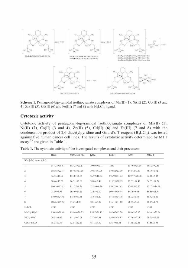
35
Scheme 1. Pentagonal-bipyramidal isothiocyanato complexes of Mn(II) (1), Ni(II) (2), Co(II) (3 and 4), Zn(II) (5), Cd(II) (6) and Fe(III) (7 and 8) with H2LCl2 ligand.
Cytotoxic activity
Cytotoxic activity of pentagonal-bipyramidal isothiocyanato complexes of Mn(II) (1), Ni(II) (2), Co(II) (3 and 4), Zn(II) (5), Cd(II) (6) and Fe(III) (7 and 8) with the condensation product of 2,6-diacetylpyridine and Girard’s T reagent (H2LCl2) was tested against five human cancer cell lines. The results of cytotoxic activity determined by MTT assay 23 are given in Table 1.
Table 1. The cytotoxic activity of the investigated complexes and their precursors.
HeLa MDA-MB-453 K562 LS174 A549 MRC-5
IC50 [μM] mean ±S.D.
1 187.28±18.91 183.33±23.57 190.93±15.71 >200 187.66±21.38 198.35±2.86
2 186.85±22.77 187.85±17.18 194.51±7.76 170.82±33.19 144.42±7.69 46.79±1.52
3 96.75±11.82 135.01±1.55 76.99±10.54 176.90±11.44 129.77±28.30 92.08±7.82
4 70.46±13.59 76.51±17.69 38.66±3.49 115.25±28.19 79.53±14.47 54.57±14.24
5 190.10±17.15 111.37±4.74 122.80±0.30 170.72±41.42 130.85±5.77 121.78±14.49
6 71.06±5.95 59.40±14.21 72.98±6.10 160.46±16.44 84.76±3.08 86.89±13.94
7 110.98±24.45 115.69±7.86 75.94±5.20 171.84±34.78 98.72±1.55 80.42±0.06
8 106.61±15.92 87.27±4.06 48.33±6.87 116.11±31.00 70.45±7.60 40.19±0.73
H2LCl2 >200 >200 >200 >200 >200 >200
MnCl2·4H2O 156.84±38.49 138.40±30.35 85.07±21.12 192.67±12.70 189.62±7.17 143.62±23.04
NiCl2·6H2O 76.31±1.09 111.59±2.08 77.76±2.91 136.61±28.97 127.68±27.82 78.75±15.03
CoCl2·6H2O 95.37±9.56 92.01±12.11 45.73±3.57 136.79±0.45 97.98±12.81 57.58±1.98
36
ZnCl2·2H2O >200 191.32±12.27 169.87±28.50 >200 200.00±0.00 105.61±25.82
Cd(NO3)2·4H2O 79.16±1.62 56.40±11.08 49.16±13.70 112.57±22.16 126.80±15.25 57.27±14.68
FeCl3·6H2O >200 >200 >200 >200 >200 >200
cisplatin 4.73±0.88 6.05±1.12 5.63±0.21 24.86±3.41 9.43±0.60 8.56±1.58
Changes in the cell cycle phase distribution
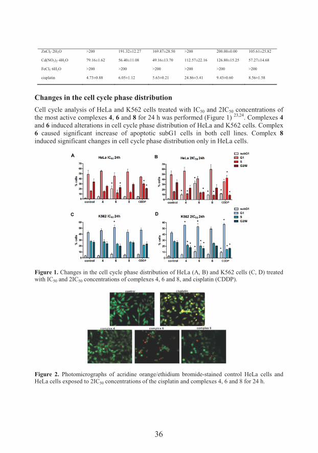
Cell cycle analysis of HeLa and K562 cells treated with IC50 and 2IC50 concentrations of the most active complexes 4, 6 and 8 for 24 h was performed (Figure 1) 23,24. Complexes 4and 6 induced alterations in cell cycle phase distribution of HeLa and K562 cells. Complex 6 caused significant increase of apoptotic subG1 cells in both cell lines. Complex 8induced significant changes in cell cycle phase distribution only in HeLa cells.
Figure 1. Changes in the cell cycle phase distribution of HeLa (A, B) and K562 cells (C, D) treated with IC50 and 2IC50 concentrations of complexes 4, 6 and 8, and cisplatin (CDDP).
Figure 2. Photomicrographs of acridine orange/ethidium bromide-stained control HeLa cells and HeLa cells exposed to 2IC50 concentrations of the cisplatin and complexes 4, 6 and 8 for 24 h.
37
Morphological evaluation of HeLa cell death mode
Morphological changes in HeLa cells were also indicative of apoptosis, with complex 6showed the most pronounced effect (Figure 2).
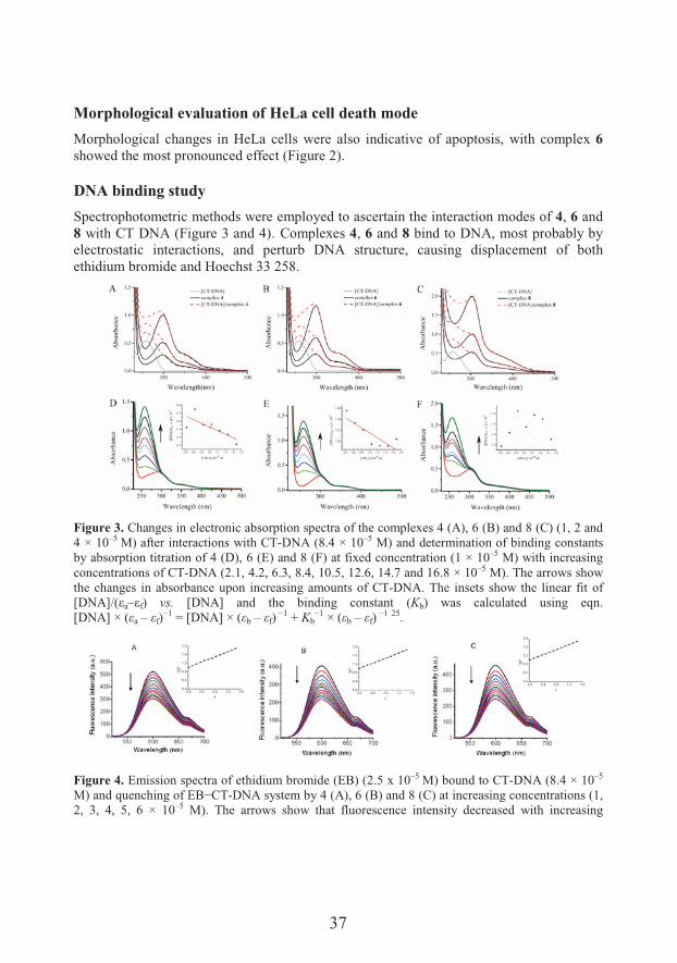
DNA binding study
Spectrophotometric methods were employed to ascertain the interaction modes of 4, 6 and 8 with CT DNA (Figure 3 and 4). Complexes 4, 6 and 8 bind to DNA, most probably by electrostatic interactions, and perturb DNA structure, causing displacement of both ethidium bromide and Hoechst 33 258.
Figure 3. Changes in electronic absorption spectra of the complexes 4 (A), 6 (B) and 8 (C) (1, 2 and 4 × 10⁻⁵ M) after interactions with CT-DNA (8.4 × 10⁻⁵ M) and determination of binding constants by absorption titration of 4 (D), 6 (E) and 8 (F) at fixed concentration (1 × 10⁻⁵ M) with increasing concentrations of CT-DNA (2.1, 4.2, 6.3, 8.4, 10.5, 12.6, 14.7 and 16.8 × 10⁻⁵ M). The arrows show the changes in absorbance upon increasing amounts of CT-DNA. The insets show the linear fit of J��+KLFMa–Mf) vs. [DNA] and the binding constant (Kb) was calculated using eqn. [DNA] × (6a – 6f)
I9 = [DNA] × (6b – 6f)I9 + Kb
I9 × (6b – 6f)I9 25.
Figure 4. Emission spectra of ethidium bromide (EB) (2.5 x 10⁻⁵ M) bound to CT-DNA (8.4 × 10⁻⁵
�@� � �D#���'�������G�I,*-DNA system by 4 (A), 6 (B) and 8 (C) at increasing concentrations (1, 2, 3, 4, 5, 6 × 10⁻⁵ M). The arrows show that fluorescence intensity decreased with increasing
38
concentration of the complex. The insets show fluorescence quenching curves of EB bound to DNA at 7max=600 nm by 4 (A), 6 (B) and 8 (C). The quenching constants K were calculated using eqn. I0/I= 1 + Kr by linear regression of the plot I0/I against [r]/[CT-DNA],where I0 and I represent the fluorescence �����&����&� ��� G�I,*-DNA in absence and presence of the complex, and r =[complex]/[CT-DNA] 26.
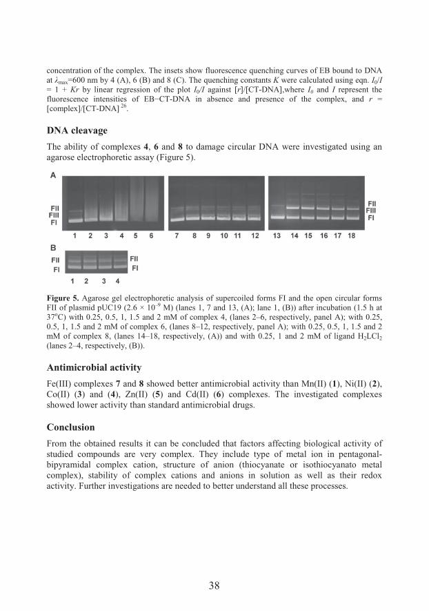
DNA cleavage
The ability of complexes 4, 6 and 8 to damage circular DNA were investigated using an agarose electrophoretic assay (Figure 5).
Figure 5. Agarose gel electrophoretic analysis of supercoiled forms FI and the open circular forms FII of plasmid pUC19 (2.6 × 10⁻⁹ M) (lanes 1, 7 and 13, (A); lane 1, (B)) after incubation (1.5 h at 37oC) with 0.25, 0.5, 1, 1.5 and 2 mM of complex 4, (lanes 2–6, respectively, panel A); with 0.25, 0.5, 1, 1.5 and 2 mM of complex 6, (lanes 8–12, respectively, panel A); with 0.25, 0.5, 1, 1.5 and 2 mM of complex 8, (lanes 14–18, respectively, (A)) and with 0.25, 1 and 2 mM of ligand H2LCl2
(lanes 2–4, respectively, (B)).
Antimicrobial activity
Fe(III) complexes 7 and 8 showed better antimicrobial activity than Mn(II) (1), Ni(II) (2), Co(II) (3) and (4), Zn(II) (5) and Cd(II) (6) complexes. The investigated complexes showed lower activity than standard antimicrobial drugs.
Conclusion
From the obtained results it can be concluded that factors affecting biological activity of studied compounds are very complex. They include type of metal ion in pentagonal-bipyramidal complex cation, structure of anion (thiocyanate or isothiocyanato metal complex), stability of complex cations and anions in solution as well as their redox activity. Further investigations are needed to better understand all these processes.
39
Acknowledgements
This work was supported by the Ministry of Education, Science and Technological development of the Republic of Serbia (Grant OI 172055 and Grant OI 175011).
References
1. Popov LD, Morozov AN, Shcherbakov IN, Tupolova YP, Lukov VV, Kogan VA. Metal complexes with polyfunctional ligands based of bis(hydrazones) of dicarbonyl compounds. Russ Chem Rev 2009;78:643–58.
2. Neto BAD, et al. Condensed, solution and gas phase behaviour of mono- and dinuclear 2,6-diacetylpyridine (dap) hydrazone copper complexes probed by X-ray, mass spectrometry and theoretical calculations. Dalton Trans 2013;42:11497–506.
3. Batchelor LJ, et al. Pentanuclear cyanide-bridged complexes based on highly anisotropic CoII sevencoordinate building blocks: synthesis, structure, and magnetic behavior. Inorg Chem 2011;50:12045–52.
4. " ����-�#�� N���� "%� +� �������� ��� *� �&������ ��� �� �����H�&� 7��'� 8�&F'$ � N���@�ligands of 2,6-diacetylpyridine. Hepta-coordination of 3d metals. Adv Inorg Chem 2004;55:315–60.
5. Ferraz KSO, et al. Investigation on the pharmacological profile of 2,6-diacetylpyridine bis(benzoylhydrazone) derivatives and their antimony(III) and bismuth(III) complexes. Eur J Med Chem 2012;53:98–106.
6. Nomiya K, et al. Syntheses, crystal structures and antimicrobial activities of monomeric 8-coordinate, and dimeric and monomeric 7-coordinate bismuth(III) complexes with tridentate and pentadentate thiosemicarbazones and pentadentate semicarbazone ligands. J Inorg Biochem 2004;98:601–15.
7. Mazza P, Orcesi M, Pelizzi C, Pelizzi G, Predieri G, Zani F. Synthesis, structure, antimicrobial, and genotoxic activities of organotin compounds with 2,6-diacetylpyridine nicotinoyl and Isonicotinoylhydrazones. J Inorg Biochem 1992;48:251–70.
8. Kasuga NC, et al. Synthesis, structural characterization and antimicrobial activities of 12 zinc(II) complexes with four thiosemicarbazone and two semicarbazone ligands. J Inorg Biochem 2003;96:298–310.
9. Liu GF, �������� �%� O����� ��� FP%� " ����-�#�� N���� "�� ����-coordinate iron and manganese complexes with acyclic and rigid pentadentate chelates and their superoxide dismutase activity. Inorg Chem 2007;46:8825–35.
10. Gutman CT, Brunold TC. Spectroscopic and computational studies of a smallmolecule functional mimic of iron superoxide dismutase, iron 2,6-diacetylpyridinebis(semioxamazide). Inorg Chem 2012;51:12729–37.
11. Gutman CT, Guzei IA, Brunold TC. Structural, spectroscopic, and computational characterization of the azide adduct of FeIII(2,6-diacetylpyridinebis(semioxamazide)), a functional analogue of iron superoxide dismutase. Inorg Chem 2013;52:8909–18.
12. ��NQ�7&��� �%� ����N��� �%� �#8����� �%� � ��� -Paryzek W. Metal-promoted synthesis, characterization, crystal structure and RNA cleavage ability of 2,6-diacetylpyridine bis(2-aminobenzoylhydrazone) lanthanide complexes. J Inorg Biochem 2013;126:38–45.
13. Gökçe C, Dilek N, Gup R. Seven coordinated cobalt(II) complexes with 2,6-diacetylpyridine bis(4-acylhydrazone) ligands: synthesis, characterization, DNAbinding and nuclease activity. Inorg Chim Acta 2015;432:213–20.
14. Gup R, Gökçe C, Dilek N. Seven-coordinated cobalt(II) complexes with 2,6-diacetylpyridine bis(4-hydroxybenzoylhydrazone): synthesis, characterisation, DNA binding and cleavage properties. Supramol Chem 2015;27:629–41.
15. 3�������-������L�%��� �����SB, Leovac V�%����������VI. Transition metal complexes with Girard reagents and their hydrazones. J Serb Chem Soc 2012;77:1129–55.

40
16. ��8�������%���� ���"��&��� �������� ����#���������� �������F""@������H�&�7��'���� ��� ���.�Oacylhydrazones of 2-(diphenylphosphino)benzaldehyde and monodentate pseudohalides. J Biol Inorg Chem 2016;21:145–62.
17. ������������%���8�������%�+�/��������%�*#���� "�������#� �����#��#��&� � ����-States of Pseudohalide Metal Complexes with Hydrazones of Girard's T Reagent. Eur J Inorg Chem 2018,838–46.
18. Chandler JD, Day BJ. Thiocyanate: a potentially useful therapeutic agent with host defense and antioxidant properties. Biochem Pharmacol 2012;84:1381–7.
19. 3�������-������5S, et al. Transition metal complexes with Girard reagent-based ligands. Part V. Synthesis, characterization and crystal structure of pentagonal-bipyramidal manganese(II) complex with 2,6-diacetylpyridine bis (Girard-T hydrazone). Inorg Chem Commun 2010;13:1085–8.
20. �� / �� R%� ��� ��� �$��'�&�&, characterization and antimicrobial activity of pentagonal-bipyramidal isothiocyanato Co(II) and Ni(II) complexes with 2,6-diacetylpyridin bis(trimethylammoniumacetohydrazone). J Coord Chem 2016;69:801–11.
21. �� / �� R%� ��� ��� �$��'�&�&%� �' � �����N ����%� ��T calculations and antimicrobial activity of pentagonal-bipyramidal Zn(II) and Cd(II) complexes with 2,6-diacetylpyridine-bis(trimethylammoniumacetohydrazone). JCoord Chem 2016;69: 2754–65.
22. +�/������� �%� ��� ��� �$��'�&�&%� �' � �����N ����� � � ��$&� �� &��#�tures of two pentagonal-bipyramidal Fe(III) complexes with dihydrazone of 2,6-diacetylpyridine and Girard's T reagent. Anticancer properties of various metal complexes of the same ligand. J Inorg Biochem 2017;174:137–49.
23. � ��2�"�Z, et al. In vitro antitumor actions of extracts from endemic plant Helichrysum zivojinii. BMC Complement Altern Med 2013;13:36–48.
24. Ormerod MG. Flow Cytometry. A Practical Approach. Oxford University Press, Oxford, UK, 2000. 2.
25. Vijayalakshmi R, Kanthimathi M, Subramanian V, Unni Nair B. DNA cleavage by a chromium(III) complex. Biochem Biophys Res Commun 2000;271:731–4.
26. Lakowicz JR, Weber G. Quenching of fluorescence by oxygen. A probe for structural fluctuations in macromolecules. Biochemistry 1973;12:4161–70.
41
Phytotherapy of cisplatin side effects: A case of two Filipendula species
������������*
Department of Chemistry, Faculty of Science, University of Kragujevac, Kragujevac,Serbia
*e-mail: [email protected]
Cisplatin (CP), an inorganic complex of platinum, has been effectively used as a potent chemotherapeutic agent against various malignancies, but it causes a number of side effects, e.g., digestive tract disorders, vomiting, and toxic effects on different organs, particularly on kidneys. Besides that, CP use in cancer chemotherapy may be responsible for secondary malignancies. Therefore, a number of scientific studies are focused on ameliorating potential of medicinal plant products (phytotherapeutics) on reducing or preventing the negative effects of anticancer drugs, namely cisplatin. Especially polyphenolic compounds from plant origin (flavonoids, phenolic acids, tannins, terpenes, etc.) showed significant activity towards modulation of cisplatin-induced toxicity. Genus Filipendula Mill. consists of over 20 plant species, two of which are growing in Serbia, Filipendula ulmaria (L.) Maxim. and F. vulgarisMoench. Both plants are in use in traditional medicine based on their antirheumatic, astringent, diuretic, and anti-inflammatory properties and potential to treat kidney problems. Hence, the effects of aerial parts and roots methanolic extracts of F. ulmaria and F. vulgaris were investigated against cisplatin-induced kidney and liver injuries in rats along with the determination of their phytochemical composition. The obtained results showed that tested extracts, rich in polyphenolic compounds, attenuate cisplatin-induced liver and kidney oxidative stress, reduce tissue damage, and enhance the antioxidative status of experimental animals during cisplatin application. Therefore, F. ulmaria and F. vulgaris extracts may be used as supportive agents for the prevention and amelioration of cisplatin side effects.
Cisplatin: activity and toxicity
Cisplatin (cis-[Pt(NH3)2Cl2] (cis-diamminedichlorplatinum(II)) is an inorganic complex with a square planar geometry, which consists of an atom of platinum surrounded with two ammonia groups and two chlorine atoms in cis position. This inorganic complex has been in clinical usage for cancer treatments, since 1978 when it was approved by the American Food and Drug Administration 1, and today it is on the World Health Organization's List of Essential Medicines 2. Cisplatin was first synthesized and described by the Italian chemist Michele Peyrone as early as in 1845, but in 1965 American biophysicist Barnett Rosenberg
42
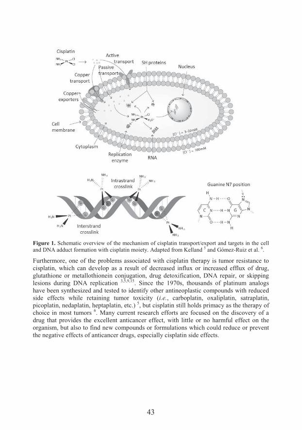
managed to characterize the powerful antiproliferative effects of this complex 3–5. For 40 years cisplatin has been effectively used as a potent and one of the most common chemotherapeutic agents against various malignancies, mainly for testicular, ovarian, head and neck, bladder, cervical, esophageal as well as small cell lung cancer 6,7, but also for breast, stomach, prostate cancers, Hodgkin’s and non-Hodgkin’s lymphomas, neuroblastoma, sarcomas, multiple myeloma, melanoma and mesothelioma 4.The mechanism of cisplatin action is based on targeting cancer cell DNA (Figure 1). Upon entering a cell, cisplatin become hydrated, after the dissociation of two chlorides, and a reactive complex is formed and it is then able to interact with nucleophilic molecules within the cell, including DNA, RNA, and proteins. When this positively charged moleculeinteracts with deoxyribonucleic acids it causes interstrand and intrastrand covalent crosslinking with local denaturation of the DNA chain 8,9. This process primarily occurs due to the favoring crosslinking between N7 and O6 atoms of the adjacent imidazole ring of the purine base guanine, and to a minor extent via N7 and N1 of the adenine molecules or via N3 atom of the cytosine. The main product responsible for the anticancer activity of cisplatin is intrastrand crosslink 1,2-(guanine deoxydinucleotide) (1,2-GpG, about 65%), where platinum is coordinated to N(7) of two guanine molecules from one DNA strand 9–
11. The final cellular outcome is generally apoptotic cell death, although the pathway(s) from platinum–DNA binding to apoptosis remains incompletely elucidated. The platinum–DNA adducts can impede cellular processes, such as replication and transcription, but also signal-transduction pathways, that control growth, differentiation and stress responses, have also been implicated 5.Although cisplatin has this very important role in cancer treatment and has had a major clinical impact, it causes a number of side effects, such as vomiting, gastrointestinal tract disorders, and toxic effects on different organs 1. Cisplatin frequently causes notorious kidney damage (nephrotoxicity) because it is mainly excreted via the kidneys (27-45%). Cisplatin-induced nephrotoxicity can also lead to acute renal failure 4,8,12. Besides nephrotoxicity, also ototoxicity (adult: 23-50%; children: >50%) and peripheral neurotoxicity (adult: 30-86%, children: ~10%) are considered to be the most serious toxicities associated with cisplatin treatment 9. Moreover, cisplatin can provoke less frequent toxic effects like hepato- and cardiotoxicity 1. Since the target of cisplatin is DNA, its use in cancer chemotherapy may be responsible for secondary malignancies. After application of cisplatin, DNA damage may lead to mutagenesis, carcinogenesis, and to apoptotic cell death 13. Generally, toxic side effects of cisplatin arise because this complex has a high affinity for sulfur-containing compounds like glutathione, and these newly formed compounds are highly reactive and generally responsible for toxic effects in the organism 10,11. Besides binding of cisplatin to various cytoplasmic molecules, some of the suggested mechanisms of cisplatin-induced toxicity are a generation of reactive oxygen species and inhibition of antioxidant enzymes (superoxide dismutase, glutathione peroxidase, and glutathione S-transferase). Taking this into consideration, oxidative stress plays a significant role in cisplatin-induced toxicity 14.
43
Figure 1. Schematic overview of the mechanism of cisplatin transport/export and targets in the cell and DNA adduct formation with cisplatin moiety. Adapted from Kelland 5 and Gómez-Ruiz et al. 6.
Furthermore, one of the problems associated with cisplatin therapy is tumor resistance to cisplatin, which can develop as a result of decreased influx or increased efflux of drug, glutathione or metallothionein conjugation, drug detoxification, DNA repair, or skipping lesions during DNA replication 3,5,9,15. Since the 1970s, thousands of platinum analogs have been synthesized and tested to identify other antineoplastic compounds with reduced side effects while retaining tumor toxicity (i.e., carboplatin, oxaliplatin, satraplatin, picoplatin, nedaplatin, heptaplatin, etc.) 5, but cisplatin still holds primacy as the therapy of choice in most tumors 6. Many current research efforts are focused on the discovery of a drug that provides the excellent anticancer effect, with little or no harmful effect on the organism, but also to find new compounds or formulations which could reduce or prevent the negative effects of anticancer drugs, especially cisplatin side effects.
44
Phytotherapy of cisplatin side effects
Despite the fact that cisplatin has been used for 40 years as part of the treatment of various solid malignancies its side effects are still unavoidable. Today, besides many synthesized drugs, numerous traditional medicinal plants, dietary vegetables, and fruits, as well as their constituents, still play a key role in the prevention and treatment of different diseases, including the protective role against oxidative stress in the organism 16. Most of the medicinal plants’ constituents, i.e., polyphenolics, alkaloids, carotenoids, vitamins, are known by their significant antioxidant potential 17.Since the oxidative and nitrosative stresses are main mechanisms involved in cisplatin toxicity 1,8, numerous recent studies are dealing with the beneficial effects of different plant extracts administration on the alleviation of cisplatin-induced toxicity. In particular, plant extracts rich in polyphenols, such as Zingiber officinale rhizome extract 18,Matricaria chamomilla aerial part extract 19, Hypericum perforatum aerial part extract 20,Stevia rebaudiana extract 21, as well as standardized extracts like silymarin 22 and ginseng extract 23, showed ameliorating effects on hepato-, oto-, and/or nephrotoxicity caused by cisplatin in vivo. The protective activities of these extracts involve, among others, antioxidant and anti-inflammatory mechanisms. A wide range of pure compounds from plant origin was also tested in vivo for the amelioration of cisplatin side effects. Different polyphenolic compounds, e.g., ellagic acid, caffeic acid, rosmarinic acid, ferulic acid, quercetin, rutin, curcumin, resveratrol, chrysin, hesperidin, luteolin, naringenin, epigallocatechin-3-O-gallate, cyanidin, genistein, gingerol, terpenes: ginsen�&� �&� >-caryophyllene, and artemisinin; alkaloids: berberine, capsaicin, and noscapine; and vitamins C and E showed significant alleviation of oxidative stress parameters during the cisplatin treatment and modulation of cisplatin-induced toxicity on various levels 24–29.The comprehensive analysis of literature suggests that phytotherapy using herbal medicines and/or plant-derived natural products (phytochemicals) can be widely implemented to prevent the cisplatin-induced toxicity. It is evident that phytomedicines exhibited potentially effective nephro-, oto- and hepato-protection in preclinical studies, primarily based on their antioxidative properties. Substantially, these antioxidant compounds not only target oxidative stress but also other events involved in cisplatin pathology, such as inflammation, mitochondrial damage and endoplasmic reticulum stress 25. Furthermore, phytomedicines have been widely documented to directly or indirectly target multiple signaling pathways and networks in cancer cells, so a combination of anticancer drugs and polypharmacological plant-derived extracts or compounds may offer a significant advantage in the efficacy of monotherapy and overcoming drug-induced resistance in cancer patients 24.
A case of two Filipendula species
Genus Filipendula Mill. (fam. Rosaceae) is consisted of around 20 plant species that are predominantly widespread the Northern hemisphere. Plants of this genus are growing in
45
Europe, North America, Siberia, and Asia 30. The genus name derives from two Latin words: “filium” - a thread or a string and “pendulus” - hanging, referring to the root of some species that are consisted of rhizomes associated with thin strings 31. Genus Filipendula in the territory of Serbia, as well as on the entire European continent, is represented by two species: Filipendula ulmaria (L.) Maxim. (syn. Spiraea ulmaria 5�@�SFilipendula vulgaris Moench (syn. Filipendula hexapetala Gilib., Spiraea filipendula L.) 32–34.F. ulmaria (meadowsweet, queen of the meadow) is a perennial herb with creamy-white flowers, a short, pink rhizome and stems 50–120 cm high. F. ulmaria is used in traditional European medicine for treatment of various ailments due to its antipyretic, astringent, diuretic, antacid, stomachic, antiseptic, analgesic, antirheumatic, and anti-inflammatory properties 35–38. Dried flowering tops are used for the treatment of common cold, minor painful articular conditions, and to facilitate renal and digestive elimination functions 35,39.Based on traditional use and proven pharmacological effects, the herb (aerial parts) of F. ulmaria was registered in European Pharmacopoeia 5th Edition (PhEur 5.0) as Filipendulae ulmariae herba 40, and now it is an integral part of the latest 9th Edition (PhEur 9.0) from 2017. F. vulgaris (dropwort) is up to 80 cm high plant, with pinkish-white flowers and characteristic rhizomes with tuberous roots 32. F. vulgaris is also used in traditional medicine of most European countries 41, and sometimes, it is used as a substitute for F. ulmaria due to their similar bioactive effects, in particular, anti-inflammatory properties 38.Based on the literature data it can be concluded that mentioned Filipendula species are characterized by the presence of the three main classes of phenolic compounds: phenolic acids and their derivatives (gallic acid, ellagic acid, salicylic acid, methyl salicylate, salicylaldehyde), flavonoid aglycones and glycosides (quercetin, kaempferol, catechin, epicatechin, rutoside, hyperoside, spiraeoside, quercitrin, apigenin, astragalin), and tannins (mainly tellimagrandins and rugosins) 36,42–45.Our previous investigations of F. ulmaria and F. vulgaris aerial part and root methanolic extracts showed that they possessed high antioxidant and anti-inflammatory activity in vitro, low genotoxicity, antigenotoxic activity, moderate antimicrobial activity, and good stability at different pH values and thermal conditions 46–49. The extracts were also subjected to a wide range of spectrophotometric and chromatographic methods (TLC, HPTLC, HPLC, LC-DAD-MSn) in order to elucidate their phytochemical composition. The results showed that all four extracts had a high content of phenolic compounds, mainly flavonoids (particularly quercetin and its derivatives, e.g., spiraeoside, rutin, hyperoside, quercitrin, isoquercitrin, Figure 2) and phenolic acids in aerial part extracts, along withhydrolyzable tannins in root extracts 46,48–52.With regard to reported biological activities and traditional uses of Filipendula spp., their phytochemical composition, and our previous studies which confirmed potent antioxidant activity of the methanolic extracts of aerial parts and roots of F. ulmaria and F. vulgaris,we aimed to further investigate those extracts for their potential in amelioration cisplatin-induced toxicity in vivo, using albino Wistar rats. All animal procedures were in
46
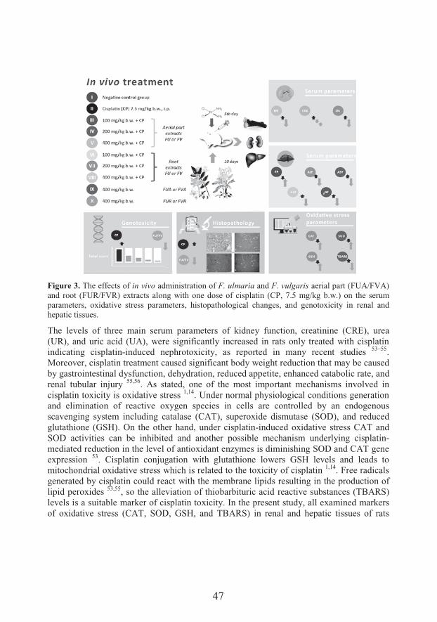
compliance with the EEC Directive (86/609/EEC) on the protection of animals used for experimental and other scientific purposes. The study was designed as follows (Figure 3): I - negative control group where animals were treated with normal saline; II - positive control/cisplatin group where toxicity was induced with cisplatin; III-V groups treated with F. ulmaria and F. vulgaris aerial part extracts (FUA or FVA) per os (p.o.) at three different concentrations 100, 200, and 400 mg/kg body weight (b.w.); VI-VII groups treated with F. ulmaria and F. vulgaris root extracts (FUR or FVR) at 100, 200, and 400 mg/kg b.w.; and two last groups were treated only with extracts at the highest concentration (400 mg/kg b.w.). The extracts were administered for 10 days and in groups II-VIII toxicity was induced on the 5th day of treatment by intraperitoneal (i.p.) administration of a single dose of CP dissolved in normal saline (7.5 mg/kg b.w.) 50,51.
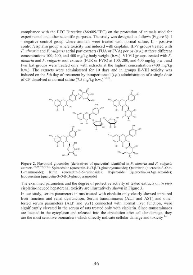
Figure 2. Flavonoid glucosides (derivatives of quercetin) identified in F. ulmaria and F. vulgarisextracts 36,44–46,50–52: Spiraeoside (quercetin-?T-O->-D-glucopyranoside); Quercitrin (quercetin-3-O-;-L-rhamnoside); Rutin (quercetin-3-O-rutinoside); Hyperoside (quercetin-3-O-galactoside); Isoquercitrin (quercetin-3-O->-D-glucopyranoside)
The examined parameters and the degree of protective activity of tested extracts on in vivocisplatin-induced hepatorenal toxicity are illustratively shown in Figure 3.In our study, serum parameters in rats treated with cisplatin only clearly showed impaired liver function and renal dysfunction. Serum transaminases (ALT and AST) and other��&�� � &��#�� � �����&� F+5.� � � UR*@� �������� � 7��'� ���� �� ����� �#������%� 7����significantly elevated in the serum of rats treated only with cisplatin. Since transaminases are located in the cytoplasm and released into the circulation after cellular damage, they are the most sensitive biomarkers which directly indicate cellular damage and toxicity 14.
1
3
2
47
Figure 3. The effects of in vivo administration of F. ulmaria and F. vulgaris aerial part (FUA/FVA) and root (FUR/FVR) extracts along with one dose of cisplatin (CP, 7.5 mg/kg b.w.) on the serum parameters, oxidative stress parameters, histopathological changes, and genotoxicity in renal and hepatic tissues.
The levels of three main serum parameters of kidney function, creatinine (CRE), urea (UR), and uric acid (UA), were significantly increased in rats only treated with cisplatin indicating cisplatin-induced nephrotoxicity, as reported in many recent studies 53–55.Moreover, cisplatin treatment caused significant body weight reduction that may be caused by gastrointestinal dysfunction, dehydration, reduced appetite, enhanced catabolic rate, and renal tubular injury 55,56. As stated, one of the most important mechanisms involved in cisplatin toxicity is oxidative stress 1,14. Under normal physiological conditions generationand elimination of reactive oxygen species in cells are controlled by an endogenous scavenging system including catalase (CAT), superoxide dismutase (SOD), and reduced glutathione (GSH). On the other hand, under cisplatin-induced oxidative stress CAT andSOD activities can be inhibited and another possible mechanism underlying cisplatin-mediated reduction in the level of antioxidant enzymes is diminishing SOD and CAT gene expression 53. Cisplatin conjugation with glutathione lowers GSH levels and leads to mitochondrial oxidative stress which is related to the toxicity of cisplatin 1,14. Free radicals generated by cisplatin could react with the membrane lipids resulting in the production of lipid peroxides 53,55, so the alleviation of thiobarbituric acid reactive substances (TBARS) levels is a suitable marker of cisplatin toxicity. In the present study, all examined markers of oxidative stress (CAT, SOD, GSH, and TBARS) in renal and hepatic tissues of rats
48
treated with cisplatin only, indicated that cisplatin-treatment disrupted normal kidney and liver function. As expected, GSH levels in liver and kidneys were markedly decreased. The serum and tissue parameters of oxidative stress and tissue damage in rats treated with three different doses of tested Filipendula spp. methanolic extracts for 10 consecutive days, in addition to cisplatin (on the 5th day of treatment), indicate a significant beneficial effect, compared to the group treated with cisplatin alone. The extracts were able to markedly reduce serum parameters, in some cases in a dose-dependent manner. Further, they ameliorated the oxidative status in kidney and liver tissues by increasing CAT and SOD activities and reducing the concentration of TBARS, as well as prevent depletion of GSH by cisplatin treatment in liver tissue. In kidneys, GSH was the most affected element of the antioxidative defense. The induction of hepato- and nephrotoxicity by cisplatin were also confirmed by histological analysis of liver and kidney tissues. It was evident that the extracts exerted tissue-protective effects, with a significant reduction of liver and kidney tissue injury caused by cisplatin administration 50,51.Considering previously reported antioxidant activity and radical scavenging capacity of F. ulmaria and F. vulgaris 46,47, as well as the high content of antioxidant phenolic compounds, our data strongly imply that their extracts potently protect against liver and kidney toxicity induced by cisplatin via antioxidant activity. The results of phytochemical analysis of all extracts confirmed the presence of a wide range of phenolic compounds, which have been shown to act as antioxidants. Procyanidins (condensed tannins) and catechins, that were detected in all extracts, have been demonstrated to possess an array of beneficial health effects 57–60. Also, hydrolyzable tannins, detected in aerial parts, have been shown to possess a variety of biological activities, including antioxidant and anticancer effects 61,62. Flavonoids and flavonoid derivatives detected in all extracts are also highly bioactive and for many of them hepato- and nephroprotective effects in vivowere reported 63–67.Another potential route of protection against cisplatin-induced oxidative stress is the proven anti-inflammatory activity of both Filipendula species 49,52 since cisplatin-induced oxidative stress leads to the activation of pro-inflammatory mediators 14.The third mechanism possibly underlying the observed protective effect of tested extracts against cisplatin-induced toxicity may be suppression of DNA damage in normal cells. Cisplatin can react with many structures in the cell, but of course, the most important intracellular target is DNA. DNA damage is a major trigger for cell cycle arrest and apoptosis and cisplatin has demonstrated induction of apoptosis in multiple tissue types 11.Although the antineoplastic effect of cisplatin is mediated through the binding to nuclear DNA, cisplatin also induces damage of mitochondrial DNA 14. Results of our study revealed significant cisplatin-induced DNA damage both in the liver and, in particular, kidney cells. Interestingly, the potency of DNA damage reduction in liver and kidneys decreased with increasing concentration of all tested extracts. At lower doses (100 and 200 mg/kg), the extracts were the most effective in reducing cisplatin-induced DNA damage 50,51.
49
Conclusions and future perspectives
Co-treatment with methanolic extracts of aerial parts and roots of F. ulmaria and F. vulgaris attenuated cisplatin-induced toxicity in kidneys and liver, regulated serum and tissue parameters related to oxidative stress and tissue damage, and helped to maintain tissue architecture, along with reducing genotoxicity caused by cisplatin. Therefore, it can be concluded that Filipendula species may be used as a supportive agent in cancer patients under cisplatin therapy to improve the oxidative stress defense of the organism and to diminish toxic side effects of cisplatin. However, further investigations are still required to completely evaluate their protective effect on the side-effects of cisplatin and obtaining the appropriate therapeutic dose, particularly obtaining evidence of extracts’ bioavailability in human subjects. Also, the extracts and phytochemical compounds should be thoroughly investigated in preclinical models for further pharmaceutical development. This approach may offer the best chance of clinically meaningful prevention of cisplatin toxicity.
Acknowledgments
This study was supported by the Ministry of Education, Science and Technological Development of the Republic of Serbia (Grant No. III 43004).
References1. Dasari S, Tchounwou P. Cisplatin in Cancer therapy: Molecular mechanisms of action. Eur J
Pharmacol 2014;740:364–78.2. World Health Organization. “Comparative Table of Medicines on the WHO Essential
Medicines Lists from 1977–2011” (XLS). Retrieved. 2011. 3. Galluzzi L, et al. Systems biology of cisplatin resistance: past, present and future. Cell Death
Dis 2014;5:e1257.4. Manohar S, Leung N. Cisplatin nephrotoxicity: a review of the literature. J Nephrol
2018;31:15–25. 5. Kelland L. The resurgence of platinum-based cancer chemotherapy. Nat Rev Cancer
2007;7:573–84. 6. Gómez-Ruiz S, Maksimovi�-" ���� �%� ��� ����� �%� � �# ������ R��� 1�� �'�� �&����$%�
biological effects, and use of cisplatin and metallocenes in anticancer chemotherapy. Bioinorg Chem Appl 2012;2012:15–7.
7. Jamieson ER, Lippard SJ. Structure, Recognition, and Processing of Cisplatin-DNA Adducts. Chem Rev 1999;99:2467–98.
8. Chirino YI, Pedraza-Chaverri J. Role of oxidative and nitrosative stress in cisplatin-induced nephrotoxicity. Exp Toxicol Pathol 2009;61:223–42.
9. Rabik CA, Dolan ME. Molecular mechanisms of resistance and toxicity associated with platinating agents. Cancer Treat Rev 2007;33:9–23.
10. �#� �2���V�%�������&����%�.��������%�O��'��#�'����%� ��G� ���������' ��&����&�# ��&�����'��reactions of platinum(II) complexes with nitrogen- and sulfur-donor biomolecules. Dalt Trans2012;41:12329–45.
11. Crul M, Schellens JH, Beijnen JH, Maliepaard M. Cisplatin resistance and DNA repair. Cancer Treat Rev 1997;23:341–66.
12. Jin J, et al. Protective effect of Wuzhi tablet (Schisandra sphenanthera extract) against cisplatin-induced nephrotoxicity via Nrf2-mediated defense response. Phytomedicine 2015;22:528–35.
50
13. Nersesyan A, Muradyan R. Sea-buckthorn juice protects mice against genotoxic action of cisplatin. Exp Oncol 2004;26:153–5.
14. Karasawa T, Steyger PS. An integrated view of cisplatin-induced nephrotoxicity and ototoxicity Toxicol Lett 2015;237:219–27.
15. Sarin N, et al. Key players of cisplatin resistance: towards a systems pharmacology approach. Int J Mol Sci 2018;19:E767.
16. Atanasov AG, et al. Discovery and resupply of pharmacologically active plant-derived natural products: A review. Biotechnol Adv 2015;33:1582–614.
17. Shahidi F, Ambigaipalan P. Phenolics and polyphenolics in foods, beverages and spices: Antioxidant activity and health effects – A review. J Funct Foods 2015;18:820–97.
18. Ajith TA, Nivitha V, Usha S. Zingiber officinale Roscoe alone and in combin ����� 7��'� ;-tocopherol protect the kidney against cisplatin-induced acute renal failure. Food Chem Toxicol2007;45:921–7.
19. Abad ANA, Nouri MHK, Gharjanie A, Tavakoli F. Effect of Matricaria chamomillaHydroalcoholic Extract on Cisplatin-induced Neuropathy in Mice. Chin J Nat Med 2011;9:126–31.
20. Shibayama Y, Kawachi A, Onimaru S, Tokunaga J, Ikeda R. Effect of pre-treatment with St John’s Wort on nephrotoxicity of cisplatin in rats. Life Sci 2007;81:103–8.
21. .���2�� ��"%����N�����%���� ���%��������%�3� ���r-���0�����%������������������ � � �stevioside protect against cisplatin nephrotoxicity through inhibition of ERK1/2, STAT3, and NF-W�� ��� ��������� �,'���*�H�����XY9<A9Y<=X9B–25.
22. Cho S Il, Lee JE, Do NY. Protective effect of silymarin against cisplatin-induced ototoxicity. Int J Pediatr Otorhinolaryngol 2014;78:474–8.
23. Yousef MI, Hussien HM. Cisplatin-induced renal toxicity via tumor necrosis factor-;%�interleukin 6, tumor suppressor P53, DNA damage, xanthine oxidase, histological changes, oxidative stress and nitric oxide in rats: Protective effect of ginseng. Food Chem Toxicol 2015;78:17–25.
24. Cheng YT, Yang CC, Shyur LF. Modulating oxidative stress and the tumor microenvironment for cancer therapy. Pharmacol Res 2016;114:128–43.
25. Gómez-Sierra T, Eugenio-Pérez D, Sánchez-Chinchillas A, Pedraza-Chaverri J. Role of food-derived antioxidants against cisplatin induced-nephrotoxicity. Food Chem Toxicol 2018;120:230–42.
26. Domitro��� �%� .���2�� �� "%� ,��2���-1����� V%� Z�� � ��� ��'����������� �������&� ���rosmarinic acid against cisplatin-induced kidney injury in mice. Food Chem Toxicol2014;66:321–8.
27. ���������� �%� ,�� �����1%� .#���� G.%� [ ��� ��R�%�� '�#����� ���O%� Z�� ���� 5#�eolin ameliorates cisplatin-induced nephrotoxicity in mice through inhibition of platinum accumulation, inflammation and apoptosis in the kidney. Toxicology 2013;310:115–23.
28. Shahid F, Farooqui Z, Khan F. Cisplatin-induced gastrointestinal toxicity: An update on possible mechanisms and on available gastroprotective strategies. Eur J Pharmacol 2018;827:49–57.
29. Ojha S, Venkataraman B, Kurdi A, Mahgoub E, Sadek B, Rajesh M. Plant-Derived Agents for Counteracting Cisplatin-Induced Nephrotoxicity. Oxid Med Cell Longev 2016;4320374:1–27.
30. Shimizu T. Taxonomical notes on the genus Filipendula Adans. (Rosaceae). J Fac Text Sci Technol 1961;26:1–31.
31. Giebelmann R. Kulturgeschichtliches zu Rosengewächsen. 2004;71:54–63. 32. *#� ������5�2�����8�������"N 2� �� � ���� nizacija RAD, Beograd, 1984. 33. ��&������������� �������8� �"-X. Serbian Academy of Sciences and Arts, Belgrade, 1973. 34. � ���� �%� ��� ��� +�� ��'��8�� ��� �� &�# $� ��� �'�� #& ��� ���7�� ��� ���� �� '��8&� ������� �����
Mountain (Central Serbia). J Ethnopharmacol 2007;111:160–75. 35. Barros L, Cabrita L, Boas MV, Carvalho AM, Ferreira ICFR. Chemical, biochemical and
electrochemical assays to evaluate phytochemicals and antioxidant activity of wild plants. Food Chem 2011;127:1600–8.
51
36. Bijttebier S, et al. A first step in the quest for the active constituents in Filipendula ulmaria(Meadowsweet): Comprehensive phytochemical identification by liquid chromatography coupled to quadrupole-orbitrap mass spectrometry. Planta Med 2016;82:559–72.
37. Harbourne N, Jacquier JC, O’Riordan D. Optimisation of the aqueous extraction conditions of phenols from meadowsweet (Filipendula ulmaria L.) for incorporation into beverages. Food Chem 2009;116:722–7.
38. Barros L, et al. Characterization of phenolic compounds in wild medicinal flowers from Portugal by HPLC–DAD–ESI/MS and evaluation of antifungal properties. Ind Crops Prod 2013;44:104–10.
39. Assessment report on Filipendula ulmaria (L.) Maxim., herba and Filipendula ulmaria (L.) Maxim., flos. European Medicines Agency. 2011, pp. 1–18.
40. Papp I, et al. Monitoring volatile and non-volatile salicylates in Filipendula ulmaria by different chromatographic techniques. Chromatographia 2008;68:125–9.
41. Vogl S, et al. Ethnopharmacological in vitro studies on Austria’s folk medicine - An unexplored lore in vitro anti-inflammatory activities of 71 Austrian traditional herbal drugs. J Ethnopharmacol 2013;149:750–71.
42. Fecka I. Qualitative and quantitative determination of hydrolysable tannins and other polyphenols in herbal products from meadowsweet and dog rose. Phytochem Anal2009;20:177–90.
43. Olennikov DN, Kruglova MY. A new quercetin glycoside and other phenolic compounds from the genus Filipendula. Chem Nat Compd 2013;49:610–6.
44. .#� �&����\� �%� 3��&�#����&� .�%� .#� �&� &� +�� .'$���'���� �� ����&������ � � ����Hidant properties of Filipendula vulgaris as a source of healthy functional ingredients. J Funct Foods 2015;15:233–42.
45. .#� �&����\��%�3��&�#����&�.�%�.#� �&� &�+��.'$���'���� �� �' � �����N ��������Filipendula ulmaria by UPLC/Q-TOF-MS and evaluation of antioxidant activity. Rec Nat Prod 2015;9:451–5.
46. � � �����%���� ������ �����$%�&� 8����$� � �'��������' � �����N ��������Filipendula ulmaria (L.) Maxim. Food Funct 2015;6(4):1164–75.
47. � � ���� �%� ��� ��� ���7���� FFilipendula hexapetala Gilib.): Potential role as antioxidant and antimicrobial agent. EXCLI J 2015;14:1–20.
48. � �����%���� ��� In vitro and in vivo assessment of the genotoxicity and antigenotoxicity of the Filipendula hexapetala and Filipendula ulmaria methanol extracts. J Ethnopharmacol 2015;174:287–92.
49. � � �����%���� ���In vitro and in vivo assessment of meadowsweet (Filipendula ulmaria) as anti-inflammatory agent. J Ethnopharmacol 2016;193:627–36.
50. � � ���� �%� ��� ��� *'�� ������ ����� ������� ��� Filipendula hexapetala extracts on hepatorenal toxicity of cisplatin. J Funct Foods 2015;18:198–212.
51. � � ���� �%� ��� ��� Filipendula ulmaria extracts attenuate cisplatin-induced liver and kidney oxidative stress in rats: In vivo investigation and LC-MS analysis. Food Chem Toxicol 2017;99:86–102.
52. � � �����%����al. Phytochemical analysis and anti-inflammatory effects of Filipendula vulgarisMoench extracts. Food Chem Toxicol 2018; In press.
53. Almaghrabi OA. Molecular and biochemical investigations on the effect of quercetin on oxidative stress induced by cisplatin in rat kidney. Saudi J Biol Sci 2015;22:227–31.
54. Yamabe N, et al. Protective effects of protocatechuic acid against cisplatin-induced renal damage in rats. J Funct Foods 2015;19:20–7.
55. Sinha S, Jothiramajayam M, Ghosh M, Jana A, Chatterji U, Mukherjee A. Vetiver oil (Java) attenuates cisplatin-induced oxidative stress, nephrotoxicity and myelosuppression in Swiss albino mice. Food Chem Toxicol 2015;81:120–8.
56. Hassan I, Chibber S, Naseem I.Ameliorative effect of riboflavin on the cisplatin induced nephrotoxicity and hepatotoxicityunder photoillumination. Food Chem Toxicol 2010;48:2052–8.
57. Lluís L, et al. Toxicology evaluation of a procyanidin-rich extract from grape skins and seeds. Food Chem Toxicol. 2011;49:1450–4.
52
58. Spranger I, Sun B, Mateus AM, Freitas V de, Ricardo-da-Silva JM. Chemical characterization and antioxidant activities of oligomeric and polymeric procyanidin fractions from grape seeds. Food Chem 2008;108:519–32.
59. Zanwar AA, Badole SL, Shende PS, Hegde M V., Bodhankar SL. Antioxidant Role of Catechin in Health and Disease. in Polyphenols in Human Health and Disease. Elsevier Inc., London, 2013, pp. 267-271
60. Sutherland BA, Rahman RMA, Appleton I. Mechanisms of action of green tea catechins, with a focus on ischemia-induced neurodegeneration. J Nutr Biochem 2006;17:291–306.
61. Koleckar V, et al. Condensed and hydrolysable tannins as antioxidants influencing the health. Mini-Reviews Med Chem 2008;8:436–47.
62. Kähkönen M, Kylli P, Ollilainen V, Salminen JP, Heinonen M. Antioxidant activity of isolated ellagitannins from red raspberries and cloudberries. J Agric Food Chem. 2012;60:1167–74.
63. Ham Y-M, et al. Quercitrin protects against oxidative stress-induced injury in lung fibroblast cells via up-regulation of Bcl-xL. J Funct Foods 2012;4:253–62.
64. Babujanarthanam R, Kavitha P, Mahadeva Rao US, Pandian MR. Quercitrin a bioflavonoid improves the antioxidant status in streptozotocin: induced diabetic rat tissues. Mol Cell Biochem 2011;358:121–9.
65. Choi J-H, et al. Protective effects of hyperoside against carbon tetrachloride-induced liver damage in mice. J Nat Prod 2011;74:1055–60.
66. Arjumand W, Seth A, Sultana S. Rutin attenuates cisplatin induced renal inflammation and apoptosis by reducing NFkB, TNF-a and caspase-3 expression in wistar rats. Food Chem Toxicol 2011;49:2013–21.
67. 3 �����]� �%� 3�8 � �%� � ��^_�]� �%� `����'�]� �%� �_��� 3�� "&�D#��������=� .' �� �����$%�toxicology, and metabolism. Food Chem Toxicol 2014;68:267–82.
53
Polypharmacologically active esters of N-methylanthranilic acid from Mexican orange (Choisya ternata Kunth): from the discovery to panacea-like properties
)��+$���� ���1*�-!��$#�������2�-!����$��� �����3�8�����$#��3�����4
1Faculty of Occupational Safety, University of Niš, Niš, Serbia2Department of Chemistry, Faculty of Sciences and Mathematics, University of Niš3Faculty of Medicine, University of Niš4Department of Physiology, Faculty of Medicine, University of Niš
*e-mail: [email protected]
Choisya ternata Kunth (Rutaceae) is a plant species with an ethnopharmacological application in Mexico. A new natural protoalkaloid, isopropyl N-methylanthranilate (IMA), named ternanthranin, was identified among 157 different constituents, by a detailed GC-MS analysis of the essential oil of C. ternata leaves. IMA was synthesized via the reduction of in situ generated imine of methyl anthranilate and formaldehyde, leading to methyl N-methylanthranilate (MMA) (also a constituent of the analyzed oil), which was subsequently transesterified with a suitable alkoxide. Prompted by the ethnopharmacology of C. ternata, antioxidant, antimicrobial and various in vivopharmacological activities of IMA and MMA were assayed. Both esters exhibited significant antinociceptive and anti-inflammatory activities (even at 0.3 mg kg-1, p.o.)in mice. They, at 50-200 mg kg-1 (i.p.), showed anxiolytic and antidepressant activities while not inducing sleep, but significantly prolonging the diazepam-induced sleep in mice. Although IMA and MMA do not produce significant antioxidant activity, in the CCl4-induced liver/kidney damage models in rats they demonstrated, at 200 mg kg-1
(i.p.), a hepatoprotective effect, while only MMA demonstrated a nephroprotective potential. Both alkaloids were shown to be gastroprotective by exhibiting a significant anti-ulcer effect even at 50 mg kg-1. Additionally, they displayed a selective antifungal activity in vitro. Urinary metabolic profile of IMA and MMA, and the distribution in organs of rats, revealed that they both undergo analogous xenobiotic biotransformation routes. The major metabolic pathway of IMA was the aromatic core hydroxylation, whereas MMA predominantly underwent ester group alterations.
Introduction
Since ancient times, plants have been used as food, for cosmetic, medicinal and many other purposes. In the beginning, people distinguished useful, edible and medicinal, plants
54
from harmful and poisonous ones based on instincts, observations, and experiences, and they conveyed their findings to the successive generations. Today, the application of modern analytical techniques and methods enables a detailed chemical analysis of the composition of plant species, as well as the isolation and determination of the structure of molecules that are present in plant tissues even in minor quantities, while different in silico, in vitro and in vivo testing of biological activities, enables the discovery of new therapeutics. Nowadays, more than three-fourths of active principles that are used for medical purposes are of plant origin, and a quarter is still isolated from plant drugs. In recent years, there has been a growing interest in natural products in the prevention and treatment of many diseases 1,2. The search for new biologically active plant-derived secondary metabolites includes several steps: (1) determining the presence of an unknown compound in the plant material, (2) isolation, (3) or synthesis of an unknown compound in order to determine its structure, and (4) testing of its biological activity/activities.Mexican orange (Choisya ternata Kunth, Rutaceae, subfamily Rutoideae) is a small evergreen ornamental shrub with leaves which when bruised give off a strong and pungent smell. The abundantly produced and highly fragrant flowers make Mexican orange an important and popular horticultural plant 3,4. Mexican people utilized the infusion of leaves of this plant species for its antispasmodic and “simulative” properties. The first pharmacological test of C. ternata was undertaken by Boudoresques, in 1895. He evaluated the effects of an aqueous extract of leaves on himself and found it to be tonic and appetizing. C. ternata was registered in the 4th and 5th editions of the Mexican Pharmacopoeia 3. The chemistry of this plant taxon received little attention to date. Some phytochemical work has been carried out on flavonoids, coumaroids, and alkaloids. The conducted studies indicated the importance of C. ternata non-volatile anthranilate-derived quinoline alkaloid constituents 3. The volatile constituents of this species have only been investigated on one previous occasion 5. Due to the obvious lack of detailed studies of the volatile chemistry of this Choisya species, we set our goal to determine the chemical composition of the leaf essential oil of Choisya ternata, as well as to assess the antioxidant and various biological activities of the selected constituents of the oil including antimicrobial, antinociceptive, anti-inflammatory, gastro-, hepato- and nephro-protective activities, anxiolytic and antidepressant properties, as well as an effect on diazepam-induced sleep and to possibly correlate them with the ethnopharmacological use of this plant species.
Chemical composition of Choisya ternata essential oil
Leaves of C. ternata were collected from the Trinity College Botanical Gardens, Dublin, Ireland. Detailed GC and GC–MS analyses of the hydrodistilled essential oils of C. ternatafresh and dried leaves enabled the identification of 157 constituents among which sabinene (ca. one-third of the oils), terpinen-4-��%��$�����%�>-'��� � ����%� � �U-terpinene were the major ones. In contrast, the previously investigated leaf oil of C. ternata resulted in the identification of only eighteen components 5.
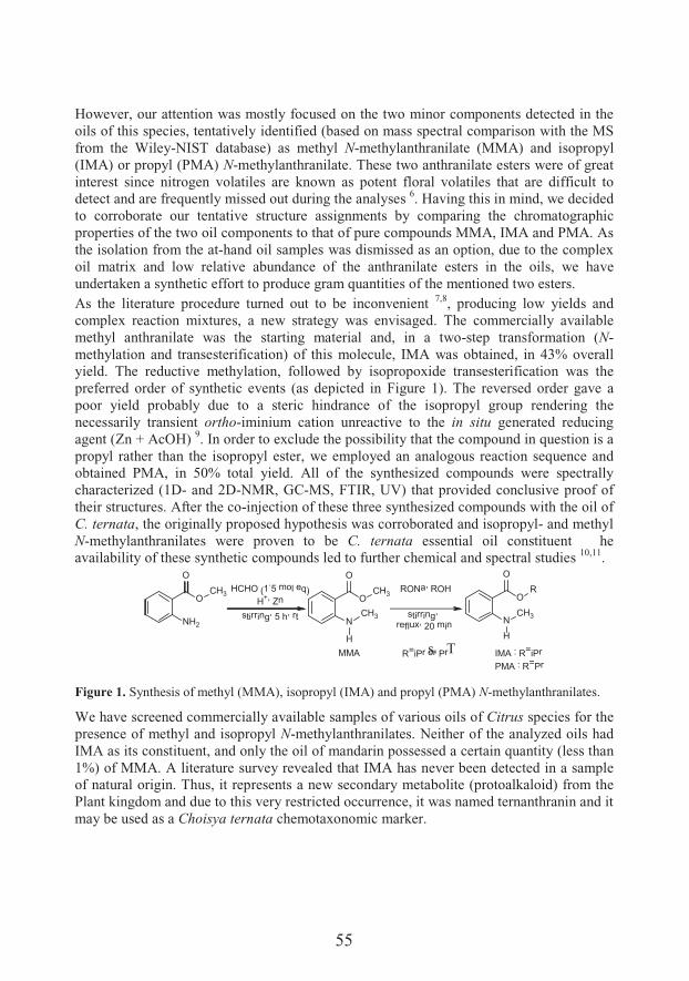
55
However, our attention was mostly focused on the two minor components detected in the oils of this species, tentatively identified (based on mass spectral comparison with the MS from the Wiley-NIST database) as methyl N-methylanthranilate (MMA) and isopropyl (IMA) or propyl (PMA) N-methylanthranilate. These two anthranilate esters were of great interest since nitrogen volatiles are known as potent floral volatiles that are difficult to detect and are frequently missed out during the analyses 6. Having this in mind, we decided to corroborate our tentative structure assignments by comparing the chromatographic properties of the two oil components to that of pure compounds MMA, IMA and PMA. As the isolation from the at-hand oil samples was dismissed as an option, due to the complex oil matrix and low relative abundance of the anthranilate esters in the oils, we have undertaken a synthetic effort to produce gram quantities of the mentioned two esters.As the literature procedure turned out to be inconvenient 7,8, producing low yields and complex reaction mixtures, a new strategy was envisaged. The commercially available methyl anthranilate was the starting material and, in a two-step transformation (N-methylation and transesterification) of this molecule, IMA was obtained, in 43% overall yield. The reductive methylation, followed by isopropoxide transesterification was the preferred order of synthetic events (as depicted in Figure 1). The reversed order gave a poor yield probably due to a steric hindrance of the isopropyl group rendering the necessarily transient ortho-iminium cation unreactive to the in situ generated reducing agent (Zn + AcOH) 9. In order to exclude the possibility that the compound in question is a propyl rather than the isopropyl ester, we employed an analogous reaction sequence and obtained PMA, in 50% total yield. All of the synthesized compounds were spectrally characterized (1D- and 2D-NMR, GC-MS, FTIR, UV) that provided conclusive proof of their structures. After the co-injection of these three synthesized compounds with the oil of C. ternata, the originally proposed hypothesis was corroborated and isopropyl- and methyl N-methylanthranilates were proven to be C. ternata essential oil constituent
s. T
he availability of these synthetic compounds led to further chemical and spectral studies 10,11.
RONa, ROH
stirring,reflux, 20 min
O
OR
NCH3
H
O
O
CH3
N
H
CH3
O
NH2
OCH3 HCHO (1
.5 mol eq)H+, Zn
stirring, 5 h, rt
MMA IMA : R=iPrPMA : R=Pr
R=iPr or Pr
Figure 1. Synthesis of methyl (MMA), isopropyl (IMA) and propyl (PMA) N-methylanthranilates.
We have screened commercially available samples of various oils of Citrus species for the presence of methyl and isopropyl N-methylanthranilates. Neither of the analyzed oils had IMA as its constituent, and only the oil of mandarin possessed a certain quantity (less than 1%) of MMA. A literature survey revealed that IMA has never been detected in a sample of natural origin. Thus, it represents a new secondary metabolite (protoalkaloid) from the Plant kingdom and due to this very restricted occurrence, it was named ternanthranin and it may be used as a Choisya ternata chemotaxonomic marker.
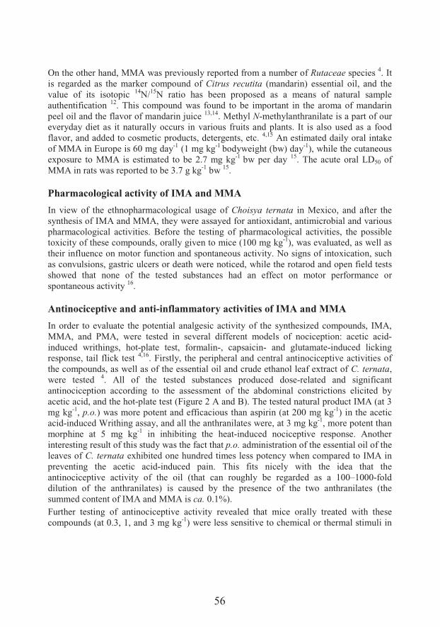
56
On the other hand, MMA was previously reported from a number of Rutaceae species 4. It is regarded as the marker compound of Citrus recutita (mandarin) essential oil, and the value of its isotopic 14N/15N ratio has been proposed as a means of natural sample authentification 12. This compound was found to be important in the aroma of mandarin peel oil and the flavor of mandarin juice 13,14. Methyl N-methylanthranilate is a part of our everyday diet as it naturally occurs in various fruits and plants. It is also used as a food flavor, and added to cosmetic products, detergents, etc. 4,15 An estimated daily oral intake of MMA in Europe is 60 mg day-1 (1 mg kg-1 bodyweight (bw) day-1), while the cutaneous exposure to MMA is estimated to be 2.7 mg kg-1 bw per day 15. The acute oral LD50 of MMA in rats was reported to be 3.7 g kg-1 bw 15.
Pharmacological activity of IMA and MMA
In view of the ethnopharmacological usage of Choisya ternata in Mexico, and after the synthesis of IMA and MMA, they were assayed for antioxidant, antimicrobial and various pharmacological activities. Before the testing of pharmacological activities, the possible toxicity of these compounds, orally given to mice (100 mg kg-1), was evaluated, as well as their influence on motor function and spontaneous activity. No signs of intoxication, such as convulsions, gastric ulcers or death were noticed, while the rotarod and open field tests showed that none of the tested substances had an effect on motor performance or spontaneous activity 16.
Antinociceptive and anti-inflammatory activities of IMA and MMA
In order to evaluate the potential analgesic activity of the synthesized compounds, IMA, MMA, and PMA, were tested in several different models of nociception: acetic acid-induced writhings, hot-plate test, formalin-, capsaicin- and glutamate-induced licking response, tail flick test 4,16. Firstly, the peripheral and central antinociceptive activities of the compounds, as well as of the essential oil and crude ethanol leaf extract of C. ternata,were tested 4. All of the tested substances produced dose-related and significant antinociception according to the assessment of the abdominal constrictions elicited by acetic acid, and the hot-plate test (Figure 2 A and B). The tested natural product IMA (at 3 mg kg-1, p.o.) was more potent and efficacious than aspirin (at 200 mg kg-1) in the acetic acid-induced Writhing assay, and all the anthranilates were, at 3 mg kg-1, more potent than morphine at 5 mg kg-1 in inhibiting the heat-induced nociceptive response. Another interesting result of this study was the fact that p.o. administration of the essential oil of the leaves of C. ternata exhibited one hundred times less potency when compared to IMA in preventing the acetic acid-induced pain. This fits nicely with the idea that the antinociceptive activity of the oil (that can roughly be regarded as a 100–1000-fold dilution of the anthranilates) is caused by the presence of the two anthranilates (the summed content of IMA and MMA is ca. 0.1%).Further testing of antinociceptive activity revealed that mice orally treated with these compounds (at 0.3, 1, and 3 mg kg-1) were less sensitive to chemical or thermal stimuli in
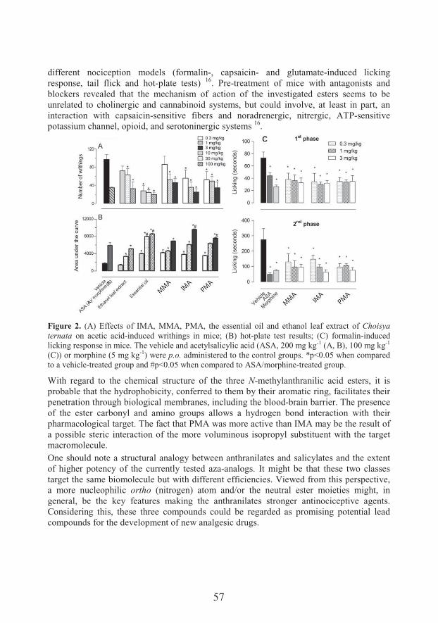
57
different nociception models (formalin-, capsaicin- and glutamate-induced licking response, tail flick and hot-plate tests) 16. Pre-treatment of mice with antagonists and blockers revealed that the mechanism of action of the investigated esters seems to be unrelated to cholinergic and cannabinoid systems, but could involve, at least in part, an interaction with capsaicin-sensitive fibers and noradrenergic, nitrergic, ATP-sensitive potassium channel, opioid, and serotoninergic systems 16.
Figure 2. (A) Effects of IMA, MMA, PMA, the essential oil and ethanol leaf extract of Choisya ternata on acetic acid-induced writhings in mice; (B) hot-plate test results; (C) formalin-induced licking response in mice. The vehicle and acetylsalicylic acid (ASA, 200 mg kg-1 (A, B), 100 mg kg-1
(C)) or morphine (5 mg kg-1) were p.o. administered to the control groups. *p<0.05 when compared to a vehicle-treated group and #p<0.05 when compared to ASA/morphine-treated group.
With regard to the chemical structure of the three N-methylanthranilic acid esters, it is probable that the hydrophobicity, conferred to them by their aromatic ring, facilitates their penetration through biological membranes, including the blood-brain barrier. The presence of the ester carbonyl and amino groups allows a hydrogen bond interaction with their pharmacological target. The fact that PMA was more active than IMA may be the result of a possible steric interaction of the more voluminous isopropyl substituent with the target macromolecule.One should note a structural analogy between anthranilates and salicylates and the extent of higher potency of the currently tested aza-analogs. It might be that these two classes target the same biomolecule but with different efficiencies. Viewed from this perspective, a more nucleophilic ortho (nitrogen) atom and/or the neutral ester moieties might, in general, be the key features making the anthranilates stronger antinociceptive agents. Considering this, these three compounds could be regarded as promising potential lead compounds for the development of new analgesic drugs.
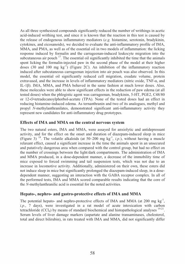
58
As all three synthesized compounds significantly reduced the number of writhings in acetic acid-induced writhing test, and since it is known that the reaction in this test is caused by the release of endogenous inflammatory mediators (e.g. histamine, serotonin, bradykinin, cytokines, and eicosanoids), we decided to evaluate the anti-inflammatory profile of IMA, MMA, and PMA, as well as of the essential oil in two models of inflammation: the licking response induced by formalin and the carrageenan-induced leukocyte migration into the subcutaneous air pouch 17. The essential oil significantly inhibited the time that the animals spent licking the formalin-injected paw in the second phase of the model at their higher doses (30 and 100 mg kg-1) (Figure 2C). An inhibition of the inflammatory reaction induced after subcutaneous carrageenan injection into air pouch was also observed. In this model, the essential oil significantly reduced cell migration, exudate volume, protein extravased, and the increase in levels of inflammatory mediators (nitric oxide, TNF-;%� � �IL-9>@�� "�+%���+%� � �.�+�8�' � � ��� �'�� & ��� � &'���� ���#�'� ��7��� �&�&��+�&�%�these molecules were able to show significant effects in the reduction of paw edema (at all tested doses) when the phlogistic agent was carrageenan, bradykinin, 5-HT, PGE2, C48/80 or 12-O-tetradecanoylphorbol-acetate (TPA). None of the tested doses had an effect in reducing histamine-induced edema. As ternanthranin and two of its analogues, methyl and propyl N-methylanthranilates, demonstrated significant anti-inflammatory activity they represent new candidates for anti-inflammatory drug prototypes.
Effects of IMA and MMA on the central nervous system
The two natural esters, IMA and MMA, were assayed for anxiolytic and antidepressant activity, and for the effect on the onset and duration of diazepam-induced sleep in mice (Figure 3) 18. The volatile alkaloids (at 50–200 mg kg-1, i.p.), without having a muscle relaxant effect, caused a significant increase in the time the animals spent in an unsecured and putatively dangerous area when compared with the control group, but had no effect on the number of crossings between the light/dark compartments. The administration of IMA and MMA produced, in a dose-dependent manner, a decrease of the immobility time of mice exposed to forced swimming and tail suspension tests, which was not due to an increase in locomotive activity. Additionally, administered on their own, these esters did not induce sleep in mice but significantly prolonged the diazepam-induced sleep, in a dose-dependent manner, suggesting an interaction with the GABA receptor complex. In all of the performed tests, IMA and MMA scored comparable results indicating that the core of the N-methylanthranilic acid is essential for the noted activities.
Hepato-, nephro- and gastro-protective effects of IMA and MMA
The potential hepato- and nephro-protective effects of IMA and MMA (at 200 mg kg-1,i.p., 7 days), were investigated in a rat model of acute intoxication with carbon tetrachloride (CCl4) by means of standard biochemical and histopathological analyses 19-21.Serum levels of liver damage markers (aspartate and alanine transaminases, cholesterol, total and direct bilirubin), in rats treated with IMA and MMA, did not significantly differ

59
from the ones in the control group. Histopathological evaluation of the livers of the test animals also revealed that IMA and MMA significantly reduced the incidence of liver lesions caused by CCl4 (Figure 4 (left A-D)). These findings are of great interest as the liver plays a central role in xenobiotic biotransformation and can be susceptible to possible toxic effects from the xenobiotics or their metabolites and is of great interest considering the potential use of IMA and MMA as therapeutics 20.
Figure 3. Effect of IMA and MMA (50, 100 and 200 mg kg-1, i.p.) on the behavior of mice in the open field (*p<0.001; **p<0.01; ***p<0.005 vs. vehicle), light/dark (diazepam at 2 mg kg-1 was the positive control; *p<0.01; **p<0.001 vs. vehicle; #animals did not enter the dark chamber), forced swimming and tail suspension (*p<0.001 vs. vehicle; #p<0.001 vs. imipramine (15 mg kg-1); ##p<0.01 vs. imipramine (15 mg kg-1)), and diazepam-induced sleep (*p<0.01 vs. diazepam (2 mg kg-1); **p<0.001 vs. diazepam (2 mg kg-1)) tests.
IMA and MMA applied on their own in high doses did not cause any significant damage to kidney tissue 21. A pretreatment with MMA, but not with IMA, prior to the administration of CCl4 significantly prevented the increase of serum levels of decreased kidney function markers (urea and creatinine). Histopathological evaluation of the kidneys also revealed that MMA reduced the incidence of kidney lesions induced by CCl4 (Figure 4 (right A-D)). Thus, both volatile alkaloids, showed hepatoprotective potential in the model of CCl4-induced liver damage, while only MMA exhibited a nephroprotective potential in the same model of kidney damage in rats.
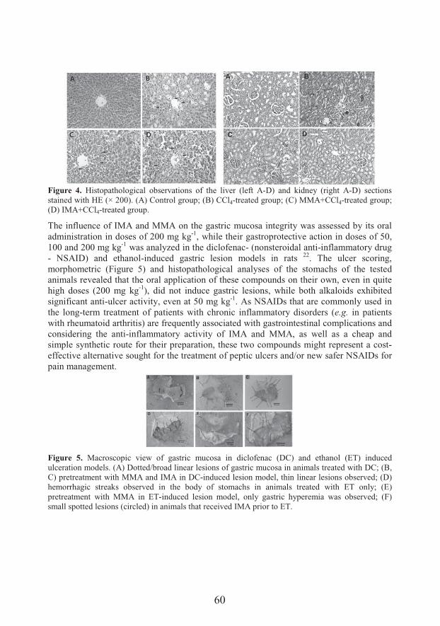
60
Figure 4. Histopathological observations of the liver (left A-D) and kidney (right A-D) sections stained with HE (× 200). (A) Control group; (B) CCl4-treated group; (C) MMA+CCl4-treated group; (D) IMA+CCl4-treated group.
The influence of IMA and MMA on the gastric mucosa integrity was assessed by its oral administration in doses of 200 mg kg-1, while their gastroprotective action in doses of 50, 100 and 200 mg kg-1 was analyzed in the diclofenac- (nonsteroidal anti-inflammatory drug - NSAID) and ethanol-induced gastric lesion models in rats 22. The ulcer scoring, morphometric (Figure 5) and histopathological analyses of the stomachs of the tested animals revealed that the oral application of these compounds on their own, even in quite high doses (200 mg kg-1), did not induce gastric lesions, while both alkaloids exhibited significant anti-ulcer activity, even at 50 mg kg-1. As NSAIDs that are commonly used in the long-term treatment of patients with chronic inflammatory disorders (e.g. in patients with rheumatoid arthritis) are frequently associated with gastrointestinal complications and considering the anti-inflammatory activity of IMA and MMA, as well as a cheap and simple synthetic route for their preparation, these two compounds might represent a cost-effective alternative sought for the treatment of peptic ulcers and/or new safer NSAIDs for pain management.
Figure 5. Macroscopic view of gastric mucosa in diclofenac (DC) and ethanol (ET) induced ulceration models. (A) Dotted/broad linear lesions of gastric mucosa in animals treated with DC; (B, C) pretreatment with MMA and IMA in DC-induced lesion model, thin linear lesions observed; (D) hemorrhagic streaks observed in the body of stomachs in animals treated with ET only; (E) pretreatment with MMA in ET-induced lesion model, only gastric hyperemia was observed; (F) small spotted lesions (circled) in animals that received IMA prior to ET.
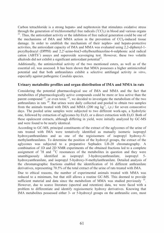
61
Carbon tetrachloride is a strong hepato- and nephrotoxin that stimulates oxidative stress through the generation of trichloromethyl free radicals (•CCl3) in blood and various organs21. Thus, the antioxidant activity or the inhibition of free radical generation could be one of the mechanisms of IMA and MMA action in the prevention of CCl4-induced tissue damage. In order to establish the mechanism of their nephro- and hepato-protective activities, the antioxidant capacity of IMA and MMA was evaluated using 2,2-diphenyl-1-���$�'$ � N$�� F�..O@� � � X%XT-azino-bis(3-ethylbenzthiazoline-6-sulphonic acid radical cation (ABTS+) assays and superoxide scavenging test. However, these two volatile alkaloids did not exhibit a significant antioxidant potential21.Additionally, the antimicrobial activity of the two mentioned esters, as well as of the essential oil, was assessed. It has been shown that MMA possesses a higher antimicrobial potential and that both anthranilates exhibit a selective antifungal activity in vitro,especially against pathogenic Candida species.
Urinary metabolite profiles and organ distribution of IMA and MMA in rats
Considering the potential pharmacological use of IMA and MMA and the fact that metabolites of pharmacologically active compounds could be more or less active than the parent compound 23, or even toxic 24, we decided to investigate the metabolism of the two anthranilates in rats 20. Rat urines were daily collected and pooled to obtain two samples from the animals treated with IMA and MMA (200 mg kg-1, i.p.) for seven consecutive days. The pooled urine samples were subjected to two different work-ups, a hydrolytic one, followed by extraction of aglycones by Et2O, or a direct extraction with Et2O. Both of these opalescent extracts, although differing in yield, were initially analyzed by GC-MS and were found to be nearly identical.According to GC-MS, principal constituents of the extract of the aglycones of the urine of � �&� ��� �� � 7��'� "�+� 7���� ���� ����$� � ����a� � &� �#�# ��$� �&������� �&���$��hydroxyanthranilates and as one of the regioisomers of isopropyl hydroxy-N-methylanthranilates. To determine the position of the hydroxyl groups, the extract of the aglycones was subjected to a preparative Sephadex LH-20 chromatography. A combination of 1D and 2D NMR experiments of the obtained fractions led to a complete assignment of 1H and 13C resonances of the metabolites in question and they were unambiguously identified as isopropyl 3-hydroxyanthranilate, isopropyl 5-hydroxyanthranilate, and isopropyl 5-hydroxy-N-methylanthranilate. Detailed analysis of the chromatographic fractions enabled the identification of 16 different anthranilate derivatives, representing 70.5% of the total extract of the urine of rats treated with IMA.Due to ethical reasons, the number of experimental animals treated with MMA was reduced to a minimum, but that still allows a routine GC-MS. This deemed to provide sufficient material and data since the metabolism of MMA was studied previously 15.However, due to scarce literature (spectral and retention) data, we were faced with a problem to differentiate and identify regioisomeric hydroxy derivatives. Knowing that IMA metabolites possessed either 3- or 5-hydroxyl groups on the anthranilic core, most
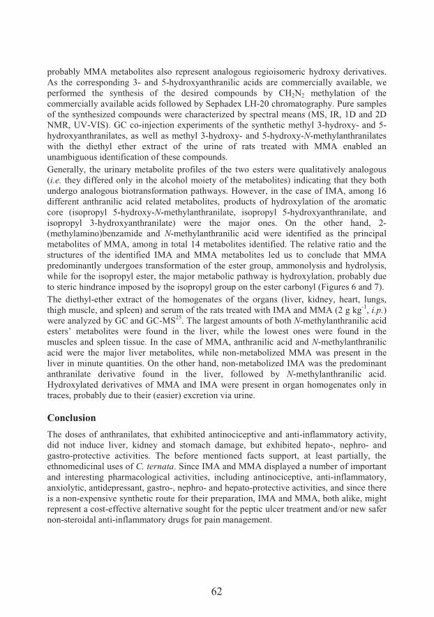
62
probably MMA metabolites also represent analogous regioisomeric hydroxy derivatives. As the corresponding 3- and 5-hydroxyanthranilic acids are commercially available, we performed the synthesis of the desired compounds by CH2N2 methylation of the commercially available acids followed by Sephadex LH-20 chromatography. Pure samples of the synthesized compounds were characterized by spectral means (MS, IR, 1D and 2D NMR, UV-VIS). GC co-injection experiments of the synthetic methyl 3-hydroxy- and 5-hydroxyanthranilates, as well as methyl 3-hydroxy- and 5-hydroxy-N-methylanthranilates with the diethyl ether extract of the urine of rats treated with MMA enabled an unambiguous identification of these compounds.Generally, the urinary metabolite profiles of the two esters were qualitatively analogous (i.e. they differed only in the alcohol moiety of the metabolites) indicating that they both undergo analogous biotransformation pathways. However, in the case of IMA, among 16 different anthranilic acid related metabolites, products of hydroxylation of the aromatic core (isopropyl 5-hydroxy-N-methylanthranilate, isopropyl 5-hydroxyanthranilate, and isopropyl 3-hydroxyanthranilate) were the major ones. On the other hand, 2-(methylamino)benzamide and N-methylanthranilic acid were identified as the principal metabolites of MMA, among in total 14 metabolites identified. The relative ratio and the structures of the identified IMA and MMA metabolites led us to conclude that MMA predominantly undergoes transformation of the ester group, ammonolysis and hydrolysis, while for the isopropyl ester, the major metabolic pathway is hydroxylation, probably due to steric hindrance imposed by the isopropyl group on the ester carbonyl (Figures 6 and 7).The diethyl-ether extract of the homogenates of the organs (liver, kidney, heart, lungs, thigh muscle, and spleen) and serum of the rats treated with IMA and MMA (2 g kg-1, i.p.)were analyzed by GC and GC-MS25. The largest amounts of both N-methylanthranilic acid esters’ metabolites were found in the liver, while the lowest ones were found in the muscles and spleen tissue. In the case of MMA, anthranilic acid and N-methylanthranilic acid were the major liver metabolites, while non-metabolized MMA was present in the liver in minute quantities. On the other hand, non-metabolized IMA was the predominant anthranilate derivative found in the liver, followed by N-methylanthranilic acid. Hydroxylated derivatives of MMA and IMA were present in organ homogenates only in traces, probably due to their (easier) excretion via urine.
Conclusion
The doses of anthranilates, that exhibited antinociceptive and anti-inflammatory activity, did not induce liver, kidney and stomach damage, but exhibited hepato-, nephro- and gastro-protective activities. The before mentioned facts support, at least partially, the ethnomedicinal uses of C. ternata. Since IMA and MMA displayed a number of importantand interesting pharmacological activities, including antinociceptive, anti-inflammatory, anxiolytic, antidepressant, gastro-, nephro- and hepato-protective activities, and since there is a non-expensive synthetic route for their preparation, IMA and MMA, both alike, might represent a cost-effective alternative sought for the peptic ulcer treatment and/or new safer non-steroidal anti-inflammatory drugs for pain management.
63
O
O
R1
NH
R2
O
O
R1
NH
R2O
O
O
R1
NH
R2
O
O
O
R1
NH
R2O O
O
R1
NH
R2
O
O
O
R1
NH
R2
O
O
O
R1
NH
R2
O
O
O
R1
NH
R2
O
O
R1
NH
R2O
O
O
O
R1
NH
R2O
O
R1
NH
R2
OO
O
O
R1
NH
R2
O
O
R1
NH
R2
O
O
O
O
R1
NH
R2
OH
O
O
R1
NH
R2
HO
NH
R2
Cyt P450O2
NADPH + H+
?
R1 = Me or iPrR2 = H or Me
Figure 6. Proposed cytochrome P450-catalyzed hydroxylation of anthranilic esters.
O
O
R1
NH
O
O
R1
NH
O
O
R1
NH2CH2HOCH3
H
O H+
Cyt P450
R1 = Me or iPrR2 = H or OH
R2 R2R2
Figure 7. Proposed cytochrome P450-catalyzed N-demethylation of N-methylanthranilic acid.
Acknowledgements
This work was supported by the Ministry of Education, Science and Technological Development of the Republic of Serbia [Project No. 172061].
64
References
1. Duke JA. Promising Phytomedicinals. In: Janick J, Simon JE (eds) Advances in New Crops,Timber Press, Portland, 1990, pp 491–8.
2. Newman DJ, Cragg GM. Natural products as sources of new drugs over the last 25 years. J Nat Prod 2007;70:461–77.
3. Creche J, Tremouillaux-Guiller J, Groeger D, Chenieux JC, Rideau M. Choisya ternata (Mexican orange): In vitro Culture and the Production of Alkaloids. In: Bajaj YPS (ed) Biotechnology in Agriculture and Forestry 24 (Medicinal and Aromatic Plants V), Springer, Berlin, Heidelberg, New York, 1992, pp 107–21.
4. � #����� ��%� ��� ��� " ������� ����� ��� � ��7� ������������� �� ��� � �&���$�� �-methylanthranilate from the essential oil of Choisya ternata Kunth. J Ethnopharm 2011;135:610–9.
5. Respaud M-J, Moulis C, Fouraste I, Bessiere J-M. Essential oil composition of Choisya ternata Kunth (Rutaceae) leaves. J Essent Oil Res 1997;9:475–6.
6. Jabalpurwala FA, Smoot JM, Rouseff RL, Russell L. A comparison of citrus blossom volatiles. Phytochemistry 2009;70:1428–34.
7. Staiger RP, Miller EB, Isatoic anhydride IV. Reactions with various nucleophiles. J Org Chem 1959;24:1214–9.
8. Dembele A, Deshayes H, Pete JP. Reduction and photoreduction of carbonic acid derivatives: effect of hexamethylphosphorotriamide. B Soc Chim Fr 1988;4:671–80.
9. da Silva RA, Estevam IHS, Bieber LW. Reductive methylation of primary and secondary amines and amino acids by aqueous formaldehyde and zinc. Tetrahedron Lett 2007;48:7680–2.
10. ���������� +�%� � #����� ���� ���#��#� �� ��#�� ����� ��� �'�����$&�&� �� #��&� ��� ���'$�� �-methyl-N-nitrosoanthranilate. RSC Adv 2015;5:53569–85.
11. ���������� +�%� � #����� ���� ,������� &&�������� ��� 9O- and 13C-NMR spectra of anthranilic acid and its hydroxy derivatives and salicylic acid and its amino derivatives. FU Phys Chem Tech 2015;13:121–32.
12. Faulhaber S, Hener U, Mosandl A. GC/IR���+� �$&�&����� � �����&&���� �����&��9��b9:,.��� � �b9B�+"�� �#�&�������'$���-methylanthranilate. J Agric Food Chem 1997;45:2579–83.
13. Fanciullino A-L, Tomi F, Luro F, Desjobert JM, Casanova J. Chemical variability of peel and leaf oils of mandarins. Flavour Fragr J 2006;21:359–67.
14. Wilson III CW, Shaw PE. Importance of thymol, methyl N-methylanthranilate, and monoterpene hydrocarbons to the aroma and flavor of mandarin cold-pressed oils. J Agr Food Chem 1981;29:494–6.
15. SCCS (Scientific Committee on Consumer Safety) Opinion on Methyl N-methylanthranilate (Phototoxicity Only). https://ec.europa.eu/health/scientific_committees/consumer_ safety/docs/sccs_o_075.pdf. (Accessed 08 October 2018).
16. Gomes Pinheiro MM, Radulovi����%�����������+�%���$� ���%����� � �&�.���+�������������esters of N-methylanthranilic acid: Mechanism of action in heat-mediated pain. Eur J Pharmacol 2014;727:106–14.
17. R���&�.��'�������%�����������+�%�� #�������%�+8 #�-Wahab IR, Boylan F, FernandesPD. Anti-inflammatory activity of Choisya ternata Kunth essential oil, ternanthranin, and its two synthetic analogs (methyl and propyl N-methylanthranilates). PLoS One 2015;10:e0121063.
18. � #�������%�����������+�%�� � �������.�%����� �������%���$� ���� Effects of methyl and isopropyl N-methylanthranilates from Choisya ternata Kunth (Rutaceae) on experimental anxiety and depression in mice. Phytother Res 2013;27:1334–8.
19. � #����� �%� � � ������� .%� ���� ����� �%� "���� "%� ���������� +�� "���#����� ��� ���'$�� nd isopropyl N-methylanthranilates on carbon tetrachloride-induced changes in rat liver and kidney morphology and function. FU Phys Chem Tech 2013;11:67–73.
20. � #����� ��%� ���������� +�%� ���� ����� ��%� � � ������� .��� ��&������ #��� �$� ��� 8������profiles of two pharmacologically active N-methylanthranilates: Three approaches to xenobiotic metabolite identification. Food Chem Toxicol 2017;109:341–55.
21. � #�������%���� ���G����������7���&���&�����-methylanthranilic acid from Rutaceae species on impaired kidney morphology and function in rats caused by CCl4. Life Sci 2015;135:110–7.
65
22. � #�������%��� �����"%�"����"�%�� � �������.�%����� �������%�����������+������'$�� � �isopropyl N-methylanthranilates attenuate diclofenac- and ethanol-induced gastric lesions in rats. Life Sci 2013;93:840–6.
23. Mitra AK, Kwatra D, Vadlapudi AD. Drug Delivery. Jones & Bartlett Publishers, Burlington,2014.
24. Macherey A-C, Dansette PM. Biotransformations Leading to Toxic Metabolites: Chemical Aspect. In: Wermuth CG (ed) The Practice of Medicinal Chemistry, Academic Press, Burlington, 2008, pp 674-696.
25. ���������� +�%� � #����� ��%� ���� ����� ��%� � � ������� .��� ��� 8���&�� ��� �&&���� �-oil constituents: Determination of methyl and isopropyl N-methylanthranilates and their metabolites in rat organs. FU Phys Chem Tech 2018;16:106.

67
IGFBP-3/transferrin complex: Crossroad for the IGF system and iron metabolism
Goran Miljuš1*�'���������!���2�-!���������0�3,4�9�,��-���1
1Department for Metabolism, Institute for the Application of Nuclear Energy (INEP),University of Belgrade, Belgrade, Serbia2Clinical Medical Centre “Bežanijska Kosa”, Belgrade, Serbia3Department for Reproduction, Institute for the Application of Nuclear Energy (INEP),University of Belgrade4Division of Women’s Health-Stem Cell Research, King’s College, London, UnitedKingdom
*e-mail: [email protected]
The insulin-like growth factor (IGF) system plays an important role in the regulation of cell growth, development and differentiation, in both physiological and pathophysiological conditions. It is comprised of two peptide hormones IGF-I and IGF-II, and regulatory elements: six IGF-binding proteins (IGFBP-1 to -6), specific receptors (IGF-1R and IGF-2R, IR, IGF-R/IR) as well as IGFBP specific proteases. IGFBP-3 is the most abundant IGFBP in the circulation and has the main role in regulation of the amount of free, receptor-reactive IGFs. Besides being the principal carrier of IGF molecules, IGFBP-3 exerts a number of activities which are IGF-independent. This comes as a result of interaction with binding partners other than IGFs, and consequently activate mechanisms and signalling cascades with independent or even opposite effects to those of IGF-I/II. One of these binding partners is transferrin (Tf), the principal iron transporter in the blood. Although the existence of IGFBP-3/Tf complexes has already been reported, a little is known on it’s role and function. The results presented offer the first complete structural and functional characterisation of the complexes, together with the analysis of their potential role. The fully optimised method for the isolation of intact IGFBP-3/Tf complexes from serum and tissue samples has been described, structural characteristics have been defined, the effect of iron and other factors on the formation of complexes was studied, their concentration in sera from healthy persons and patients with impaired iron metabolism was measured, as well as their subcellular distribution in colon tissue (healthy and tumor tissue).
Introduction
Insulin-like growth factors (IGF-I and IGF-II) are peptides that are involved in a broad range of metabolic and mitogenic activities, acting in endocrine, paracrine or autocrine
68
fashion 1-3. Effects of IGFs are expressed after their binding to insulin or IGF receptors and the amount of free, receptor-reactive IGF is regulated by a family of six homologous binding proteins (IGFBPs) 4-6. IGFBP-3 is the most abundant IGFBP in the circulation. It is a glycoprotein (40 - 45 kDa) and two naturally occurring isoforms differ in the amount and the site of carbohydrate binding 7. Majority of the total IGF and IGFBP-3 form ternary complexes (150 kDa) together with an acid-labile subunit (ALS), serving as an IGF reservoir 8-10. IGFBP-3 was reported to interact with several other biomolecules forming complexes that might have IGF-dependent or independent functions. Formation of these complexes involves specific protein domains. In the circulation, IGFBP-3 was found to interact with fibrinogen and plasminogen 11,12, in the extracellular matrix with collagen 13
and heparin 14, whereas at the cell surface, IGFBP-:�7 &� ������ �8�#� ����*R�>�-V 15,caveolin 16 and to several other receptors 17,18. Transport of IGFBP-3 molecule through cytoplasm is facilitated by interaction with importin-> 19, directing it to the nucleus where it triggers apoptosis after interaction with retinoid X receptor (RXR)-;� ��� ��������� �� �receptor (RAR) 20,21. Other ways of IGFBP-3 to assist in apoptosis include p53 and NF-W��dependent mechanisms 22,23. Thus, IGFBP-3 is predominantly growth-inhibiting and pro-apoptotic agent. There are, however, results that point also to the survival-promoting role of IGFBP-3 in certain situations. The group of Baxter has proposed IGFBP-3 to be positioned at the crossroad between the cell survival and death in response to a specific stimuli 18,24,25.Transferrin (Tf) (70-80 kDa) is an iron-transporting protein that possesses two iron binding sites 26,27. It is the third most abundant protein of the circulation (after albumin and immunoglobulins) with primary role in transport and sequestration of iron ions. Through specific interaction with highly selective transferrin receptors 28,29 (TfR1 and TfR2) on the cell membrane, Tf loaded with iron is internalised into the cells, where iron is further utilised for the cellular processes. High specificity and dynamic of this interaction could be useful in targeted medication approaches 30. Besides Fe3+, Tf is able to carry many other metal ions including Cr3+, Ni2+,Cu2+, Bi3+, Ti4+, Ru3+, V4+ (as VO2+) and others 31-34.Attempts were made to examine the potential physiological role of Tf in transportation and clearance of ions other than Fe3+, but the general significance has not been fully documented. Even less is known about possible mechanisms underlying these processes. We know that IGFBP-3 itself possesses metal binding domain 35 in the C-region, which candidates it an ideal partner for other metal-binding proteins such as Tf is. Both IGFBP-3and Tf are important metabolic factors. This protein complex presents a connecting point for both protein systems in their regulation of growth and anabolism. While, as said previously, IGFBP-3 can have both proliferative and pro-apoptotic activity, Tf is mostly mentioned as “positive” protein, with strong impact on iron metabolism and protection against oxidative damage and microbial infection. Formation of the IGFBP-3/Tf complexes may be expected to influence metabolism of metal ions and/or mitogenic/metabolic roles in which IGFBP-3 is involved. Data on these protein complexes are scarce, so the aim of our study was to quantify these complexes and to investigate possible importance of specific metal ion for the formation and isolation of IGFBP-3/Tf
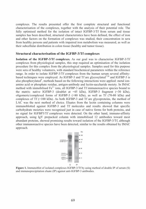
69
complexes. The results presented offer the first complete structural and functional characterisation of the complexes, together with the analysis of their potential role. The fully optimised method for the isolation of intact IGFBP-3/Tf from serum and tissue samples has been described, structural characteristics have been defined, the effect of iron and other factors on the formation of complexes was studied, their concentration in sera from healthy persons and patients with impaired iron metabolism was measured, as well as their subcellular distribution in colon tissue (healthy and tumor tissue).
Structural characterisation of the IGFBP-3/Tf complexes
Isolation of the IGFBP-3/Tf complexes. As our goal was to characterise IGFBP-3/Tf complexes from physiological samples, this step required an optimisation of the isolation procedure for this complex from the physiological samples. Samples used for this purpose were sera of healthy volunteers, with standard biochemical parameters within the reference range. In order to isolate IGFBP-3/Tf complexes from the human serum several affinity-based techniques were employed. As IGFBP-3 and Tf are glycosylated7,36 and IGFBP-3 is also phosphorylated4, methods based on the following interactions were applied: metal ion-amino acid or phosphate residue, antigen-antibody and lectin-saccharide moiety. In IMAC method with immobilised Fe3+ ions, all IGFBP-3 and Tf immunoreactive species bound to the matrix: native IGFBP-3 (doublet at ~45 kDa), IGFBP-3 fragment (~30 kDa), oligomeric/complexed forms of IGFBP-3 (>80 kDa), as well as Tf (70-80 kDa) and complexes of Tf (>100 kDa). As both IGFBP-3 and Tf are glycoproteins, the method of LAC was the next method of choice. Eluates from the lectin containing columns were immunoblotted against IGFBP-3 and Tf molecules and results showed that specific carbohydrate moieties were recognised just in case of native forms for both proteins, and no signal for IGFBP3/Tf complexes were detected. On the other hand, immuno-affinity approach, using IgY prepacked column with immobilised 12 antibodies toward most abundant proteins, showed promising results toward isolation of the IGFBP-3/Tf, although other immunoreactive species have been detected, similar to the results obtained by IMAC approach.
Figure 1. Immunoblot of isolated complexes IGFBP-3/Tf by using method of double IP of serum (S) and immunoprecipitation eluate (IP) against anti-IGFBP-3 antibodies.
70
None of the applied affinity-chromatography methods was able to selectively recognise IGFBP-3/Tf complexes and separate them from the monomeric, oligomeric and fragmented forms of IGFBP-3 and Tf. IgY-C seemed the most promising tool, so other immunochemical techniques were tested for the isolation of the complexes: immunoelectrophoresis (IEP), preparative immunoaffinity chromatography and immunoprecipitation (IP) method. Preparative chromatography gave no visible results on immunobotting for IGFBP-3/Tf, instead it has shown that this complex retains the possibility to bind ligands (IGF-I in this case). Finally, a co-immunoprecipitation method was employed, using commercial activated resin to immobilise separately anti-IGFBP-3and anti-Tf antibodies. Immunoprecipitation method is among the most specific techniques to isolate target protein, with the least amount of the contaminating substances. In this study, a double immunoprecipitation was performed. Samples were allowed to interact with immobilised anti-Tf antibodies in the first step and, after elution of the bound proteins, this fraction was allowed to interact with anti-IGFBP-3 antibodies37. Proteins eluted after the second step contained almost exclusively IGFBP-3/Tf complexes (Figure 1). Large aggregates and fragments (~30 kDa) immunoreactive with anti-IGFBP-3antibodies were present in the miniature amounts and they were completely removed by the ultrafiltration. Densitometric analysis showed that the amount of isolated IGFBP-3/Tf complexes by using the double IP approach was more than 80% of total signal, which belongs to a band at MW slightly over 110 kDa. This result approved double IP as the method of choice for the isolation and purification of complexes out of physiological samples. Metal ion effect on IGFBP-3/Tf complexes. After optimisation of the most suitable method for the isolation of the IGFBP-3/Tf protein complex, its structural analysis was possible. Since no data were available on the structure of this complex, our direction was to analyse it on those originating from healthy individuals. Within this group all parameters of iron metabolism (iron concentration, Hb, TIBC, ferritin, total proteins and Tf) were within reference range. The concentration of IGFBP-3 was also within expected range (data not shown). The effect of the type of metal ion on the efficiency of the isolation of IGFBP-3/Tf complexes in IMAC was analysed. Samples were randomly incubated with matrices saturated with Fe2+, Fe3+, Ni2+, Mg2+ Zn2+ or Cu2+ ions as major physiological elements. Eluted proteins were analyzed by IGFBP-3 immunoblotting. Representative results are shown in Figure 2A. As it can be seen, matrices saturated with Fe2+, Fe3+ or Cu2+ were capable of binding IGFBP-3 immunoreactive molecules almost completely, while other matrices were less efficient (especially those saturated with Mg2+
or Zn2+). In the case of Fe2+, Fe3+, Mg2+ and Zn2+ resins, bound proteins were almost completely eluted with phosphate buffer pH 8.0, whereas in the case of Cu2+ and Ni2+
complete elution was achieved with EDTA solution.Having on mind the fact that IMAC column packed with iron ions specifically bound IGFBP-3 immunoreactive species, it can be postulated that presence of this metal plays a role in forming the interaction of these two molecules. In the experiment to confirm this role of iron (and other metal) ions, serum filtrates containing only monomeric forms of
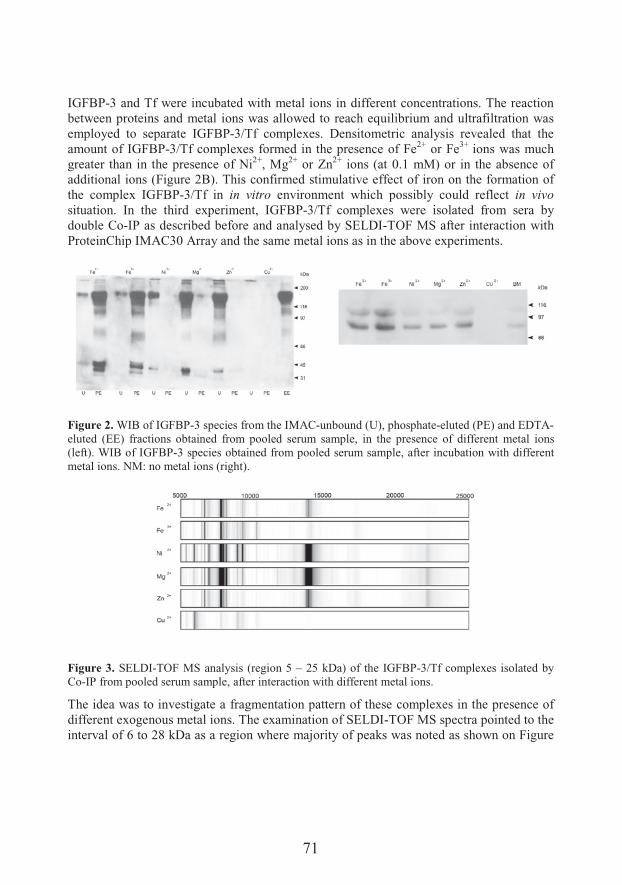
71
IGFBP-3 and Tf were incubated with metal ions in different concentrations. The reaction between proteins and metal ions was allowed to reach equilibrium and ultrafiltration was employed to separate IGFBP-3/Tf complexes. Densitometric analysis revealed that the amount of IGFBP-3/Tf complexes formed in the presence of Fe2+ or Fe3+ ions was much greater than in the presence of Ni2+, Mg2+ or Zn2+ ions (at 0.1 mM) or in the absence of additional ions (Figure 2B). This confirmed stimulative effect of iron on the formation of the complex IGFBP-3/Tf in in vitro environment which possibly could reflect in vivosituation. In the third experiment, IGFBP-3/Tf complexes were isolated from sera by double Co-IP as described before and analysed by SELDI-TOF MS after interaction withProteinChip IMAC30 Array and the same metal ions as in the above experiments.
Figure 2. WIB of IGFBP-3 species from the IMAC-unbound (U), phosphate-eluted (PE) and EDTA-eluted (EE) fractions obtained from pooled serum sample, in the presence of different metal ions (left). WIB of IGFBP-3 species obtained from pooled serum sample, after incubation with different metal ions. NM: no metal ions (right).
Figure 3. SELDI-TOF MS analysis (region 5 – 25 kDa) of the IGFBP-3/Tf complexes isolated by Co-IP from pooled serum sample, after interaction with different metal ions.
The idea was to investigate a fragmentation pattern of these complexes in the presence of different exogenous metal ions. The examination of SELDI-TOF MS spectra pointed to the interval of 6 to 28 kDa as a region where majority of peaks was noted as shown on Figure
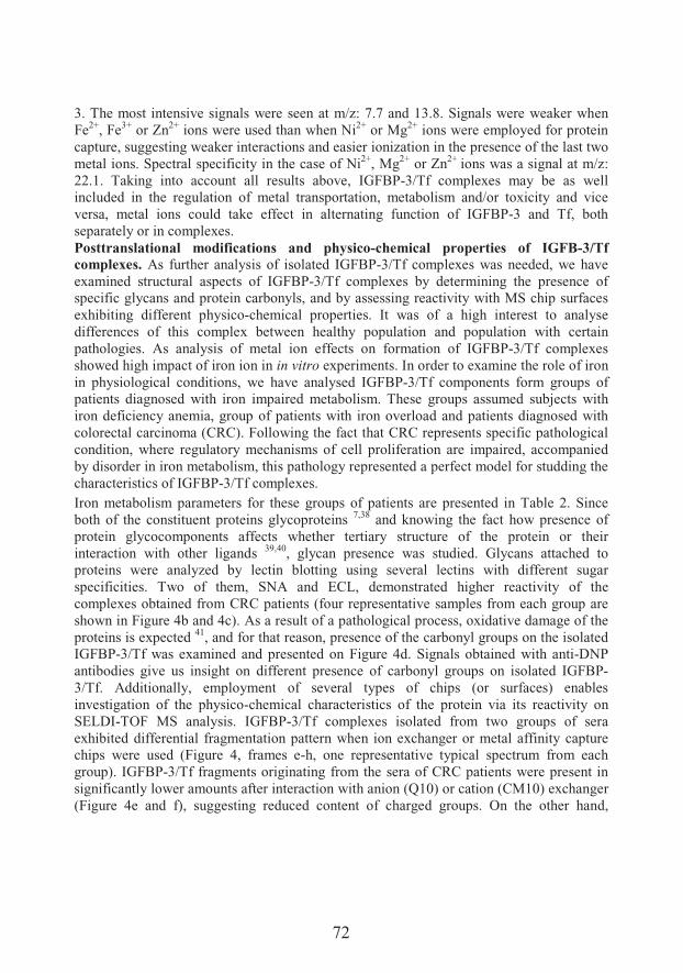
72
3. The most intensive signals were seen at m/z: 7.7 and 13.8. Signals were weaker when Fe2+, Fe3+ or Zn2+ ions were used than when Ni2+ or Mg2+ ions were employed for protein capture, suggesting weaker interactions and easier ionization in the presence of the last two metal ions. Spectral specificity in the case of Ni2+, Mg2+ or Zn2+ ions was a signal at m/z: 22.1. Taking into account all results above, IGFBP-3/Tf complexes may be as well included in the regulation of metal transportation, metabolism and/or toxicity and vice versa, metal ions could take effect in alternating function of IGFBP-3 and Tf, both separately or in complexes.Posttranslational modifications and physico-chemical properties of IGFB-3/Tf complexes. As further analysis of isolated IGFBP-3/Tf complexes was needed, we have examined structural aspects of IGFBP-3/Tf complexes by determining the presence of specific glycans and protein carbonyls, and by assessing reactivity with MS chip surfaces exhibiting different physico-chemical properties. It was of a high interest to analyse differences of this complex between healthy population and population with certain pathologies. As analysis of metal ion effects on formation of IGFBP-3/Tf complexes showed high impact of iron ion in in vitro experiments. In order to examine the role of iron in physiological conditions, we have analysed IGFBP-3/Tf components form groups of patients diagnosed with iron impaired metabolism. These groups assumed subjects with iron deficiency anemia, group of patients with iron overload and patients diagnosed with colorectal carcinoma (CRC). Following the fact that CRC represents specific pathological condition, where regulatory mechanisms of cell proliferation are impaired, accompanied by disorder in iron metabolism, this pathology represented a perfect model for studding the characteristics of IGFBP-3/Tf complexes. Iron metabolism parameters for these groups of patients are presented in Table 2. Since both of the constituent proteins glycoproteins 7,38 and knowing the fact how presence of protein glycocomponents affects whether tertiary structure of the protein or their interaction with other ligands 39,40, glycan presence was studied. Glycans attached to proteins were analyzed by lectin blotting using several lectins with different sugar specificities. Two of them, SNA and ECL, demonstrated higher reactivity of the complexes obtained from CRC patients (four representative samples from each group are shown in Figure 4b and 4c). As a result of a pathological process, oxidative damage of the proteins is expected 41, and for that reason, presence of the carbonyl groups on the isolated IGFBP-3/Tf was examined and presented on Figure 4d. Signals obtained with anti-DNP antibodies give us insight on different presence of carbonyl groups on isolated IGFBP-3/Tf. Additionally, employment of several types of chips (or surfaces) enables investigation of the physico-chemical characteristics of the protein via its reactivity on SELDI-TOF MS analysis. IGFBP-3/Tf complexes isolated from two groups of sera exhibited differential fragmentation pattern when ion exchanger or metal affinity capture chips were used (Figure 4, frames e-h, one representative typical spectrum from each group). IGFBP-3/Tf fragments originating from the sera of CRC patients were present in significantly lower amounts after interaction with anion (Q10) or cation (CM10) exchanger (Figure 4e and f), suggesting reduced content of charged groups. On the other hand,
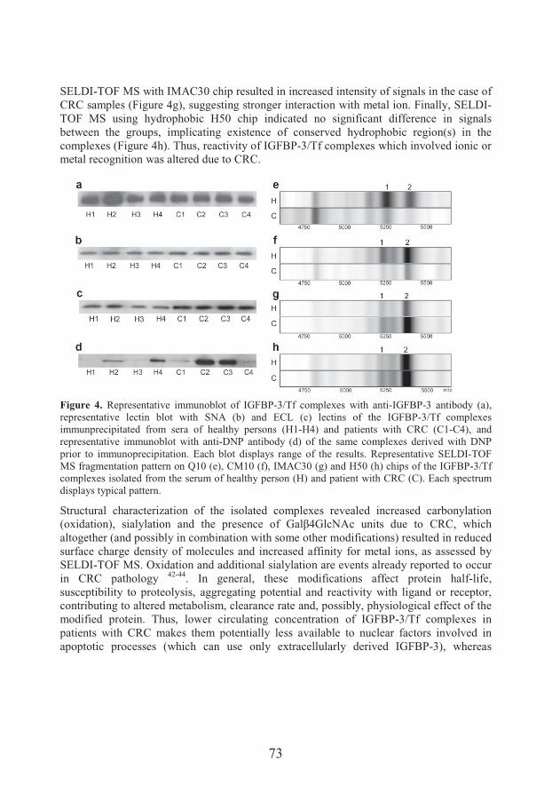
73
SELDI-TOF MS with IMAC30 chip resulted in increased intensity of signals in the case of CRC samples (Figure 4g), suggesting stronger interaction with metal ion. Finally, SELDI-TOF MS using hydrophobic H50 chip indicated no significant difference in signals between the groups, implicating existence of conserved hydrophobic region(s) in the complexes (Figure 4h). Thus, reactivity of IGFBP-3/Tf complexes which involved ionic or metal recognition was altered due to CRC.
Figure 4. Representative immunoblot of IGFBP-3/Tf complexes with anti-IGFBP-3 antibody (a), representative lectin blot with SNA (b) and ECL (c) lectins of the IGFBP-3/Tf complexes immunprecipitated from sera of healthy persons (H1-H4) and patients with CRC (C1-C4), and representative immunoblot with anti-DNP antibody (d) of the same complexes derived with DNP prior to immunoprecipitation. Each blot displays range of the results. Representative SELDI-TOF MS fragmentation pattern on Q10 (e), CM10 (f), IMAC30 (g) and H50 (h) chips of the IGFBP-3/Tf complexes isolated from the serum of healthy person (H) and patient with CRC (C). Each spectrum displays typical pattern.
Structural characterization of the isolated complexes revealed increased carbonylation F�H� ����@%� &� �$� ����� � � �'�� ��&����� ��� R �>?R���+�� #���&� #�� ��� ,�,%� 7'��'�altogether (and possibly in combination with some other modifications) resulted in reduced surface charge density of molecules and increased affinity for metal ions, as assessed by SELDI-TOF MS. Oxidation and additional sialylation are events already reported to occur in CRC pathology 42-44. In general, these modifications affect protein half-life, susceptibility to proteolysis, aggregating potential and reactivity with ligand or receptor, contributing to altered metabolism, clearance rate and, possibly, physiological effect of the modified protein. Thus, lower circulating concentration of IGFBP-3/Tf complexes in patients with CRC makes them potentially less available to nuclear factors involved in apoptotic processes (which can use only extracellularly derived IGFBP-3), whereas
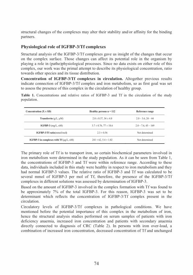
74
structural changes of the complexes may alter their stability and/or affinity for the binding partners.
Physiological role of IGFBP-3/Tf complexes
Structural analysis of the IGFBP-3/Tf complexes gave us insight of the changes that occur on the complex surface. Those changes can affect its potential role in the organism by playing a role in (patho)physiological processes. Since no data exists on either role of this complex, our work was the primal attempt to describe its physiological concentration, ratio towards other species and its tissue distribution. Concentration of IGFBP-3/Tf complexes in circulation. Altogether previous results indicate connection of IGFBP-3/Tf complex and iron metabolism, so as first goal was set to assess the presence of this complex in the circulation of healthy group.
Table 1. Concentrations and relative ratios of IGFBP-3 and Tf in the circulation of the study population.
The primary role of Tf is to transport iron, so certain biochemical parameters involved in iron metabolism were determined in the study population. As it can be seen from Table 1, the concentrations of IGFBP-3 and Tf were within reference range. According to these data, individuals included in this study were healthy in respect to iron metabolism and they had normal IGFBP-3 values. The relative ratio of IGFBP-3 and Tf was calculated to be several mmol of IGFBP-3 per mol of Tf, therefore, the presence of the IGFBP-3/Tf complexes in different solutions was assessed by determination of IGFBP-3. Based on the amount of IGFBP-3 involved in the complex formation with Tf was found to be approximately 7% of the total IGFBP-3. For this reason, IGFBP-3 was set to be determinant which reflects the concentration of IGFBP-3/Tf complex present in the circulation. Circulatory levels of IGFBP-3/Tf complexes in pathological conditions. We have mentioned before the potential importance of this complex in the metabolism of iron, hence the structural analysis studies performed on serum samples of patients with iron deficiency anaemia, increased iron concentration and patients with secondary anaemia directly connected to diagnosis of CRC (Table 2). In persons with iron over-load, a combination of increased iron concentration, decreased concentration of Tf and unchanged
Concentration (X ± SD) Healthy persons n = 112 Reference range
Transferrin F�L5%�c�@ 2.8 ± 0.37, 34 ± 4.0 2.0 – 3.6, 24 – 44
IGFBP-3 (mg/L, nM) 3.7 ± 0.76, 77 ± 18.6 2.0 – 7.6, 45 – 169
IGFBP-3/Tf ratio(mmol/mol) 2.3 ± 0.56 Not determined
IGFBP-3 in complexes with Tf Fc�L5%���@ 241 ± 62, 5.4 ± 1.02 Not determined
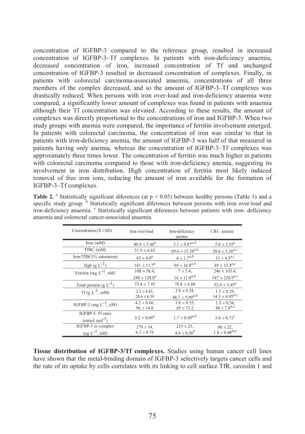
75
concentration of IGFBP-3 compared to the reference group, resulted in increased concentration of IGFBP-3–Tf complexes. In patients with iron-deficiency anaemia, decreased concentration of iron, increased concentration of Tf and unchanged concentration of IGFBP-3 resulted in decreased concentration of complexes. Finally, in patients with colorectal carcinoma-associated anaemia, concentrations of all three members of the complex decreased, and so the amount of IGFBP-3–Tf complexes was drastically reduced. When persons with iron over-load and iron-deficiency anaemia were compared, a significantly lower amount of complexes was found in patients with anaemia although their Tf concentration was elevated. According to these results, the amount of complexes was directly proportional to the concentrations of iron and IGFBP-3. When two study groups with anemia were compared, the importance of ferritin involvement emerged. In patients with colorectal carcinoma, the concentration of iron was similar to that in patients with iron-deficiency anemia, the amount of IGFBP-3 was half of that measured in patients having only anemia, whereas the concentration of IGFBP-3–Tf complexes was approximately three times lower. The concentration of ferritin was much higher in patients with colorectal carcinoma compared to those with iron-deficiency anemia, suggesting its involvement in iron distribution. High concentration of ferritin most likely induced removal of free iron ions, reducing the amount of iron available for the formation ofIGFBP-3–Tf complexes.
Table 2. a Statistically significant diferences (at p < 0.05) between healthy persons (Table 1) and a specific study group. b Statistically significant diferences between persons with iron over-load and iron-deficiency anaemia. c Statistically significant diferences between patients with iron- deficiency anaemia and colorectal cancer-associated anaemia.
Concentration (X ±SD) Iron over-load Iron-deficiencyanemia
CRC anemia
Iron (mM) 40.8 ± 3.46a 3.1 ± 0.87a,b 5.0 ± 1.55a
TIBC (mM) 51.9 ± 6.85 69.6 ± 13.34a,b 30.6 ± 5.36a,c
Iron/TIBC(% saturation) 83 ± 8.0a 4 ± 1.7a,b 15 ± 4.3a,c
Hgb (g L-1) 141 ± 11.7a 94 ± 16.8a,b 85 ± 13.4a,c
Ferritin (mg L-1, nM) 108 ± 58.4,
240 ± 128.8a 7 ± 5.4,
16 ± 11.0a,b246 ± 103.4,
547 ± 228.9a,c
Total protein (g L-1) 73.4 ± 7.95 70.8 ± 6.88 52.9 ± 5.45a
Tf (g L-1, mM) 2.3 ± 0.43,28.6 ± 6.39
3.8 ± 0.38,
48.7 ± 5.99a,b 1.3 ± 0.29,
14.3 ± 4.95a,c
IGFBP-3 (mg L-1, nM) 4.2 ± 0.66,96 ± 14.0
3.8 ± 0.55,85 ± 13.2
1.9 ± 0.36,40 ± 7.6a,c
IGFBP-3–Tf ratio
(mmol mol-1)3.2 ± 0.69a 1.7 ± 0.30a,b 3.0 ± 0.71c
IGFBP-3 in complex
Fc� L-1, nM)279 ± 34,6.2 ± 0.76
215 ± 25,
4.6 ± 0.56b80 ± 22,
1.8 ± 0.48a,c
Tissue distribution of IGFBP-3/Tf complexes. Studies using human cancer cell lines have shown that the metal-binding domain of IGFBP-3 selectively targets cancer cells and the rate of its uptake by cells correlates with its linking to cell surface TfR, caveolin 1 and

76
integrin p 45,46. It triggers apoptosis in stressed cells 34. Functions that IGFBP-3 exerts via interaction of its metal-binding domain followed by IGFBP-3 internalization and nuclear localization (which includes nuclear localization sequence) are termed IGF independent 47.Lee et al. 48 have hypothesized that IGFBP-3 is first secreted then taken up again into cells to be translocated to the nucleus. The definitive role of IGFBP-3 in the nucleus has yet to be fully supported, but it most likely modulates the gene expression 49. Existence of a new cell death receptor, IGFBP-3R, was recently reported 50. In vivo and in vitro studies have demonstrated that the activation of IGFBP-3R mediates apoptosis in cancer cells via induction of caspase 8. Therefore, at least two different mechanisms and receptors are involved in the cellular functions of IGFBP-3. Tf bound to TfR undergoes clathrin-mediated endocytosis and is directed in endosomes along a recycling pathway, not a degradation pathway. Proteins intended for recycling are returned to the cell surface 51.IGFBP-3 bound in complexes with Tf may be following the same route but this is, at the moment, only an experimentally unsupported hypothesis. Pro-apoptotic effect of IGFBP-3 was confirmed in colon carcinoma22,23. Although many cells can synthetize IGFBP-3, liver cells are responsible for the production of the majority of IGFBP-3 present in the circulation 52. Some tumour cells express IGFBP-3, whereas others only acquire IGFBP-3 via its interaction with surface receptors. Colorectal cancer arises from the colon epithelial cells which were reported to produce IGFBP-3 53, but they can also internalize IGFBP-3 48. The metabolism of iron was found disturbed in certain types of tumour, including CRC 54,55. Low concentrations of free iron ion and iron ion bound to Tf, together with increased concentrations of ferritin were measured in the circulation of patients with CRC 56. In this study we have focused on the interaction of IGFBP-3/Tf complexes with TfR and the cellular localization of the ternary assembly IGFBP-3/Tf/TfR. Two separate experiments, one involving immunoprecipitation of the membrane and cytosol samples using anti-TfR antibody and the other based on immunofluorescent staining of the IGFBP-3 and TfR in tissue slides have documented the presence of IGFBP-3/Tf complexes in both sample types from the cancer and non-cancer colon tissues. The immunoprecipitation experiment revealed on average more TfR on membranes than in cytosols (as expected) and they were proportionally less occupied with IGFBP-3/Tf complexes than in cytosols. Thus, the efficiency of the internalization process which concentrates IGFBP-3/Tf complexes inside the cells seems to have similar final outcome in both cancer and non-cancer tissue. It may be postulated that increased expression of TfR on membranes aims to compensate for the reduced extracellular availability of IGFBP-3/Tf complexes and possibly altered affinity due to their posttranslational modifications. However, one must bear in mind that this result was achieved by using pooled samples, as there were significant inter-individual diferences most likely reflecting different stages of a disease and different involvement of the surrounding tissue.The existence of the IGFBP-3/Tf/TfR ternary complexes in colon tissue was confirmed by immunohistochemistry as well. Since it is known that IGFBP-3 does not interact directlywith TfR, but only via Tf 30, the experiment was performed by two-phase application of
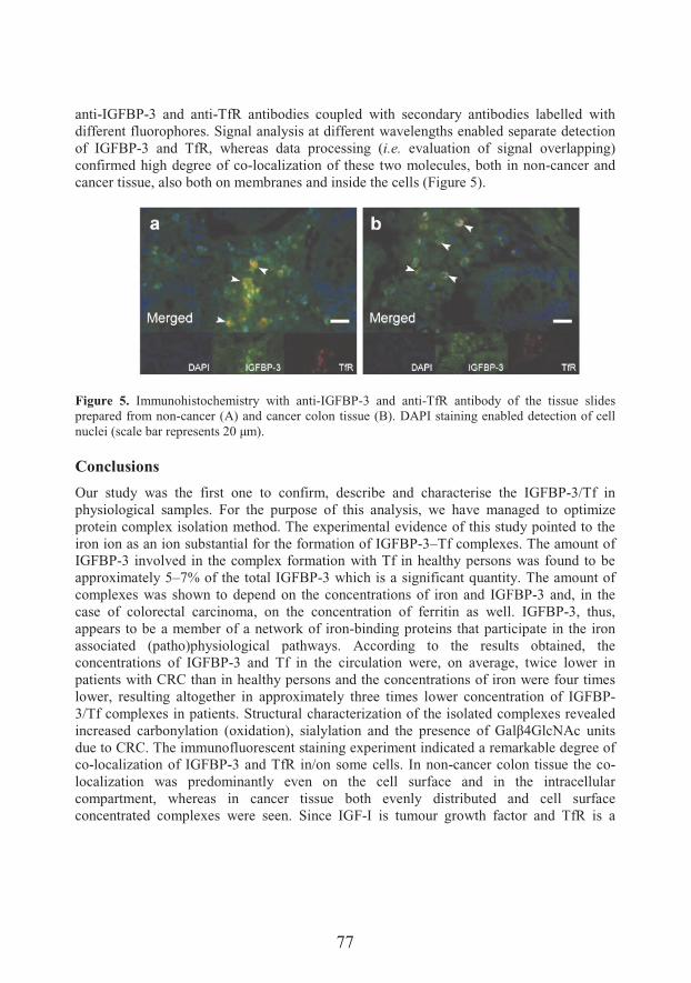
77
anti-IGFBP-3 and anti-TfR antibodies coupled with secondary antibodies labelled with different fluorophores. Signal analysis at different wavelengths enabled separate detection of IGFBP-3 and TfR, whereas data processing (i.e. evaluation of signal overlapping) confirmed high degree of co-localization of these two molecules, both in non-cancer and cancer tissue, also both on membranes and inside the cells (Figure 5).
Figure 5. Immunohistochemistry with anti-IGFBP-3 and anti-TfR antibody of the tissue slidesprepared from non-cancer (A) and cancer colon tissue (B). DAPI staining enabled detection of cell �#�����F&� ���8 ������&���&�XY�c�@�
Conclusions
Our study was the first one to confirm, describe and characterise the IGFBP-3/Tf in physiological samples. For the purpose of this analysis, we have managed to optimizeprotein complex isolation method. The experimental evidence of this study pointed to the iron ion as an ion substantial for the formation of IGFBP-3–Tf complexes. The amount of IGFBP-3 involved in the complex formation with Tf in healthy persons was found to be approximately 5–7% of the total IGFBP-3 which is a significant quantity. The amount of complexes was shown to depend on the concentrations of iron and IGFBP-3 and, in the case of colorectal carcinoma, on the concentration of ferritin as well. IGFBP-3, thus, appears to be a member of a network of iron-binding proteins that participate in the iron associated (patho)physiological pathways. According to the results obtained, the concentrations of IGFBP-3 and Tf in the circulation were, on average, twice lower in patients with CRC than in healthy persons and the concentrations of iron were four times lower, resulting altogether in approximately three times lower concentration of IGFBP-3/Tf complexes in patients. Structural characterization of the isolated complexes revealed increased carbonylation (oxidation), sialylation and the pre&����� ��� R �>?R���+�� #���&�due to CRC. The immunofluorescent staining experiment indicated a remarkable degree of co-localization of IGFBP-3 and TfR in/on some cells. In non-cancer colon tissue the co-localization was predominantly even on the cell surface and in the intracellular compartment, whereas in cancer tissue both evenly distributed and cell surface concentrated complexes were seen. Since IGF-I is tumour growth factor and TfR is a
78
binding partner of IGFBP-3 via Tf, increased expression of TfR may be a (patho)physiological response to ensure extracellular capture of IGFBP-3 and its direction towards pro-apoptotic pathways.
Acknowledgements
This study was supported by the ministry of Education, Science and Technological Development under grant agreement No. 173042.
References1. Cohick WS, Clemmons DR. The insulin-like growth factors. Annu Rev Physiol 1993;55:131-
53.2. Stewart CEH, Rotwein P. Growth, differentiation, and survival: Multiple physiological
functions for insulin-like growth factors. Physiol Rev 1996;76:1005-26.3. Annunziata M, Granata R, Ghigo E. The IGF system. Acta Diabetol 2011;48:1-9.4. Firth SM, Baxter RC. Cellular actions of the insulin-like growth factor binding proteins. Endocr
Rev 2002;23:824-54.5. Collett-Solberg PF, Cohen P. Genetics, chemistry, and function of the IGF/IGFBP system.
Endocrine 2000;12:121-36.6. Rajaram S, Baylink DJ, Mohan S. Insulin-like growth factor-binding proteins in serum and
other biological fluids: regulation and functions. Endocr Rev 1997;18:801-31.7. Firth SM, Baxter RC. Characterisation of recombinant glycosylation variants of insulin-like
growth factor binding protein-3. J Endocrinol 1999;160:379-87.8. Khosravi MJ, Diamandi A, Mistry J, Krishna RG, Khare A. Acid-labile subunit of human
insulin-like growth factor-binding protein complex: measurement, molecular, and clinical evaluation. J Clin Endocrinol Metab 1997;82:3944-51.
9. Janosi JBM, Ramsland PA, Mott MR, Firth SM, Baxter RC, Delhanty PJD. The acid-labile subunit of the serum insulin-like growth factor-binding protein complexes. Structural determination by molecular modeling and electron microscopy. J Biol Chem 1999;274:23328-32.
10. Biosclair YR, Rhoads RP, Ueki I, Wang J, Ooi GT. The acid-labile subunit (ALS) of the 150 kDa IGF-binding protein complex: an important but forgotten component of the circulating IGF system. J Endocrinol 2001;170:63-70.
11. Campbell PG, Durham SK, Hayes JD, Suwanichkul A, Powell DR. Insulin-like growth factor-binding protein-3 binds fibrinogen and fibrin. J Biol Chem 1999;274:30215-21.
12. Oesterreicher S, Blum WF, Schmidt B, Braulke T, Kübler B. Interaction of insulin-like growth factor II (IGF-II) with multiple plasma proteins. J Biol Chem 2005;280:9994-10000.
13. Liu B, Weinzimer SA, Gibson TB, Mascarenhas D, ,�'���.��*$��";����� �����&� ��"R��.-3binding protein. Growth Horm IGF Res 2003;13:89-97.
14. Fowlkes JL, Serra DM. Characterization of Glycosaminoglycan-binding domains present in insulin-like growth factor-binding protein-3. J Biol Chem 1996;21:14676–79.
15. 5� ����%�5�#�d%�O# �����%�O# �������*'���$��3��� �&�����������7�'�� �����>����������&��'��putative insulin-like growth factor-binding protein 3 receptor. J Biol Chem 1997;272:20572–6.
16. Couet J, Li S, Okamotoi T, Ikezui T, Lisanti MP. Identification of peptide and protein ligands for the caveolin-scaffolding domain. J Biol Chem 1997;272:6525–33.
17. Yamanaka Y, Fowlkes JL, Wilson EM, Rosenfeld RG, Oh Y. Characterization of insulin-like growth factor binding protein-3 (IGFBP-3) binding to human breast cancer cells: Kinetics of IGFBP-3 binding and identification of receptor binding domain on the IGFBP-3 molecule. Endocrinology 1999;140:1319-28.
18. Baxter RC. Insulin-like growth factor binding protein-3 (IGFBP-3): Novel ligands mediate unexpected functions. J Cell Commun Signal 2013;7:179-89.
79
19. Schedlich LJ, Le Page SL, Firth SM, Briggs LJ, Jans DA, Baxter RC, Nuclear import of insulin-like growth factor-binding protein-3 and -B��&��� � �� �8$��'����������>��#8#������������,'���2000;275:23462-70.
20. Lee KW, Ma L, Yan X, Liu B, Zhang XK, Cohen P. Rapid apoptosis induction by IGFBP-3involves an insulin-like growth factor-independent nucleomitochondrial translocation of �e�;L�#�<<���������,'���XYYBAXfY=9Cg?X-48.
21. Liu B, Lee HY, Weinzimer SA, Powell DR, Clifford JL, Kurie JM, Cohen P. Direct functional interactions between insulin-like growth factor-binding protein-3 and retinoid X receptor-aregulate transcriptional signaling and apoptosis. J Biol Chem 2000;275:33607-13.
22. Williams AC, Collard TJ, Perks CM, Newcomb P, Moorghen M, Holly JMP, Paraskeva C. Increased p53-dependent apoptosis by the insulin-like growth factor binding protein IGFBP-3 in human colonic adenoma-derived cells. Cancer Res 2000;60:22-7.
23. Williams AC, Smartt H, H-Zadeh AM, MacFarlane M, Paraskeva C, Collard TJ. Insulin-like growth factor binding protein 3 (IGFBP-3) potentiates TRAIL-induced apoptosis of human colorectal carcinoma cells through inhibition of NF-W���,������ �'��������XYY<A9?=9:<-45.
24. R������ �%� 1h�����$� 3,%� O �� �%� O���� �%� � H���� �,%� ����'� ���� "R��.-3 binds GRP78, stimulates autophagy and promotes the survival of breast cancer cells exposed to adverse microenvironments. Oncogene 2013;32:2412-20.
25. Weinzimer SA, Gibson TB, Collett-Solberg PF, Khare A, Liu B, Cohen P. Transferrin is an insulin-like growth factor-binding protein-3 binding protein. J Clin Endocrinol Metab 2001;86:1806-13.
26. Gomme PT, McCann KB. Transferrin: structure, function and potential therapeutic actions. Drug Discov Today 2005;10:267-73.
27. De Jong G, Van Dijk JP, Van Eijk HG. The biology of transferrin. Clin Chim Acta 1990;190:1-46.
28. Chloupková M, Zhang AS, Enns CA. Stoichiometries of transferrin receptors 1 and 2 in human liver. Blood Cells Mol Dis 2010;44:28–33.
29. Paterson S, Armstrong NJ, Iacopetta BJ, McArdle HJ, Morgan EH. Intravesicular pH and iron uptake by immature erythroid cells. J Cell Physiol 1984;120:225–32.
30. Li H, Qian ZM. Transferrin/transferrin receptor-mediated drug delivery. Inc Med Res Rev 2002;22:225–50.
31. Sinicropi MS, Amantea A, Caruso A, Saturnino C. Chemical and biological properties of toxic metals and use of chelating agents for the pharmacological treatment of metal poisoning. Arch Toxicol 2010;84:501-20.
32. Quarles Jr CD, Marcus RK, Burmaghim JL. Competitive binding of Fe3+, Cr3+, and Ni2+ to transferrin. J Biol Inorg Chem 2011;16:913-21.
33. El-Hage-Chahine JM, Hémadi M, Ha Duong NT. Uptake and release of metal ions by transferrin and interaction with receptor 1. Biochim Biophys Acta-Gen Subj 2012;1820:334-47.
34. Vincent JB, Love S. The binding and transport of alternative metals by transferrin. Biochim Biophys Acta- Gen Subj 2012;1820:362-78.
35. Singh B, Charkowicz D, Mascarenhas D. Insulin-like growth factor-independent effects mediated by a C-terminal metal-binding domain of insulin-like growth factor binding protein-3. J Biol Chem 2004;279:477-87.
36. Bergen HR, Lacey JM, O’Brien JF, Naylor S. Online single-step analysis of blood proteins: The transferrin story. Anal Biochem 2001;296:122-29.
37. ����#�� R%� .��������%� �� ��� 1�� "&�� ����� ��� �����H�&� ����� � 8��7���� ��&#���-like growth factor-binding protein-3 and transferrin from the human serum. J Serb Chem Soc 2012;77:607-17.
38. ��'������%�+ ���������� � ��������'#� ��&��#���� �&��������&�����&�8y high-performance pellicular anion-exchange chromatography. Protein Expres Purif 1996;7:39–44.
39. Fiedler K, Simons K. The role of N-glycans in the secretory pathway. Cell 1995;81:309–12.40. Gagneux P, Varki A. Evolutionary considerations in relating oligosaccharide diversity to
biological function. Glycobiology 1999;9:747–55.41. Suzuki YJ, Carini M, Butterfield DA. Protein carbonylation. Antioxid Redox Signal
2010;12:323–25.
80
42. Oberreuther-Moschnera DL, Rechkemmerb G, Pool-Zobelc BL. Basal colon crypt cells are more sensitive than surface cells toward hydrogen peroxide, a factor of oxidative stress. Toxicol Lett 2005;159:212-18.
43. Perše M. Oxidative stress in the pathogenesis of colorectal cancer: Cause or consequence? BioMed Res Int 2013;725710.
44. Sethi MK, Thaysen-Andersen M, Smith JT, Baker MS, Packer NH, Hancock WS, Fanayan S. Comparative Niglycan profiling of colorectal cancer cell lines reveals unique bisecting GlcNAc � � ;i2,3-linked sialic acid determinants are associated with membrane proteins of the more metastatic/aggressive cell lines. J Proteome Res 2014;13:277-88.
45. Burrows C, Holly JMP, Laurence NJ, Vernon EG, Carter JV, Clark MA, McIntosh J et al. Insulin-like growth factor binding protein 3 has opposing actions on malignant and nonmalignant breast epithelial cells that are each reversible and dependent upon cholesterol-stabilized integrin receptor complexes. Endocrinology 2006; 147:3484–500.
46. Huq A, Singh B, Meeker T, Mascarenhas D. The metal-binding domain of IGFBP-3 selectively delivers therapeutic molecules into cancer cells. Anti-Cancer Drugs 2009;20:21–31.
47. Oufattole M, Lin SWJ, Liu B, Mascarenhas D, Cohen P, Rodgers BD. Ribonucleic Acid Polymerase II Binding Subunit 3 (Rpb3), a Potential Nuclear Target of Insulin-Like Growth Factor Binding Protein-3. Endocrinology 2006;147:2138–46.
48. Lee KW, Liu B, Ma L, Li H, Bang P, Koeffler HP, P. Cohen P. Cellular internalization of insulin-like growth factor binding protein-3 distinct endocytic pathways facilitate re-uptake and nuclear localization. J Biol Chem 2004;279:469–76.
49. Jogie-Brahim S, Feldman D, Oh Y. Unraveling insulin-like growth factor binding protein-3actions in human disease. Endocr Rev 2009;30:417–37.
50. Ingermann AR, Yang YF, Han J, Mikami A, Garza AE, Mohanraj L, Fan L et al. Identification of a novel cell death receptor mediating IGFBP-3-induced anti-tumor effects in breast and prostate cancer. J Biol Chem 2010;285:30233–46.
51. Mayle KM, Le AM, Kamei DT. The intracellular trafficking pathway of transferrin. Biochim Biophys Acta 2012;1820:264–81.
52. Jones JI, Clemons DR. Insulin-like growth factors and their binding proteins: Biological actions. Endocr Rev 1995;16:3-34.
53. Mehta HH, Gao Q, Galet C, Paharkova V, Wan J, Said J, Sohn JJ, et al. IGFBP-3 is a metastasis suppression gene in prostate cancer. Cancer Res 2011;71:5154-63.
54. Gackowski D, Kruszewski M, Banaszkiewicz Z, Jawien A, Olinski R. Lymphocyte labile iron pool, plasma iron, transferrin saturation and ferritin levels in colon cancer patients. Acta Biochim Pol 2002;49:269-72.
55. Beale AL, Penney MD, Allison MC. The prevalence of iron deficiency among patients presenting with colorectal cancer. Colorectal Dis 2005;7:398-402.
56. ����#��R%�� ��������3%��� ���1��*'������� ���������� �����&������'������ ����� � ��&�� �����of insulin-like growth factor-binding protein 3–transferrin (IGFBP-3–Tf) complexes, and the analysis of their physiological involvement. Metallomics 2013;5:251-8.
81
Transport and metabolism of vanadium in filamentous fungi with emphasis on fungus Phycomyces blakesleeanus
����������1*�: ���;����)���2���1���<��0 �������3�����������2,Joanna Zakrzewska4, Milan Ž0�1
1Department of Life Sciences, Institute for Multidisciplinary Research, University ofBelgrade, Belgrade, Serbia2Institute of Physiology and Biochemistry, Faculty of Biology, University of Belgrade3Elementary School “Knezova ����� ��<= Nova Varoš, Serbia4Institute of General and Physical Chemistry, Belgrade, Serbia
*e-mails: [email protected]; [email protected]
Even though potential therapeutic effects of vanadium were discovered back in the late 19th century, this transition metal is still not significantly represented in the R&D of pharmaceutical companies due to its very complex chemistry. Vanadium redox properties and stereochemistry are the crucial aspects in considering its effects on biological systems. Vanadium in the oxidation states IV and V are the most important forms of this element in physiological conditions, and can also have a variety of geometries and polymerization forms, depending on local pH, concentration, ionic strength and interactions with various ligands. In many groups of organisms it is an essential trace element, but it becomes toxic at higher concentrations. Due to anthropogenic-activity related increase of its level in the environment, it may pose an environmental threat in the future. Water and soil are the major sources of vanadium uptake by living organisms, with fungi as one of the main routes of its entrance into the biotic component of the ecosystem. Having in mind the abundance of vanadium in nature, it is baffling that the data on its transport and metabolism in fungi are scarce. To right this wrong, we have chosen a filamentous fungus Phycomyces blakesleanus as a model for a detailed study of vanadium metabolism, transport and accumulation. Our findings show that this fungus has a variety of mechanisms for overcoming the potentially toxic effects of various forms of vanadium and high accumulation potential, which makes it an appropriate candidate for vanadium bioremediation.
Introduction
Vanadium is a naturally occurring transition metal which is found in high concentrations in the Earth’s crust, oceans, fossil fuels and soil. In the crust, vanadium is 22nd in abundance, while in the oceans, where it is mostly present as Na+H2VO4
- , its average concentration is 30 nM, which makes it a second most abundant metal, surpassed only by molibdenum 1. In
82
nature, vanadium is found as vanadium compounds with oxidation states ranging from -1to +5 2. It is a trace element, but essential for many groups of living organisms 3, which is not surprising having in mind its prevalence in nature. In higher concentrations, it can be toxic 4. Vanadium in the oxidation states V, IV and to some extent III is of biological relevance. The most common forms of vanadium under physiological conditions are protonated vanadate anions (H2VO4
- and HVO42-) and vanadyl cation (VO2+) 5.
Physiologically most stable vanadium (V) draws particular attention due to its prominent metabolic effects in many living system 6, and its biological activity is in strong correlation with its oligomeric form. Depending on pH, concentration, and ionic strength, vanadium(V) can exist mostly as a monomer, a dimer, or a tetramer 1.In the past couple of decades, there has been a remarkable increase in the investigation of vanadium behavior in biological systems, which can be associated with discoveries of its potential therapeutis effects 1,3, existence of vanadium dependent enzymes 7,8 and identification of several groups of vanadium-accumulating organisms 9. Many biological activities of vanadium are based on the fact that vanadium in oxidation state V - vanadate (VV) is a structural and electronic analogue of phosphate, especially in their tetrahedral trianionic forms – VO4
3- and PO43- 3. Hence, vanadate is able to influence phosphate-
metabolizing systems 10, often by inhibiting different enzymes such as phosphatases, phosphodiesterases, ribonucleases, ATPases, phosphoglucomutase etc. The inhibitory effect is actually based on slight differences between vanadate and phosphate. Phosphorus can only attain the coordination number 4 in transitional states of relatively high energy, while vanadium easily forms stable complexes with coordination numbers 4, 5 or 6. Once incorporated into the active site of a phosphate-dependent enzyme, taking the place of phosphate, the activity of this enzyme may be inhibited 11. A well known insulin mimetic activity of vanadate is related to this substitution. Cellular uptake of glucose is mediated byglucose transporter GLUT4 which is activated by a signal cascade involving extracellular bi� ���������&#���������&#������������F"�@� � �'�&'��$� ������������ ����#� ��>�#���&�of IR. In the absence of insulin or insufficient responce of IR a protein tyrosine phosphatase (PTP) ��#���� ��&� �'�� '�&'���� ����� ��� "�>� � � �& 8��&� � &��� �� � &�ade which leads to glucose uptake 12. Vanadate restores the signaling path by coordinating to cystein residue of PTP preventing dephosphorilation of IR> 12. Another important difference between vanadate and phosphate is that under physiological conditions vanadate can undergo redox chemistry 3 and the interconversion between vanadate (VV) and vanadyl (VIV) can play an important role in cellular redox balance, acting as either generator or eliminator of reactive oxygen species 13. This is presumed as a possible mode of action of vanadium as anti-cancer drug 14. Vanadium also shows promissing therapeutic effects against protozoa causing leischmaniasis, Chagas’ desease, as well as antiviral and anti-bacterial effects 1. Despite all of its potential, vanadium is still not the subject of research in big pharmaceutical companies. Its complex chemistry rises the fear of serious adverse effects, and very little is known about its effects on the immune system, oxidative stress, inflamatory response and toxicity, especally during long term administration 15.
83
Some bacterial strains can use vanadate as a terminal electron acceptor in respiratory or dissimilatory reduction 12. Bacteria and cyanobacteria such as Anabaena azotica and Azotobacter vinelandii 16 have alternative, vanadium-dependent nitrogenases for the conversion of dinitrogen into ammonium anions. Vanadium is also detected in the active center of vanadium-dependent haloperoxidases, that catalise two-electron reduction of halides by hidrogen peroxide. The hipohalous acids and halogenetic intermediates that are produced, react nonezymatically and nonspecifically with the array of organic targets producing halogenated compounds RX 11. Apart from bacteria, vanadium-dependent haloperoxidases were identified in fungi, algae and lychens. Nitrate reductase, enzymes that commonly catalize two-electron reduction of nitrate to nitrite and contain Mo in the active center, were found with V instead of Mo in bacterium Thioalkalivibrio nitratireducens 17.Three groups of organisms that accumulate vanadium with no apparent benefit have been identified so far: ascidians, polychaetes and several Amanita mushrooms. Vanadium accumulating ability was first discovered by German physiologist Martin Henze in 1911 who detected high ammounts of vanadium in the blood cells of ascidian (sea squirt) Phallusia mamillata 1. Specialized blood cells – vanadocites take up vanadium, and the highest concentration (up to 350 mM – 107 times the concentration in seawater) was found in Ascidia gemmata 18. There is some disagreement among authors with regard to oxidation states of vanadium in vanadocites, but it appeares that the Aplousobranchiasuborder contains mostly VIV, while Phlebobranchia suborder contains mostly VIII 3.Vanadium uptake ocurs in VV form, in the cytoplasm it is reduced to VIV by NADPH and glutathione from pentose phosphate pathway, and then it may be further reduced to VIII in the vacuoles 19, as the requirement to stabilize VIII in the physiological conditions is either high acidity or a powerful coordinating ligand 3. Vanadocytes contain proteins vanabines that are involved in vanadium uptake, act as metalochapperones and are responsible for the transport of VIV to vacuoles 11. In polychaeta or fan worms, also marine organisms, vanadium concentration is highest in the epidermis covering the radioles3. Most of it is believed to be in the oxidation state III, and is associated with the high ammounts of sulfur, predominantly in the form of sulphate20. Fungi from genus Amanita, especially Amanita muscaria (fly agaric), accumulate high ammounts of vanadium in the form of non-oxo VIV,termed amavadin, in which V is octa-coordinated to two hidpa3- ligands12. Vanadium in amavadin can be oxidized to VV and in that form it exhibits peroxidase- (in the presence of thiol and H2O2) or catalase-type (absence of thiol) activity 9. Minor structural differences between the oxidized and reduced form and its redox behavior suggest electron-transfer biological function 9.As allready mentioned, fungi have the ability to take up and accumulate vanadium, and therefore represent one of the the main routes of its entrance into the biotic component of the ecosystem 21,22. However, available data on vanadium metabolism in fungi are scarce and concern mostly ascomycetous yeast species Saccharomyces cesevisiae and Hansenula polymorpha 23. Filamentous fungi represent a vast majority of fungal spesies, but till recently, there were only two studies of vanadium uptake concerning this group, both on
84
Neurospora crassa and by the same authors 24,25. Previous research has shown that both VV
and VIV can enter the cell of S. cerevisiae. VV enters the cell trough phosphate transport system, and is then reducer to VIV by cytoplasmatic reducing systems such as thiols 26. The entrance of VV into the cell via phosphate transport system was also shown in N. crassa24,25, while other authors showed that a fraction of intracellularly produced VIV can be transported out of the cell or into the vacuole as a detoxification mechanism 26-29. Another study showed that the incubation of yeast cells with VV results in the appearance of cell associated VIV, and the authors suggested a mechanism of extracellular reduction followed by a slower VIV uptake 30. The principal mechanism of vanadium effects is not known, but there is a hypothesis that oxidative stress related to its redox chemistry is responsible 31-34.Considering the aspects of VV influence on fungal metabolism, role of polyphosphates (PolyP) must be mentioned, as they play a central role in the vanadate detoxification in H. polymorpha 27. Sugar phosphate metabolism may also be modulated by vanadium 35.
Vanadium(V) triggers changes in metabolism of phosphate compounds in the mycelium of P. blakesleeanus
For a deeper insight into vanadium interactions with filamentous fungi, we have chosen Phycomyces blakesleeanus, a saprotrophic filamentous fungus of the phylum Zygomycota,which is placed near the base of fungal phylogenetic tree. All experiments were performed on 24 h old mycelium (exponential phase), as the effect of vanadate is most pronounced in this phase of development.The action of vanadium on phosphate compounds was monitored by 31P NMR. Minimal concentration of externally added VV for initial changes in the spectrum was 20 μmol/gFW.Addition of VV at concentration of 80 μmol/gFW (~ 20 mM) led to pronounced changes in the intensity of almost all signals in the spectrum (Figure 1). Most pronounced effects were noticed in the part of the spectrum assigned to sugar phosphates, whose intensity increased by 60%. Signal of core PolyP residues (PPc) increased by 40% while Pi signal decreased by 21.5%. PPc/Pi ratio, which increased by 67.8%, may be used as an indicator of cellular energy status in fungi, together with ATP/(ATP+ADP) ratio which decreased, indicating increase in ATP consumption. These results were not surprising, as VV is known as a potent inhibitor of many enzymes involved in sugar phosphate metabolism, such as glucose-6-phosphatase, phosphoglucomutase, phosphoglyceromutase, fructose-2,6-bisphospatase, while it stimulates glucose-6-phosphate dehydrogenase, ribulose-5-phosphate epimerase, phosphoglucose isomerase and adenylyl cyclase 36-40. It is important to note that the addition of VIV had no effect on 31P NMR spectrum of the mycelium. HPLC and 31P NMR analysis of perchloric acid extracts of control and treated mycelium showed that phosphorylated sugar primarily responsible for the increase in SP part of the spectrum was glucose-6-phosphate (G6P), with contribution of glucose-1-phosphate (G1P), fructose-6-phosphate (F6P), fructose-1,6-bisphosphate (F1,6P) and fructose-2,6-bisphosphate (F2,6P). There is a slight disagreement between these results and those obtained on fresh mycelium as the position of G6P in 31P NMR spectrum is far from position of the signal most affected by VV, and spiking experiments showed that it belongs
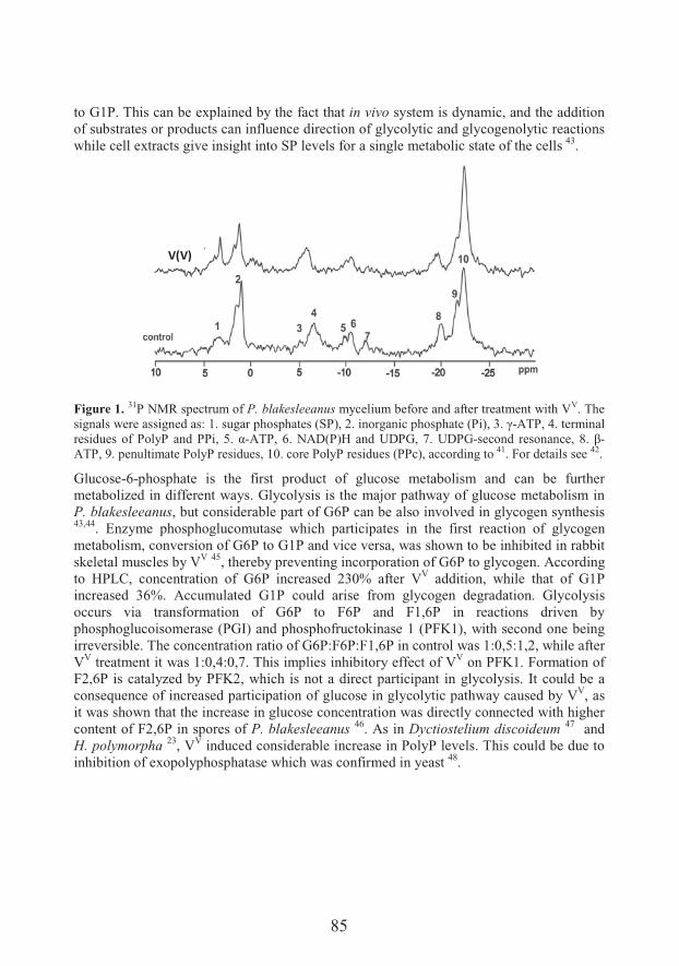
85
to G1P. This can be explained by the fact that in vivo system is dynamic, and the addition of substrates or products can influence direction of glycolytic and glycogenolytic reactions while cell extracts give insight into SP levels for a single metabolic state of the cells 43.
V(V)
Figure 1. 31P NMR spectrum of P. blakesleeanus mycelium before and after treatment with VV. The signals were assigned as: 1. sug ��'�&' ��&�F�.@%�X������� ����'�&' ���F.�@%�:��U-ATP, 4. terminal ��&� #�&����.��$.� � �..�%�B�� ;-ATP, 6. NAD(P)H and UDPG, 7. UDPG-&���� � ��&�� ���%� f�� >-ATP, 9. penultimate PolyP residues, 10. core PolyP residues (PPc), according to 41. For details see 42.
Glucose-6-phosphate is the first product of glucose metabolism and can be further metabolized in different ways. Glycolysis is the major pathway of glucose metabolism in P. blakesleeanus, but considerable part of G6P can be also involved in glycogen synthesis43,44. Enzyme phosphoglucomutase which participates in the first reaction of glycogen metabolism, conversion of G6P to G1P and vice versa, was shown to be inhibited in rabbit skeletal muscles by VV 45, thereby preventing incorporation of G6P to glycogen. According to HPLC, concentration of G6P increased 230% after VV addition, while that of G1P increased 36%. Accumulated G1P could arise from glycogen degradation. Glycolysis occurs via transformation of G6P to F6P and F1,6P in reactions driven byphosphoglucoisomerase (PGI) and phosphofructokinase 1 (PFK1), with second one being irreversible. The concentration ratio of G6P:F6P:F1,6P in control was 1:0,5:1,2, while after VV treatment it was 1:0,4:0,7. This implies inhibitory effect of VV on PFK1. Formation of F2,6P is catalyzed by PFK2, which is not a direct participant in glycolysis. It could be a consequence of increased participation of glucose in glycolytic pathway caused by VV, as it was shown that the increase in glucose concentration was directly connected with higher content of F2,6P in spores of P. blakesleeanus 46. As in Dyctiostelium discoideum 47 and H. polymorpha 23, VV induced considerable increase in PolyP levels. This could be due to inhibition of exopolyphosphatase which was confirmed in yeast 48.
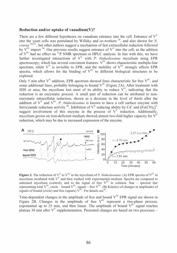
86
Reduction and/or uptake of vanadium(V)?
There are a few different hypotheses on vanadium entrance into the cell. Entrance of VV
into the yeast cells was postulated by Willsky and co-workers 26, and also shown for N. crassa 24,25 , but other authors suggest a mechanism of fast extracellular reduction followed by VIV import 30. Our previous results suggest entrance of VV into the cell, as the adittion of VIV had no effect on 31P NMR spectrum or HPLC analysis. In line with this, we have further investigated interactions of VV with P. blakesleeanus mycelium using EPR spectroscopy, which has several convinient features. VIV shows characteristic multiple-line spectrum, while VV is invisible to EPR, and the mobility of VIV strongly affects EPR spectra, which allows for the binding of VIV to different biological structures to be explored. Only 5 min after VV addition, EPR spectrum showed lines characteristic for free VIV, and some additional lines, probably belonging to bound VIV (Figure 2A). After treatment with SDS or urea, the mycelium lost most of its ability to reduce VV, indicating that the reduction is an enzymatic process. A small part of reduction can be attributed to non-enzymatic intracellular reduction, shown as a decrease in the level of thiols after the addition of VV and VIV. P. blakesleeanus is known to have a cell surface enzyme with ferricyanide reductase activity 49. Inhibition of VV reducing ability by Cd+ and [Fe(CN)6]
3-
suggest involvement of this enzyme in the process of VV reduction. Additionally, mycelium grown on iron-deficient medium showed almost two-fold higher capacity for VV
reduction, which may be due to increased expression of the enzyme.
Figure 2. The reduction of VV to VIV in the mycelium of P. blakesleeanus. (A) EPR spectra of VIV in mycelium incubated with VV and then washed with experimental medium. Spectra are compared to untreated mycelium (control), and to the signal of free VIV in solution. Star – spectral line representing total VIV, circle – bound VIV, square – free VIV. (B) Kinetics of changes in amplitudes of signals of bound (circle) and free (square) VIV. For details see50.
Time-dependent changes in the amplitude of free and bound VIV EPR signal are shown in Figure 2B. Changes in the amplitude of free VIV represent a two-phase process, exponential up to 25 min, and then linear. The amplitude of bound VIV signal reaches plateau 30 min after VV supplementation. Presented changes are based on two processes –
B
free V(IV)
bound V(IV)
V(V)
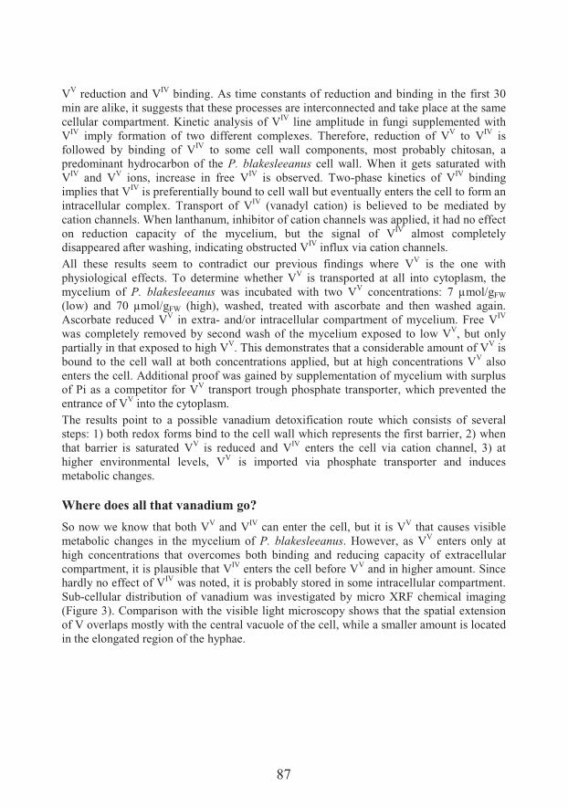
87
VV reduction and VIV binding. As time constants of reduction and binding in the first 30 min are alike, it suggests that these processes are interconnected and take place at the same cellular compartment. Kinetic analysis of VIV line amplitude in fungi supplemented with VIV imply formation of two different complexes. Therefore, reduction of VV to VIV is followed by binding of VIV to some cell wall components, most probably chitosan, a predominant hydrocarbon of the P. blakesleeanus cell wall. When it gets saturated with VIV and VV ions, increase in free VIV is observed. Two-phase kinetics of VIV binding implies that VIV is preferentially bound to cell wall but eventually enters the cell to form an intracellular complex. Transport of VIV (vanadyl cation) is believed to be mediated by cation channels. When lanthanum, inhibitor of cation channels was applied, it had no effect on reduction capacity of the mycelium, but the signal of VIV almost completely disappeared after washing, indicating obstructed VIV influx via cation channels. All these results seem to contradict our previous findings where VV is the one with physiological effects. To determine whether VV is transported at all into cytoplasm, the mycelium of P. blakesleeanus was incubated with two VV concentrations: 7 μmol/gFW
(low) and 70 μmol/gFW (high), washed, treated with ascorbate and then washed again. Ascorbate reduced VV in extra- and/or intracellular compartment of mycelium. Free VIV
was completely removed by second wash of the mycelium exposed to low VV, but only partially in that exposed to high VV. This demonstrates that a considerable amount of VV is bound to the cell wall at both concentrations applied, but at high concentrations VV also enters the cell. Additional proof was gained by supplementation of mycelium with surplus of Pi as a competitor for VV transport trough phosphate transporter, which prevented the entrance of VV into the cytoplasm. The results point to a possible vanadium detoxification route which consists of several steps: 1) both redox forms bind to the cell wall which represents the first barrier, 2) when that barrier is saturated VV is reduced and VIV enters the cell via cation channel, 3) at higher environmental levels, VV is imported via phosphate transporter and induces metabolic changes.
Where does all that vanadium go?
So now we know that both VV and VIV can enter the cell, but it is VV that causes visible metabolic changes in the mycelium of P. blakesleeanus. However, as VV enters only at high concentrations that overcomes both binding and reducing capacity of extracellular compartment, it is plausible that VIV enters the cell before VV and in higher amount. Since hardly no effect of VIV was noted, it is probably stored in some intracellular compartment. Sub-cellular distribution of vanadium was investigated by micro XRF chemical imaging (Figure 3). Comparison with the visible light microscopy shows that the spatial extension of V overlaps mostly with the central vacuole of the cell, while a smaller amount is located in the elongated region of the hyphae.
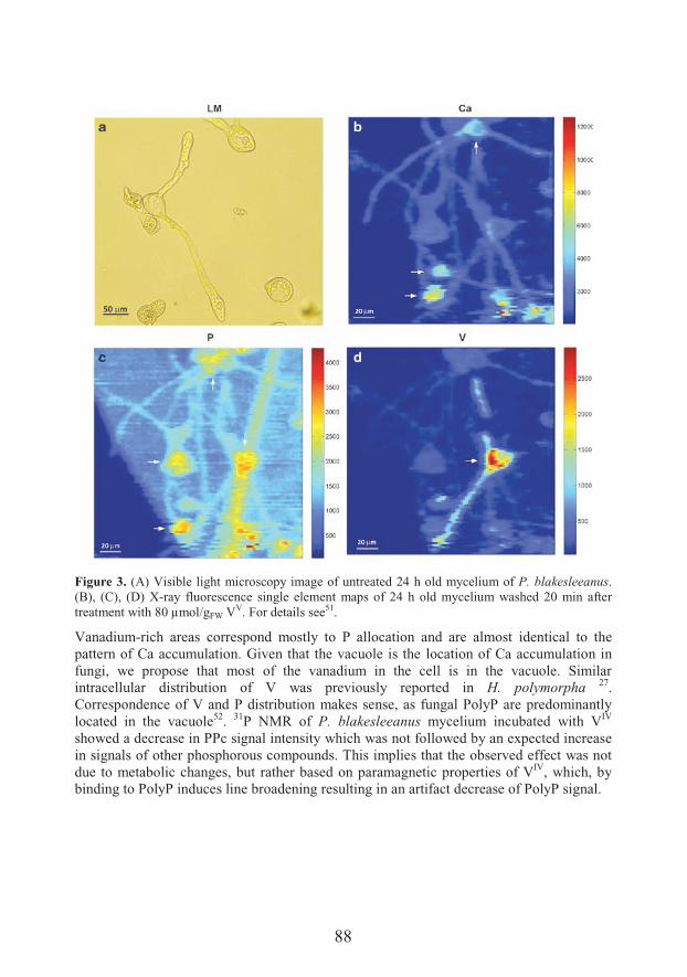
88
Figure 3. (A) Visible light microscopy image of untreated 24 h old mycelium of P. blakesleeanus.(B), (C), (D) X-ray fluorescence single element maps of 24 h old mycelium washed 20 min after treatment with 80 μmol/gFW VV. For details see51.
Vanadium-rich areas correspond mostly to P allocation and are almost identical to the pattern of Ca accumulation. Given that the vacuole is the location of Ca accumulation in fungi, we propose that most of the vanadium in the cell is in the vacuole. Similar intracellular distribution of V was previously reported in H. polymorpha 27.Correspondence of V and P distribution makes sense, as fungal PolyP are predominantly located in the vacuole52. 31P NMR of P. blakesleeanus mycelium incubated with VIV
showed a decrease in PPc signal intensity which was not followed by an expected increase in signals of other phosphorous compounds. This implies that the observed effect was not due to metabolic changes, but rather based on paramagnetic properties of VIV, which, by binding to PolyP induces line broadening resulting in an artifact decrease of PolyP signal.
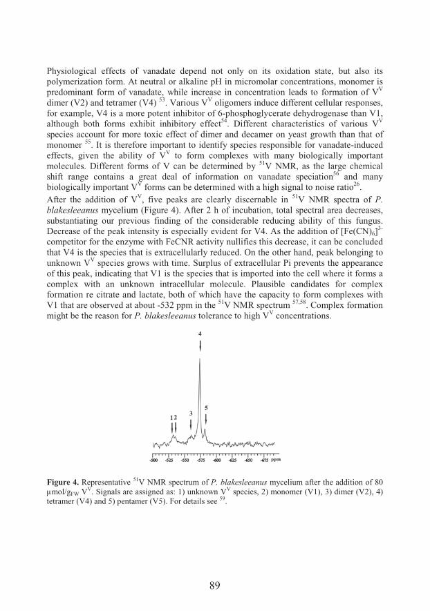
89
Physiological effects of vanadate depend not only on its oxidation state, but also its polymerization form. At neutral or alkaline pH in micromolar concentrations, monomer is predominant form of vanadate, while increase in concentration leads to formation of VV
dimer (V2) and tetramer (V4) 53. Various VV oligomers induce different cellular responses, for example, V4 is a more potent inhibitor of 6-phosphoglycerate dehydrogenase than V1, although both forms exhibit inhibitory effect54. Different characteristics of various VV
species account for more toxic effect of dimer and decamer on yeast growth than that of monomer 55. It is therefore important to identify species responsible for vanadate-induced effects, given the ability of VV to form complexes with many biologically important molecules. Different forms of V can be determined by 51V NMR, as the large chemical shift range contains a great deal of information on vanadate speciation56 and many biologically important VV forms can be determined with a high signal to noise ratio26.After the addition of VV, five peaks are clearly discernable in 51V NMR spectra of P. blakesleeanus mycelium (Figure 4). After 2 h of incubation, total spectral area decreases, substantiating our previous finding of the considerable reducing ability of this fungus. Decrease of the peak intensity is especially evident for V4. As the addition of [Fe(CN)6]
3-
competitor for the enzyme with FeCNR activity nullifies this decrease, it can be concluded that V4 is the species that is extracellularly reduced. On the other hand, peak belonging to unknown VV species grows with time. Surplus of extracellular Pi prevents the appearance of this peak, indicating that V1 is the species that is imported into the cell where it forms a complex with an unknown intracellular molecule. Plausible candidates for complex formation re citrate and lactate, both of which have the capacity to form complexes with V1 that are observed at about -532 ppm in the 51V NMR spectrum 57,58. Complex formation might be the reason for P. blakesleeanus tolerance to high VV concentrations.
Figure 4. Representative 51V NMR spectrum of P. blakesleeanus mycelium after the addition of 80 μmol/gFW VV. Signals are assigned as: 1) unknown VV species, 2) monomer (V1), 3) dimer (V2), 4) tetramer (V4) and 5) pentamer (V5). For details see 59.
90
Conclusion
Physiological response of the cell to vanadium depends on its oxidation state and polymerization form (speciation). When high concentrations of VV are applied to P.blakesleeanus mycelium at close to neutral pH, V1, V2, V4 and V5 are observed, with no V10 formation. P. blakesleeanus exhibits an array of mechanisms for overpowering the toxicity of this metal, with all of its different forms. The first line of defense is a physical barrier in the form of a cell wall that has binding capacity for both VV and VIV. When this barrier is overwhelmed, a cell surface enzyme with ferricyanide reductase activity reducesVV in its most abundant form V4, to VIV. VIV enters the cell through cationic channels, andis further sequestered in the vacuole where it binds to PolyP, a well-known cationic chelator. Reducing capacity of P. blakesleeanus for VV in the form of V1 was not observed, instead, this form of vanadium enters the cell aided by the phosphate transporter where it leads to changes in the metabolism of phosphorylated compounds. However, complexation of V1 with some unknown intracellular compounds, probably lactate or citrate, decreases its toxicity in the cytoplasm. Owing to all the above, P. blakesleeanus is capable of accumulating high concentrations of both VV and VIV, much higher than those recorded for fungus Coprinus commatus, a well-known V accumulator 60.
Acknowledgements
This study was supported by the Ministry of Education, Science and Technological Development of the Republic of Serbia (Grant No 173040)
References1. Rehder D. Bioinorganic vanadium chemistry. 2008. Wiley and Sons, Chichester, New York2. Barceloux DG. Vanadium. Clin Toxicol 1999;37:265-78.3. Crans D, Jason JS, Gaidamauskas E, Yang L. The chemistry and biochemistry of vanadium and
the biological activities exerted by vanadium compounds. Chem Rev 2004;104:849-902.4. Hope BK, 1994. A global biogeochemical budget for vanadium. Sci Total Environ 1994;141:1-
10.5. Rehder D. Structure and function of vanadium compounds in living organisms. Biometals
1992;5:1-12.6. Aureliano M. Decavanadate toxicology and pharmacological activities: V10 or V1 or none?
Oxid Med Cell Longev 2016; 2016:6103457.7. Bernroitner M, Zamocky M, Furtmüller PG, Peschek GA, Obinger C. Occurrence, phylogeny,
structure, and function of catalases and peroxidases in cyanobacteria. J Exp Bot 2009;60:423-40.
8. Boison G, Steingen C, Stal LJ, Bothe H. The rice field cyanobacteria Anabaena azotica and Anabaena sp. CH1 express vanadium-dependent nitrogenase. Arch Microbiol 2006;186:367-76.
9. ���� ��+5%���j����%� ���� ��%�.��8�����+�5��+� ��%� � � �#��� �#� �������H=�"�&�role and applications. Coord Chem Rev 2013;257:2388-400.
10. Plass W. Phosphate and Vanadate in Biological Systems: Chemical Relatives or More? Angew Chem 1999;38:909-12.
11. Pessoa JC, Garribba E, Santos MFA, Santos-Silva T. Vanadium and proteins: Uptake, transport, structure, activity and function. Coord Chem Rev 2015;301-302:49-86.
12. Rehder D. The role of vanadium in biology. Metallomics 2015;7:730-42.13. Rehder D. The future of/for vanadium. Dalton Trans 2013;42:11749.
91
14. Bishayee A, Waghray A, Patel MA, Chatterjee M. Vanadium in the detection, prevention and treatment of cancer: the in vivo evidence. Cancer Lett 2010;294:1-12.
15. Scior T, Guevara-Garcia JA, Do QT, Bernard P, Lauferd S. Why Antidiabetic Vanadium Complexes are Not in the Pipeline of “Big Pharma” Drug Research? A Critical Review. Curr Med Chem 2016;23:2874-91.
16. Baran EJ. Vanadium detoxification: chemical and biochemical aspects. Chem Biodivers 2008;5:1475-84.
17. Antipov AN, Sorokin DY, L’vov NP, Kuenen JG. New enzyme belonging to the family of molybdenum-free nitrate reductases. Biochem J 2003;369:185-9.
18. Michibata H, Iwata Y, Hirata J. "&�� ��������'��'�$� �� ��� � � � �#�k���� ������8��� �����&������ ����� &��� �� �$�&� ��� 8��� � ����� ����� &�� �� �� &����&� 8$� ��&��$k�� �����centrifugation. J Exp Zool 1991;257:306-13.
19. Kitayama H, Yamamoto S, Michibata H, Ueki T. Metal ion selectivity of the vanadium(V)-reductase Vanabin2. Dalton Trans 2013;42:11921-5.
20. Uteki T, Michibata H. Molecular mechanism of the transport and reduction pathway of vanadium in ascidians. Coord Chem Rev 2011:255:2249-57.
21. Lepp NW, Harrison SCS, Morrell BG. A role for Amanita muscaria L. in the circulation of cadmium and vanadium in a non-polluted woodland. Environ Geochem Health 1987;9:61-4.
22. Kalac P, Svoboda L. A review of trace element concentrations in edible mushrooms. Food Chem 2000;69:273-81.
23. Mannazzu I. Vanadium detoxification and resistance in yeast: a minireview. Ann Microbiol 2001;51:1-9.
24. Bowman BJ. Vanadate uptake in Neurospora crassa occurs via phosphate system II. J Bacteriol 1983;153:286-91.
25. Bowman BJ, Allen KE, Slayman CW. Vanadate-resistant mutants of Neurospora crassa are deficient in a high-affinity phosphate transport system. J Bacteriol 1983;153:292-6.
26. Willsky GR, White DA, McCabe BC. Metabolism of added orthovanadate to vanadyl and high-molecular-weight vanadate by Saccharomyces cerevisiae. J Biol Chem 1984;21:13273-81.
27. Mannazzu I, Guerra E, Strabbioli R, Masia A, Maestrale GB, Zoroddu MA, Fatichenti F. Vanadium affects vacuolation and phosphate metabolism in Hansenula polymorpha. FEMS Microbiol Lett 1997;147:23-8.
28. Mannazzu I, Guerra E, Strabbioli R, Pediconi D, Fatichenti F. The vanadate tolerant yeast Hansenula polymorpha undergoes cellular reorganization during growth in, and recovery from, the presence of vanadate. Microbiology 1998;144:2589-97.
29. Zoroddu MA, Fruianu M, Dallocchio R, Masia A. Electron paramagnetic resonance studies and effects of vanadium in Saccharomyces cerevisiae. BioMetals 1996;9:91-7.
30. Bode HP, Friebel C, Fuhrmann GF. Vanadium uptake by yeast cells. Biochim Biophys Acta 1990;1022:163-70.
31. Lu B, et al.. Enhanced sensitivity of insulinresistant adipocytes to vanadate is associated with oxidative stress and decreased reduction of vanadate(+5) to vanadyl(+4). J Biol Chem 2001;276:35589-98.
32. Mannazzu I, Guerra E, Ferretti F, Pediconi D, Fatichenti F. Vanadate and copper induce overlapping oxidative stress responses in the vanadate tolerant yeast Hansenula polymorpha. Biochim Biophys Acta 2000;1475:151-6.
33. Minasi LA, Chang A, Willsky GR. Plasma membrane-stimulated vanadate-dependent NADH oxidation is not the primary mediator of vanadate toxicity in Saccharomyces cerevisiae. J Biol Chem 1990;265:14907-10.
34. Minasi LA, Willsky GR. Characterization of vanadate-dependent NADH oxidation stimulated by Saccharomyces cerevisiae plasma membranes. J Bacteriol 1991;173:834-41.
35. Tsiani E, Fantus IG. Vanadium compounds. Biological actions and potential as pharmacological agents. Trends Endocrinol Metab 1997;8:51-8.
36. Stankiewicz PJ, Tracey AS, Crans DC. Inhibition of phosphatemetabolizingenzymes by oxovanadiumcomplexes. Met Ions Biol Syst 1995;31:287–324.
37. Rider MH, Bartrons R, Hue L. Vanadate inhibits liver fructose-2,6-bisphosphatase. Eur J Biochem 1990;190:53–6.
92
38. Benabe J, Echegoyen LA, Pastranall B, Martinez-Maldonado M. Mechanism of inhibition of glycolysis by vanadate. J Biol Chem 1987;262:9555–60.
39. Gomez-Foix AM, Rodriguez-Gil JE, Fillat C, Guinovart JJ, Bosch F. Vanadate raises fructose 2,6-bisphosphate concentrations and activates glycolysis in rat hepatocytes. Biochem J 1988;255:507-12.
40. Rehder D. Biological and medical aspects of vanadium. Inorg Chem Commun 2003;6:604–17.41. V���� �%� [ ��N�7&� � �%� V�0��� �%� � 2��� R�� 31P NMR study of polyposphate levels during
different growth phases of Phycomyces blakesleeanus. Antonie van Leeuwenhoek 2007;91:169–77.
42. V�0����%�V�����%�� �&������3%��� �����%����0 ���%�,�����+�����*%�[ ��N�7&� J. Vanadate influence on metabolism of sugar phosphates in fungus Phycomyces blakesleeanus. PLoS One 2014;9:e102849.
43. Shanks JV, Bailey JE. Estimation of intracellular sugar phosphate concentrations in Saccharomyces cerevisiae using 31P nuclear magnetic resonance spectroscopy. Biotechnol Bioeng 1988;32:1138–52.
44. Rua J, Busto F, De Arriaga D, Soler J. Glycogen in Phycomyces blakesleeanus: influence of growth conditions and nutrient limit action. J. Gen. Microbiol 1993;139:217–22.
45. Climent F, Bartrons R, Pons G, Carreras J. Effect of vanadate on phosphoryl transfer enzymes involved in glucose metabolism. Biochem Bioph Res Co 1981;101:570–6.
46. Van Laere A, Van Schaftingeen E, Hers HG. Fructose 2,6-bisphosphate and germination of fungal spores. Proc Natl Acad Sci USA 1983;80:6601–5.
47. Klein G, Cotter DA, Martin JB, Satre M. Vanadate, an inhibitor of growth, development and endocytosis in Dictyostelium discoideum amoebae. J Cell Sci 1989;94:127–34.
48. R#� ��7&���+%��� �N$l&� �G%�� ���&�5�%���8��&���+�%�5�#����+ ���&����B6-tetraphosphate phosphohydrolase activity is an inherent property of soluble exopolyphosphatase from yeast Saccharomyces cerevisiae. Biochim Biophys Acta 1998;1380:232-8.
49. Baroja-Mazo A, Del Valle P, Rúa J, Busto F, De Cima S, De Arriaga D.. A transplasma membrane redox system in Phycomyces blakesleeanus: properties of a ferricyanide reductase activity regulated by iron level and vitamin K3. J Bioenerg Biomembr 2004;36:481-92.
50. V�0����%�V�����%�� &������"%���� �����.��&����%��� �����%�,�����+�����*%�[ ��N�7&� ����The interactions of vanadium with Phycomyces blakesleeanus mycelium: enzymatic reduction, transport and metabolic effects. Res Microbiol 2013;164:61–9.
51. V�0����%� ��� ��� e-ray absorption near-edge structure micro-spectroscopy study on vanadium speciation on fungus Phycomyces blakesleeanus mycelium. Anal Bioanal Chem 2015;407:7487-96.
52. Klionsky DJ, Herman PK, Emr SD. The fungal vacuole: composition, function, and biogenesis. Microbiol Rev 1990;54:266–92.
53. Csermely P, Martonosi A, Levy GC, Ejchart AJ. 51V NMR analysis of the binding of vanadium (V) oligoanions to sarcoplasmic reticulum. Biochem J 1985;230:807-15.
54. Levina A, McLeod AI, Lay PA. Vanadium speciation by XANES spectroscopy: a three-dimensional approach. Chem Eur J 2014;20:12056-60.
55. Wong J, Messmer RP, Maylotte DH. K-edge absorption spectra of selected vanadium compounds. Phys Rev B 1984;30:5596–5610.
56. Crans DC. Fifteen years of dancing with vanadium. Pure Appl Chem 2005;77:1497-527.57. Gorzsás A, Getty K, Andersson I, Pettersson L. Speciation in the aqueous H+/H2VO4
-
/H2O2/citrate system of biomedical interest. Dalton Trans 2004:2873-82.58. Gorzsás A, Getty K, Andersson I, Pettersson L. Speciation in the aqueous H+/H2VO4
-
/H2O2/lactate system. Dalton Trans 2003:2503-11.59. V�0����%���� ������ [%� �� �����%� O 0�8� '�������%� V���� �%� [ ��N�7&� � ��� 51V NMR
investigation of cell-associated vanadate species in Phycomyces blakesleeanus mycelium. Res Microbiol 2016;167:521–8.
60. Hadžibrahimov����%��#0�������%�. &�����%�,�����+�����*%�V�0����%�[ ��N�7&� ��%�V�������The interactions of vanadate monomer with the mycelium of fungus Phycomyces blakesleeanus: reduction or uptake? Antonile van Leeuwenhoek 2017;110:365-73.
93
Unraveling mechanisms of Si action: modulation of gene expression in plants under abiotic stresses
6��,���-!���*���� �-�"��6��,���+����, ������:$������0�
Laboratory for Plant Molecular Biology, Institute of Molecular Genetics and GeneticEngineering, University of Belgrade, Belgrade, Serbia
*e-mail: [email protected]
Si is known as a beneficial element, shown to alleviate various abiotic and biotic stresses in plants. However, the underlying processes are poorly understood. Due to disparities among the effects of Si observed in different plant species and under different growth conditions, there are still some open questions. One is the question of silicon’s role in primary metabolism. The most of the data suggest minor role of Si in plants grown in optimal conditions. On the other hand, there are some Si-mediated changes in gene expression and physiology in non-stressed plants that should be taken into a consideration when discussing its functioning. Another question concerns the role of Si in stress conditions. Majority of the studies support the opinion that detected changes in gene expression, during stress, arise indirectly as a consequence of Si activity on biochemical and physicochemical levels. However, analyses of Si influence on transcript and protein levels can give us valuable information on molecular processes which occur during Si-mediated alleviation of stress and give us directions for further investigations.Herein we give an overview of the reported Si-mediated modulation of gene expression in non-stressed and plants under different abiotic stresses, including salt, heavy metals and nutritional deficiency. Recent discoveries related to the regulation of genes involved in polyamine synthesis, photosynthesis, nutrient transport and retranslocation are discussed. In addition, some new data on influence of Si on gene expression in iron deficiency stress are presented.
Introduction
���7��'&� � �����#����#&� �����&� &'�7����8���a�� �� ������&� ��� � ����$���� &����&� � �environmental circumstances%�&�������F��@��&������� &&�a� � &� ���&&���� ��� ����#�������Earths’ crust is mainly composed of silicon that is present primarily as silicate minerals, secondary alumino silicates and various forms of silicon dioxide. However, the plant available silicon is in the form of monosilicic acid (H4SiO4), which is found both in liquid and adsorbed phases of silicon in soils. The concentration of the H4SiO4 in the soil solution is influenced by the soil properties. The beneficial role of Si will be greatly influenced by
94
the ability of the plant species under treatment to absorb the element. One of the most intriguing properties of Si is its differential absorption by plants. Depending on the plant species, soil properties, Si source and Si amount, in planta Si contents can vary from 0.1%to 10% (on a dry weight basis 1,2). This biological limitation is reason why some plant species will not respond to a Si treatment, and the protective effect of Si is absent. Based on Si concentrations found in their tissues, plants were ranked as low, intermediate and high Si accumulators 3.Although its nutritional role in plants seems limited, there has been increasing evidence that Si presence in plants plays an important function in alleviating adverse effects of biotic and abiotic stresses. For instance, Si improved plant growth and fitness in nutrient deficiency or excess 4-6, under stress imposed by several diseases 7-10. Despite the accumulating scientific evidence of the benefits of Si there is no conclusive evidence showing exactly how Si plays a role in stress tolerance. The approaches to resolve this puzzle involve comprehensive analyses of the effect of Si supplementation, biochemical and physiological investigations, co-localization and distribution of mineral elements, as well as transcriptomic and metabolomic analyses.
Modulation of gene expression by Si in plants in the absence of stress
Although it is widely accepted that Si alleviates numerous biotic and abiotic stresses, opinions on the silicon’s role in unstressed plants disagree to a large extent. It has been suggested that Si has no effect when plants are grown in optimal conditions 11, 12. As reported in several studies, physiological parameters such as biomass, membrane potential, photosynthetic activity, respiration, ion and water transport and enzyme activity were not altered upon Si amendment, in the absence of stress (summarized in Coskun et al. 12). Accordingly, some transcriptomic and proteomic analyses did not detect great changes in respect to Si supply. Gene regulation by Si was first investigated in rice, using microarray analysis 13. Alterations in expression were detected for more than 20 of c. 9000 clones analyzed and confirmed for 5 clones by RT PCR, however, changes were modest. Transcriptomic analyses in Arabidopsis, an non-accumulating plant, showed that only two of the c. 28500 analyzed genes were altered under Si 14. Proteomic analysis determined only four proteins significantly changed in Si supplied rice 15. Some analyses revealed moderate influence of Si on gene expression, mostly affecting stress related genes 16, 17
.
On the other hand, in some studies it has been reported that Si amendment led to a substantial change in gene expression 18 or promoted plant growth and yield 19-22. Si significantly increased fresh and dry weight, as well as relative water content in cotton, canola and wheat 23. Transcriptomic analysis in unstressed rice revealed Si-induced changes in expression of more than 200 genes, which were associated with defense and stress, as well as with primary metabolic processes 24.Recently, we have observed moderate increases in the expression of Strategy II genes for Fe acquisition under influence of Si, in non-stressed barley. As a member of Gramineaefamily (grasses), barley predominantly uses chelation-based strategy (Strategy II) for Fe
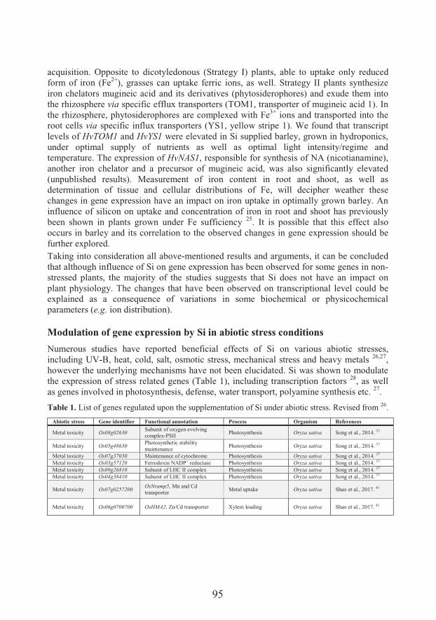
95
acquisition. Opposite to dicotyledonous (Strategy I) plants, able to uptake only reduced form of iron (Fe2+), grasses can uptake ferric ions, as well. Strategy II plants synthesize iron chelators mugineic acid and its derivatives (phytosiderophores) and exude them into the rhizosphere via specific efflux transporters (TOM1, transporter of mugineic acid 1). In the rhizosphere, phytosiderophores are complexed with Fe3+ ions and transported into the root cells via specific influx transporters (YS1, yellow stripe 1). We found that transcript levels of HvTOM1 and HvYS1 were elevated in Si supplied barley, grown in hydroponics, under optimal supply of nutrients as well as optimal light intensity/regime and temperature. The expression of HvNAS1, responsible for synthesis of NA (nicotianamine), another iron chelator and a precursor of mugineic acid, was also significantly elevated (unpublished results). Measurement of iron content in root and shoot, as well as determination of tissue and cellular distributions of Fe, will decipher weather these changes in gene expression have an impact on iron uptake in optimally grown barley. An influence of silicon on uptake and concentration of iron in root and shoot has previously been shown in plants grown under Fe sufficiency 25. It is possible that this effect also occurs in barley and its correlation to the observed changes in gene expression should be further explored.Taking into consideration all above-mentioned results and arguments, it can be concluded that although influence of Si on gene expression has been observed for some genes in non-stressed plants, the majority of the studies suggests that Si does not have an impact on plant physiology. The changes that have been observed on transcriptional level could be explained as a consequence of variations in some biochemical or physicochemical parameters (e.g. ion distribution).
Modulation of gene expression by Si in abiotic stress conditions
Numerous studies have reported beneficial effects of Si on various abiotic stresses, including UV-B, heat, cold, salt, osmotic stress, mechanical stress and heavy metals 26,27,however the underlying mechanisms have not been elucidated. Si was shown to modulate the expression of stress related genes (Table 1), including transcription factors 28, as well as genes involved in photosynthesis, defense, water transport, polyamine synthesis etc. 27.
Table 1. List of genes regulated upon the supplementation of Si under abiotic stress. Revised from 26.
Abiotic stress Gene identifier Functional annotation Process Organism References
Metal toxicity Os08g02630Subunit of oxygen evolving complex-PSII Photosynthesis Oryza sativa Song et al., 2014. 37
Metal toxicity Os05g48630Photosynthetic stability maintenance
Photosynthesis Oryza sativa Song et al., 2014. 37
Metal toxicity Os07g37030 Maintenance of cytochrome Photosynthesis Oryza sativa Song et al., 2014. 37
Metal toxicity Os03g57120 Ferrodoxin NADP+ reductase Photosynthesis Oryza sativa Song et al., 2014. 37
Metal toxicity Os09g26810 Subunit of LHC II complex Photosynthesis Oryza sativa Song et al., 2014. 37
Metal toxicity Os04g38410 Subunit of LHC II complex Photosynthesis Oryza sativa Song et al., 2014. 37
Metal toxicity Os07g0257200 OsNramp5, Mn and Cd transporter Metal uptake Oryza sativa Shao et al., 2017. 41
Metal toxicity Os06g0700700 OsHMA2, Zn/Cd transporter Xylem loading Oryza sativa Shao et al., 2017. 41
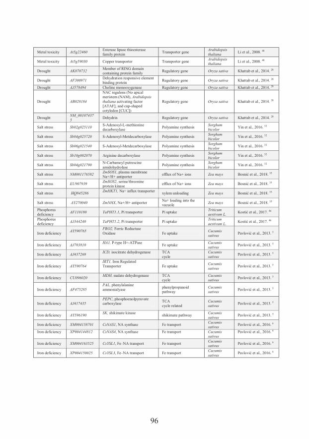
96
Metal toxicity At5g22460Esterase lipase thioesterase family protein
Transporter geneArabidopsis thaliana
Li et al., 2008. 48
Metal toxicity At5g59030 Copper transporter Transporter gene Arabidopsis thaliana
Li et al., 2008. 48
Drought AK070732Member of RING domain containing protein family Regulatory gene Oryza sativa Khattab et al., 2014. 28
Drought AF300971Dehydration responsive element binding protein Regulatory gene Oryza sativa Khattab et al., 2014. 28
Drought AJ578494 Choline monooxygenase Regulatory gene Oryza sativa Khattab et al., 2014. 28
Drought AB028184
NAC regulons (No apical meristem (NAM), Arabidopsis thaliana activating factor [ATAF], and cup-shaped cotyledon [CUC])
Regulatory gene Oryza sativa Khattab et al., 2014. 28
DroughtNM_001074375
Dehydrin Regulatory gene Oryza sativa Khattab et al., 2014. 28
Salt stress Sb02g025110S-Adenosyl-L-methionine decarboxylase
Polyamine synthesis Sorghum bicolor
Yin et al., 2016. 32
Salt stress Sb04g025720 S-Adenosyl-Metdecarboxylase Polyamine synthesis Sorghum bicolor
Yin et al., 2016. 32
Salt stress Sb06g021540 S-Adenosyl-Metdecarboxylase Polyamine synthesisSorghum bicolor
Yin et al., 2016. 32
Salt stress Sb10g002070 Arginine decarboxylase Polyamine synthesisSorghum bicolor
Yin et al., 2016. 32
Salt stress Sb04g021790N-Carbamoyl putrescine amidohydrolase
Polyamine synthesisSorghum bicolor
Yin et al., 2016. 32
Salt stress NM001176582ZmSOS1, plasma membrane Na+/H+ antiporter
efflux of Na+ ions Zea mays Bosni����� ��%�XY9f��35
Salt stress EU907939ZmSOS2, serine/threonine protein kinase
efflux of Na+ ions Zea mays ��&������� ��%�XY9f��35
Salt stress HQ845286ZmHKT1, Na+ influx transporter xylem unloading Zea mays ��&������� ��%�XY9f��35
Salt stress AY270040 ZmNHX, Na+/H+ antiporterNa+ loading into the vacuole Zea mays ��&������� ��%�XY9f��35
Phosphorus deficiency AF110180 TaPHT1.1, Pi transporter Pi uptake
Triticum aestivum L.
��&������� ��%�XY9<��46
Phosphorus deficiency
AJ344240 TaPHT1.2, Pi transporter Pi uptakeTriticum aestivum L.
��&������� ��%�XY9<��46
Iron deficiencyAY590765
FRO2, Ferric Reductase Oxidase Fe uptake
Cucumis sativus
. �������� ��%�XY9:��5
Iron deficiency AJ703810HA1, P-type H+-ATPase
Fe uptakeCucumis sativus
. ������� al., 2013. 5
Iron deficiency AJ437269ICD, isocitrate dehydrogenase TCA
cycleCucumis sativus
. �������� ��%�XY9:��5
Iron deficiency AY590764IRT1, Iron Regulated Transporter Fe uptake
Cucumis sativus
. �������� ��%�XY9:��5
Iron deficiency CU096020MDH, malate dehydrogenase TCA
cycleCucumis sativus
. �������� ��%�XY9:��5
Iron deficiency AF475285PAL, phenylalanine ammonialyase
phenylpropanoid pathway
Cucumis sativus
. �������� ��%�XY9:��5
Iron deficiency AJ417435PEPC, phosphoenolpyruvatecarboxylase
TCAcycle related
Cucumis sativus
. �������� ��%�XY9:��5
Iron deficiency AY596190SK, shikimate kinase
shikimate pathwayCucumis sativus
. �������� ��%�XY9:��5
Iron deficiency XM004158701 CsNAS1, NA synthase Fe transport Cucumis sativus
. �������� ��%�XY9C��6
Iron deficiency XP004144812 CsNAS4, NA synthase Fe transport Cucumis sativus
. �������� ��%�XY9C��6
Iron deficiency XM004163525 CsYSL1, Fe–NA transport Fe transportCucumis sativus
. �������� ��%�XY9C��6
Iron deficiency XP004150025 CsYSL3, Fe–NA transport Fe transportCucumis sativus
. �������� ��%�XY9C��6
97
Salt stress
Soil salinity is recognized as one of the most threatening abiotic stresses, greatly reducing the yield of agricultural crops. Low osmotic potential of soil solution results in decreased water and nutrient uptake, whereas ion toxicity and oxidative stress can seriously damage plant cells 29,30. It has been reported that deposition of Si in exodermis and endodermis of rice root limits apoplastic transport of sodium 19, while accumulation of Si in the cell wall of rice leaves reduces transpiration, resulting in a decreased sodium uptake 31. Hence, it has been suggested that Si alleviates salt stress merely by providing mechanical barrier in apoplast. However, these lines of evidences have been provided only for rice, and the proposed mechanisms are questioned for other species 32. Recent studies, comprising gene expression and biochemical analysis, pointed to a more complex mode of action. Yin and co-workers emphasized the role of polyamines (PAs) in Si-induced salt tolerance. Polyamines are a group of compounds containing two or more amine groups, involved in many growth and developmental processes. There is a growing amount of evidence showing importance of PAs in plant tolerance to various stresses 33. PAs improve salt stress tolerance by regulation of osmotic potential and K+/Na+ homeostasis, as well as antioxidant capacity in plants 32. As reported, Si treatment of salt stressed sorghum led to accumulation of PAs and a decrease of ethylene precursor ACC (1-aminocyclopropane-1-carboxylic acid). Moreover, when synthesis of PAs was blocked by applying the inhibitor of their synthesis (dicyclohexylammonium sulfate, DCHA), the beneficial effect of Si was lost. Correspondingly, Si enhanced the expression of S-adenosyl-L-methionine decarboxylase (SAMDC), a gene encoding a key and a rate-limiting enzyme in PA biosynthesis. SAMDC is also responsible for a decrease in accumulation of ethylene precursor ACC, contributing that way to the reduction of salt-induced leaf senescence. Si also regulated a set of genes involved in polyamine biosynthesis, both under salt stress and control conditions 32. However, the proposed link between Si and ethylene is seemingly at odds with the results obtained in salt stressed BY-2 cells 34. In cultured cells under salt treatment, Si actually increased ethylene emission and the expression of ethylene biosynthesis genes. Moreover, ethylene was necessary for Si mitigation effect. Although Si enhances salt tolerance in various plant species, it seems that mechanisms of its action differ greatly. Opposite to rice and sorghum where Si protects plants by decreasing sodium uptake and translocation to the shoot, sodium accumulation in maize shoot and root-to-shoot translocation are actually increased 35. In this study, a step forward has been made in elucidation of underlying mechanisms by employing gene expression analysis and confocal imaging of subcellular localization of Na. As reported, Si increased the expression of ZmSOS1 encoding a plasma membrane Na+/H+ antiporter responsible for efflux of Na+ ions out of root cortex and its regulator ZmSOS2, encoding a serine/threonine protein kinase. In addition, in the stele, Si down-regulated the expression of HKT1, Na+
influx transporter responsible for xylem unloading. As a result, sodium concentration was decreased in root apex and cortex, but increased in xylem and leaves. Moreover, Si up-regulated the expression of ZmNHX, an Na+/H+ antiporter responsible for Na+ loading into
98
the leaf vacuole. This result correlated well with Si-induced elevated vacuolar sequestration of Na+ and a concomitant decrease of Na+ in chloroplasts.
Metal toxicity
Zn is an essential microelement involved in vital cellular processes; nevertheless, when present in excess it becomes toxic, inhibiting seed germination, plant growth and development. One of the most prominent symptoms of Zn toxicity is leaf chlorosis, disturbed chloroplast ultrastructure and inhibition of photosynthesis 36. Si nutrition of rice under Zn toxicity has led to recovery and reversal of all these symptoms 37. Interestingly, Si improved chlorophyll content and photosynthetic parameters, as well as shoot and root dry weight, not only in Zn-intoxicated plants, but also in those grown in optimal Zn nutrition. Si-induced decrease in root-to-shoot transport of Zn was very important for plant protection in high-Zn conditions, however, influence of Si on photosynthesis related gene expression was also suggested to play an important role. Si increased the expression of genes important for functioning of photosynthetic electron transport chain: PsbY (a low-molecular-mass subunit of oxygen-evolving photosystem II), PsaH (a subunit of photosystem I), PetC (protein binding the Rieske FeS center of cytochrome b6f complex), PetH (ferredoxin NADP+ reductase) and two genes encoding subunits of the LHCII complex 37. Si-induced increase of the expression of all mentioned genes not only under high Zn but also in stress-free conditions suggests that this effect could be an important part of the mechanism of silicon’s action, and not just indirect consequence of structural and physiological changes brought by Si.Cd is not an essential element and presence of this toxic metal is detrimental for plant growth and development. Soil contamination with Cd leads to its accumulation in plant tissues, and once in the food chain it can have dangerous impact on human health. Deposition of Si in the cell wall of the endodermis and epidermis decreases apoplastic transport of Cd 38. It is also proposed that the formation of a complex of Cd with Si–hemicellulose matrix is important for alleviation of Cd stress and reduction of its accumulation in plant cells 39,40. Moreover, Si down-regulated the expression of genes involved in Cd uptake and translocation in plants 41.Since Cd is toxic, non-essential element, no specific Cd transporters have evolved in plants. However, due to similar chemical properties, Cd uses transporters for essential elements like Mn and Zn to enter root cells and to be translocated toward shoot. Silicon significantly down-regulated the expression of OsNramp5, the gene encoding for the transporter that mediates uptake of Mn and Cd into root cells. The expression of OsHMA2 transporter involved in xylem loading was also found to be decreased. These results correlate with lowered root-to-shoot translocation of Cd in Si treated rice. Sufficient accumulation of Si in shoots is required for regulation of OsNramp5 and OsHMA2, since this effect is absent in mutants deficient in Si uptake 41.Targeted transcript analyses have also been used to investigate influence of Si on plants under Cd stress 42. Authors of this study showed the ability of Si to recover plant tissues
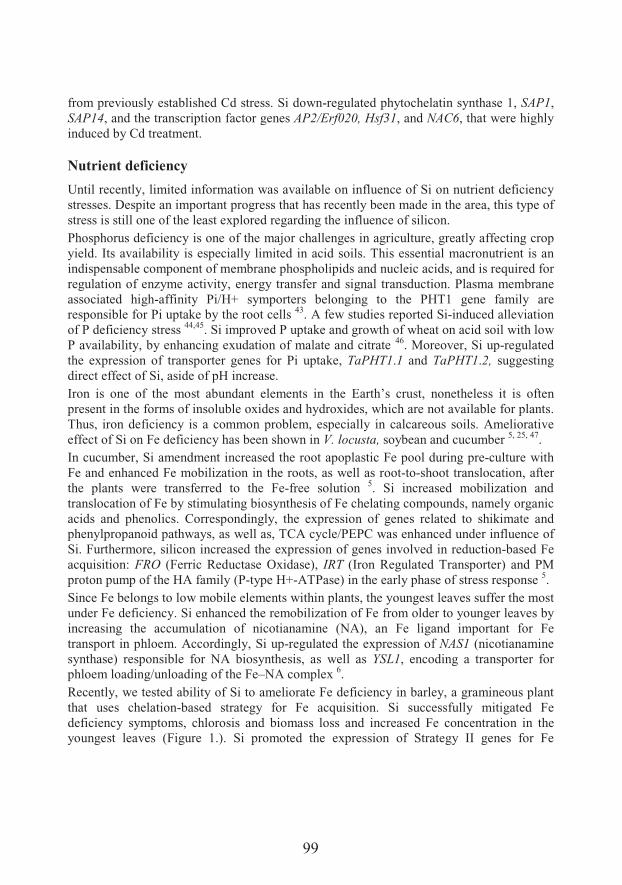
99
from previously established Cd stress. Si down-regulated phytochelatin synthase 1, SAP1,SAP14, and the transcription factor genes AP2/Erf020, Hsf31, and NAC6, that were highly induced by Cd treatment.
Nutrient deficiency
Until recently, limited information was available on influence of Si on nutrient deficiency stresses. Despite an important progress that has recently been made in the area, this type of stress is still one of the least explored regarding the influence of silicon. Phosphorus deficiency is one of the major challenges in agriculture, greatly affecting crop yield. Its availability is especially limited in acid soils. This essential macronutrient is an indispensable component of membrane phospholipids and nucleic acids, and is required for regulation of enzyme activity, energy transfer and signal transduction. Plasma membrane associated high-affinity Pi/H+ symporters belonging to the PHT1 gene family are responsible for Pi uptake by the root cells 43. A few studies reported Si-induced alleviation of P deficiency stress 44,45. Si improved P uptake and growth of wheat on acid soil with low P availability, by enhancing exudation of malate and citrate 46. Moreover, Si up-regulated the expression of transporter genes for Pi uptake, TaPHT1.1 and TaPHT1.2, suggesting direct effect of Si, aside of pH increase.Iron is one of the most abundant elements in the Earth’s crust, nonetheless it is often present in the forms of insoluble oxides and hydroxides, which are not available for plants. Thus, iron deficiency is a common problem, especially in calcareous soils. Ameliorative effect of Si on Fe deficiency has been shown in V. locusta, soybean and cucumber 5, 25, 47.In cucumber, Si amendment increased the root apoplastic Fe pool during pre-culture with Fe and enhanced Fe mobilization in the roots, as well as root-to-shoot translocation, after the plants were transferred to the Fe-free solution 5. Si increased mobilization and translocation of Fe by stimulating biosynthesis of Fe chelating compounds, namely organic acids and phenolics. Correspondingly, the expression of genes related to shikimate and phenylpropanoid pathways, as well as, TCA cycle/PEPC was enhanced under influence of Si. Furthermore, silicon increased the expression of genes involved in reduction-based Fe acquisition: FRO (Ferric Reductase Oxidase), IRT (Iron Regulated Transporter) and PM proton pump of the HA family (P-type H+-ATPase) in the early phase of stress response 5.Since Fe belongs to low mobile elements within plants, the youngest leaves suffer the most under Fe deficiency. Si enhanced the remobilization of Fe from older to younger leaves by increasing the accumulation of nicotianamine (NA), an Fe ligand important for Fe transport in phloem. Accordingly, Si up-regulated the expression of NAS1 (nicotianamine synthase) responsible for NA biosynthesis, as well as YSL1, encoding a transporter for phloem loading/unloading of the Fe–NA complex 6.Recently, we tested ability of Si to ameliorate Fe deficiency in barley, a gramineous plant that uses chelation-based strategy for Fe acquisition. Si successfully mitigated Fe deficiency symptoms, chlorosis and biomass loss and increased Fe concentration in the youngest leaves (Figure 1.). Si promoted the expression of Strategy II genes for Fe
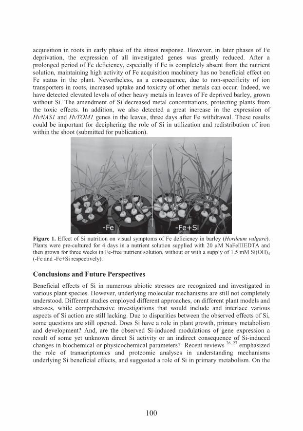
100
acquisition in roots in early phase of the stress response. However, in later phases of Fe deprivation, the expression of all investigated genes was greatly reduced. After a prolonged period of Fe deficiency, especially if Fe is completely absent from the nutrient solution, maintaining high activity of Fe acquisition machinery has no beneficial effect on Fe status in the plant. Nevertheless, as a consequence, due to non-specificity of ion transporters in roots, increased uptake and toxicity of other metals can occur. Indeed, we have detected elevated levels of other heavy metals in leaves of Fe deprived barley, grown without Si. The amendment of Si decreased metal concentrations, protecting plants from the toxic effects. In addition, we also detected a great increase in the expression of HvNAS1 and HvTOM1 genes in the leaves, three days after Fe withdrawal. These results could be important for deciphering the role of Si in utilization and redistribution of iron within the shoot (submitted for publication).
Figure 1. Effect of Si nutrition on visual symptoms of Fe deficiency in barley (Hordeum vulgare). Plants were pre-cultured for 4 days in a nutrient solution supplied with 20 μM NaFeIIIEDTA and then grown for three weeks in Fe-free nutrient solution, without or with a supply of 1.5 mM Si(OH)4
(-Fe and -Fe+Si respectively).
Conclusions and Future Perspectives
Beneficial effects of Si in numerous abiotic stresses are recognized and investigated in various plant species. However, underlying molecular mechanisms are still not completely understood. Different studies employed different approaches, on different plant models and stresses, while comprehensive investigations that would include and interlace various aspects of Si action are still lacking. Due to disparities between the observed effects of Si, some questions are still opened. Does Si have a role in plant growth, primary metabolism and development? And, are the observed Si-induced modulations of gene expression a result of some yet unknown direct Si activity or an indirect consequence of Si-induced changes in biochemical or physicochemical parameters? Recent reviews 26, 27 emphasized the role of transcriptomics and proteomic analyses in understanding mechanisms underlying Si beneficial effects, and suggested a role of Si in primary metabolism. On the
101
other hand, Coskun 12 questioned the role of Si in plant’s metabolism in unstressed conditions. They also suggested that Si alleviates different stresses in plants mainly as a result of its apoplastic deposition, and that all Si-induced changes in plant metabolism and gene expression in fact indirectly arise from this feature. A majority of studies support the opinion that detected changes in gene expression, during stress, occur indirectly as a consequence of Si activity on biochemical and physicochemical levels. However, analyses of Si influence on transcript and protein levels can give us valuable information on molecular processes which appear during Si-mediated alleviation of stress and direct us toward further investigations. Future research that would be more comprehensive and include omics approach as well as mutant analyses on various plant models, could provide answers on these questions and make our understanding of Si action deeper.
Acknowledgements
This study was supported by the Serbian Ministry of Education, Science and Technological Development (ON-173005).
References1. Epstein E. The anomaly of silicon in plant biology. Proc Natl Acad Sci 1994;91:11-7.2. Epstein E. Silicon in plants: facts vs. concepts. In: Datnoff LE, Snyder GH, Korndörfer GH
(eds) Studies in Plant Science, Elsevier, 2001, pp 1-15.3. Jones L, Handreck K. Silica in soils, plants, and animals. In: Norman AG (eds) Adv Agron,
Elsevier, 1967, pp 107-49.4. Datnoff LE, Snyder GH, Korndörfer GH. Silicon in agriculture. In: Datnoff LE, Snyder GH,
Korndörfer GH (eds) Studies in Plant Science, Elsevier, New York, 2001.5. Pavlovic J, et al. Silicon alleviates iron deficiency in cucumber by promoting mobilization of
iron in the root apoplast. New Phytol 2013;198:1096-107.6. Pavlovic J, et al. Silicon enhances leaf remobilization of iron in cucumber under limited iron
conditions. Ann Bot 2016;118:271-80.7. Belanger R, Bowen P, Ehret D, Menzies J. Soluble silicon: its role in crop and disease
management of greenhouse crops. Plant Dis 1995;79:329-368. Rodrigues FÁ, Benhamou N, Datnoff LE, Jones JB, Bélanger RR. Ultrastructural and
cytochemical aspects of silicon-mediated rice blast resistance. Phytopathology 2003;93:535-46.9. Fauteux F, Rémus-Borel W, Menzies JG, Bélanger RR. Silicon and plant disease resistance
against pathogenic fungi. FEMS Microbiol Lett 2005;249:1-6.10. Vivancos J, Labbé C, Menzies JG, Bélanger RR. Silicon-mediated resistance of Arabidopsis
against powdery mildew involves mechanisms other than the salicylic acid (SA)-dependent defence pathway. Mol Plant Pathol 2015;16:572-82.
11. Ma JF. Role of silicon in enhancing the resistance of plants to biotic and abiotic stresses. Soil Sci Plant Nutr 2004;50:11-18.
12. Coskun D, et al. The controversies of silicon's role in plant biology. New Phytol 2018.13. Watanabe S, et al. Identification of several rice genes regulated by Si nutrition. Soil Sci Plant
Nutr 2004;50:1273-6.14. Fauteux F, Chain F, Belzile F, Menzies JG, Bélanger RR. The protective role of silicon in the
Arabidopsis–powdery mildew pathosystem. Proc Natl Acad Sci 2006;103:17554-9.15. Nwugo CC, Huerta AJ. The effect of silicon on the leaf proteome of rice (Oryza sativa L.)
plants under cadmium-stress. J Proteome Res 2010;10:518-28.
102
16. Rasoolizadeh A, et al. Silicon protects soybean plants against Phytophthora sojae by interfering with effector-receptor expression. BMC Plant Biol 2018;18:97.
17. Chain F, Côté-Beaulieu C, Belzile F, Menzies J, Bélanger R. A comprehensive transcriptomic analysis of the effect of silicon on wheat plants under control and pathogen stress conditions. Mol Plant-Microbe Interact 2009;22:1323-30.
18. Van Bockhaven J, et al. Primary metabolism plays a central role in moulding silicon-inducible brown spot resistance in rice. Mol Plant Pathol 2015;16:811-24.
19. Gong H, Randall D, Flowers T. Silicon deposition in the root reduces sodium uptake in rice (Oryza sativa L.) seedlings by reducing bypass flow. Plant, Cell Environ 2006;29:1970-9.
20. Flam-Shepherd R, Huynh WQ, Coskun D, Hamam AM, Britto DT, Kronzucker HJ. Membrane fluxes, bypass flows, and sodium stress in rice: the influence of silicon. J Exp Bot 2018;69:1679-92.
21. Detmann KC, et al. Silicon nutrition increases grain yield, which, in turn, exerts a feed-forward stimulation of photosynthetic rates via enhanced mesophyll conductance and alters primary metabolism in rice. New Phytol 2012;196:752-62.
22. Frew A, Allsopp PG, Gherlenda AN, Johnson SN. Increased root herbivory under elevated atmospheric carbon dioxide concentrations is reversed by silicon-based plant defences. J Appl Ecol 2017;54:1310-9.
23. Mehrabanjoubani P, Abdolzadeh A, Sadeghipour HR, Aghdasi M. Silicon affects transcellular and apoplastic uptake of some nutrients in plants. Pedosphere 2015;25:192-201.
24. Brunings A, et al. Differential gene expression of rice in response to silicon and rice blast fungus Magnaporthe oryzae. Ann Appl Biol 2009;155:161-70.
25. Gonzalo MJ, Lucena JJ, Hernández-Apaolaza L. Effect of silicon addition on soybean (Glycine max) and cucumber (Cucumis sativus) plants grown under iron deficiency. Plant Physiol Biochem 2013;70:455-61.
26. Manivannan A, Ahn Y-K. Silicon regulates potential genes involved in major physiological processes in plants to combat stress. Front Plant Sci 2017;8:1346.
27. Frew A, Weston LA, Reynolds OL, Gurr GM. The role of silicon in plant biology: a paradigm shift in research approach. Ann Bot 2018;121:1265-73.
28. Khattab H, Emam M, Emam M, Helal N, Mohamed M. Effect of selenium and silicon on transcription factors NAC5 and DREB2A involved in drought-responsive gene expression in rice. Biol Plant 2014;58:265-73.
29. Ashraf M. Organic substances responsible for salt tolerance in Eruca sativa. Biol Plant 1994;36:255-9.
30. Zhu J-K. Plant salt tolerance. Trends Plant Sci 2001;6:66-71.31. Matoh T, Kairusmee P, Takahashi E. Salt-induced damage to rice plants and alleviation effect
of silicate. Soil Sci Plant Nutr 1986;32:295-304.32. Yin L, et al. Silicon-mediated changes in polyamines participate in silicon-induced salt
tolerance in Sorghum bicolor L. Plant, Cell Environ 2016;39:245-58.33. Alcázar R, et al. Polyamines: molecules with regulatory functions in plant abiotic stress
tolerance. Planta 2010;231:1237-49.34. Liang X, et al. Silicon does not mitigate cell death in cultured tobacco BY-2 cells subjected to
salinity without ethylene emission. Plant Cell Rep 2015;34:331-43.35. Bosnic P, Bosnic D, Jasnic J, Nikolic M. Silicon mediates sodium transport and partitioning in
maize under moderate salt stress. Environ Exp Bot 2018;155:681-7.36. Chen W, Yang X, He Z, Feng Y, Hu F. Differential changes in photosynthetic capacity, 77 K
chlorophyll fluorescence and chloroplast ultrastructure between Zn-efficient and Zn-inefficient rice genotypes (Oryza sativa) under low zinc stress. Physiol Plant 2008;132:89-101.
37. Song A, Li P, Fan F, Li Z, Liang Y. The effect of silicon on photosynthesis and expression of its relevant genes in rice (Oryza sativa L.) under high-zinc stress. PLoS One 2014;9:e113782.
38. Shi X, Zhang C, Wang H, Zhang F. Effect of Si on the distribution of Cd in rice seedlings. Plant Soil 2005;272:53-60.
39. Liu J, et al. Inhibition of cadmium ion uptake in rice (Oryza sativa) cells by a wall-bound form of silicon. New Phytol 2013;200:691-9.
103
40. Ma J, Cai H, He C, Zhang W, Wang L. A hemicellulose-bound form of silicon inhibits cadmium ion uptake in rice (Oryza sativa) cells. New Phytol 2015;206:1063-74.
41. Feng Shao J, Che J, Yamaji N, Fang Shen R, Feng Ma J. Silicon reduces cadmium accumulation by suppressing expression of transporter genes involved in cadmium uptake and translocation in rice. J Exp Bot 2017;68:5641-51.
42. Farooq MA, Detterbeck A, Clemens S, Dietz K-J. Silicon-induced reversibility of cadmium toxicity in rice. J Exp Bot 2016;67:3573-85.
43. Gu M, Chen A, Sun S, Xu G. Complex regulation of plant phosphate transporters and the gap between molecular mechanisms and practical application: what is missing? Molecular plant 2016;9:396-416.
44. Ma J, Takahashi E. Effect of silicon on the growth and phosphorus uptake of rice. Plant Soil 1990;126:115-9.
45. Neu S, Schaller J, Dudel EG. Silicon availability modifies nutrient use efficiency and content,C: N: P stoichiometry, and productivity of winter wheat (Triticum aestivum L.). Sci Rep 2017;7:40829.
46. Kostic L, Nikolic N, Bosnic D, Samardzic J, Nikolic M. Silicon increases phosphorus (P) uptake by wheat under low P acid soil conditions. Plant Soil 2017;419:447-55.
47. Gottardi S, et al. Beneficial effects of silicon on hydroponically grown corn salad (Valerianella locusta (L.) Laterr) plants. Plant Physiol Biochem 2012;56:14-23.
48. Li J, Leisner SM, Frantz J. Alleviation of copper toxicity in Arabidopsis thaliana by silicon addition to hydroponic solutions. J Am Soc Hort Sci 2008;133:670-7.

105
Distribution and role of metals in sclerotic hippocampi of patients with mesial temporal lobe epilepsy
6�� �������1*����"9&�/�2�)��!�������$#���3, Dragoslav S�!�3, Vladimir +�"/�����4, ����#�/���4, ��� ��������5, Marko Živin6, Vid Simon Šelih7,���0����&���8�4����&��� ���2
1������#��������+�` ��w=���� �+������! �� ����������{�|� � ��������� �=<�University of Belgrade, Belgrade, Serbia 2Life Sciences Department, Institute for Multidisciplinary Research, University of Belgrade3Center for Epilepsy and Sleep Disorders, Neurology Clinic, Clinical Center of Serbia, Belgrade, Serbia 4Institute for Neurosurgery, Clinical Center of Serbia5Institute of Forensic Medicine, Medical School, University of Belgrade6Brain Research Laboratory, Institute of Pathophysiology, Medical Faculty, University of Ljubljana, Ljubljana, Slovenia7Department of Analytical Chemistry, National Institute of Chemistry Slovenia, Ljubljana,Slovenia8Department of Chemistry, Institute of Chemistry, Technology and Metallurgy, University of Belgrade
*e-mail: [email protected]
The accumulating evidence on the relation between the disturbed metal homeostasis and epilepsy urges the need for data regarding the total metal concentrations, as well as metal distribution in the brain itself, in order to indicate where to direct the potential therapy, to metal supplementation or chelation. This paper summarizes our results on the measurements of some important essential metals in hippocampi of patients with mesial temporal lobe epilepsy (mTLE) who underwent amigdalohippocampectomy. The key findings point out that levels of copper and manganese are deficient in hippocampi of mTLE patients, and that their concentrations correlated positively with neuronal loss in affected regions of sclerotic hippocampus. In addition, the Cu concentration was decreased in the areas of total neuronal loss. Iron and zinc total hippocampal levels were neither accumulated nor deficient compared to control. Our results contribute to deeper insight of metals biology in the epilepsy and may represent the initial point of new and non-invasive therapy of drug resistant epilepsy.

106
Introduction
Epilepsy is neurological condition which is clinically manifested as spontaneous, recurrent epileptic seizures, due to short-term, uncontrolled and highly synchronized outburst of electrical activity of larger group of neurons in the brain. As a result, balance between brain excitability and inhibition is disturbed and shifted in favor of excitatory state. Epilepsy is accounted for one of the most common among serious neurological disorders. It affects about 50 million people worldwide, with 50 new cases per 100,000 per annum 1.It is estimated that 1% of each population suffers from epilepsy at any time point. Despite that, many uncertainties considering the pathophysiology follow this condition, making therapeutic strategy inadequate or insufficiently good for one third of patients that enter chronic state of this disorder. In line with that, there are multiple causes of epilepsy which may have a genetic, structural or metabolic background and often the underlying cause is indefinite and remains to be identified. Epileptic seizures could be generalized, when rapidly affecting neuronal networks of both hemispheres, or they could be focal when affecting neuronal networks unilaterally 2.Focal epileptic seizures frequently arise in temporal lobe, mostly in its mesial part comprised from amygdala, hippocampus, uncus, gyrus dentatus and parahippocampal gyrus. Although mesial temporal lobe epilepsy (mTLE) is one of the most common types of focal epilepsies, portion of the mTLE patients is difficult to treat with available antiepileptic drugs. Unfortunately, in time they develop drug resistant epilepsy. The most common cause of drug resistant mTLE is structural changes within hippocampus that involve different level of neuronal cell degeneration and loss (mTLE with hippocampal sclerosis – mTLE-HS), as well as abnormal reorganization of neuronal circuits 3.Therefore, surgical treatment, i.e. removal of sclerotic hippocampus, was shown to be helpful in attaining the control over epileptic seizures in 60 – 80% of patients 4. Surgical failures emphasize the complexity of the condition indicating the role of other mesial structures in seizure development, as well as the constant need for new therapies. On the other side, specimens gained through these surgical interventions offer direct look into the “crime scene”, which could contribute to better understanding of the pathophysiological processes associated with mTLE-HS. There are four types of HS based on the affected regions and the level of neuronal loss according to International League Against Epilepsy (ILAE) 4.
Metals biology in epilepsy
More and more studies, point that loss of regulation of metal homeostasis in the central nervous system contributes to pathophysiological processes in various neurodegenerative diseases. For example: it is hypothesized that iron loading and beta-amyloid plaque pathology are synergistic events in Alzheimer’s disease (AD) 5,6; perturbations in iron and copper metabolism contribute to the pathology of Parkinson’s disease (PD) 7,8; mutations in copper-transporter protein ATP7A in Menkes disease result in severe neurodevelopmental deficiencies and epileptic seizures as a result of copper deficiency in
107
the brain 9; extensive research on zinc signaling in epileptic seizures indicate that interplay between intracellular and extracellular concentrations of this metal may contribute to harmful or protective effects 10. Therefore, targeting the key players of metal homeostasis could be helpful in treatment of neurodegenerative diseases. However, it is very demanding to identify precise targets responsible in maintaining balance of essential metals in healthy or diseased brain due to complexity of interactions among the metals themselves and the plethora of metalloproteins. Nevertheless, field of metal biology is gaining its place under the spotlight of neurosciences.Perturbations in metal homeostasis are possible cofactor in the onset and/or progress of epilepsy. Meta-analysis performed by Saghazadeh and co-workers indicate differences in concentrations of trace elements in sera of the epilepsy patients (on or without therapy), and control subjects 11. However, what happens in relation of various metals concentrations in the brain itself exposed to epileptic seizures, remained to be determined and systematically analyzed. Therefore, the goal of our investigations is to contribute to the field of metal biology in epilepsy by determining total concentrations and regional distribution of some essential trace elements in sclerotic hippocampus resected from drug resistant mTLE-HS patents as surgical therapeutic approach. We have used two powerful techniques: inductively coupled plasma optical emission spectrometry (ICP-OES), to measure total concentrations of metals; and laser ablation inductively coupled plasma mass spectrometry (LA-ICP-MS) imaging, in order to provide detailed metal maps of epilepsy related sclerotic hippocampus. In our research we have investigated HS ILAE type 1 (HS1) and type 2 (HS2). HS1 is the most common among all mTLE patients that undergo surgical intervention, and represent severe lost of neurons in all segments of cornu amonis (CA1 – CA4), as well as in gyrus dentatus (GD). HS2 is the rare type seen in 5-10% of surgical cases of mTLE and affects exclusively CA1 region. Our strategy was to: compare epilepsy-induced HS with control tissue; compare regions with neuronal degeneration with preserved ones. Measurements of total concentrations of metals gained by ICP-OES can be observed in table 1 (adapted from 12). The profile of sodium, potassium, copper, and manganese in hippocampi of patients with mTLE-HS differed considerably from the control tissue. Disturbances in levels of main extracellular and intracellular electrolyte, i.e. increased sodium and lowered potassium concentration point out to the neuronal cell loss and changed ratio between intracellular and extracellular volume of sclerotic hippocampus. Further, hippocampus of mTLE patients is marked by significant copper and manganese deficiency 12. Spatial distribution of copper and manganese in hippocampi of patients with mTLE-HS acquired by LA-ICP-MS shown in figure 1, revealed that these metals mostly concentrate in areas reach in neuronal somata, which are the following regions of hippocampus: subiculum (SUB), CA4, GD, and stratum pyramidale (SPy) in CA1 and CA2; while alveus and other regions with axons and dendrites generally showed lower levels of Cu, and Mn 13. What is important is that Cu concentrations were significantly lower in areas of hippocampus affected with neuronal loss (SPy of CA1) compared to preserved regions (SUB). Furthermore, Cu and Mn concentrations positively correlated
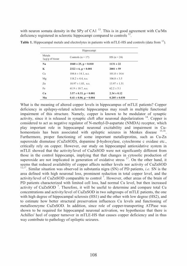
108
with neuron somata density in the SPy of CA1 13. This is in good agreement with Cu/Mn deficiency registered in sclerotic hippocampi compared to controls 12.
Table 1. Hippocampal metals and electrolytes in patients with mTLE-HS and controls (data from 12).
What is the meaning of altered copper levels in hippocampus of mTLE patients? Copper deficiency in epilepsy-related sclerotic hippocampus may result in multiple functional impairment of this structure. Namely, copper is known to be modulator of synaptic activity, since it is released in synaptic cleft after neuronal depolarization 14. Copper is considered to act as negative regulator of N-methyl-D-aspartate (NMDA) receptor, which play important role in hippocampal neuronal excitability and impairment in Cu-homeostasis has been associated with epileptic seizures in Menkes disease 15,16.Furthermore, proper functioning of some important metalloprotins, such as Cu-Zn superoxide dismutase (CuZnSOD), dopamine >-hydroxylase, cytochrome c oxidase etc., critically rely on copper. However, our study on hippocampal antioxidative system in mTLE showed that the activity/level of CuZnSOD were not significantly different from those in the control hippocampi, implying that that changes in cytosolic production of superoxide are not implicated in generation of oxidative stress 17. On the other hand, it seems that reduced availability of copper affects neither levels nor activity of CuZnSOD 12,17. Similar situation was observed in substantia nigra (SN) of PD patients, i.e. SN is the area defined with high neuronal loss, prominent reduction in total copper level, and the activity/level of CuZnSOD comparable to control 7. However, other areas of the brain of PD patients characterized with limited cell loss, had normal Cu level, but then increased activity of CuZnSOD 7. Therefore, it will be useful to determine and compare total Cu concentrations and activity/level of CuZnSOD in two subgroups of mTLE patients, the one with high degree of hippocampal sclerosis (HS1) and the other with low degree (HS2), and to estimate how better structural preservation influences Cu levels and functioning of metalloenzyme CuZnSOD. In addition, since role of copper-transporting ATPase was shown to be required for hippocampal neuronal activation, we hypothesize that there is Achilles' heel of copper turnover in mTLE-HS that causes copper deficiency and in thus way contribute to pathology of epileptic seizures.
Hippocampi
Metals1μg/g of tissue
Controls (n = 17) HS (n = 24)
Na 1040 ± 25, p = 0.010 1131 ± 22
K 2322 ± 6, p < 0.001 2001 ± 59
Ca 104.6 ± 14.1, n.s. 101.0 ± 14.6
Mg 118.2 ± 4.4, n.s. 106.8 ± 3.5
Zn 10.97 ± 1.03, n.s. 13.97 ± 1.51
Fe 61.9 ± 10.7, n.s. 62.2 ± 5.1
Cu 3.57 ± 0.33, p < 0.001 2.34 ± 0.12
Mn 0.41 ± 0.06, p = 0.004 0.205 ± 0.030
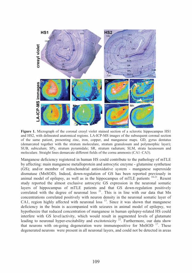
109
Figure 1. Micrograph of the coronal cresyl violet stained section of a sclerotic hippocampus HS1 and HS2, with delineated anatomical regions. LA-ICP-MS images of the subsequent coronal section of the same patient, presenting zinc, iron, copper, and manganese maps. GD, gyrus dentatus (demarcated together with the stratum moleculare, stratum granulosum and polymorphic layer); SUB, subiculum; SPy, stratum pyramidale; SR, stratum radiatum; SLM, strata lacunosum and moleculare. Straight lines demarcate different fields of the cornu ammonis (CA1–CA3).
Manganese deficiency registered in human HS could contribute to the pathology of mTLE by affecting: main manganese metalloprotein and astrocytic enzyme - glutamine synthetase (GS); and/or member of mitochondrial antioxidative system - manganese superoxide dismutase (MnSOD). Indeed, down-regulation of GS has been reported previously in animal model of epilepsy, as well as in the hippocampus of mTLE patients 18-21. Resent study reported the almost exclusive astrocytic GS expression in the neuronal somatic layers of hippocampus of mTLE patients and that GS down-regulation positively correlated with the degree of neuronal loss 21. This is in line with our data that Mn concentrations correlated positively with neuron density in the neuronal somatic layer of CA1, region highly affected with neuronal loss 13. Since it was shown that manganese deficiency in the brain is accompanied with seizures in animal model of epilepsy, wehypothesize that reduced concentration of manganese in human epilepsy-related HS could interfere with GS level/activity, which would result in augmented levels of glutamate leading to neuronal hyper-excitability and excitotoxicity 22. Furthermore, our data show that neurons with on-going degeneration were immunopositive for MnSOD 17. These degenerated neurons were present in all neuronal layers, and could not be detected in areas
110
of total neuronal loss, and in regions with no neuron somata, such as alveus, which is againin good agreement with Mn spatial organization that positively correlates with density of neuronal bodies 13,17.Our results show that iron accumulation is not characteristic of sclerotic hippocampus of mTLE patients 12,13. Comparable iron concentrations of mTLE and control hippocampi support this statement. However, metal maps showed that iron mainly follows the paths of hippocampal blood vessels. It was hard to draw any conclusions regarding local iron accumulation and/or deficiency, since high amounts of this metal was resent in thevasculature. Although, the phenomenon of hippocampal iron accumulation is the feature associated with other neurodegenerative diseases, such as AD and PD, it is accepted that epilepsy- and neurodegeneration-related pathological changes of hippocampus are markedly different mostly due to preserved neurogenic capacity of epileptogenic hippocampus 3,23.Considering the role of metals in epilepsy, zinc signalling is the most extensively investigated. However, owing to the complex relationship between generation of seizures and zinc signalling, the data about the role of intracellular/extracellular zinc are often conflicting 10. Therefore, information considering total concentration, as well as providing data about regional distribution of this metal would help to resolve in which direction zinc will act. Our results show that the total level of zinc in human hippocampi obtained from patients with drug resistant epilepsy does not differ significantly from the levels of zinc in control hippocampi 12. Analysis of zinc regional distribution revealed that Zn is mainly located in mossy fiber reach regions: GD, CA4, and CA3. Rank of order in zinc levels in $� �� �� � $����������#� ����&�7���� &� �����7&=�,+?�m�,+:�m�,+X�n�,+9�13. Except validating previous findings on Zn hippocampal distribution, gained with histochemical staining, our results also imply that we need to go beyond regional distribution, and to map Zn at cellular resolution, using synchrotron-based x-ray fluorescence microscopy for example. That way may provide information on fine tuning of Zn levels inside and outside of the cell, which may give the deeper insights at the role of Zn signalling in mTLE 24-27.
Conclusion
Determining the total concentrations, as well as spatial distribution of essential metals in crime scene of mTLE, which is sclerotic hippocampus, contribute to overall knowledge about pathology of epilepsy and point to the much needed non-invasive therapeutic directions. Among the examined metals, we confirmed the deficiency of copper in sclerotic hippocampus on two levels: (i) in whole structure (ii) and locally in the areas of neuronal loss, with significant correlation between copper concentration and neuron density.Therefore, our findings identify members of copper turnover, including proteins that serve as copper transporters, carriers and chaperons, as crucial targets of future investigation.
111
Acknowledgements
This study was funded by the Ministry of Education, Science and Technological Development of the Republic of Serbia, grant numbers III41014 and OI173014, and by Slovenian Research Agency (ARRS), Project numbers P3-0171 and P1-0034.
References
1. Stafstrom CE, Carmant L. Seizures and epilepsy: an overview for neuroscientists. Cold Spring Harb Perspect Med 2015;5(6).
2. National Clinical Guideline C. National Institute for Health and Clinical Excellence: Guidance. The Epilepsies: The Diagnosis and Management of the Epilepsies in Adults and Children in Primary and Secondary Care: Pharmacological Update of Clinical Guideline 20. London: Royal College of Physicians (UK) National Clinical Guideline Centre 2012.
3. Blumcke I, Coras R, Miyata H, Ozkara C. Defining clinico-neuropathological subtypes of mesial temporal lobe epilepsy with hippocampal sclerosis. Brain Pathol 2012;22:402-11.
4. Blumcke I, et al. International consensus classification of hippocampal sclerosis in temporal lobe epilepsy: a Task Force report from the ILAE Commission on Diagnostic Methods. Epilepsia 2013;54:1315-29.
5. Adlard PA. Metals and Alzheimer’s disease: How far have we come in the clinic? J Alzheimers Dis 2018;62:1369-79.
6. Peters DG, Connor JR, Meadowcroft MD. The relationship between iron dyshomeostasis and amyloidogenesis in Alzheimer's disease: Two sides of the same coin. Neurobiol Dis 2015;81:49-65.
7. Davies KM, et al. Copper pathology in vulnerable brain regions in Parkinson's disease. Neurobiol Aging 2014;35:858-66.
8. Devos D, et al. Targeting chelatable iron as a therapeutic modality in Parkinson's disease. Antioxid Redox Signal 2014;21:195-210.
9. Verrotti A, Carelli A, Coppola G. Epilepsy in children with Menkes disease: a systematic review of literature. J Child Neurol 2014;29:1757-64.
10. Doboszewska U, Mlyniec K, Wlaz A, Poleszak E, Nowak G, Wlaz P. Zinc signaling and epilepsy. Pharmacol Ther 2018;10.1016/j.pharmthera.2018.08.013.
11. Saghazadeh A, Mahmoudi M, Meysamie A, Gharedaghi M, Zamponi GW, Rezaei N. Possible role of trace elements in epilepsy and febrile seizures: a meta-analysis. Nutr Rev 2015;73:760-79.
12. Ristic AJ, et al. Metals and electrolytes in sclerotic hippocampi in patients with drug-resistant mesial temporal lobe epilepsy. Epilepsia 2014;55:e34-7.
13. Opacic M, et al. Metal maps of sclerotic hippocampi of patients with mesial temporal lobe epilepsy. Metallomics 2017;9:141-8.
14. Mathie A, Sutton GL, Clarke CE, Veale EL. Zinc and copper: pharmacological probes and endogenous modulators of neuronal excitability. Pharmacol Ther 2006;111:567-83.
15. Schlief ML, Craig AM, Gitlin JD. NMDA receptor activation mediates copper homeostasis in hippocampal neurons. J Neurosci 2005;25:239-46.
16. Schlief ML, West T, Craig AM, Holtzman DM, Gitlin JD. Role of the Menkes copper-transporting ATPase in NMDA receptor-mediated neuronal toxicity. Proc Natl Acad Sci U S A 2006;103:14919-24.
17. Ristic AJ, et al. Hippocampal antioxidative system in mesial temporal lobe epilepsy. Epilepsia 2015;56:789-99.
18. Eid T, et al. Loss of glutamine synthetase in the human epileptogenic hippocampus: possible mechanism for raised extracellular glutamate in mesial temporal lobe epilepsy. Lancet 2004;363:28-37.
19. Hammer J, Alvestad S, Osen KK, Skare O, Sonnewald U, Ottersen OP. Expression of glutamine synthetase and glutamate dehydrogenase in the latent phase and chronic phase in the kainate model of temporal lobe epilepsy. Glia 2008;56:856-68.
112
20. Eid T, Tu N, Lee TS, Lai JC. Regulation of astrocyte glutamine synthetase in epilepsy. Neurochem Int 2013;63:670-81.
21. Papageorgiou IE, et al. Astrocytic glutamine synthetase is expressed in the neuronal somatic layers and down-regulated proportionally to neuronal loss in the human epileptic hippocampus. Glia 2018;66:920-33.
22. Carl GF, Critchfield JW, Thompson JL, Holmes GL, Gallagher BB, Keen CL. Genetically epilepsy-prone rats are characterized by altered tissue trace element concentrations. Epilepsia 1990;31:247-52.
23. Winner B, Winkler J. Adult neurogenesis in neurodegenerative diseases. Cold Spring Harb Perspect Biol 2015;7:a021287.
24. Adams B, et al. Nerve growth factor accelerates seizure development, enhances mossy fiber sprouting, and attenuates seizure-induced decreases in neuronal density in the kindling model of epilepsy. J Neurosci 1997;17:5288-96.
25. Mitsuya K, Nitta N, Suzuki F. Persistent zinc depletion in the mossy fiber terminals in the intrahippocampal kainate mouse model of mesial temporal lobe epilepsy. Epilepsia 2009;50:1979-90.
26. Popescu BF, Nichol H. Mapping brain metals to evaluate therapies for neurodegenerative disease. CNS Neurosci Ther 2011;17:256-68.
27. Proper EA, et al. Immunohistochemical characterization of mossy fibre sprouting in the hippocampus of patients with pharmaco-resistant temporal lobe epilepsy. Brain 2000;123:19-30.
113
Intrinsic disorder and insect diapause – A first look
Miloš Avramov1*, Iva Uzelac1, Éva Schád2, Ágnes R. Révész3, Lilla Turiák3, Ágnes Tantos2, László Drahos3���� !�6$8�&���1
1Department of Biology and Ecology, Faculty of Sciences, University of Novi Sad,Novi Sad, Serbia
2Institute of Enzymology, Research Centre for Natural Sciences, Hungarian Academy ofSciences, Budapest, Hungary3Institute of Organic Chemistry, Research Centre for Natural Sciences, HungarianAcademy of Sciences
*e-mail: [email protected]
During their life cycles, organisms encounter different environmental challenges that they need to overcome in order to survive. These challenges include high or low temperatures, food and water availability, presence of predators and similar. Numerous organisms have developed different strategies to cope with these challenges. Certain insect species, for example, enter a state of arrested development called diapause in order to wait out these harsh ecological conditions. Diapause is a type of dormancy consisting of several overlapping, successive phases – induction, preparation, initiation, maintenance, termination and post-diapausal quiescence 1. During diapause organisms obtain an increased tolerance to stress, achieved by modifying gene expression, metabolic pathways, energy production and accumulation of protective proteins, such as molecular chaperones, and different metabolic products 2. One category of proteins would be of particular interest in the study of diapause. Intrinsically disordered proteins (IDPs), in contrast to globular proteins, do not possess a defined secondary or tertiary structure. Despite this fact, these proteins are functional and are key parts of many signalling pathways, are involved in the reparation and recycling of aberrant proteins, as well as salvage of toxic compounds. They do not get denatured by high or low temperatures, acidic or basic pH and similar conditions. In fact, these conditions cause the IDPs to obtain transient secondary and tertiary structures, making them extremely important for the survival of organisms when these conditions occur 3.In this study, IDP content assessment, as well as the analysis of the effect of temperature on the distribution of different IDPs, were carried out using homogenates of non-cold hardy and cold hardy diapausing 5th instar larvae of the European corn borer (ECB), Ostrinia nubilalis Hbn. It is an economically important species of moths in Europe and North America 4. Moreover, the ECB is used as a model organism for studying overwintering diapause 5. Homogenized samples were submitted for protein identification by way of shotgun LC-MS/MS chromatography. Using the Mascot Search Engine 6,obtained peptide sequences were run against known lepidopteran protein sequences in the
114
NCBI database. Using the IUPRED platform 7, identified proteins were analysed for intrinsic disorder (>50% sequence disorder), as well as partial disorder (30-50% sequence disorder). In the control group, 294 unique proteins were identified, out if which 44 were disordered and 130 partially disordered. In the cold hardy group, there were 120 unique proteins, with 14 disordered ones and 51 partially disordered. 214 proteins were common for both groups, out of which 30 were disordered and 89 were partially disordered. This study has laid the groundwork for future research on the poorly understood role ofintrinsically disordered proteins in insect diapause. All of the identified IDPs, including the partially disordered ones, can be classified into five broad groups, depending on their biological/molecular functions – cytoskeletal elements, HSPs, cell fate proteins, regulatory factors and carbohydrate/lipid metabolism proteins, suggesting the importance of selected processes in altered and hypometabolic states of arrested development, such as the diapause of the ECB. By identifying candidate proteins, it will be possible to focus on them individually, in order to ascertain their functions in the cold hardiness of insects, mechanisms of insect diapause, as well as the post-diapausal recovery from cold temperatures and depression of metabolism.
Acknowledgements
This study was supported by the Ministry of Education, Science and Technological Development of the Republic of Serbia through the grant no. 173014 - “Molecular mechanisms of redox signalling in homeostasis: adaptation and pathology” and by the International Visegrad Fund (Bratislava, Slovakia) through the post-master research scholarship no. 51700031 – “Intrinsically disordered proteins (IDPs) in adaptation to low temperatures.
References
1. Koštál V, Eco-physiological phases of insect diapause. J Insect Physiol 2006;52:113-27.2. MacRae TH. Gene expression, metabolic regulation and stress tolerance during diapause. Cell
Mol Life Sci 2010;67:2405-24.3. Tompa P. Intrinsically disordered proteins: a 10-year recap. Trends Biochem Sci 2012;37:509-
16.4. Krumm JT, et al. Genetic variability of the European corn borer, Ostrinia nubilalis, suggests
gene flow between populations in the Midwestern United States. J Insect Sci 2008;8:72.5. Beck SD. Photoperiodic induction of diapause in an insect. Biol Bul 1962;122:1-12.6. Perkins DN, Pappin DJ, Creasy DM, Cottrell JS. Pro8 8����$k8 &� � ������� � ������� ����� 8$�
searching sequence databases using mass spectrometry data. Electrophoresis: Int J1999;20:3551-67.
7. Dosztányi Z. Prediction of protein disorder based on IUPred. Prot Sci 2018;27:331-40.
115
Classification and elucidation of Anthriscus sylvestrislignans using spectral information
��� �+���0��6� ��9�/�*, Neda Mimica-6�!�
Department of Chemistry, Biochemistry and Environmental protection, Faculty ofSciences, University of Novi Sad, Novi Sad, Serbia
*e-mail: [email protected]
Anthriscus sylvestis (Apiaceae) is a rich source of anticancer and anti-inflammatory lignans, mainly belonging to aryltetralins (AT), saturated and unsaturated dibenzobutyrolactones (sDBBL and uDBBL). Additionally, some representatives possesshydroxy group (e.g. podophyllotoxin) or oxo group (e.g. podophyllotoxone) in position 7. Based on literature and our preliminary studies, there are over 50 different lignans in A. sylvestris root 1–4 . Since both mechanism and potency of anticancer and anti-inflammatory activity depend on stereochemistry and nature of substituents, we have devised a hierarchical scheme for rapid classification and structural elucidation of lignans. Based on general look of UV and positive ionization ESI-MS2 spectra, the main lignan classes – DBBL (saturated and unsaturated) and AT (unsubstituted, 7-hydroxy and 7-oxo) – can all be differentiated.It is possible to distinguish lignans with extended delocalization (uDBBL and 7-oxo AT) from the others (sDBBL, AT and their 7-OH derivatives) based on UV spectra. Namely, the former exhibit absorption maximum at 315–330 nm, while the latter do not have maxima at >300 nm 5.Using characteristic losses and diagnostic fragments in MS2, it is possible not only to differentiate lignan classes, but also to obtain information about their substitution. Based on MS, AT without C7 substituents have abundant [C+H]+ ion resulting from the loss of pendant ring, followed by parallel CO and CO2 loses. AT with 7-oxo supstituent also have have abundant [C+H]+, but the most significant is loss of crotonolactone, which does not occur in unsubstituted AT. AT with 7-OH substituent additionally differ from all other lignan classes by extensive fragmentation and prominent H2O loss from [M+H]+.uDBBL have very similar fragmentation as AT due to AT/uDBBL interconversion. sDBBL have characteristic [A]+ and [B]+ ions, formed by cleavage of C7–C8 and C8–C8’ in [M+H–H2O]+, respectively 6.
Acknowledgements
This study was supported by a research grant from the Ministry of Education and Science, Republic of Serbia (Grant No. 172058).
116
References
1. Hendrawati O, Woerdenbag HJ, Michiels PJA, Aantjes HG, Van Dam A, Kayser O.Identification of lignans and related compounds in Anthriscus sylvestris by LC–ESI-MS/MS and LC-SPE–NMR, Phytochemistry 2011:72:2172–79.
2. Jeong GS, et al. Lignans and Coumarins from the Roots of Anthriscus sylvestris and Their Increase of Caspase-3 Activity in HL-60 Cells. Biol Pharm Bull 2007;30:1340–3.
3. Ikeda R, Nagao T, Okabe H, Nakano Y, Matsunaga H, Katano M, Mori M. Antiproliferative constituents in Umbelliferae plants. III. Constituents in the root and the ground part of Anthriscus sylvestris Hoffm. Chem Pharm Bull 1998;46:871–4.
4. Ikeda R, Nagao T, Okabe H, Nakano Y, Matsunaga H, Katano M, Mori M. Antiproliferative constituents in Umbelliferae plants. IV. Constituents in the fruits of Anthriscus sylvestris Hoffm. Chem Pharm Bull 1998;46:875–8.
5. Ayres DC, Loike JD. Lignans: Determination of Structure. University Press, Cambridge, 1990, pp. 166–268.
6. Schmidt TJ, Alfermann AW, Fuss E. High-performance liquid chromatography/mass spectrometric identification of dibenzylbutyrolactonetype lignans: insights into electrospray ionization tandem mass spectrometric fragmentation of lign-7-eno-9,90-lactones and application to the lignans of Linum usitatissimum L. (Common Flax). Rapid Commun Mass Spectrom 2008:22:3642–50.
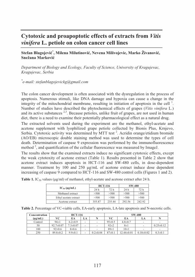
117
Cytotoxic and proapoptotic effects of extracts from Vitis vinifera L. petiole on colon cancer cell lines
������+��,� ���*���������������-��������� �������!������������0������!���
Department of Biology and Ecology, Faculty of Science, University of Kragujevac, Kragujevac, Serbia
*e-mail: [email protected]
The colon cancer development is often associated with the dysregulation in the process of apoptosis. Numerous stimuli, like DNA damage and hypoxia can cause a change in the integrity of the mitochondrial membrane, resulting in initiation of apoptosis in the cell 1.Number of studies have described the phytochemical effects of grapes (Vitis vinifera L.) and its active substances 2,3. Because petioles, unlike fruit of grapes, are not used in human diet, there is a need to examine their potentially pharmacological effect as a natural drug.The extracted solvents used during the experiment are the methanol, ethyl-acetate and acetone supplement with lyophilized grape petiole collected by Bionis Plus, Krnjevo, Serbia. Cytotoxic activity was determined by MTT test 2. Acridin orange/etidium bromide (AO/EB) microscopic double staining method was used to determine the types of cell death. Determination of caspase 9 expression was performed by the immunofluorescence method 2, and quantification of the cellular fluorescence was measured by ImageJ.The results show that the examined extracts induce no significant cytotoxic effects, except the weak cytotoxity of acetone extract (Table 1). Results presented in Table 2 show that acetone extract induces apoptosis in HCT-116 and SW-480 cells, in dose-dependent manner. *�� ������ 8$� 9YY� � � XBY� c�L�L of acetone extract induce dose dependent increasing of caspase 9 compared to HCT-116 and SW-480 control cells (Figures 1 and 2).
Table 1. IC50 �#�&�Fc�L��@�������' ���%���'$�-acetate and acetone extract after 24 h.
IC50 =>,/mL)HCT-116 SW-480
24 h 72 h 24 h 72 hMethanol extract >500 >500 >500 >500
Ethyl acetate extract >500 >500 >500 >500
Acetone extract 355.87 235.44 292.56 242.92
Table 2. Percentage of VC-viable cells, EA-early apoptosis, LA-late apoptosis and N-necrotic cells.
Concentration (μg/mL)
HCT-116 SW-480VC EA LA N VC EA LA N
Control 94±1 6.2±0.9 - - 95±0.5 4.8±0.7 - -50 92.7±0.07 7.3±0.07 - - 92±2 8±1 - 0.25±0.12
100 92±0.6 8±0.6 - - 89±1 10±1 - -250 89.8±0.2 9.9±0.1 - 0.2±0.04 87±0.1 12.44±0.01 0.18±0.02 0.3±0.1
118
Figure 1. Expression of caspase 9 in control HCT-116 and SW-480 and treated cells in a �������� ��������9YY� � �XBY�c�L�L. The nucleus is colored blue and caspases 9 are red (secondary antibody conjugated to Cy3).
Figure 2. The relative fluorescence of control and treated cells, as an indicator of Caspase 9 protein expression, calculated by ImageJ computer program. *P < 0.05 compared to control.
In conclusion, acetone extract of petiole (Vitis vinifera) produces dose-dependent proapoptotic activity on the HCT-116 and SW480 cell lines, with higher effect on SW-480 cells. Also, this treatment induces increased caspase 9 protein expression. The Vitis vinifera petioles are available source of phytochemicals and deserve further examination.
Acknowledgements
This study was supported by Ministry of Education and Science of the Republic of Serbia for financial support (Projects No. III41010).
References
1. Wong RSY. Apoptosis in cancer: from pathogenesis to treatment. Exp Clin Cancer Res 2011;30:87.
2. ���#�������o%��� ������o%�,��������%�� �&������3%�Z�����%�. ������%�� ���������The molecular mechanisms of apoptosis induced by Allium flavum L. and synergistic effects with new-synthesized Pd(II) complex on colon cancer cells. J Food Biochem.2015;39:238-50.
3. Flamini R, Mattivi F, De Rosso M, Arapitsas P, Bavaresco L. Advanced knowledge of three important classes of grape phenolics: anthocyanins, stilbenes and flavonols. Int J Mol Sci.2013;14:19651–69.
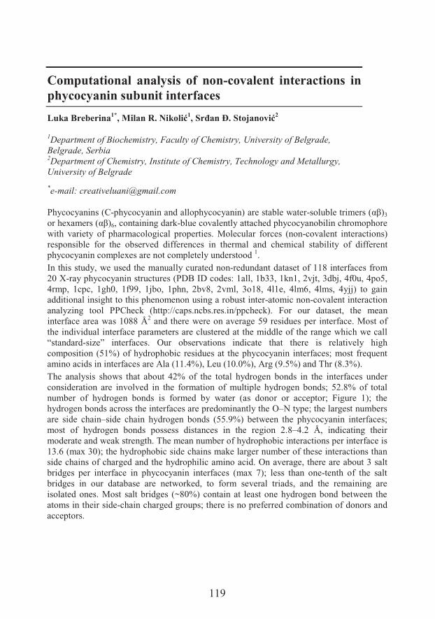
119
Computational analysis of non-covalent interactions in phycocyanin subunit interfaces
Luka Breberina1*, ����#$-!���1���3��.$��� �����2
1Department of Biochemistry, Faculty of Chemistry, University of Belgrade,Belgrade, Serbia2Department of Chemistry, Institute of Chemistry, Technology and Metallurgy,University of Belgrade
*e-mail: [email protected]
Phycocyanins (C-phycocyanin and allophycocyanin) are stable water-&��#8���������&�F;>@3
���'�H ���&�F;>@6, containing dark-blue covalently attached phycocyanobilin chromophore with variety of pharmacological properties. Molecular forces (non-covalent interactions) responsible for the observed differences in thermal and chemical stability of different phycocyanin complexes are not completely understood 1.In this study, we used the manually curated non-redundant dataset of 118 interfaces from 20 X-ray phycocyanin structures (PDB ID codes: 1all, 1b33, 1kn1, 2vjt, 3dbj, 4f0u, 4po5, 4rmp, 1cpc, 1gh0, 1f99, 1jbo, 1phn, 2bv8, 2vml, 3o18, 4l1e, 4lm6, 4lms, 4yjj) to gain additional insight to this phenomenon using a robust inter-atomic non-covalent interaction analyzing tool PPCheck (http://caps.ncbs.res.in/ppcheck). For our dataset, the mean interface area was 1088 Å2 and there were on average 59 residues per interface. Most of the individual interface parameters are clustered at the middle of the range which we call “standard-size” interfaces. Our observations indicate that there is relatively high composition (51%) of hydrophobic residues at the phycocyanin interfaces; most frequent amino acids in interfaces are Ala (11.4%), Leu (10.0%), Arg (9.5%) and Thr (8.3%).The analysis shows that about 42% of the total hydrogen bonds in the interfaces under consideration are involved in the formation of multiple hydrogen bonds; 52.8% of total number of hydrogen bonds is formed by water (as donor or acceptor; Figure 1); the hydrogen bonds across the interfaces are predominantly the O–N type; the largest numbers are side chain–side chain hydrogen bonds (55.9%) between the phycocyanin interfaces; most of hydrogen bonds possess distances in the region 2.8–4.2 Å, indicating their moderate and weak strength. The mean number of hydrophobic interactions per interface is 13.6 (max 30); the hydrophobic side chains make larger number of these interactions than side chains of charged and the hydrophilic amino acid. On average, there are about 3 salt bridges per interface in phycocyanin interfaces (max 7); less than one-tenth of the salt bridges in our database are networked, to form several triads, and the remaining are isolated ones. Most salt bridges (~80%) contain at least one hydrogen bond between the atoms in their side-chain charged groups; there is no preferred combination of donors and acceptors.
120
Figure 1. View of the water-bridged hydrogen bonds (KL interface) of Allophycocyanin from Mastigocladus laminosus (PDB 1b33). The flattened diagram places atoms and bonds on the 2D page to minimize the overlap of atoms and the crossing of bonds in the final diagram. Hydrogen bonds are indicated by dashed green lines (with the distance between donor and acceptor printed in the middle) between the atoms involved. The Figure was prepared using program LigPlot+ v.2.1.
The stability of a non-covalent complex is usually related to the complexation energy, which is proportional to the strength of the interactions involved. Analysis shows that hydrogen bond energies contribute to about 88% to the total energy. Van der Waals and electrostatic energy contributes to 9.3% and 1.9% on average in these complexes, respectively. Thus, hydrogen bonds contribute maximally towards the stability of protein–protein complexes. Results show the total binding energy is more for large phycocyanin ������ ��&��*'������ ��N� ������$������&� #��7 &���&&��' ��I9C�������I9, while most of �'��� ' �� �����$� ��� �'�� � ���� ����� IC� ��� I9?� ��� ���I9. The non-covalent interacting residues in phycocyanin protein interfaces were found to be highly conserved (ConSurf server: http://consurf.tau.ac.il/2016/); salt bridge forming residues have average conservation scores 7.3; for those involved in hydrogen bonds is 7.0; the amino acid residues forming hydrophobic interactions and water-bridged hydrogen bonds both have average conservation scores of 5.9 (on scale 1–9). Obtained results might contribute to the understanding of structural stability of this class of evolutionary essential proteins with increased practical application and future designs of novel protein–bioactive compound interactions.
Acknowledgements
This work was supported by the Ministry of Education, Science and Technological Development of the Republic of Serbia (Grants numbers 172001 and 172035).
References
1. McGregor A, Klartag M, David L, Adir N. Allophycocyanin trimer stability and functionality are primarily due to polar enhanced hydrophobicity of the phycocyanobilin binding pocket. J Mol Biol 2008;384:406–421.
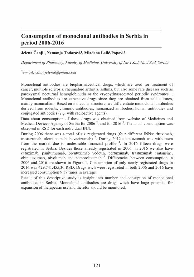
121
Consumption of monoclonal antibodies in Serbia in period 2006-2016
������7�� *�-���� �:���������������(���-8�&���
Department of Pharmacy, Faculty of Medicine, University of Novi Sad, Novi Sad, Serbia
*e-mail: [email protected]
Monoclonal antibodies are biopharmaceutical drugs, which are used for treatment of cancer, multiple sclerosis, rheumatoid arthritis, asthma, but also some rare diseases such as paroxysmal nocturnal hemoglobinuria or the cryopyrinassociated periodic syndromes 1.Monoclonal antibodies are expencive drugs since they are obtained from cell cultures, mainly mammalian. Based on molecular structure, we differentiate monoclonal antibodies derived from rodents, chimeric antibodies, humanized antibodies, human antibodies and conjugated antibodies (e.g. with radioactive agents). Data about consumption of these drugs was obtained from website of Medicines and Medical Devices Agency of Serbia for 2006 2, and for 2016 3. The anual consumption was observed in RSD for each individual INN.During 2006 there was a total of six registrated drugs (four different INNs: rituximab, trastuzumab, alemtuzumab, bevacizumab) 2. During 2012 alemtuzumab was withdrawn from the market due to undesirable financial profile 4. In 2016 fifteen drugs were registrated in Serbia. Besides those already registrated in 2006, in 2016 we also have cetuximab, panitumumab, brentuximab vedotin, pertuzumab, trastuzumab emtansine, obinutuzumab, nivolumab and pembrolizumab 3. Differencies between consumption in2006 and 2016 are shown in Figure 1. Consumption of only newly registrated drugs in 2016 was 429.741.455,30 RSD. Drugs wich were registreted in both 2006 and 2016 have increased consumption 9.57 times in avarage. Result of this descriptive study is insight into number and consuption of monoclonal antibodies in Serbia. Monoclonal antibodies are drugs witch have huge potential for expansion of therapeutic use and therefor should be monitored.
122
Figure 1. Consumption of three drugs in RSD for 2006 (dark) and 2016 (pale).
Acknowledgements
This study was supported by The Ministry of Education, Science and Technological Development, Republic of Serbia (project OI 172058).
References1. Ecker DM, Jones SD, Levine HL. The therapeutic monoclonal antibody market. MAbs
2015;7:9-14.2. Agencija za lekove i medicinska sredstva Srbije. Promet i potrošnja lekova za 2006. [Online].
Available from: https://www.alims.gov.rs/ciril/files/2018/08/PPL2006.pdf. [12.10.2018.]3. Agencija za lekove i medicinska sredstva Srbije. Promet i potrošnja lekova za 2016. [Online].
Available from: https://www.alims.gov.rs/ciril/files/2018/08/PPL2016.pdf. [12.10.2018.]4. European Medicines Agency. MabCampath (alemtuzumab), Withdrawal of the marketing
authorisation in the European Union [Online]. Available from: https://www.ema.europa.eu/documents/public-statement/public-statement-mabcampath-alemtuzumab-withdrawal-marketing-authorisation-european-union_en.pdf [12.10.2018.]
0
20000000
40000000
60000000
80000000
1E+09
1.2E+09
1.4E+09
1.6E+09
1.8E+09
2E+09
rituksimab trastuzumab bevacizumab
RSD
123
Excipients in iron medications as potential causes of side effects in pediatric population
+� ���7���*�-���� �:�������
Department of Pharmacy, Faculty of Medicine, University of Novi Sad, Novi Sad, Serbia
*e-mail: [email protected]
Iron, one of the basic microelements, is necessary for the normal functioning of biochemical processes. A vast variety of enzymes have iron in their composition such as hemoglobin, myoglobin, cytochrome, peroxidase, indicating that its surplus, and especially deficit, can lead to serious health problems 1. The most common form of iron deficiency in the organism is sideropenic anemia, which is manifested by various symptoms of fatigue, immunity and skin fading, while in children can cause slow growth and development 2.The pediatric population is the most vulnerable for anemia development, due to increased iron requirements. Medications for pediatric usage which contain iron as an active substance are considered in this paper. Type and safety of excipients used in the given formulations were observed. Data on selected medicines (n = 6) were obtained from the website of the Agency for Medicines and Medical Devices of Serbia; and the statements are extracted from Section 2 (Qualitative and Quantitative Composition) and Subsection 6.1 (List of Excipients) 3.Six excipients (parabens, ethanol, propylene glycol, sorbitol, saccharose, saccharin sodium) were detected, which may result in appearance of side effects. Use of excipients is based on the improvement of dosage form characteristics. Parabene is a preservative, ethanol and propylene glycol are co-solvent, and sorbitol, saccharose and saccharin sodium improve the organoleptic properties of the dosage form, and in this way increase children’s compliance.In preterm infants, the use of ethanol at a dose of 0.2-1.8 mL per week is safe. If a dose is calculated per kilogram of body weight, it is concluded that preterm infants are exposed to a dose of ethanol higher than permitted 4. Propylene glycol intoxication may lead to gastrointestinal tract disorders. Considering the frequency of the occurrence of adverse effects of ethanol, as well as propylene glycol (1/3 of ethanol intoxication), attention should be paid to the dosing of drugs for pediatric populations. The maximum safe dose for sorbitol does not exist, but side effects such as diarrhea and malabsorbia 5,6 have been reported. It is also not recommended to take medication containing sorbitol in fructose-intolerant individuals. Saccharin and sucrose can rely lead to urticaria. Parabens can cause allergic reactions (possibly delayed) as well as contact dermatitis 7.Comparing side effects of drugs contained in the summary of drug characteristics and undesirable effects of excipients, it has been concluded that the occurrence of allergic
124
reactions, urticaria and dermatitis are present in preparations 2, 3 and 4 containing parabens in their composition. All selected drugs 1-6 can cause gastrointestinal side effects due to the presence of iron as an active substance, which can be more common in drugs 1,3, 4 due to the presence of propylene glycol.
Acknowledgement
This study was supported by The Ministry of Education, Science and Technological Development, Republic of Serbia (project OI 172058).
References1. Rodrigues VC, Mendes BD, Gozzi A, Sandrini F, Santana RG, Matioli G. Deficiência de ferro,
prevalência de anemia e fatores associados em crianças de creches públicas do oeste do Paraná, Brasil. Rev Nutr 2011;24:407-20.
2. World Health Organization (WHO). Growth Reference 5–19 Years. Genava: WHO; 2007. [acessado 2015 Jul 15] Disponível em http: // who.org.int/growthref/who.pdf. [2018 Oct 15]
3. Agencija za lekove i medicinska sredstva Srbije. Promet i potrošnja lekova za 2016. [Online]. Available from: https://www.alims.gov.rs/ciril/files/2018/08/PPL2016.pdf. [15.10.2018.]
4. Whittaker A, Currie A, Turner M, Field D, Mulla H, Pandya H. Toxic additives in medication for preterm infants. Arch Dis Child - Fetal Neonat Ed 2009;94:F236-40.
5. Committee on Nutrition American Academy of Pediatrics. The use and misuse of fruit juice in pediatrics. Pediatrics 2001;107:1210–13.
6. Duro D, et al. Association between infantile colic and carbohydrate malabsorption from fruit juices in infancy. Pediatrics 2002;109:797–805.
7. Rowe RC, Quinn ME, Sheskey PJ. Handbook of Pharmaceutical Excipients, 6th ed., Pharmaceutical Press, London, 2009.
125
Biliverdin-copper complex at the physiological pH
�����6��� ���1*�������+�,������8�����1������0�1, 6�� ������!���2,3,Danica Bajuk-+�,������4�����������1, Snežana Spasi�5, Wilfred Hagen6, Ivan �&��� ���1
1Department of Life Sciences, Institute for Multidisciplinary Research, University of Belgrade, Belgrade, Serbia2&{��, �)����� �+������+�������� ����=�}� ��� �w���!�����$�3Innovation Center of the Faculty of Chemistry, University of Belgrade4Faculty of Physical Chemistry, University of Belgrade5Department of Chemistry, Institute of Chemistry, Technology and Metallurgy, University of Belgrade6Department of Biotechnology, Delft University of Technology, Delft, The Netherlands
*e-mail: [email protected]
Biliverdin (BV) is a degradation product of heme catabolism, which is rapidly converted to bilirubin (BR) by BV reductase 1. Biliverdin and unconjugated BR, commonly named bile pigments, have important function in biochemical processes. The presence of copper and other biological and toxic transitional metals at significant concentrations in bile implies the possibility that metal complexes with bile pigments can be formed 2. Consequently, our interest was to study the complex of BV with copper in physiological conditions –phosphate buffer with pH 7.4.UV-Vis spectrophotometry was applied to investigate formation/degradation of complex of BV with copper ions and to check stoichiometry by titration, showing that BV interacted with Cu2+ in 1:1 stoichiometry. Mass spectroscopy analysis confirmed this – ion at m/z 643.36 was detected. The results of Raman spectroscopy of BV were in good agreement with previous reports 3. Comparing spectra of BV and BV-Cu complex, the following differences were observed: a new band at low wave number is emerged for the complex may be attributed to Cu-N bond vibration; the band which was shifted to lower energies implicates increased stability of BV in the complex; intensity changes imply a more planar structure of BV in the complex, while stronger bands in complex imply higher ���� ��N ����� ��� p-electrons and consequently a higher stability of the BV structure. Pertinent to this, it has been proposed that complexes of BV model compounds with Cu2+
may show unusual electronic structures that exhibit a significant ligand radical character. 1H NMR spectrum of BV in phosphate buffer had a poor resolution of signals, which may originate from aggregation, but this was of little relevance here, since the addition of ��������&��� ���� ���$�&�������������q��'������������&&���� ���&�� �������&��*'����&&����signals represents the result of strong paramagnetic effects that may come from an unpaired e- �' ���&� ���� ��N� ����p���8�� �s of the ring/ligand influencing all protons in the
126
complex. The EPR spectrum of Cu2+ (S = 1/2; I = 3/2) in phosphate buffer shows that Cu2+
is weakly coordinated in an axial symmetry with one gr line and four lines coming from hyperfine coupling along gs. The addition of BV in equimolar concentration led to the loss of Cu2+ signal. The remaining signal in the [BV]/[Cu2+] = 1 system was broad, and did not show hyperfine structure. The g-value of the isotropic signal of BV-Cu complex was significantly lower than the average g-value of Cu2+ in the phosphate buffer indicating delocalization of the spin away from the metal nucleus. Similar EPR signals have been reported previously 4. Parallel-mode EPR showed no signal. Furthermore, the spectra were run over a wide field range and no half field lines were observed, either in parallel or in perpendicular mode. These results are consistent with S = 0 for the copper center. Further, redox properties of the complex were examined. BV showed a well-defined anodic peak. The [BV]/[Cu2+] = 2 system showed two additional oxidation peaks at much lower potentials than BV. The former potential corresponds to the oxidation of Cu1+, as we have shown previously 5. There was a slight consumption of O2 in [BV]/[Cu2+] = 1 system, which may be explained by traces of ‘free’ copper. However, in the presence of an excess of copper ([BV]/[Cu2+] = 0.5), the consumption of O2 was significant. This implies that ‘free’ Cu2+ reacts with the complex and ‘shuttles’ an e- to O2. The complex was susceptibleto oxidizing agents but not to reducing agents.Considering the results obtained we conclude that, at physiological pH, BV builds a complex with copper ions in 1:1 stoichiometry. The formation of complex involves the rearrangement of electronic structure which provides increased energetic stability and strong paramagnetic effects. We believe that a complex with a highly delocalized unpaired e- and the formal BV•+-Cu1+ character best suites the outlined properties, but other structures of the complex cannot be completely ruled out. The presented results may shed new light on long-standing issues of BV chemistry and catalysis in biological systems.
Acknowledgements
This study was supported by the Ministry of Education, Science and Technological Development of the Republic of Serbia (III43010).
References1. Zhang L. Heme Biology: The Secret Life of Heme in Regulating Diverse Biological Processes.
World Scientific Publishing Company, Singapore, 2011.2. Suzuki N. On bilirubin-metal complex compounds in relation to black pigments of gallstones.
Tohoku J Exp Med 1966;90:195-05.3. Celis F, Campos-Vallette M, Gómez-Jeria J, Clavijo R, Jara G, Garrido C. Surface-enhanced
Raman scattering and theoretical study of the bilichromes biliverdin and bilirubin. SpectroscLett 2016;49:336-42.
4. Peeks M, et al. Electronic delocalization in the radical cations of porphyrin oligomer molecular wires. J Am Chem Soc 2017;139:10461-71.
5. ��0����%� ��� ������' ��&�&� ��� �� �H� ����� �����&� ��� 8����#8���7��'� ����� � � �'�� ������&� ���penicillamine. Chem Biol Interact 2017; 25:129-34.
127
Modified androstane and estrane steroids as novel ligands of cytochromes P450
Yaraslau U. Dzichenka1*, Aleksei V. Yantsevich1, Sergey A. Usanov1, Sofija +�!�2,Maja �������3, Marina ����2, Jovana Ajduko��2, Suzana ��������-Šanta2
1Institute of Bioorganic Chemistry of National Academy of Sciences, Minsk, Belarus2Department of Chemistry, Biochemistry and Environmental protection, Faculty of Sciences, University of Novi Sad, Novi Sad, Serbia3Department of Biology and Ecology, Faculty of Sciences, University of Novi Sad
*e-mail: [email protected]
Cytochromes P450 (CYPs) are widely known as monooxygenases involved in many metabolic processes – metabolism of steroids, unsaturated fatty acids, xenobiotics/drugs and other biomolecules 1,2. Normal conditions do not need any impact on these heme-containing proteins, but in pathological ones inhibitors of these enzymes are of great interest, since they could decrease or eliminate enzymes activity. That is the reason for screening of ligand libraries in order to find good lead compound for further drug development. In silico experiments jointly with in vitro binding tests could represent good base for search of lead compound. Here we present results of in vitro screening, provided in order to test binding of a library of modified androstane and estrane derivatives to the selected palette of human cytochromes P450: CYP7A1, CYP7B1 and CYP21A. Among tested compounds some have shown biomedical potential via enzyme inhibition, receptor binding or antiproliferative effect against the cancer cells of reproductive tissues. After initial screening, the mode of binding was determined by spectrophotometric titration and Kdswere calculated for the compounds with highest potential of binding to the tested proteins. For the most promising candidates activity was measured in reconstituted system and products of enzymatic reaction were analyzed.Obtained results present base platform for drug development, since any information aboutinteraction of cytochromes P450 with any class of ligands is very important for “smart” drug-design. Potential of binding of other modified steroids towards other CYPs are the aims of our further work.
Acknowledgements
Presented results are obtained in the frame of Belarus- Serbia bilateral project “Target-specific screening of new activity modulators of human sterol-'$ ��H$� &�&)� F���e9ftuvw-002) which is being realized between Institute of Bioorganic Chemistry of NAS of Belarus and University of Novi
128
Sad Faculty of Sciences. Authors acknowledge Dr. N.V. Strushkevich for the plasmid constructions used for expression of recombinant CYPs in E. coli cells.
References1. Danielson PB. The cytochrome P450 superfamily: biochemistry, evolution and drug metabolism
in humans. Curr Drug Metab 2002;3:561-97.2. Estabrook RW. A passion for P450s (rememberances of the early history of research on
cytochrome P450). Drug Metab Dispos 2003;31:1461-73.
129
Ethyl pyruvate has tolerogenic effects on dendritic cells
-���.�����1, María José Mansilla2,3, Bojan Jevtic1, Juan Navarro-Barriuso2,3, Eva Martínez-Cáceres2,3�.��3��� !���1
1Department of Immunology, Institute for Biological Research “Siniša ������ �<=University of Belgrade, Belgrade, Serbia2Immunology Division, Germans Trias i Pujol University Hospital and Research Institute,Campus Can Ruti, Badalona, Spain3Department of Cellular Biology, Physiology and Immunology, Universitat Autònoma deBarcelona, Cerdanyola del Vallés, Spain
*email:[email protected]
Dendritic cells (DC) are professional antigen presenting cells that have a key role in shaping the immune response. Tolerogenic DC (tolDC) maintain T-cell tolerance either directly by inducing anergy, apoptosis, or indirectly by induction of regulatory T cells 1-2.Therefore, tolDC have immuno-regulatory properties and they are a promising prospective therapy for multiple sclerosis (MS) 3. Ethyl pyruvate (EP) is a redox analogue of dimethyl fumarate (Tecfidera), a drug for MS treatment 4. We have recently shown that EP ameliorates experimental autoimmune encephalomyelitis (EAE), a MS animal model 5.Also, EP showed beneficial effects within the CNS, where reactivity of microglia and astrocytes was reduced 6. Therefore, we expanded an investigation focus in order to see if EP has tolerogenic influence on DC and showed that it induces tolerogenicity in mice and human DC. When applied every third day during propagation of mice bone marrow derived DC, EP reduced expression of molecules required for efficient T cell activation, including MHC class II molecules and co-stimulatory proteins CD40 and CD86 on DC. Importantly, expression of CD11c, a marker of dendritic cells, remained at high level. Further, DC treated with EP showed decreased production of pro-inflammatory cytokines: IL-12, IL-1beta, IL-6 and TNF. Similar results were obtained with human DC. There, EP was applied every other day during the propagation of monocyte derived DC obtained from healthy individuals and MS patients. Phenotypic analysis has shown that DC treated with EP have significantly reduced levels of molecules required for T cell activation such as CD86, CD83, and HLA-DR whereby CD11c expression and viability of DC were not affected. There was no difference in response to EP by DC of patients and healthy controls. Inhibition of allogeneic lymphocyte proliferation by tEP DC demonstrated their functional tolerogenic properties. These results demonstrate that ethyl pyruvate has the ability to direct DC towards tolDC. It is shown to be a potent tolerogenic agent in both murine and human DC. In vivo experiments on application of EP-induced tolDC in EAE and detailed molecular characterization of these cells are warranted. These steps should complete pre-clinical studies on tEPDC as potential MS therapy.
130
Acknowledgements
This study was supported by MPNTR Republic of Serbia, OI173013/OI173035. Project PI14/01175, PI17/01521, integrated in the Plan Nacional de I+D+I and co-supported by the ISCIII-Subdirección General de Evaluación and the FEDER.
References
1. Marland G, Bakker AB, Adema GJ, Figdor CG. Dendritic cells in immune response induction. Stem Cells 1996;14:501-7.
2. Hivroz C, Chemin K, Tourret M, Bohineust A. Crosstalk between T lymphocytes and dendritic cells. Crit Rev Immunol 2012;32:139-55.
3. Phillips BE, Garciafigueroa Y, Trucco M, Giannoukakis N. Clinical tolerogenic dendritic cells: Exploring therapeutic impact on human autoimmune disease. Front Immunol 2017;8:1279.
4. Xu Z, Zhang F, Sun F, Gu K, Dong S, He D. Dimethyl fumarate for multiple sclerosis. Cochrane Database Syst Rev 2015;CD011076.
5. M�������� �%� et al. A comparative analysis of multiple sclerosis-relevant anti-inflammatory properties of ethyl pyruvate and dimethyl fumarate. J Immunol 2015;194:2493-503.
6. ��� ���� �%� �� ��& ������ �%� ������ �%� ���2������ �%� 5 ��� � "%� ��������� ��� +���-encephalitogenic effects of ethyl pyruvate are reflected in the central nervous system and the gut. Biomed Pharmacother 2017;96:78-85.
131
Human pharmacokinetics of rosmarinic acid
Maja Hitl1*����0���+�!�2,3�*������6��,���4, Nebojša Kladar1, -���*�����1,Biljana Božin1
1 Department of Pharmacy, Faculty of Medicine, University of Novi Sad, Novi Sad, Serbia2 Department of Infectious Diseases, Faculty of Medicine, University of Novi Sad3 Clinic for Infectious Diseases, Clinical Centre of Vojvodina, Novi Sad, Serbia4 Department of Pharmacology, Clinical Pharmacology and Toxicology, School ofMedicine, University of Belgrade, Belgrade, Serbia
*e-mail: [email protected]
Rosmarinic acid (RA) represents ester of caffeic acid and 3,4-dihydroxyphenyllactic acid. It was originally isolated from Rosmarinus officinalis, and it is commonly detected in plants from subfamily Nepetoideae, family Lamiaceae, as well as in other plant families 1.Biological activities recorded for RA include antioxidant, antibacterial, anti-inflamatory, antidepressant and many other effects 2; this compound is believed to be responsible for prominent antiviral effects of some Lamiceae plants (e.g. lemon balm use in herpes labialis), including antiretroviral activity 3.Pharmacokinetics of RA was first studied in animal models, mainly in rats2. Main data were discovered this way, after which human studies showed similarities and differences in absorption, distribution, metabolism and elimination.Phenolic compounds with complex structure, including RA, generally have limited absorption in digestive tract2. Current knowledge suggests that intestinal microbials are significant in processes of RA metabolism coupled with absorption. Catabolic activity is mediated by enzymes specific for gut bacteria results in less complex compounds, further more easily available in human organism 2,4,5. Simultaneous intake of food slightly reduces Cmax and delays Tmax of RA, with high interindividual differences 4. Concerning topical application of RA, penetration through human skin in relatively poor, suggesting only local effects of applied RA pharmaceutical formulations 6.Data suggest that distribution of RA in mediated by human serum albumin, and that hydrophobic interactions are responsible for bonding 7.RA undergoes several steps of metabolism. Initial step in metabolism includes process of conjugation, as it can be seen by reached peaks of free and conjugated forms of RA.This metabolism is fast, as these levels are achieved in 0.5 hour. Another process of metabolism is methylation, and methyl-RA reached peak after 2 hours5. With increase of ingested amount of RA, concentrations of free forms are elevated, suggesting saturation kinetics of conjugating process 4. Degradational product ferulic acid was also detected after 0.5 h 5.
132
Studies mainly investigated urinary elimination of RA. The compound was mainly excreted as free and conjugated forms RA and methyl-RA, although some conjugates of degradational products such as caffeic acid, m-coumaric acid and ferulic acid were also detected. Again, as well as metabolism, process of renal elimination was fast, approximately within 6 h 5.In conclusion, human pharmacokinetics of RA remains to be fully investigated. Further researches will give more data on this compound’s kinetics and explain its beneficialeffects for human health.
Acknowledgements
This study was supported by The Ministry of Education, Science and Technological Development, Republic of Serbia (grant number OI 172058).
References
1. Petersen M, Simmonds MS. Rosmarinic acid. Phytochemistry 2003;62:121-5.2. Nunes S, et al. Therapeutic and nutraceutical potential of rosmarinic acid- Cytoprotective
properties and pharmacokinetic profile. Crit Rev Food Sci Nutr 2017;57:1799-806.3. ���#���%��������%��� ���%��� �����R%�R �����%���0������.������ �����&������ �5 �� �� ��
plants in anti(retro)viral therapy. Pharmacol Res 2018;133:301-14.4. Noguchi-Shinohara M, et al. Pharmacokinetics, safety and tolerability of Melissa officinalis
extract which contained rosmarinic acid in healthy individuals: A randomized controlled trial. PLoS One 2015;10:e0126422.
5. Baba S, et al. Absorption, metabolism, degradation and urinary excretion of rosmarinic acid after intake of Perilla frutescens extract in humans. Eur J Nutr 2005;44:1-9.
6. ����� ����\� +%� � � � #&����\� �%� ���� �&� 3�� ���� &�� ��� ��&� ������ �� � ����� &���&��� �formulations and its penetration through human skin ex vivo. Acta Pharm 2015;65:199-205.
7. Peng X, Wang X, Qi W, Su R, He Z. Affinity of rosmarinic acid to human serum albumin and its effect on protein conformation stability. Food Chem 2016;192:178-87.
133
Effects of Acetamiprid on gene-specific DNA methylation in zebrafish (Danio rerio) embryos
+� ���4�!���*�-������������, Svetlana Fa�-� � "�)����
Department of Biology and Ecology, Faculty of Sciences, University of Novi Sad, NoviSad, Serbia
*e-mail: [email protected]
Acetamiprid is one of the most widely used pesticides belonging to the neonicotinoid group. It is used in controlling sucking insect pests and it is applied to crops of vegetables and fruits. Acetamiprid causes oxidative stress and mitochondrial damage in Leydig cells and inhibits the synthesis of testicular ATP and cAMP 1.It is well known that embryonic development is extremely susceptible to unfavorableenvironmental conditions, and by the concept "Developmental Origins of Health and Disease" (DOHAD or DOHaD) it is hypothesized that negative effects occurring during embryogenesis are the cause of later in life diseases. Xenobiotics play relevant role in environmental influences on embryogenesis 2. Via DOHAD theory it is considered that xenobiotics affect epigenetic modifications occurring during embryo development.Epigenetic modifications, such as DNA methylation, are maintained during cell divisionwhich leads to conclusion that epigenetic changes are possible mechanism responsible for “the memory” of the early exposure to harmful stimulus. DNA methylation plays a key role in gene expression. Generally, change in DNA methylation of promoter region inversely affects genes expression 3.Primordial germ cells (PGCs) of zebrafish are formed and migrate during the first 24 h of embryogenesis. In zebrafish, germ line lineage can be traced by means of dazl, gene with conserved functions in PGC migration, germ stem cell proliferation, differentiation and meiosis progression 4.Development of zebrafish can be traced with p53 and p21 genes, which are structurally and functionally conserved and play major roles in embryogenesis 5, as well as cyp19a1,which is considered the most conserved ovary-factor in vertebrates.Our study aims to investigate whether the reprogramming of DNA methylation, which occurs during the early development of zebrafish, is sensitive to the effects of xenobiotic acetamiprid. Emphasis is on genes related to reproductive success, dazl and cyp19a1 and tumor protective genes, p53 and p21 related to general proper development.Embryos of zebrafish (Danio rerio, AB wild-type strain) were treated with acetamiprid (AC) which stock solutions were prepared in dimethylsulfoxide (DMSO). Desired concentrations for the treatment were prepared by dissolving AC stock solutions in ISO-water, where all treatment groups, including control (ISO-water) contained 0.1% DMSO.
134
One control and five experimental groups were used: control (0.1% DMSO), 10-8 M AC, 10-7 M AC, 5x10⁻⁷ M AC, 10-6 M AC and 10-5 M AC. Treatment was applied from 2 h post fertilization until 5 days post fertilization (dpf). 5 dpf larvae were collected and stored at -80°C prior the further analysis. Genomic DNA was isolated using phenol-chloroform-isoamyl alcohol method. DNA was digested with methylation sensitive restriction enzymes (MSRE) HpaII and BstUI, which cut at certain restriction sites, only when those are unmethylated. MSRE-qPCR was used for detection of DNA methylation on dazl, cyp19a1,p53 and p21 genes. The primers are designed by using NCBI primer blast tool (https://www.ncbi.nlm.nih.gov/tools/primer-blast/) and sequences obtained from the UCSC genome browser (https://genome.ucsc.edu/). They are designed so that they amplify promoter regions of target genes which at the same time contain HpaII and BstUI restriction sites.Preliminary results revealed that methylation status of dazl gene promoter was 61.8% in control embryos, and was unaffected by the AC treatment. The methylation status of cyp19a1a gene was 64.4% in control embryos and AC treatment affected its methylation in terms of hypermethyilation, which ranged up to 100% depending on the pesticide concentration. Percentages of methylation of the p53 and p21 gene promoters were significantly lower, 0.5% and 0.2%, in controls, respectively. Both gene promoters were hypermethylated by the AC treatment. Hypermethylation of both promoters was detected already in 10-7 M group, and ranged up to 3.9% for p53 and 2.3% for p21 in the highest concentration group.Our results show that acetamiprid has the ability to disrupt DNA methylation status of certain genes during the early embryonic development of zebrafish. Persistent nature ofDNA methylation may lead to speculations that observed AC induced changes in the level of DNA methylation may have lasting effects on expression of cyp19a1, p53 and p21which can be reflected in the further development of the organism as well as the functioning of the reproductive system.
Acknowledgements
This study was supported by Ministry of Education, Science and Technological Development, Project No. 173037.
References1. Keshta AT, Hataba AA, Mead HMI, El-Shafey NM. Oxidative stress and biochemical changes
induced by thiamethoxam and actamiprid insecticides in rats. World J Pharma Pharmacol Sci 2016;5:44-60.
2. Bonde JP, Giwercman A. Environmental xenobiotics and male reproductive health. Asia J Androl 2014;16:3–4.
3. Hamilton JP. Epigenetics: Principles and Practise. Digestice Deasase 2011;29:130-5.4. Mingyou L, Feng Z, Zhendong L, Ni H, Yunhan H. Dazl is a critical player for primordial germ
cell formation in medaka. Sci Rep 2016;6:28317.5. Abukhdeir AM, Park BH. p21 and p27:roles in carcinogenesis and drug resistance. Expert Rev
Mol Med 2009;10:e19.
135
Does peppermint’s post distilation waste can reduce lipid peroxidation?
�������������1*�-���*�����1�)��!������-!���2, Nebojša Kladar1, Maja Bekut1�-���� �:�������1, Biljana Božin1
1Department of Pharmacy, Faculty of Medicine, University of Novi Sad, Novi Sad, Serbia2 Department of Obstetrics and Gynecology, Vojvodina Clinical Centre, Novi Sad, Serbia
*e-mail: [email protected]
The use of medicinal herbs has been rooted in traditional medicine and pharmacy since ancient civilizations. Treatment and co-therapy with medicinal herbs successfully expands and develops nowadays. World Health Organization reported that 80% of population in developing countries uses traditional medicine and herbal remedies in primary health care. Due to the increase in consumption of dietary supplements and herbal medicinal products, there is a growing need for more rational use of plant resources.Peppermint is an aromatic plant with long tradition of medical use.This plant has numerous proven pharmacological effects - antimicrobial, anti-inflammatory, spasmolytic, antioxidant. Phenol compounds are responsible for most of these effects. The official drug is Menthae x piperitae folium and from this drug, with different distillation techniques, an essential oil is obtained. In order to achieve a more rational use of plant material, scientists tests possiblity of use of residues after the extraction of essential oil, for the isolation of other active principles 1-3.Male Swiss Albino mice, six weeks old, weighing 25–35 g were used in this investigation. All efforts were made to minimize animal discomfort and the experimental procedures were approved by Ethical Committee for Animal Use in Experiments, University of Novi Sad. Standard peppermint leaf extract (Ph. Eur. IV, 2002) was prepared by macerationprocedure in 45% ethanol, as a solvent during 24 h (1:10 w/v, 10 g dried leaves) at room temperature (N1). Essential oil was isolated by hydrodistillation technique (Ph. Eur. IV, 2002) 4. The waste material, remaining after isolation, was filtered and deodorised leaves were used the preparation of N2 (45% ethanol, 24 h) and N3 (75% ethanol, 24 h). Lipid peroxidation and total protein content were determined spectrophotometrically in brain homogenate and in serum of experimental animals 5.Values of biochemical parameters in brain homogenate and in serum of group of experimental animals treated with selected peppermint extracts were compared with parameters in control group of animals by using Student t-test. The difference between compared groups was considered to be significant when p< 0.05.By monitoring the effect on lipid peroxidation in the brain, it has been established that all three investigated extracts exhibit a protective effect in the state of induced oxidative stress
136
(after application of CCl4 when compared with the group with one-time application of CCl4), as they lead to a statistically significant decrease of LP intensity.In the serum, only the N3 extract shows a statistically significant reduction of the lipid peroxidation intensity.Based on our results and previous investigations, we can conclude that examined waste extracts exhibit comparable antioxidant effect to officially prepared pepppermint leaf extract. This waste and extracts may be used for preparation of herbal medicines or in pharmaceutical and food industry as preservatives of natural origin.
Acknowledgements
The authors are thankful toThe Ministry of Education, Science and Technological Development, Republic of Serbia (project OI 172058).
References
1. �� ��������#����*%���0����%������l&���+��,'���������� &�# $ of biological activities of 10 aromatic Lamiaceae species' essential oils. J Chemometrics 2016;30:88-196.
2. Bozin B, Mimica-Dukic N, Simin N, Anackov G. Characterization of the volatile composition of essential oils of some Lamiaceae spices and the antimicrobial and antioxidant activities of the entire oils. J Agri Food Chem 2006;54:1822-8.
3. R ���� �%� �� �� �%� ��� �� +%� �������� +%� � ������� "%� ����� -Duki�� �% Božin B. Postdistillation waste material of thyme (Thymus vulgaris L., Lamiaceae) as a potential source of biologically active compounds. Ind Crops Prod 2015;74:457-64.
4. European Pharmacopeia. 2002. 4th ed. Council of Europe, Strasbourg, pp. 183–4.5. Buege AL, Aust D. Microsomal Lipid Peroxidation. In: Fleisher S, Parker L. Methods in
Enzimology. Academic Press, New York, 1978, pp. 306-10.
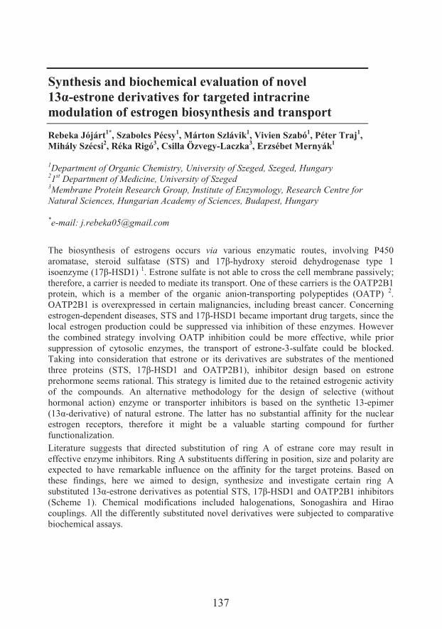
137
Synthesis and biochemical evaluation of novel ���-estrone derivatives for targeted intracrine modulation of estrogen biosynthesis and transport
Rebeka Jójárt1*, Szabolcs Pécsy1, Márton Szlávik1, Vivien Szabó1, Péter Traj1,Mihály Szécsi2, Réka Rigó3, Csilla Özvegy-Laczka3, Erzsébet Mernyák1
1Department of Organic Chemistry, University of Szeged, Szeged, Hungary21st Department of Medicine, University of Szeged3Membrane Protein Research Group, Institute of Enzymology, Research Centre for Natural Sciences, Hungarian Academy of Sciences, Budapest, Hungary
*e-mail: [email protected]
The biosynthesis of estrogens occurs via various enzymatic routes, involving P450 ��� � &�%� &����� � &#�� � &�� F�*�@� � � 9<>-hydroxy steroid dehydrogenase type 1 �&���N$���F9<>-HSD1) 1. Estrone sulfate is not able to cross the cell membrane passively; therefore, a carrier is needed to mediate its transport. One of these carriers is the OATP2B1 protein, which is a member of the organic anion-transporting polypeptides (OATP) 2.OATP2B1 is overexpressed in certain malignancies, including breast cancer. Concerning estrogen-dependent diseases, STS and 9<>-HSD1 became important drug targets, since the local estrogen production could be suppressed via inhibition of these enzymes. However the combined strategy involving OATP inhibition could be more effective, while prior suppression of cytosolic enzymes, the transport of estrone-3-sulfate could be blocked. Taking into consideration that estrone or its derivatives are substrates of the mentioned �'���� ������&� F�*�%� 9<>-HSD1 and OATP2B1), inhibitor design based on estrone prehormone seems rational. This strategy is limited due to the retained estrogenic activity of the compounds. An alternative methodology for the design of selective (without hormonal action) enzyme or transporter inhibitors is based on the synthetic 13-epimer F9:;-derivative) of natural estrone. The latter has no substantial affinity for the nuclear estrogen receptors, therefore it might be a valuable starting compound for further functionalization. Literature suggests that directed substitution of ring A of estrane core may result in effective enzyme inhibitors. Ring A substituents differing in position, size and polarity are expected to have remarkable influence on the affinity for the target proteins. Based on these findings, here we aimed to design, synthesize and investigate certain ring A &���#�� �9:;-�&������ ��� ���&� &������� ���*�%�9<>-HSD1 and OATP2B1 inhibitors (Scheme 1). Chemical modifications included halogenations, Sonogashira and Hirao couplings. All the differently substituted novel derivatives were subjected to comparative biochemical assays.
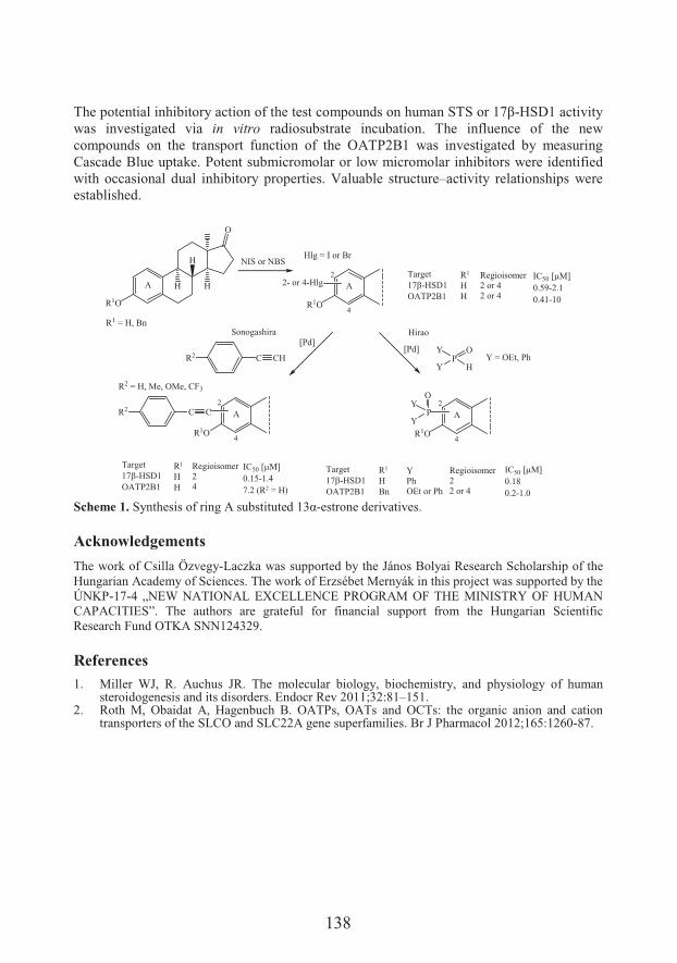
138
*'�������� ����'�8����$� ����������'����&������#� &����'#� ���*�����9<>-HSD1 activity was investigated via in vitro radiosubstrate incubation. The influence of the new compounds on the transport function of the OATP2B1 was investigated by measuring Cascade Blue uptake. Potent submicromolar or low micromolar inhibitors were identified with occasional dual inhibitory properties. Valuable structure–activity relationships were established.
O
R1O
A
R1O
2- or 4-Hlg2
4
A
Sonogashira Hirao[Pd]
C CHR2 PY
Y H
O[Pd]
A
R1O
2
4
C CR2A
R1O
2
4
P
OY
Y
Target17�-HSD1OATP2B1
R1
HBn
YPhOEt or Ph
Regioisomer22 or 4
IC50 [�M]0.180.2-1.0
Target17�-HSD1OATP2B1
IC50 [�M]0.15-1.47.2 (R2 = H)
R1
HH
Regioisomer24
NIS or NBSTarget17�-HSD1OATP2B1
R1
HH
Regioisomer2 or 42 or 4
IC50 [�M]0.59-2.10.41-10
R1 = H, Bn
R2 = H, Me, OMe, CF3
Hlg = I or Br
Y = OEt, Ph
HH
H
Scheme 1. �$��'�&�&���������+�&���#�� �9:;-estrone derivatives.
Acknowledgements
The work of Csilla Özvegy-Laczka was supported by the János Bolyai Research Scholarship of the Hungarian Academy of Sciences. The work of Erzsébet Mernyák in this project was supported by the ÚNKP-17-4 „NEW NATIONAL EXCELLENCE PROGRAM OF THE MINISTRY OF HUMAN CAPACITIES”. The authors are grateful for financial support from the Hungarian Scientific Research Fund OTKA SNN124329.
References1. Miller WJ, R. Auchus JR. The molecular biology, biochemistry, and physiology of human
steroidogenesis and its disorders. Endocr Rev 2011;32:81–151. 2. Roth M, Obaidat A, Hagenbuch B. OATPs, OATs and OCTs: the organic anion and cation
transporters of the SLCO and SLC22A gene superfamilies. Br J Pharmacol 2012;165:1260-87.
139
Biological effects of long-term exposure of human vascular endothelial cells to bisphenol A
Dunja Kokai1*�+� ��������2�-� � "�)����1
1Department of Biology and Ecology, Faculty of Sciences, University of Novi Sad, NoviSad, Serbia2Department of Environmental Engineering and Occupational Safety and Health, Facultyof Technical Sciences, University of Novi Sad
*e-mail: [email protected]
Cardiovascular diseases (CVDs) are the leading cause of death worldwide, accounting forover 17.9 million deaths in 2018 1. Smoking, alcohol consumption, poor diet and physical inactivity are the main risk factors for CVDs; however, these lifestyle factors alone cannot fully account for the magnitude of the epidemic. Recently, a great share of scientific attention and research has focused on environmental chemicals as potential CVD risk factors. Bisphenol A (BPA) and other plastic-associated chemicals (PACs) have a wide range of uses in products such as food containers, water pipes, medical tubing and children’s toys. Although it has been estimated that over 95% of the European population is exposed to PACs and epidemiological studies suggest a possible correlation between PACs’ exposure and CVDs 2, very few mechanistic and functional studies regarding the effect of PACs on vascular cells have been conducted to date. To that end, our goal was to identify and examine biomolecular events associated with endothelial dysfunction in human vascular endothelial cellline EA.hy926after long-term exposure to BPA.EA.hy926 cells share morphological, phenotypic and functional characteristics with human macro-vascular endothelial cells 3. The cells originating from three different cryopreserved stock vials were exposed to either control conditions or three different concentrations of BPA (10-9, 10-8 and 10-7 M) and cultured independently in cell culture flasks over 14 weeks. The abovementioned concentrations of BPA were selected based on a range of detected BPA plasma levels in human population 4. The cells were passaged and counted twice a week, whereas the media containing different treatments were replaced 4 times a week. Cell number was estimated using the dye-exclusion test with Trypan blue. Over the course of the study, there were no significant differences in cell number between the treatments (average: 3.01 to 3.24 × 10⁶ cells/flask). In parallel, AlamarBlue assay was conducted to establish whether exposure to different concentrations of BPA had impact on EA.hy926 viability. Cell viability was not significantly effected after 2, 4, 8 and 12 weeks; however, after 14 weeks we observed ~25% and 18% decrease in viability in cells exposed to 10-8
and 10-7 M BPA, respectively. Nitric oxide (NO•) is an important mediator of many biological events in the vasculature. Nitrite levels (stable oxidation product of NO•) were estimated in cell culture media using Griess method 5. The highest concentration of BPA
140
(10-7 M) caused a sustained increase in nitrite levels in all investigated time points: 8, 9, 12 and 14 weeks. To assess the extent of endothelial dysfunction after long-term exposure of EA.hy926 cells to BPA we performed adhesion studies using calcein AM-labeled human monocytic cell line U937 6. After 5 weeks, we observed ~75% decrease in adherence of U937 cells to the EA.hy926 monolayer in 10-7 M BPA group, whereas this transient effect was no longer evident after 9 and 14 weeks. Endothelial permeability after long-term exposure to selected concentrations of BPA was determined by the two-compartment permeability assay using fibronectin-coated transwell units by measuring the passage of FITC-labeled dextran 7 through the EA.hy926 cell monolayer. After 5 weeks we observed ~60% decrease in permeability in 10-7 M BPA group, whereas after 14 weeks we observed significantly diminished permeability in cells exposed to 10-8 and 10-7 M BPA (by ~55% and 60%, respectively). To implicate involvement of tight junction molecules in altered endothelial permeability after long-term exposure to BPA, protein expression of occludin and ZO-1 was determined by Western blotting. Increased expression of these proteins was observed with the highest concentration of BPA, supporting the findings from the permeability studies. In summary, obtained results indicate that long-term exposure of EA.hy926 cells to low doses of BPA causes direct biological effects, warranting further comprehensive investigation aimed at uncovering the exact mechanisms and the effects of BPA on vascular cells.
Acknowledgements
This study was supported by the grant from the Ministry of Education, Science and Technological Development of the Republic of Serbia “Endocrine disrupting compounds: reproductive, metabolic, developmental responses and mechanisms of action in selected model organism and cell lines”, #173037.
References1. World Health Organization: Noncommunicable diseases country profiles 2018;
http://www.who.int/nmh/publications/ncd-profiles-2018/en/.2. Posnack NG. The adverse cardiac effects of di(2-ethylhexyl)phthalate and bisphenol A.
CardiovascToxicol 2014;14:339-57.3. Edgel CJ, et al. Endothelium specific Weibel-Palade bodies in a continuous human cell line,
EA.hy926. In Vitro Cell Dev Biol 1990;26:1167-72.4. Posnack NG. The adverse cardiac effects of di(2-ethylhexyl)phthalate and bisphenol A.
Cardiovasc Toxicol 2014;14:339-57.5. Green LC, Wagner DA, Glogowski J, Skipper PL, Wishnok JS, Tannenbaum SR. Analysis of
nitrate, nitrite, and [15N]nitrate in biological fluids. Anal Biochem 1982;126:131-8.6. Schmitz B, et al. Increased monocyte adhesion by endothelial expression of VCAM-1 missense
variation in vitro. Atherosclerosis 2013;230:185-90.7. Simoneu B, Houle F, Hout J. Regulation of endothelial permeability and transendothelial
migration of cancer cells by tropomyosin-1 phosphorylation. Vasc Cell 2012;4:18.
141
Redox interactions of epinephrine with iron at physiological pH
�����������1?�6�� ������!���2�������+�,������8�����1�( � ���-!���3,4����&��� ���1
1Life Sciences Department, Institute for Multidisciplinary Research, University ofBelgrade, Belgrade, Serbia2Department of Analytical Chemistry, Innovation center of the Faculty of Chemistry,University of Belgrade3Institute for Biological Research “Siniša ������ �<= University of Belgrade
*e-mail: [email protected]
Epinephrine ((R)-4-(1-hydroxy-2-(methylamino)ethyl)-benzene-1,2-diol (Epi) is catecholamine that is released by the sympathetic nervous system and adrenal medulla. It is a physiologically important molecule that acts as a hormone, neurotransmitter, and medication with a broad range of effects 1-3. Coordinate and redox interaction of Epi with iron affects the interactions with other molecules and its biological effects 4. In this study,we reported details of redox interactions of Epi with Fe2+ at pH 7.4, which correspond to the pH value of human plasma Epi and Fe2+ form a complex that acts as a strong reducingagent. Cyclic voltammetry showed that the positions of Epa and Epc potentials were at approximately -480 and -1100 mV. This implies that Epi and Fe2+ build a complex with unique redox properties. E1/2 was significantly lower compared to E0' for O2/O2
•- (-350 mV). It is important to point out this because superoxide radical anion is produced via spontaneous Fe2+ reaction with O2. In other words, Epi-Fe2+ complex should be capable of reducing transition metals in (patho)physiologicaly relevant complexes that are not susceptible to reduction by O2. Our results confirmed that Epi-Fe2+ is capable of reducing the S-S group of glutathione disulfide. On the other hand, Epi acted in a catalyst-like fashion to promote Fe2+ oxidation by molecular oxygen, and to a facilitated formation of the Epi–Fe3+ complexes, at physiological pH. In addition, we examined the effects of epinepfrine and Epi/Fe3+ system on glioma cells. Epinephrine alone evokes changes in the membrane currents of glioma cells, but such effects were not observed for the complex with Fe3+. This implies that Epi-Fe3+ might modulate neural activity of Epi in CNS.
Acknowledgements
This study was supported by the Ministry of Education, Science and Technological Development of the Republic of Serbia, grant number OI173017.
142
References
1. Álvarez-Diduk R, Galano A. Adrenaline and noradrenaline: Protectors against oxidative stress or molecular targets? J Phys Chem B 2015;119:3479-91.
2. van Mourik T. The shape of neurotransmitters in the gas phase: a theoretical study of adrenaline, pseudoadrenaline, and hydrated adrenaline. Phys Chem Chem Phys 2004;6:2827–37.
3. Lee DR, Galant NJ, Wang H, Mucsi Z, Setiadi DH, Viskolcz B, Csizmadia IG. Thermodynamic functions of molecular conformations of (2-fluoro-2-phenyl-1-ethyl)ammonium ion and (2-hydroxy-2-phenyl-1-ethyl)ammonium ion as models for protonated noradrenaline and adrenaline: first-principles computational study of conformations and thermodynamic functions for the noradrenaline and adrenaline models. J Phys Chem A 2009;113:2507–15.
4. Kora��J, et al. Coordinate and redox interactions of epinephrine with ferric and ferrous iron at physiological pH. Sci Rep 2018;8:3530.
143
T-2 toxin inhibits ovulatory genes expression and steroidogenesis through cAMP signaling pathway in human granulosa cells
Gordana Košanin*�������8�,���-�� !��-� � "�)����
Department of Biology and Ecology, Faculty of Sciences, University of Novi Sad, Novi Sad, Serbia
*e-mail: [email protected]
T-2 toxin is a type A trichothecene mycotoxin produced by different Fusarium species. These mycotoxins are regularly found on various cereals including maize, wheat, barley, rice, beans, and soybeans, as well as in some cereal-based commodities 1–2. It is estimated that around 25% of the world’s agricultural products are contaminated to some degree with mycotoxins 3. T-2 toxin is one of the earliest studied and the most toxic representatives of the trichothecene family 1. Exposure to T-2 toxin occurs via the ingestion of contaminated food or through inhalation and dermal contact. T-2 toxin is remarkably stable under varying environmental conditions or during food processing and has been recorded to cause a variety of adverse effects on humans 3. A number of studies have indicated negative effects of the T-2 toxin on reproduction and development in animals 4. Our objectives were to examine whether environmentally-relevant doses of T-2 toxin can influence human ovarian granulosa cells’ function and to reveal the potential mechanism of action of T-2 toxin. For analysis of T-2 toxin’s action on the steroidogenic and ovulatory potency in human granulosa cells, we have utilized the freshly isolated cumulus granulosa cells. Human cumulus granulosa cells were collected from women undergoing in vitro fertilization (IVF) at the Clinic for Gynecology and Obstetrics, Clinical Center of Vojvodina, Novi Sad, Serbia. First, we conducted the SRB assay to ascertain whether T-2 toxin (1 nM, 10 nM, and 100 nM) affected the viability of human cumulus granulosa cells. Results show that 100 nM T-2 toxin caused a statistically significant reduction in the viability of the human cumulus granulosa cells, and 1 nM and 10 nM doses were without effect. Further, weinvestigated the impacts of lower, not cytotoxic doses of T-2 toxin (1 nM and 10 nM) on the luteinizing hormone/choriogonadotropin receptor (LHCGR) and the ovulatory gene expression, steroidogenesis and cAMP signaling. The cells were treated with 100 ng/mL FSH and T-2 toxin for 48 h and the level of LHCGR mRNA transcript was detected using qRT-PCR. Results revealed that T-2 toxin inhibits the FSH-induced LHCGR mRNA expression and the addition of human chorionic gonadotropin (hCG) was not able to obtain a maximal response of ovulatory genes amphiregulin, epiregulin and progesterone receptor. The results also showed that T-2 toxin (1 nM and 10 nM) reduced aromatase
144
(CYP19A1) and steroidogenic acute regulatory protein (STAR) mRNA expression and that 10 nM T-2 toxin lowered the FSH-stimulated estradiol and progesterone production. We also analyzed level of cAMP in the incubation medium of T-2 toxin exposed cells using enzyme immunoassays. Mechanistic experiments demonstrated that 10 nM T-2 toxin decreased the FSH-stimulated cAMP production in human cumulus granulosa cells. Furthermore, addition of the total phosphodiesterase (PDE) inhibitor IBMX prevented 10 nM T-2 toxin’s action on LHCGR, STAR and CYP19A1 mRNA expression in the FSH-stimulated human cumulus granulosa cells. Overall, our results indicate that anti-ovulatory and anti-steroidogenic effects of T-2 toxin in human cumulus granulosa cells are mediated through the activation of PDE and suppression of the cAMP signaling pathway. Furthermore, the T-2 toxin was involved in inhibition of the FSH-induced cAMP, STAR and CYP19A1 levels and estradiol andprogesterone production. These results suggest an endocrine disruptive capability of T-2toxin in human cumulus granulosa cells and indicate that exposure to this environmental contaminant can impair ovulation and female fertility in humans. Therefore, understanding the effects of the T-2 toxin on the ovary, particularly in women of reproductive age, has essential public health significance.
Acknowledgements
This study was supported by two research grants from the Provincial Secretariat for Higher Education and Scientific Research, Autonomous Province of Vojvodina, Republic of Serbia (no.114-451-2780) and the Ministry of Education, Science and Technological Development of the Republic of Serbia (no. 173037).
References
1. Creppy EE. Update of survey, regulation and toxic effects of mycotoxins in Europe. Toxicol Lett 2002;127:19–28.
2. Kos J, Hajnal E�%� Z����� ���%���� �� +%�� � ��� +%� �� ��� .%����� ����� "�� .��&����� ���Fusarium toxins in maize from Autonomous Province of Vojvodina, Serbia. Food Contr2014;46:98–101.
3. Adhikari M, Negi B, Kaushik N, Adhikari A, Al-Khedhairy A, Kaushik NK, Choi EH. T-2mycotoxin: toxicological effects and decontamination strategies. Oncotarget 2017;8:33933–52.
4. Somoskoi B, Kovacs M, Cseh S. Effects of T-2 mycotoxin on in vitro development and chromatin status of mouse embryos being in preimplantation stages. Toxicol Ind Health2016;32:1260–5.
145
Anticancer and antioxidant effects of naringenin and its semi-synthetic oxime ethers
Ahmed Latif1,2, Tímea Gonda1, Norbert Kúsz1, Ágnes Kulmány2, István Zupkó2,Attila Hunyadi1
1Institute of Pharmacognosy, University of Szeged, Szeged, Hungary2Department of Pharmacodynamics and Biopharmacy, University of Szeged
*e-mail: [email protected]
Naringenin is natural flavanone mainly found in Citrus fruits and vegetables. It has been reported to exhibit multiple biological activities including antibacterial, antifungal, antiviral, anti-inflammatory, antioxidant and anticancer effects 1-3. Due to its numerous beneficial activities, the design and synthesis of new naringenin derivatives is of continuous interest.The aim of the present study was to prepare a set of naringenin oxime derivatives, and to evaluate and compare their cytotoxic and antioxidant effects in comparison with that of their parental compound. Seven analogs have been prepared, including the two possible geometric isomers of naringenin oxime, and the methyl, ethyl, tert-butyl, allyl and benzyl ethers of naringenin E-oxime. The compounds were separated by flash chromatography, and their structure was confirmed by NMR and HRMS measurements.Anticancer effects of the compounds were evaluated by means of their antiproliferative activity against human leukemia cells and a gynecological cancer cell line panel. The tert-butyl ether of naringenin E-oxime exhibited the strongest activity with IC50 values of 19.5, 23.5, 29.7, 31.8, and 35.4 μM against MCF-7, Hela, MDA-231, HL-60 and SiHa cell lines, respectively. This compound was also found to induce apoptosis in HeLa cells through the activation of caspase-3. On the other hand, the corresponding methyl ether exerted potent antioxidant effect by means of ORAC and DPPH assays.
Acknowledgements
This work was supported by the National Research, Development and Innovation Office, Hungary (NKFIH; K119770 and K109293). A.H. acknowledges the JánosBolyai fellowship of the Hungarian Academy of Sciences and the Kálmán Szász Prize.
References1. Rui-Fang L, Feng Y, Chen J, Ge L, Xiao S, Zuo X. Naringenin suppresses K562 human
leukemia cell proliferation and ameliorates Adriamycin-induced oxidative damage in polymorphonuclear leukocytes. J Exp Ther Med 2015;9:697-706.

146
2. Bao L, Liu F, Guo H, Li Y, Tan B, Zhang W, Peng Y. Naringenin inhibits proliferation, migration, and invasion as well as induces apoptosis of gastric cancer SGC7901 cell line by downregulation of AKT pathway. J Tumor Biol 2016;37:11365-74.
3. ��NQ�7&� ��%�.�� ������%�x ��7&� ��%�+���Q�����$��'�&�&� � �8������� �� �����$���������1-alkyl derivatives of naringenin and their oximes. Molecules 2017;22:1485.
147
Plantago species as modulators of thromboxane A2 and prostaglandin E2 production in inflammation
:�� ����� !�*, Marija Lesjak, Neda Mimica-6�!��������+�!������ Ivana Beara
Department of Chemistry, Biochemistry and Environmental Protection, Faculty ofSciences, University of Novi Sad, Novi Sad, Serbia
*e-mail: [email protected]
Chronical inflammation is involved in the pathogenesis of diseases which are the leading cause of death of our time, such as atherosclerosis, arthritis, allergy, cancer, diabetes, neurodegenerative and autoimmune diseases. In the treatment of inflammation the most frequently used drugs are steroid (corticosteroids) and non-steroidal antiinflammatory drugs (NSAIDs), which, in addition to the therapeutic effect, exhibit also a number of undesired side effects. Therefore, a great challenge of modern medicine is discovering new antiinflammatory drugs, which would have a good activity with fewer contraindications. These researches are often directed towards the characterization of the antiinflammatory activity of natural products. One of the numerous approaches in treatment and prevention of inflammation is the modulation of the arachidonic acid pathway and eicosanoids production 1,2.Thus, the aim of this study was to examine the potential of Plantago species to inhibit production of prostaglandin E2 (PGE2) and thromboxane A2 (TXA2). Some of Plantagospecies, such as P. lanceolata L. and P. major L., are highly recognized in ethnomedicine as the remedy for oral or pharyngeal irritations, dry cough, treatment of the wounds and ulcers, gastrointestinal tract diseases, inflammation and conjunctivitis. Besides, there are more than 240 species in the Plantago genus, which are poorly investigated 3.The antiinflammatory activity of methanolic extract of four Plantago species (P. altissimaL., P. argentea Chaix, P. lanceolata and P. major) was examined. Human U937 monocytes (1 × 10⁶ cells/mL) were differentiated into macrophages in the presence of .�+�� "��� �� ����� 7 &� �� #�� � 8$� 5.�� ����� ����� ������ 7��'� �H�� ��&� F?BY� c�L�5@��Afterward, arachidonic acid was added and cells were incubated for 45 min. Extraction of products and internal standard was done according to the previously described procedure. Quantification of produced PGE2 and TXA2 in cell lysate was done by LC-MS/MS 4,5.Among all analyzed extracts, P. argentea had the best potential to inhibit PGE2, as well as TXA2 production. It is worth mentioning that this poorly investigated species, P. argentea,evinced better potential then P. lanceolata and P. major, famous by their antiinflammatory activity. Obtained results report novel and valuable data about the biological potential of Plantago species, in particular of P. argentea and P. altissima. Furthermore, the presented
148
results support further investigation in order to determine the potential use of these species in food, cosmetic or pharmaceutical industry.
Acknowledgements
This study was supported by The Ministry of Education, Sciences and Technological Development of the Republic of Serbia (OI 172058).
References1. Nathan C, Ding, A. Nonresolving inflammation. Cell 2010;140:871-82.2. Koeberle A, Werz O. Perspective of microsomal prostaglandin E2 synthase-1 as drug target in
inflammation-related disorders. Biochem Pharmacol 2015;98:1-15.3. Fleer H, Verspohl EJ. Antispasmodic activity of an extract from Plantago lanceolata L. and
some isolated compounds. Phytomedicine 2007;14:409-15.4. Beara I, et al. Liquid chromatography/tandem mass spectrometry study of anti-inflammatory
activity of plantain (Plantago L.) species. J Pharma Biomed 2010;52:701–6.5. Lesjak M, et al. Chemical characterisation and biological effects of Juniperus foetidissima
Willd. 1806. LWT - Food Sci Techol 2013;53:530–9.
149
Mitigating biotic stress in soybean (Glycine max L.) by plant-growth-promoting fungi Trichoderma asperellum
An����� ����?�.��3e Mal��/��������@�����������.����)��!������8������
Faculty of Agriculture, University of Novi Sad, Novi Sad, Serbia
*e-mail: [email protected]
In natural systems, plants are surrounded by an enormous number of potential enemies who may induce biotic stress in them. A wide variety of bacteria, viruses, fungi, nematodes, mites, insects are greatly responsible for heavy reduction in crop productivity 1.Among the important arthropod pests of soybean are several species of plant feeding mites. The Tetranychidae, a family of the most damaging species, are commonly called "spiders", red spiders, or spider mites 2. In response to pest invasion, plants produce reactive oxygen species (ROS) 3, which are highly reactive and in absence of any protective mechanism they can seriously damage vital biomolecules such as lipids, proteins and nucleic acids 4.Naturally, plants protect themselves by producing some compounds called as secondary metabolites 1. Flavonoids are polyphenolic compounds synthesized by plants, with a basic structure (C6-C3-C6), composed of two aromatic rings joined by a three carbon chain. The presence of multiple hydroxyl groups in their structures gives flavonoids a reducing character 5. Stress-responsive dihydroxy B-ring-substituted flavonoids have great potential to reduce the levels of ROS once they are formed 6. Trichoderma species are free-living fungi in soil and root ecosystems and commonly used as biological control agents against plant pathogens 7. It was stated that Trichoderma species affects induced systemic resistance in plants 8. The objective of this work was to study the effect of inoculation of soybean (Glycine max L., cv. Maximus) seeds with Trichoderma asperellum, followed by mites (Tetranychus urticae) exposure, on content of total flavonoids (TF). In addition, radical scavenging activity of extracts was investigated using 2,2-azinobis(3-ethyl-benzothiazoline-6-sulfonic acid) (ABTS) assay. The experimental design included inoculated and non-inoculated soybean plants – with, and without exposure to mites. Significant accumulation of TF was occurred in control group followed by mites attack (Figure 1a). During biotic stress caused by mites, inoculation successfully reduced oxidative stress. Furthermore, it seems that inoculation with T. asprellum itself produced mild stress which was beneficial to soybean plants. Stress is a factor that improves resistance and adaptive evolution. Radical scavenging activity is shown in Figure 1b. From the obtained results, it can be concluded that greatest oxidative stress occurred in control group followed by mites exposure and it seems that inoculation with T. asperellumsuccessfully reduced oxidative stress.
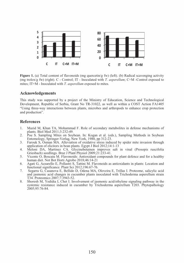
150
Figure 1. (a) Total content of flavonoids (mg quercetin/g fw) (left). (b) Radical scavenging activity (mg trolox/g fw) (right). C - Control; IT - Inoculated with T. asperellum; C+M -Control exposed to mites; IT+M - Inoculated with T. asperellum exposed to mites.
Acknowledgements
This study was supported by a project of the Ministry of Education, Science and Technological Development, Republic of Serbia, Grant No TR-31022, as well as within a COST Action FA1405 “Using three-way interactions between plants, microbes and arthropods to enhance crop protection and production”.
References
1. Mazid M, Khan TA, Mohammad F. Role of secondary metabolites in defense mechanisms of plants. Biol Med 2011;3:232-49.
2. Poe S. Sampling Mites on Soybean. In: Kogan et al. (eds.), Sampling Methods in Soybean Entomology, Springer-Verlag, New York, 1980, pp 312-23.
3. Farouk S, Osman MA. Alleviation of oxidative stress induced by spider mite invasion through application of elicitors in bean plants. Egypt J Biol 2012;14:1-13
4. Meloni DA, Martinez CA. Glycinebetainen improves salt in vinal (Prosopis ruscifolia Griesbach) seedlings. Braz J Plant Physiol 2009;21:233-41.
5. Vicente O, Boscaiu M. Flavonoids: Antioxidant compounds for plant defence and for a healthy human diet. Not Bot Horti Agrobo 2018;46:14-21
6. Agati G, Azzarello E, Pollastri S, Tattini M. Flavonoids as antioxidants in plants: Location and functional significance. Plant Sci 2012;196:67-76.
7. Segarra G, Casanova E, Bellido D, Odena MA, Oloveira E, Trillas I. Proteome, salicylic acid and jasmonic acid changes in cucumber plants inoculated with Trichoderma asperellum strain T34. Proteomics 2007;7:3943-52.
8. Shoresh M, Yedidia I, Chet I. Involvement of jasmonic acid/ethylene signaling pathway in the systemic resistance induced in cucumber by Trichoderma asperellum T203. Phytopathology 2005;95:76-84.
012345
C IT C+M IT+M0
20406080
C IT C+M IT+M
151
Expression of protein synthesis elongation factor 1A in different physiological stages of winter wheat varieties
�������$���!���1*�-�����<$.�!�1�6�������0���2
1Department of Biology and Ecology, Faculty of Science, University of Kragujevac,Kragujevac, Serbia2 Faculty of Agriculture, University of Priština, Kosovska Mitrovica, Lešak, Kosovo andMetohia, Serbia
*e-mail: [email protected]
The climate changes and the rise in temperature in the last decade can lead to substantial reduction in crop productivity worldwide. For this reason, it is important to identify proteins involved in protection of plants from heat stress and help to develop varieties of cereals tolerant to heat. Over the last few years, studies shown that the eukaryotic elongation factors have potential influence and connection with heat tolerance of plants. They prevent aggregation of proteins 1, also eukaryotic elongation factors participate in the degradation of N-terminal blocked proteins and catalyze the formation of disulfide bonds in proteins 2. Considering that numerous studies have shown that the eukaryotic elongation factor 1A (eEF1A) can significantly affect the development of the plant heat stress tolerance 3-4, the aim of our research was to examine the expression and accumulation of eEF1A in different physiological stages of winter wheat varieties and to compare relative abundance of eEF1A in different varieties of winter wheat. Statistical analysis was performed in the SPSS program. For research we used four varieties of winter wheat collected in three different physiological stages (ear emergence, milk development, anddough development stage). Following the isolation of leaf proteins, immunoblot analysis was done and eEF1A protein was detected 5. Results shown that in all varieties expression of eEF1A from ear emergence stage to dough development stage was in increase. The highest expression of eEF1A compared to all varieties was observed in variety NS 40 S in the stage of dough development, while the lowest expression of eEF1A compared to all varieties was observed in variety Zvezdana in the stage of ear emergence. Based on the results we can conclude that on expression of eEF1A beside high temperature stress, the important factor for expression of eEF1A also could be physiological stage of wheat development.
Acknowledgements
This study was supported by Ministry of Education, Science and Technological Development of the Republic of Serbia, Project Grant No. TR 31092.
152
References
1. Rao D, Momcilovic I, Kobayashi S, Callegari E, Ristic Z. Chaperone activity of recombinant maize chloroplast protein synthesis elongation factor, EF-Tu. Eur J Biochem 2004;271:3684–92.
2. Fu J, Momcilovic I, Prasad PVV. Roles of protein synthesis elongation factor EF-Tu in heat tolerance in plants. J Bot 2012;835836.
3. Ristic Z, Bukovnik `%� ���2������ "%� �#� �%� .� & � .3V. Heat-induced accumulation of chloroplast protein synthesis elongation factor, EF-Tu, in winter wheat. J Plant Physol 2008;165:192-202.
4. Bukovnik U, Fu J, Bennett M, Prasad PVV, Ristic Z. Heat tolerance and expression of protein synthesis elongation factors, EF-Tu and EF-1a, in spring wheat. Funct Plant Biol 2009;36:234–41.
5. Momcilovic I, Ristic Z. Expression of chloroplast protein synthesis elongation factor, EF-Tu, in two lines of maize with contrasting tolerance to heat stress during early stages of plant development. J Plant Physiol 2007;164:90–99.
153
Relationship between hematological parameters and glycemic control in type 2 diabetes mellitus patients
Dragana Miloševi�1,2,*�'�����(�!� Panin2
1Department of Laboratory Diagnostics, Health Care ~����������� ���$�� ���*���� ��=����ija, Serbia2Faculty of Medicine, University of Novi Sad, Novi Sad, Serbia
*e-mail: [email protected]
Diabetes mellitus with its micro- and macrocomplications is the leading global epidemic of the 21st century. The aim of the research is to determine possible changes in the complete blood count (CBC) parameters depending on glycemic control in patients with Type 2 diabetes mellitus (T2DM) 1-3.The study included a total of 178 patients with T2DM, both gender over the age of 40 years, from the Health Care Center -�������� ���� �. ����-�"�/ija, Serbia. To notice the possible correlation between the CBC parameters and glucose control in T2DM, the subjects were divided in two groups with HbA1c y 7% and with HbA1c > 7%. We analysed CBC parameters, parameters of glycoregulation, lipid status using standard biochemical methods, performed anthropometric measurements and collected patients data by questionnaire and electronic patient card.There was statistical difference between HbA1c groups for PMDW (p = 0.045), HDL (p =0.0067). Using univariate linear regression it is shown that PCT was correlated with WBC (p = 0.0005), neutrophils (p = 0.046), monocytes (p = 0.003); MPM was associated with MPV (p = 0.0005); MPC (p = 0.0005), PDW (p = 0.0005), GLU0 (p = 0.034), HDL-C (p =0.005), BW (p = 0.053); PMDW was correlated with HbA1c% (p = 0.049), GLU0 (p =0.013), GLU2 (p = 0.053), HDL-C (p = 0.001), BW (p = 0.043) in all patients.Based on our results it may be concluded that some of the parameters of CBC could be useful tools in following glycemic control of diabetics.
References1. World Health Organization (WHO). Diabetes research. Available at:
http://www.who.int/diabetes/en/ (Last accessed on May 10, 2017)2. Incidencija i mortalitet od dijabetesa u Srbiji 2016. Institut za javno zdravlje Srbije “Dr Milan
�� ����� � �#�)%� XY9C�� + �� 8��� �= http://www.batut.org.rs/index.php?content=187 (Last accessed on May 5, 2017)
3. Ziaee A, Ghorbani A, Kalbasi S, Hejrati A, Moradi S. Association of hematological indices with prediabetes: A cross-sectional study. Electronic Physician 2017;9:5206-11.

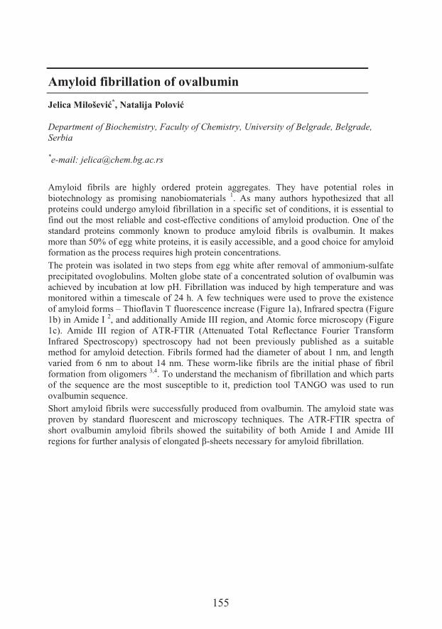
155
Amyloid fibrillation of ovalbumin
Jelica ���"���*, Natalija 8�����
Department of Biochemistry, Faculty of Chemistry, University of Belgrade, Belgrade, Serbia
*e-mail: [email protected]
Amyloid fibrils are highly ordered protein aggregates. They have potential roles in biotechnology as promising nanobiomaterials 1. As many authors hypothesized that all proteins could undergo amyloid fibrillation in a specific set of conditions, it is essential to find out the most reliable and cost-effective conditions of amyloid production. One of the standard proteins commonly known to produce amyloid fibrils is ovalbumin. It makes more than 50% of egg white proteins, it is easily accessible, and a good choice for amyloid formation as the process requires high protein concentrations. The protein was isolated in two steps from egg white after removal of ammonium-sulfate precipitated ovoglobulins. Molten globe state of a concentrated solution of ovalbumin was achieved by incubation at low pH. Fibrillation was induced by high temperature and was monitored within a timescale of 24 h. A few techniques were used to prove the existence of amyloid forms – Thioflavin T fluorescence increase (Figure 1a), Infrared spectra (Figure 1b) in Amide I 2, and additionally Amide III region, and Atomic force microscopy (Figure 1c). Amide III region of ATR-FTIR (Attenuated Total Reflectance Fourier Transform Infrared Spectroscopy) spectroscopy had not been previously published as a suitable method for amyloid detection. Fibrils formed had the diameter of about 1 nm, and length varied from 6 nm to about 14 nm. These worm-like fibrils are the initial phase of fibril formation from oligomers 3,4. To understand the mechanism of fibrillation and which parts of the sequence are the most susceptible to it, prediction tool TANGO was used to run ovalbumin sequence.Short amyloid fibrils were successfully produced from ovalbumin. The amyloid state was proven by standard fluorescent and microscopy techniques. The ATR-FTIR spectra of short ovalbumin amyloid fibrils showed the suitability of both Amide I and Amide III regions for further analysis of ����� �� �>-sheets necessary for amyloid fibrillation.
156
Figure 1. Amyloid fibrils of ovalbumin: a) Thioflavin T fluorescence increase during fibril formation; b) ATR-FTIR spectra of native (black), and amyloid (gray) ovalbumin; c) AFM microscopy of fibrils.
Acknowledgements
This work was financially supported by the Ministry of Education, Science and Technological Development, Republic of Serbia, Grant No. 172049.
References1. Rambaran R, Serpell L. Amyloid fibrils: Abnormal protein assembly. Prion 2008;2:112-7.2. Moran S, Zanni M. How to get insight into amyloid structure and formation from infrared
spectroscopy. J Phys Chem Lett 2014;5:9gf?I93.3. Lara C, Gourdin-Bertin S, Adamcik J, Bolisetty S, Mezzenga R. Self-Assembly of Ovalbumin
into Amyloid and Non-Amyloid Fibrils. Biomacromolecules 2012;13:4213-21. 4. Bhattacharya M, Jain N, Dogra P, Samai S, Mukhopadhyay S. Nanoscopic amyloid pores
formed via stepwise protein assembly. J Phys Chem Lett 2013;4:480-5.
157
Antitumor effects of herbal sesquiterpenes
Nikolett Nagy1, Noémi Bózsity1, Andrea Vasas2, Judit Hohmann2, Georg Krupitza3,Zsuzsanna Schelz1, István Zupkó1*
1Department of Pharmacodynamics and Biopharmacy, University of Szeged, Szeged,Hungary2Institute of Pharmacognosy, University of Szeged3Clinical Institute of Pathology, Medical University of Vienna, Vienna, Austria
*e-mail: [email protected]
Cervical cancer is a leading malignant disease in women and mainly attributed to human papillomavirus (HPV) infection. Sesquiterpene constituents of Neurolena lobata L.(Asteraceae) were tested on human cervical cell lines in vitro in order to evaluate their antiproliferative, antimetastatic and proapoptotic effects.The antiproliferative effects were investigated by the MTT assay, IC50 values were determined on three cervical cancer cell lines with different HPV status (SiHa, HeLa, C33A). Tumor selectivity was examined by using fibroblast cells (NIH, MRC-5). The migratory capacity of tumor cells was analyzed by the wound healing assay, the migration of the cells into the wound site was visualized by phase-contrast inverted microscope. Images were taken by a CCD camera at definite intervals and the rate of migration was calculated according to the rate of wound closure by ImageJ software. Cell cycle analysis was performed by flow cytometry in order to further elucidate the antitumor effects of the tested sesquiterpenes. Two of the twelve tested compounds showed pronounced antiproliferative effects with significant tumor selectivity (IC50 values varied between 1.83-8.14 μM). SiHa, an HPV-16 positive epithelial cervical cell line was the most responsive to the treatment. LOB-48, the most effective sesquiterpene component inhibited the cell migration in concentration dependant manner. According to the cell cycle analysis, LOB-48 slightly elevated the cell number in the hypodiploid phase and altered the distribution of the different subpopulations.Our results revealed the in vitro antitumor effects of sesquiterpenes isolated from Neurolena lobata and confirm its proper utilization in traditional medicine as an anticancer drug. The tested sesquiterpenes can be regarded as prototypes for the design of novel anticancer agents.
Acknowledgements
Financial support from the Economic Development and Innovation Operative Program (GINOP-2.3.2-15-2016-00012) is gratefully acknowledged.

159
Phenolic content and antioxidant potential of extracts of parsley (Petroselinum crispum) cultivated in the Province of Vojvodina
Ivana Nemeš1*, Nataša Simin1���� ����/��1�6� ��9�/�1, Danijela Arsenov2,-���"�-!���2, Neda Mimica-6�!�1
1Department of Chemistry, Biochemistry and Environmental Protection, Faculty ofSciences, University of Novi Sad, Novi Sad, Serbia2Department of Biology and Ecology, Faculty of Sciences, University of Novi Sad
*e-mail: [email protected]
Parsley (Petroselinum crispum) root and leaves are commonly used in diet of many cultures. Long before its use in diet, this plant has been known for its medicinal properties. In traditional medicine, parsley has been used as diuretic as well for gastrointestinal, kidney and lower urinary tract disorders. Furthermore, its use in treatment of dyspepsia, cystitis, dysmenorrhea, functional amenorrhea, and myalgia has also been reported 1.Health benefits of plants are mainly attributed to its secondary metabolites, particularly phenolic compounds. Due to different environmental factors, such as climate or agronomic factors, phenolic composition of plants can be variable, which can influence their health-promoting benefits 2.Since parsley is grown throughout the Province of Vojvodina, the aim of this study was to determine phenolic content as well as to evaluate antioxidant activity of extracts obtained from parsley cultivated within this province. Samples were collected in local markets and shops, but cultivated by different manufacturers in different geographical locations in Vojvodina. The extracts were prepared by the extraction of dried parsley roots with 80% methanol. The total phenolic and total flavonoid contents were determined by standard spectrophotometric assays 3. The presence and content of 44 selected phenolic compounds were investigated by LC-MS/MS 4. Antioxidant activity was evaluated by measuring the 2,2-diphenyl-1-picrylhydrazyl (DPPH) radical scavenging potential (DPPH-RSC) and ability to inhibit lipid peroxidation (LP) 3.The total phenolic content (5.00-9.18 mg gallic acid equivalents/g of d.e.) and total flavonoid content (0.70I9�B?� ��� D#�������� �D#� ����&L�� ��� ���@� ��� � 8��7���� �'��samples cultivated in different locations. Phenolic compounds examined by LC-MS/MS included 14 phenolic acids, 25 flavonoids, 3 coumarins and 2 lignans. Flavonoid apiin was the most dominant out of all examined compounds, but its concentration varied greatly between the samples obtained from different locations (1862-:9YY� c�L�� ��� �$� �H�� ��@��Except for apiin, flavonoids apigenin and apigenin-7-O-glucoside were also detected in the extracts. The most dominant phenolic acids were chlorogenic acid and ferulic acid. All
160
three investigated coumarins (umbelliferone, scopoletin and esculetin) were detected, but scopoletin was the most abundant, although its content was highly variable between the samples (2.9-198.4 c�L�� ��� �$� �H�� ��@�� � ���&� ����� ��������� ��� �����&� �&�� �H'�8��� �distinctive antioxidant activity, which was weak in comparison to standard antioxidant PG (IC50nY�?9BIY�<f?���L�5� vs. IC50=0.570 �g/mL in DPPH assay and IC50=1.352–3.402 mg/mL vs. IC50=0.056 mg/mL in LP assay). In this research, phenolic content of selected compounds, as well as total phenolic content, total flavonoid content and antioxidant activity of parsley root extracts cultivated in seven different locations in Vojvodina were determined. The results suggest that environmental factors, including agricultural practices and type of soil have an influence on phenolic composition. Additionally, the obtained results revealed that parsley root is a week antioxidant, while environmental factors also influence its antioxidant activity.
Acknowledgements
This study was supported by Provincial Secretariat for Science and Technological Development, Autonomous Province of Vojvodina, Serbia (Grant No. 114-451-2149/2016-03, 2016-2019).
References1. Agyare C, Appiah T, Boakye YD, Apenteng JA. Petroselinum crispum: A Review. In: Kuete V.
(ed). Medicinal Spices and Vegetables from Africa, Academic Press, Cambridge, 2017, pp. 527–47.
2. Manach C, Scalbert A, Morand C, Rémésy C, Jiménez L. Polyphenols: food sources and bioavailabiliy. Am J Clin Nutr 2004;79:727–47.
3. Simin N et al. Chemical profile and biological activities of Allium melanantherum . �2��extracts. Planta Med 2011;77;PM221.
4. 1�2�������� ���d# ���� ���� ������� ��������� ���'������&� ���`���� � �oica extracts by high-performance liquid chromatography coupled with tandem mass spectrometric detection. Food Chem 2014;143:48–53.
161
The pro-inflammatory effect of Act d 1, cysteine protease from kiwifruit (Actinidia deliciosa), on intestinal epithelial cells in vitro
Andrijan�-�"�1*�����8�&���1������7���2, Joost J Smit3���� �*������-���!�����1
1Department of Biochemistry, Faculty of Chemistry University of Belgrade, Belgrade,Serbia2Institute for Oncology and Radiology of Serbia, Belgrade, Serbia3Institute for Risk Assessment Sciences, Utrecht University, Utrecht, Netherlands
* e-mail: [email protected]
Actinidin (Act d 1) is a papain-like cysteine protease present in kiwifruit in both active and inactive form. It is able to keep its primary structure, proteolytic activity and immunological reactivity for up to 2 h under simulated gastric digestion 1. In vitro model systems have shown that Act d 1 exerts direct proteolytic cleavage of occludin and leads to disruption of epithelial barrier function 2,3. However, mechanisms underlying the induction of the immune response to Act d 1 and its mode of action on the gastrointestinal barrier remain unknown. Therefore, the aim of this research was to test if Act d 1 can induce the innate immune response in intestinal epithelial cells. Act d 1, was originally purified from kiwi fruit using previously well-established methods. Evaluation of Act d 1 influence on the permeability of the epithelial monolayer was performed on Caco-2 cells cultured as a monolayer on inserts, #&����>-lactoglobulin (BLG), one of major whey proteins. Activation of the innate immune response in terms of the production of pro-inflammatory cytokines to Act d 1 was measured both at the genetic and protein level, using qRT-PCR and ELISA assays, respectively. Activated Act d 1 influences the permeability of intestinal monolayer of Caco-2 cells, as. BLG was detected in the baso-lateral compartment with respect to the control. Treatment with Act d 1 leads to the production of pro-inflammatory cytokines atthe protein level. In addition, Act d 1 causes an increase in gene expression levels of IL-33 and TSLP. In the in vitro model tested in this work proteolytically active Act d 1 can initiate an upregulation of the transcription and synthesis of pro-inflammatory cytokines. This finding contributes to the understanding of the molecular mechanisms of kiwifruit allergy development.
Acknowledgements
This study was supported by the Ministry of Education, Science and Technological Development (Grant No. 172049)

162
References
1. Grozdanovic MM, Ostojic S, Aleksic I, Andjelkovic U, Petersen A, Gavrovic-Jankulovic M. Active actinidin retains function upon gastro-intestinal digestion and is more thermostable than the E-64-inhibited counterpart. J Sci Food Agricul 2014;94:3046–52.
2. � ��� �%� R��N ����� �%� � ���� +%� �� ��-� ���� *%� +�/�#&� .�%� R ����-� ��#����� ���Actinidin, a protease from kiwifruit, induces changes in morphology and adhesion of T84 intestinal epithelial cells. Phytochemistry 2012;77:46–52.
3. R��N �������%�� ����%�������+%�+� ��������`%�+�8 ���.%��������%�R ����-� ��#�����M. Kiwifruit cysteine protease actinidin compromises the intestinal barrier by disrupting tight junctions. Biochim Biophys Acta 2016;1860:516-26.
163
Analysis of Arabidopsis intrinsically disordered DSS1(V) protein mutants exposed to oxidative stress
4����-!���*, 6��,���+����, 6��,���-!���, ������������0�, Gordana :��� ���
Laboratory for Plant Molecular Biology, Institute of molecular genetics and geneticengineering, University of Belgrade, Belgrade, Serbia
*e-mail: [email protected]
Intrinsically disordered DSS1 protein has crucial role in persistency of genome integrity, regarding its involvement in BRCA2 mediated repair through DNA recombination 1. Also, DSS1 takes part in protein homeostasis, participating in the 26S proteasome biogenesis. Two genes encoding DSS1 protein have been revealed in the Arabidopsis thaliana genome: DSS1(I) and DSS1(V), indicating its diverse biological functions 2. Novel role of DSS1 protein has been proposed recently. Apparently, DSS1 is able to bind oxidized proteins and therefore mark them for degradation through ubiquitin-proteasome system 3. In this work we investigated susceptibility of DSS1(V) arabidopsis mutants to oxidative stress induced by methyl viologen or hydrogen peroxide.In order to elucidate plant DSS1(V) function, mature arabidopsis plants grown hydroponically were submitted to oxidative stress induced by 300 μM methyl viologen (MV) or 10 mM hydrogen peroxide (H2O2). Seedlings were grown on solid medium containing 1 μM, 10 μM, 100 μM or 200 μM MV or 10 mM H2O2. By Real time PCR, expression profiles of DSS1(V) gene was analysed using following primers: DVf: TTGAATTGCATCCTTTGGTTT, DVr: GAGAGAAGATGGTGATTATCAGAATAG. Anti-DSS1 Antibody (Santa Cruz) was used for immunoblotting. Level of lipid peroxidation was used as an indicator of oxidative stress and measured as level of malondialdehyde in roots and leaves. Chlorophyll was extracted from seedlings by methanol, and calculated as: Total Chl = 28.65 × A630nm + 12.94 × A652nm + 0.68 × A696nm +5.22 × A696nm.The results presented in this paper indicated that oxidative stress has an effect on the DSS1(V) gene response in arabidopsis. Increasing trend of lipid peroxidation was detected in plants exposed to stated stress factors. None of the tested concentrations of MV or H2O2
did not cause significant changes in the level of DSS1(V) expression in the mature plant leaves. However, particularly dramatic increase of DSS1(V) gene and protein expression was detected in the roots treated with 10 mM hydrogen peroxide. According to this preliminary results, DSS1(V) could be important player in plant response to oxidative stress.
164
Furthermore, with aim to confirm and clarify functions of plant DSS1(V) gene we have selected Arabidopsis homozygous line with T-DNA insertion in DSS1(V) gene and characterized mutant plants. Gene expression analysis revealed that mutant plants show 75% lower level of DSS1(V) mRNA than wild-type, probably due to the presence of T-DNA insertion in the second intron of the gene. To investigate the influence of oxidative stress on the early stages of Arabidopsisdevelopment, dss1(V) mutant and wild-type seedlings were exposed to mentioned stressors. It was found that dss1(V) mutants were slightly more sensitive to the stress and grow more slowly than wild-type seedlings. We have measured fresh weight per 10 seedlings grown with or without MV. Our results indicate that mutant seedlings are significantly lighter in comparision with wild-type seedlings. In addition, total chlorophyll content in dss1(V) mutant seedlings is lower than in wild-type arabidopsis, grown with MV. We have measured fresh weight per 10 seedlings and compared mutant seedlings to wild-type with or without methyl viologen. Seedlings of dss1(V) mutant are significantly lighter. Also total chlorophyll content in dss1(V) mutant seedlings is lower than in wild-type arabidopsis, grown with or without MV. In further research, generation knockout lines of DSS1 genes would be one of the choices that could contribute to revelation of role which DSS1 has in the oxidative stress. Some exciting findings can be anticipated from the parallel analysis of such loss-of-function mutants. Other issues that can be addressed are different functions between DSS1(I) and DSS1(V) proteins.
Acknowledgements
This study was supported by the Ministry of Education, Science and Technological Development of the Republic of Serbia (173005).
References1. Kragelund BB, Schenstrøm SM, Rebula CA, Panse VG, Hartmann-Petersen R. DSS1/Sem1, a
Multifunctional and Intrinsically Disordered Protein. Trends Biochem Sci 2016;41:446-59. 2. Dray E, Siaud N, Dubois E, Doutriaux MP. Interaction between Arabidopsis Brca2 and its
partners Rad51, Dmc1, and Dss1. Plant Physiol 2006;140:1059-69.3. Zhang Y, et al. DSSylation, a novel protein modification targets proteins induced by oxidative
stress, and facilitates their degradation in cells. Prot Cell 2014;5:124-40.
165
Seasonal variation in fatty acid composition of fat body lipids in worker honey bee (Apis mellifera L.)
���0���9�/�1*, Milica Bekavac1�:�� ���'$-!���1�������8����1, Elvira L. '�!�"����1���� !�6$8�&���1, 6�"!�8$+��,� ���2, 6�� ����� �1
1Department of Biology and Ecology, Faculty of Sciences, University of Novi Sad, NoviSad, Serbia2Institute for Biological Research "Siniša ������ ��= University of Belgrade, Belgrade,Serbia
*e-mail: [email protected]
Environmental factors associated with different seasons of the year, such as temperature and availability of food, influence the activity and physiological processes of honey bees (Apis mellifera L.) throughout the year. Honey bees overwintering in temperate environments adopt different adaptations to survive harsh conditions and conserve resources for the subsequent growing season 1.Fatty acids (FAs) play numerous roles in living organisms. They are the main source of energy and the building blocks of cell membranes. In insects, the majority of stored lipids are found in the fat body, where more than 90% are triacylglycerols. Previous studies on insects indicate that the composition of FAs is continually changing, adapting to environmental, developmental and dietary factors 2,3.This study aims to analyze the FAs profile of fat body lipids in honey bees related to the seasonal changes. The experimental material consisted of the Apis mellifera worker honey bees collected during the summer and winter season. After solid-liquid extraction of non-polar lipids, the FAs composition was determined by gas chromatography with mass spectrometry detection (GC-MSD). Twelve fatty acids (C16:0 to C24:0) were identified. The results showed that the fatty acid composition of the fat body lipids differed markedly between summer and winter worker honey bees (Figure 1). Considering the lipids from summer honey bees, significant increase of the percentages of saturated fatty acids was observed. In contrast, during the winter season, the increase in the level of unsaturation of the fat body lipids was noticed. In both groups, the predominant fatty acids were oleic, palmitic and stearic acid. Adjustments of fatty acid compositions in honey bees are likely to be an important component of seasonal adaptations, maintaining the fluidity of storage lipids (necessary for lipase action) and the functionality of the organism during seasonal temperature changes.

166
Figure 1. Content of saturated and unsaturated FAs (% of total FAs) of the fat body lipid extracts of worker honey bee during winter and summer season. Different letters (a, b) above the bar indicate statistically significant differences (p<0,05) between groups.
Acknowledgements
This study was supported by the Ministry of Education, Science and Technological Development of the Republic of Serbia, grant no. 173014, project entitled “Molecular mechanisms of redox signalling in homeostasis: adaptation and pathology.”
References1. Doke MA, Frazier M, Grozinger CM. Overwintering honey bees: biology and management.
Curr Opin Insect Sci 2015;10:185–93.2. Sushchik N, Yurchenko Y, Gladyshev M, Belevich O, Kalachova G, Kolmakova C.
Comparison of fatty acid contents and composition in major lipid classes of larvae and adults of mosquitoes (Diptera: Culicidae) from a steppe region. Insect Sci 2012;20:1–16.
3. Vukašinovi�� G%� .�� �P�%P��� � ���%��������%� .#� �� �%� �� ������� .�%�R�#8��-5 ����� R��Diapause induces changes in the composition and biophysical properties of lipids in larvae of the European corn borer, Ostrinia nubilalis (Lepidoptera: Crambidae). Comp Biochem Physiol B 2013;165:219–25.
167
The influence of extraction solvent and maturity stage on antioxidant capacity of fruits of sweet cherry
��� ���8��:�!�� ��1*, Dejan 8�������1, Goran +����2, Maja �����,���2,Vladislav Ognjanov2
1Department of Field and Vegetable Crops, Faculty of Agriculture, University of Novi Sad,Novi Sad, Serbia1Department o f Fruit Science, Viticulture, Horticulture and Landscape Architecture,Faculty of Agriculture, University of Novi Sad
*e-mail: [email protected]
Many studies show that the content of phenolic compounds in sweet and sour cherry fruits depends on environmental conditions 1,2, maturity stage 1,2, cultivar 1,3, postharvest conditions 4, and rootstock 5. The aim of this study was to determine the content of phenolic compounds and antioxidant activity in fruits of sweet cherry (Prunus avium L.) (cultivar Sandor) extracted at different maturity stages with two different extraction solvents (70% ethanol or acetone). Cherry fruits were picked at two maturity stages: seven days before commercial maturity and at commercial maturity, on the basis of fruit color.The content of total phenolics, total flavonoids and antioxidant capacity of the fruit extracts from sweet cherry are presented in table 1. Antioxidant activity of ethanol and acetone extracts was measured by five different assays: DPPH (2,2-diphenyl-1-picrylhydrazyl), ABTS (2,2’-azinobis-(3-ethylbenzothiazoline-6-sulphonic acid), FRAP (ferric-reducing antioxidant power), NBT (nitroblue tetrazolium assay-superoxide free radical scavenging activity) and TRC (total reduction capacity).In sweet and sour cherry fruits the ripening process is related to a change from the initial green color into red, with degradation of chlorophyll and accumulation of different phenolic compounds. The typical color of sweet cherry fruits is due to the presence of different water-soluble phenolic compounds 6. Significant variability exists among the examined sweet cherry samples, regarding their content in total phenolics, ranging from 12.36 (ripe, ethanol extract) up to 21.14 (semi-ripe, acetone extract) mg QE/g FW. The antioxidant capacity of fruit extracts may vary with the test performed. Therefore, a single assay could be inadequate. For this reason, we checked the antioxidant capacity of different extracts of sweet cherry fruits with five different assays. DPPH, FRAP and NBT tests demonstrated that both ethanol and acetone extracts of semi-ripe sweet cherry fruits possess higher antioxidant capacity compared to ripe ones.The results of the present investigation revealed that phenolic compound contents and antioxidant capacity of extracts of sweet cherry fruits are significantly affected by maturity stage, but not by the solvent system used for the extraction process.
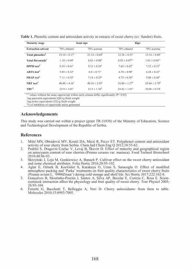
168
Table 1. Phenolic content and antioxidant activity in extracts of sweet cherry (cv. Sandor) fruits.
Maturity stage Semi ripe Ripe
Extraction solvent 70% ethanol 70% acetone 70% ethanol 70% acetone
Total phenolics1 19.10 ± 0.17a 21.14 ± 0.64b 12.36 ± 0.51c 13.14 ± 0.40c
Total flavonoids1 1.18 ± 0.09a 0.83 ± 0.08b 0.92 ± 0.07b,c 1.03 ± 0.04a,c
DPPH test2 9.10 ± 0.61a 9.12 ± 0.34a 7.65 ± 0.42b 7.52 ± 0.33b
ABTS test2 5.85 ± 0.32a 6.4 ± 0.71a 6.70 ± 0.90a 6.24 ± 0.22a
FRAP test2 7.11 ± 0.52a 7.14 ± 0.25a 4.73 ± 0.36b 5.00 ± 0.44b
NBT test3 40.40 ± 4.16a 40.18 ± 2.95a 35.80 ± 1.57b 29.44 ± 5.70b
TRC2 23.9 ± 1.01a 31.5 ± 1.34b 24.42 ± 1.01a 36.06 ± 0.74c
a-c values without the same superscript within each column differ significantly (P< 0.05)1mg quercetin equivalents (QE)/g fresh weight2mg trolox equivalents (TE)/g fresh weight3% of inhibition of superoxide anion generated
Acknowledgements
This study was carried out within a project (grant TR-31038) of the Ministry of Education, Science and Technological Development of the Republic of Serbia.
References1. ��������%�18� �����3%���&�����+%��������%�.����G*��.��$'������������� � � ����H� ���
activity of sour cherry from Serbia. Chem Ind Chem Eng Q 2012;18:53-62.2. .� ������%��� ����-Uzelac V, Levaj B, Škevin D. Effect of maturity and geographical region
on antocyanin content of sour cherries (Prunus cerasus var. marasca). Food Technol Biotechnol 2010;48:86-93.
3. ���N$l&����%�5�� ��%�R�����7��N�+%�� � �'�.��,#��� �������������'��&7�����'���$� ����H� ���and some chemical attributes. Folia Hortic 2016;28:95-102.
4. Aglar E, Ozturk B, KocGuler S, Karakaya O, Uzun S, Saracoglu O. Effect of modified atmosphere packing and ‘Parka’ treatments on fruit quality characteristics of sweet cherry fruits (Prunus avium L. ‘0900Ziraat’) during cold storage and shelf life. Sci Hortic 2017;222:162-8.
5. Gonçalves B, Moutinho-Pereira J, Santos A, Silva AP, Becelar E, Correia C, Rosa E. Scion-rootstock interaction affect the physiology and fruit quality of sweet cherry. Tree Physiol 2005; 26:93-104.
6. Ferretti G, Bacchetti T, Belleggia A, Neri D. Cherry antioxidants: from farm to table. Molecules 2010;15:6993-7005.
169
Natural vs. commercial products: comparison of antioxidant activity
6�����8����*, Milena Rašeta, Marija Lesjak, Neda Mimica-Duk�
Department of Chemistry, Biochemistry and Environmental Protection, Faculty ofSciences, University of Novi Sad, Novi Sad, Serbia
*e-mail: [email protected]
Oxidative stress is considered to be the main catalyst ally in the development of manychronic diseases, including cardiovascular and neurodegenerative disorders, diabetes, cancer and the process of aging. Maintaining a high antioxidant capacity in the organism is essential in preventing or reversing the deleterious effects of oxidative stress. This can be achieved by a balanced diet which includes regular consumption of fruits, vegetables and other plant derived food that contain a great variety of beneficial phytochemicals, where the antioxidant activity is mostly attributed to phenolic compounds 1,2. Since there is an ongoing debate on whether or not there is a difference in the biological activity and manifested health benefits of natural and commercial beverages, this study was conducted to compare the antioxidant potential of various plants and their corresponding products in the form of dietary supplements. For this purpose oregano, artichoke, spinach and kohlrabi were chosen for comparison with commercially available oregano essential oil, artichoke tincture, Ferolin C drink and Zn-Se tablets. Ferolin C and Zn-Se tablets were compared with both spinach and kohlrabi that are a good source of these micronutrients. The antioxidant assays included scavenging of the diphenylpicrylhydrazyl radical (DPPH•), inhibition of lipid peroxidation (LP) and Ferric Reducing Antioxidant Power assay (FRAP)3,4. While oregano expressed better results than the commercial product in all experiments, opposite results were obtained for artichoke. Tablets of Zn-Se only showed activity in the FRAP assay, while Ferolin C yielded better results in the scavenging of DPPH• compared to both spinach and kohlrabi, while it was not able to terminate LP. Oregano was the most potent sample in all assays followed by artichoke tincture. Although a clear distinction between natural and commercial beverages was not visible, in this study the results for plant based samples prevailed over those of commercial products concerning the antioxidant activity.
Acknowledgements
This study was supported by the Ministry of Education, Science and Technological Development of the Republic of Serbia (Grant No. 172058)

170
References
1. Dani C, Oliboni L. S, Agostini F, Funchal C, Serafini L, Henriques J. A, Salvador M. Phenolic content of grapevine leaves (Vitis labrusca var . Bordo) and its neuroprotective effect against peroxide damage.Toxicol In Vitro 2010;24:148–53.
2. Della Valle E, Cacciatore F, Farinaro E, Salvatore F, Marcantonio R, Stranges S, Trevisan M. The Mediterranean Diet in the Prevention of Degenerative Chronic Diseases. In: Shiomi N, Waisundara V (eds) ,Superfood and Functional Food, IntechOpen, London, 2017, pp 115–32.
3. 5�&� ���%��� � �"%�1�2����%�+� 2���R%�� �����%��� ����������%������ -�#��������#����#&�sibirica Burgsdorf. as a novel source of antioxidant and anti-inflammatory agents. Food Chem 2011;124:850–6.
4. 1�2��� �%� ����� -�#��� �%� �� ��������� �%� .������� �% Jovin E. Antioxidant activity relationship of phenolic compounds in Hypericum perforatum L. Chem Cent J 2011;5:34.
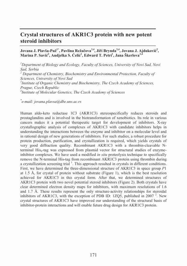
171
Crystal structures of AKR1C3 protein with new potent steroid inhibitors
Jovana J. Plavša-Puž1*�8�����#�0�/���3,4��AB+�����3,4��������$) ��!���2,�����8$����2�)�� ��!��$C���1, Edward T. Petri1, Jana Škerlova3,4
1Department of Biology and Ecology, Faculty of Sciences, University of Novi Sad, NoviSad, Serbia2 Department of Chemistry, Biochemistry and Environmental Protection, Faculty ofSciences, University of Novi Sad3Institute of Organic Chemistry and Biochemistry, The Czech Academy of Sciences,Prague, Czech Republic4Institute of Molecular Genetics, The Czech Academy of Sciences
*e-mail: [email protected]
Human aldo-keto reductase 1C3 (AKR1C3) stereospecifically reduces steroids and prostaglandins and is involved in the biotransformation of xenobiotics. Its role in various cancers makes it a potential therapeutic target for development of inhibitors. X-ray crystallographic analysis of complexes of AKR1C3 with candidate inhibitors helps in understanding the interactions between the enzyme and inhibitor on a molecular level and in rational design of new generations of inhibitors. For such studies, a robust procedure for protein production, purification, and crystallization is required, which yields crystals of very good diffraction quality. Recombinant AKR1C3 with a thrombin-cleavable N-terminal His6-tag was expressed from plasmid vector for structural studies of enzyme-inhibitor complexes. We have used a modified in situ proteolysis technique to specifically remove the N-terminal His-tag from recombinant AKR1C3 protein using thrombin during a crystallization screening trial 1. This approach resulted in crystals in different conditions. First, we have determined the three-dimensional structure of AKR1C3 in space group P1at 1.5 Å, for crystal of protein without substrate (Figure 1), which is the best resolution achieved for AKR1C3 in this crystal form. After that, we determined structures of AKR1C3 protein with two novel potential steroid inhibitors (Figure 2). Both crystals have clear determined electron density maps for inhibitors, with maximum resolutions of 1.6 and 1.7 Å. These results represent the only structure-activity relationships for steroidal inhibitors of AKR1C3, with the exception of PDB ID: 1ZQ5, published in 2007 2. New crystal structures of AKR1C3 have improved our understanding of the structural basis of inhibitor-protein interactions and will enable future drug design for AKR1C3 protein.
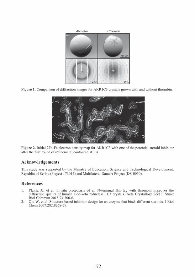
172
Figure 1. Comparison of diffraction images for AKR1C3 crystals grown with and without thrombin.
Figure 2. Initial 2Fo-Fc electron density map for AKR1C3 with one of the potential steroid inhibitor after the first round of refinement, �����#�� � ��9�z�
Acknowledgements
This study was supported by the Ministry of Education, Science and Technological Development, Republic of Serbia (Project 173014) and Multilateral Danube Project (DS-0050).
References
1. Plavša JJ, et al. In situ proteolzsis of an N-terminal His tag with thrombin improves the diffraction qualitz of human aldo-keto reductase 1C3 crystals. Acta Crystallogr Sect F Struct Biol Commun 2018:74:300-6.
2. Qiu W, et al. Structure-based inhibitor design for an enzyme that binds different steroids. J Biol Chem 2007:282:8368-79.
173
Antitumor interaction and safety of metformin and itraconazole low doses in hamster fibrosarcoma
6�"���$8�&���1�������$8�&���2�6�"��(���"���1�6� ���� !���1, Ivan 7�&�1�������$8�&���3*
1Department of Histology and Embryology, Faculty of Medicine, University of Novi Sad,Novi Sad, Serbia2Department of Pharmacy, Faculty of Medicine, University of Novi Sad3Department of Pharmacology, Toxicology and Clinical Pharmacology, Faculty ofMedicine, University of Novi Sad
*e-mail: [email protected]
Metformin exhibited anticancer effects in vitro in number of various cancer cell lines. The anticancer effects of metformin were explained by different mechanisms 1. The antifolate activity may be very important for metformin antitumor activity 1. In our previous experiment, we found anticancer effects of metformin on BHK-21/C13 fibrosarcoma in hamsters 1. In diabetic patients, metformin is usually comedicated with the most common and safest antimicotic agent itraconazole, since fungal infections are frequent in diabetes. This combination is nontoxic in humans. Itraconazole possesses anticancer properties, especially antiangiogenesis (as a multitarget angiogenesis inhibitor) 2,3, and antifolate properties 4, like metformin. Multiple common activities, such as antifolate activity 3, or diverse mechanisms of anticancer action of metformin 1, and itraconazole 2,3, were identified. The finding that nontoxic itraconazole, as nontoxic metformin, exhibits antifolate activity, like toxic chemotherapeutic methotrexate, was important for our research motivation.Syrian golden hamsters of both sexes (n = 24), weighing approximately 100 g, were randomly allocated to 4 equally sized groups. 106 BHK-21/C13 cells in 1 ml were injected subcutaneously into the animals’ back. The first group started peroral treatment with physiological solution, the second with metformin 250 mg/kg daily, the third with itraconazole 125 mg/kg daily and the fourth with a combination of metformin 250 mg/kg and itraconazole 125 mg/kg daily, via a gastric probe 3 days before tumor inoculation. After 3 weeks, when the tumors were larger than 2 cm in the first group, all animals were sacrificed. The blood was collected for glucose and other analyses. The tumors were excised and weighed and their diameters were measured. The tumor samples were pathohistologically (HE) and immunohistochemically (Ki-67, CD 34, COX IV, GLUT-1, iNOS) assessed (Figure 1) and the main organs toxicologically analyzed. Tumor volume was determined using the water displacement method. Ki-67-positive cells in the tumor samples were quantified. Images were taken and processed by software UTHSCSA Image
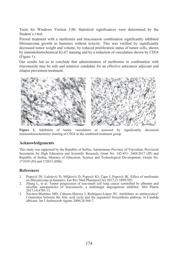
174
Tools for Windows Version 3.00. Statistical significances were determined by the Student´s t-test. Peroral treatment with a metformin and itraconazole combination significantly inhibited fibrosarcoma growth in hamsters without toxicity. This was verified by significantly decreased tumor weight and volume, by reduced proliferation status of tumor cells, shown by immunohistochemical Ki-67 staining and by a reduction of vasculature shown by CD34 (Figure 1).Our results led us to conclude that administration of metformin in combination withitraconazole may be safe and nontoxic candidate for an effective anticancer adjuvant and relapse prevention treatment.
Figure 1. Inhibition of tumor vasculature as assessed by significantly decreased immunohistochemistry staining of CD34 in the combined treatment group
Acknowledgements
This study was supported by the Republic of Serbia, Autonomous Province of Vojvodina, Provincial Secretariat for High Education and Scientific Research, Grant No. 142-451- 2469/2017 (JP) and Republic of Serbia, Ministry of Education, Science and Technological Development, Grants No. 171039 (JS) and 172013 (DM).
References
1. .�������%�5 ��������%�����������%�.�������%�� ��"%�.���������G�������������������on fibrosarcoma in hamsters. Eur Rev Med Pharmacol Sci 2017;21:5499-505.
2. Zhang L, et al. Tumor progression of non-small cell lung cancer controlled by albumin and micellar nanoparticles of itraconazole, a multitarget angiogenesis inhibitor. Mol Pharm 2017;14:4705-13.
3. Navarro-Martínez MD, Cabezas-Herrera J, Rodríguez-López JN: Antifolates as antimycotics? Connection between the folic acid cycle and the ergosterol biosynthesis pathway in Candida albicans. Int J Antimicrob Agents 2006;28:560-7.
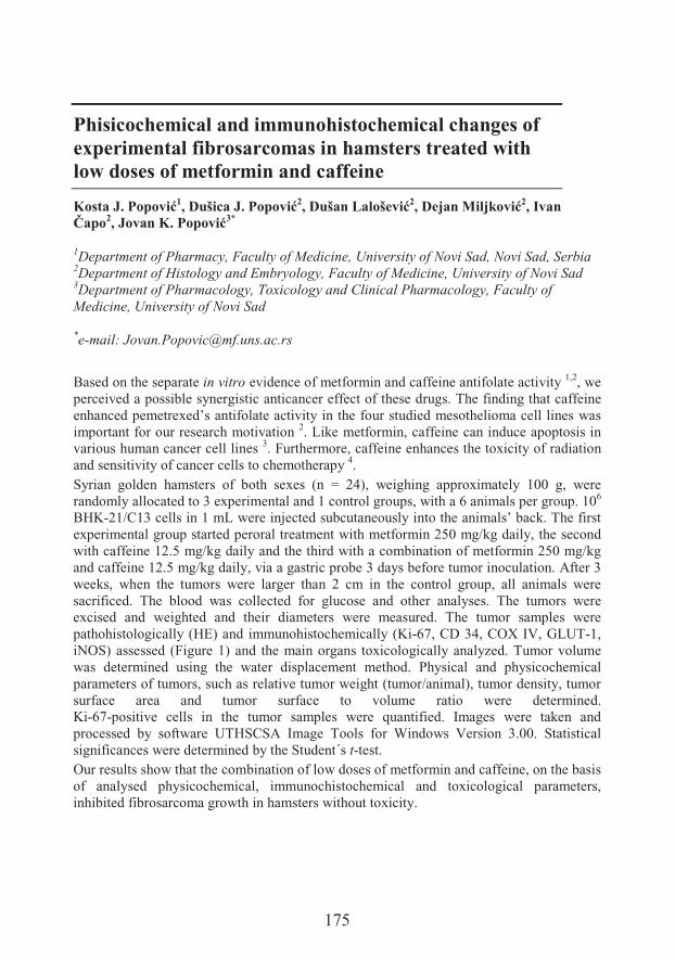
175
Phisicochemical and immunohistochemical changes of experimental fibrosarcomas in hamsters treated with low doses of metformin and caffeine
������$8�&���1�6�"���$8�&���2�6�"��(���"���2�6� ���� !���2, Ivan 7�&�2�������$8�&���3*
1Department of Pharmacy, Faculty of Medicine, University of Novi Sad, Novi Sad, Serbia2Department of Histology and Embryology, Faculty of Medicine, University of Novi Sad3Department of Pharmacology, Toxicology and Clinical Pharmacology, Faculty ofMedicine, University of Novi Sad
*e-mail: [email protected]
Based on the separate in vitro evidence of metformin and caffeine antifolate activity 1,2, we perceived a possible synergistic anticancer effect of these drugs. The finding that caffeine enhanced pemetrexed’s antifolate activity in the four studied mesothelioma cell lines was important for our research motivation 2. Like metformin, caffeine can induce apoptosis in various human cancer cell lines 3. Furthermore, caffeine enhances the toxicity of radiation and sensitivity of cancer cells to chemotherapy 4.Syrian golden hamsters of both sexes (n = 24), weighing approximately 100 g, were randomly allocated to 3 experimental and 1 control groups, with a 6 animals per group. 106
BHK-21/C13 cells in 1 mL were injected subcutaneously into the animals’ back. The first experimental group started peroral treatment with metformin 250 mg/kg daily, the second with caffeine 12.5 mg/kg daily and the third with a combination of metformin 250 mg/kg and caffeine 12.5 mg/kg daily, via a gastric probe 3 days before tumor inoculation. After 3 weeks, when the tumors were larger than 2 cm in the control group, all animals were sacrificed. The blood was collected for glucose and other analyses. The tumors were excised and weighted and their diameters were measured. The tumor samples were pathohistologically (HE) and immunohistochemically (Ki-67, CD 34, COX IV, GLUT-1,iNOS) assessed (Figure 1) and the main organs toxicologically analyzed. Tumor volume was determined using the water displacement method. Physical and physicochemical parameters of tumors, such as relative tumor weight (tumor/animal), tumor density, tumorsurface area and tumor surface to volume ratio were determined.Ki-67-positive cells in the tumor samples were quantified. Images were taken and processed by software UTHSCSA Image Tools for Windows Version 3.00. Statistical significances were determined by the Student´s t-test. Our results show that the combination of low doses of metformin and caffeine, on the basis of analysed physicochemical, immunochistochemical and toxicological parameters, inhibited fibrosarcoma growth in hamsters without toxicity.
176
We concluded that the low doses administration of metformin with caffeine might be an effective and safe approach in novel nontoxic adjuvant and relapse prevention anticancer treatment.
Figure 1. Inhibition of tummor growth as assessed by significantly decreased immunohistochemistry staining of Ki-67 in the combined treatment group.
Acknowledgements
This study was supported by the: Republic of Serbia, Autonomous Province of Vojvodina, Provincial Secretariat for High Education and Scientific Research, Grant No. 142-451- 2469/2017 (JP) and Republic of Serbia, Ministry of Education, Science and Technological Development, Grants No. 171039 (BM) and 172013 (SD)
References
1. Corominas-Faja B, et al. Metabolomic fingerprint reveals that metformin impairs one-carbon metabolism in a manner similar to the antifolate class of chemotherapy drugs. Aging 2012;4:480–98.
2. Min SH, Goldman ID, Zhao R. Caffeine markedly sensitizes human mesothelioma cell lines to pemetrexed. Cancer Chemother Pharmacol 2008;61:819-27.
3. HaQ &��%���� ���, ��������� #��&��$��&����� ���' ���&� � ������ � �'����O9Xgg�����&��,����G#����Biol 2014;9:727–38.
4. Saiki S, et al. Caffeine induces apoptosis by enhancement of autophagy via PI3K/Akt/mTOR/p70S6K inhibition. Autophagy 2011;7:176-87.
177
Production, purification and structural characterisation of recombinant BanLec-Bet v 1
4�����8����-#���1*�����8�&���1�D��")�3��!���2���� �*������-���!�����1
1Department of Biochemistry, Faculty of Chemistry, University of Belgrade, Belgrade,Serbia2Institue of Chemistry, Technology and Metallurgy, University of Belgrade
*e-mail: [email protected]
The sublingual route of allergens administration in allergen-specific immunotherapy (ASIT) is proven to be a successful way to treat patients with respiratory allergy. The trend of replacing natural extracts with purified recombinant allergens is growing. Although the purified allergens themselves are not good immunogens, the combined vector systems and adjuvans can improve their immunogenicity 1. Cell surfaces are decorated by different glycan structures, so the lectins specific for these glycans can be used to deliver particular therapeutic to target specific tissue 2. Banana lectin (BanLec) is mannose-specific protein which belongs to the subfamily of Jacalin-related lectins 3. Apart from its characteristic to bind glycans, BanLec also modulates immune cells in vitro 4. On the other hand, Bet v 1 (Betula verrucosa) is the major birch pollen allergen. T-cell epitops are distributed over almost entire protein structure 5.In the study the recombinant BanLec-Bet v 1 construct is designed, produced by the recombinant DNA technology, purified and characterized by classical biochemical methods for the application in the ASIT of birch pollen allergy. The expression of newly designed BanLec-Bet v 1 was performed in E. coli BL21 (DE3). After expression the protein was found in the inclusion bodies from which it was extracted with 4 M urea solution. After renaturation, affinity chromatography (Sephadex G-75superfine) was used for protein purification. Biochemical characterization of the chimera was performed by: SDS PAGE electrophoreses, CD spectroscopy and mass spectrometry. Biological activity of the construct was confirmed by binding of BanLecBet v 1 to ahorseradish peroxidase glycoprotein in ELISA. Purified BanLec-Bet v 1 showed molecular mass of 32 kDa. CD spectra of the recombinant construct revealed well defined secondary structures with predominant beta sheets (41.2%). By mass spectrometry 51.8% of the BanLec-Bet v 1 primary structure was confirmed.Biologicaly active recombinant BanLec-Bet v 1 was produced by the recombinant DNA technology. Further in vitro and in vivo studies will evaluate immunomodulatory potential of BanLec-Bet v 1 for application in ASIT.
178
Acknowledgements
This study was supported by the Ministry of Education, Science and Technological Development (Grant No. 172049)
References
1. Tourdot S, et. al. Efficacy of sublingual vectorized recombinant bet v 1a in a mouse model of birch pollen allergic asthma. Vaccine 2013;31:2628-37.
2. Dimitrijevic R, Jadranin M, Burazer L, Ostojic S, Gavrovic-Jankulovic M. Evaluation of the thermal stability and digestibility of heterologously produced banana lectin. Food Chem 2010;120:1113-8.
3. Dimitrijevic R, Stojanovic M, Micic M, Dimitrijevic Lj, Gavrovic-Jankulovic M. Recombinant banana lectin as mucosal immunostimulator. J Funct Food 2012;4: 636-41.
4. Sansone A, Sansone M, Dias C, Nascimento J. Oral administration of banana lectin modulates cytokine profile and abundance of T-cell populations in mice. Int J Biol Macromol 2016;89:19-24.
5. Gajhede M, et al. X-Ray and NMR structure of Bet v 1, the origin of birch pollen allergy. NatStruct Biol 1996;3:1040-5.
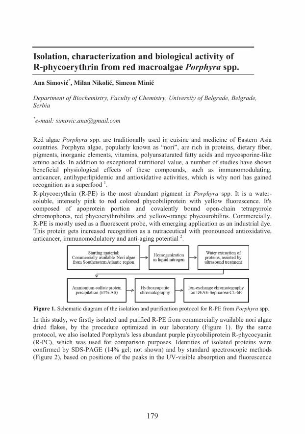
179
Isolation, characterization and biological activity ofR-phycoerythrin from red macroalgae Porphyra spp.
)�������*�����-!������������
Department of Biochemistry, Faculty of Chemistry, University of Belgrade, Belgrade,Serbia
*e-mail: [email protected]
Red algae Porphyra spp. are traditionally used in cuisine and medicine of Eastern Asia countries. Porphyra algae, popularly known as “nori”, are rich in proteins, dietary fiber, pigments, inorganic elements, vitamins, polyunsaturated fatty acids and mycosporine-like amino acids. In addition to exceptional nutritional value, a number of studies have shown beneficial physiological effects of these compounds, such as immunomodulating, anticancer, antihyperlipidemic and antioxidative activities, which is why nori has gained recognition as a superfood 1.R-phycoerythrin (R-PE) is the most abundant pigment in Porphyra spp. It is a water-soluble, intensely pink to red colored phycobiliprotein with yellow fluorescence. It's composed of apoprotein portion and covalently bound open-chain tetrapyrrole chromophores, red phycoerythrobilins and yellow-orange phycourobilins. Commercially, R-PE is mostly used as a fluorescent probe, with emerging application as an industrial dye. This protein gets increased recognition as a nutraceutical with pronounced antioxidative, anticancer, immunomodulatory and anti-aging potential 2.
Figure 1. Schematic diagram of the isolation and purification protocol for R-PE from Porphyra spp.
In this study, we firstly isolated and purified R-PE from commercially available nori algae dried flakes, by the procedure optimized in our laboratory (Figure 1). By the same protocol, we also isolated Porphyra's less abundant purple phycobiliprotein R-phycocyanin (R-PC), which was used for comparison purposes. Identities of isolated proteins were confirmed by SDS-PAGE (14% gel; not shown) and by standard spectroscopic methods (Figure 2), based on positions of the peaks in the UV-visible absorption and fluorescence
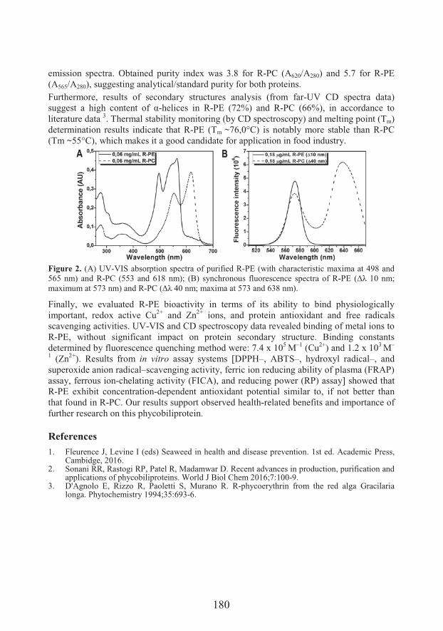
180
emission spectra. Obtained purity index was 3.8 for R-PC (A620/A280) and 5.7 for R-PE (A565/A280), suggesting analytical/standard purity for both proteins.Furthermore, results of secondary structures analysis (from far-UV CD spectra data) suggest a high content of ;-helices in R-PE (72%) and R-PC (66%), in accordance to literature data 3. Thermal stability monitoring (by CD spectroscopy) and melting point (Tm)determination results indicate that R-PE (Tm ~76,0°C) is notably more stable than R-PC (Tm ~55°C), which makes it a good candidate for application in food industry.
Figure 2. (A) UV-VIS absorption spectra of purified R-PE (with characteristic maxima at 498 and 565 nm) and R-PC (553 and 618 nm); (B) synchronous fluorescence spectra of R-.G� FE{�9Y���A�maximum at 573 nm) and R-.,�FE{�?Y���A�� H�� � ��B<:� � �C:f���@�
Finally, we evaluated R-PE bioactivity in terms of its ability to bind physiologically important, redox active Cu2+ and Zn2+ ions, and protein antioxidant and free radicals scavenging activities. UV-VIS and CD spectroscopy data revealed binding of metal ions to R-PE, without significant impact on protein secondary structure. Binding constants determined by fluorescence quenching method were: 7.4 x 10⁵ M–1 (Cu2+) and 1.2 x 10³ M–
1 (Zn2+). Results from in vitro assay systems [DPPH–, ABTS–, hydroxyl radical–, and superoxide anion radical–scavenging activity, ferric ion reducing ability of plasma (FRAP) assay, ferrous ion-chelating activity (FICA), and reducing power (RP) assay] showed that R-PE exhibit concentration-dependent antioxidant potential similar to, if not better than that found in R-PC. Our results support observed health-related benefits and importance of further research on this phycobiliprotein.
References1. Fleurence J, Levine I (eds) Seaweed in health and disease prevention. 1st ed. Academic Press,
Cambidge, 2016.2. Sonani RR, Rastogi RP, Patel R, Madamwar D. Recent advances in production, purification and
applications of phycobiliproteins. World J Biol Chem 2016;7:100-9.3. D'Agnolo E, Rizzo R, Paoletti S, Murano R. R-phycoerythrin from the red alga Gracilaria
longa. Phytochemistry 1994;35:693-6.
181
Cadmium as the main endocrine disrupter in papillary thyroid carcinoma
)��!��������� ���� ���1*�4����)������1�������+��!���-���2, Branislav #��/���3�'��������� ���3�4���8������3�+� �����4���� �*������-���!�����5�6��,������ ����1,6
1Department of Analytical Chemistry, Faculty of Chemistry, University of Belgrade,Belgrade, Serbia2Deparment of Physiology, Institute for Biological Research “Siniša Sta��� �<=University of Belgrade3Center for Endocrine Surgery, Clinical Center of Serbia, Belgrade, Serbia4 Faculty of Biology, University of Belgrade5Deparment of Biochemistry, Faculty of Chemistry, University of Belgrade6Department of Ecology and Chemical Technology, South Ural State University,Chelyabinsk, Russia
*e-mail: [email protected]
Thyroid cancer is the most frequent endocrine neoplasm and its prevalence has increased in many countries worldwide 1-4, including Serbia. It has long been hypothesis that the toxic metals, as endocrine disrupters, could play a major role in the thyroid cancer etiology. The aim of this investigation was to examine physiologically important toxic and essential trace metals in the malignant tissue samples of patients with papillary thyroid carcinoma (n = 60). Intra-variability was achieved by using the healthy tissues of the same patients as the control group, making the total sample size of 120 specimens. All samples were collected after thyroidectomy and the metal concentrations were determined by ICP-Q-MS after tissue’s decomposition with nitric acid and hydrogen-peroxide in the microwave digestion system. It was found that the level of cadmium (Cd) was significantly higher in the malignant tissues when compared to the control tissues (91.87 ± 91.43 vs.63.51 ± 54.49 ng/g; p < 0.05 by Mann-Whitney U test). Contrary, the level of selenium (Se) was significantly lower in the malignant tissues when compared to the control tissues (80.33 ± 54.01 vs. 142.11 ± 81.55 ng/g; p < 0.05). Significantly lower level of selenium could be explained by higher level of Cd and strong negative (antagonistic) correlation obtained by Spearmen’s Rho test between these elements (r = -0.75; p < 0.05). It is interesting to point out that the ratio Cd/Se separated two examined groups over 55%. According to demographic, clinical and pathophysiological data, we have noticed different metallomic pattern. Cadmium was the only metal that separated age groups (< 50 years vs.> 50 years) and smokers from the non-smokers by its higher values in the older and the smokers group (p < 0.05). The level of Cd in women and men was practically the same. On the other hand, cadmium had no effect on the clinical course of the disease. Instead of Cd,
182
copper and zinc significantly separated two groups according to size of the tumor (< 3 cm vs. > 3 cm) and the invasiveness of thyroid capsule. One-way ANOVA test was applied to separate patients according to the TNM classification. It was found that manganese (Mn) differs between T1 and T2 stage, as well as T1 and T3 stage (p < 0.05), while Cd strongly separated groups between T1 and T2 stage (p < 0.05). None of the patients was in T4 stage of disease. Manganese and cadmium also separated Nx from the No group (p < 0.05). Distant metastases were not diagnosed (Mx). Our results indicate that Cd has a role in the etiology of papillary thyroid carcinoma. Obtained higher concentrations of cadmium in the malignant tissues and the strong antagonistic interaction with selenium could explain the expulsion of essential selenium by toxic cadmium and, consequently, a significant drop in selenium levels in the malignant tissues, which is the third most common element in the healthy thyroid gland. Significantly higher ratio of Cd/Se obtained in the malignant tissues could be used in the diagnosis of papillary carcinoma as the tumor marker.
Acknowledgements
This study was supported by the Ministry of Education, Science and Technological Development ofSerbia, Grant No. 173020.
References1. Pacini F, Castagna G, Brilli L, Pentheroudakis. Thyroid cancer: EMSO clinical practice
guidelines for diagnosis, treatment and follow-up. Ann Oncol 2012;23:110-9.2. Grogan H, Mitmaker E, Clarck O. The evolution of biomarkers in thyroid cancer-from mass
screening to a personalized biosignature. Cancers 2010;2:885-912.3. Malandrino P, et al. Desctiptive epidemiology of human thyroid cancer: experience from a
regional registry and the “volcanic factor”. Front Endocrinol 2013;4:1-7.4. Chen A, Kim S, Chung E, Dietrich K. Thyroid hormones in relation to lead, mercury and
cadmium exposure in the national health and nutrition examination survey. Environ Health Persp 2013;121:181-6.
183
Synthesis and antiproliferative activities of 16-triazolyl-methyl-17-estradiol derivatives
Seyyed Ashkan Senobar Tahaei1, Anita Kiss2, Zsanett Benke2, Erzsébet Mernyák2,Gyula Schneider2, István Zupkó1*
1Department of Pharmacodynamics and Biopharmacy, University of Szeged, Szeged,Hungary2Department of Organic Chemistry, University of Szeged
*e-mail: [email protected]
Besides their crucial physiologic ���#������&�����'������ #�����&$&����9<>-estradiol may increase the proliferation of gynecological cancers. Our previous investigations indicate that modified estranes bearing triazolyl substitution in D-ring exert potent antiproliferative action on cancer cells in vitro 1,2. Based on our previous results a set of 16-triazolyl-methyl-17-estradiol derivatives have been synthesized and tested for antiproliferative action. We have previously reported the synthesis and determination of the configuration of the four possible isomers of 3-methoxy- and 3-benzyloxy-16-hydroxy-methylestra-17-ols 3.The isomers were converted into their 16-p-tosyloxy-methyl derivatives, and the succeeding azide exchange reaction led to the corresponding 16-azido-methyl 17-hydroxy estranes. The regioselective Cu(I)-catalyzed 1,3-dipolar cycloaddition of these isomers with different terminal alkines afforded novel 1’,4’-substituted -16-triazolyl-methyl-17-hydroxy hybrid molecules.In these molecules on the 16-position a methylene linker with different orientation connect the 1’,4’-substituted triazolyl ring with the sterane skeleton.The antiproliferative actions of the analogs were determined by means of MTT assay against a gynecological panel containing MCF7, MDA-MB-231 breast and Hela, SiHa cervical cancer cell lines in order to describe strucure-activity relationships.
Acknowledgements
Financial supports from the Economic Development and Innovation Operative Program (GINOP-2.3.2-15-2016-00012) and National Research, Development and Innovation Office (NKFIH; K109293) are gratefully acknowledged.
184
References
1. Mernyák E, et al. Synthesis of trans-16-triazolyl-9:;-methyl-17-estradiol diastereomers and the effects of structural modifications on their in vitro antiproliferative activities. J Steroid Biochem Mol Biol 2015;134:123-34.
2. Bózsity N, et al. Mechanism of antiproliferative action of a new d-secoestrone-triazole derivative in cervical cancer cells and its effect on cancer cell motility. J Steroid Biochem Mol Biol 2017;165:247-57.
3. Tapolcsányi P, et al. Synthesis and receptor-binding examination of 16-hydroxymethyl-3,17-estradiol stereoisomers. Steroids 2002;67:671-8.
185
The effects of fructose-rich diet and/or chronic unpredictable stress on antioxidant enzymes function in the rat kidney
6����:���1����"9&�/�2*�6�� ���'� ������������1�)��!������-!���-��!�3�4�������!���1�*����������1, Jelena Nestorov1
1Department of Biochemistry, Institute for Biological Research “Siniša ������ �<=University of Belgrade, Belgrade, Serbia2 Life Sciences Department, Institute for Multidisciplinary Research, University ofBelgrade3Department of Physiology, Institute for Biological Research “Siniša ������ �<=University of Belgrade
*e-mail: [email protected]
Modern lifestyle is characterized by unhealthy high-calorie diet, lack of physical activity and daily exposure to various stressors. Since the introduction of high-fructose corn syrup in 1970s, fructose daily intake has raised rapidly. This was parallel with an increasing prevalence of metabolic disorders, suggesting fructose overconsumption, along with the chronic exposure to stress, as factors contributing to development and progression of metabolic disorders including obesity, insulin resistance, nonalcoholic fatty liver disease, cardiovascular diseases, disturbed kidney function, etc. Also, it was suggested that oxidative stress participates in the development and progression of these disturbances. Both, chronic stress and fructose overconsumption can induce oxidative stress in various tissues. Nevertheless, a relation between fructose consumption and oxidative stress appear to be rather complex, since fructose was shown to produce both pro- and anti-oxidative effects, depending on the dose, duration of consumption and (patho)physiological milieu. Fructose is mostly metabolized in the liver, but a significant part of ingested sugar can be metabolized in other organs, especially kidneys. The aim of this study was to investigate the effects of fructose, chronic unpredictable stress and their combination on the function of antioxidant enzymes in the kidney. Our hypothesis was that the combination of these factors induces more pronounced alterations in the renal redox setting, in comparison to separate treatments.To test this hypothesis, adult male Wistar rats were randomly divided in four experimental groups: control group (C) – rats fed with commercial standard chow and drinking water, fructose group (F) – rats kept at 9-week long diet with ad libitum access to standard laboratory chow and 20% fructose solution instead of drinking water, Stress group (S) –rats subjected to 4-week long protocol of chronic unpredictable stress, and Stress + Fructose groups (SF) – rats subjected to both stress and fructose diet. The stress protocol
186
included: forced swimming in cold water for 10 min, physical restraint (1 h), cold room (4°C, 1 h), wet bedding (4h), switching cages (overnight), rocking cages (2 h), cage tilt (45°, overnight). The time and type of daily stressors was randomly selected at the beginning of the treatment. The effects of fructose-enriched diet and unpredictable stress on physiological and biochemical parameters, as well as on renal expression of fructose transporter Glut5, fructolytic enzymes (ketohexokinase and aldolase B), and function of antioxidant enzymes (superoxide dismutase, catalase, glutathione peroxidase, and glutathione reductase), were evaluated.A decrease in food intake, increase in liquid intake, and triglyceridemia were observed in all fructose-fed rats, regardless of stress exposure. Fructose induced the expression of Glut5, ketohexokinase and aldolase B in the kidney. Stress however, had no effect on fructose import and catabolism. Exposure to stress increased only catalase activity, with no effect on other antioxidant enzymes. At the same time the activity and expression of antioxidant enzymes in the kidney of all fructose-fed rats remained unaltered.In conclusion, fructose is imported and metabolized in the kidney, and exposure to stress had no effect on renal fructose metabolism. Chronic stress, fructose overconsumption and their combination have not altered renal antioxidant enzymes function. The only exception was catalase, whose activity was increased only by chronic unpredictable stress in rats subjected to standard dietary regime. This effect of stress was not detected in fructose-fed stressed rats, which might be attributed to antioxidative properties of fructose. These results disputed the initial hypothesis, and raised new questions regarding the relation of fructose and chronic unpredictable stress, as well as possibility of their tissue-specific effects on redox settings.
Acknowledgements
This study was supported by the Ministry of Education, Science and Technological Development of the Republic of Serbia, Grant III41009.
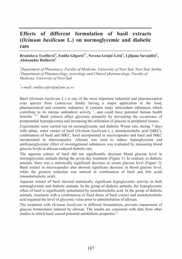
187
Effects of different formulation of basil extracts (Ocimum basilicum L.) on normoglycemic and diabetic rats
+��������:�������1�����*�,���1*�-�����*�� �-(���1�( � ������� �0�1,)��!������#�"!���2
1Department of Pharamacy, Faculty of Medicine, University of Novi Sad, Novi Sad, Serbia2Department of Pharmacology, toxicology and Clinical pharmacology, Faculty ofMedicine, University of Novi Sad
*e-mail: [email protected]
Basil (Ocimum basilicum L.) is one of the most important industrial and pharmaceutical crop species from Lamiaceae family having a major application in the food, pharmaceutical and cosmetic industries. It contains many antioxidant substances which contribute to its intense antiradical activity 1, and could have potential human health benefits 2,3. Basil extracts affect glycemia primarily by preventing the occurrence of postprandial hyperglycemia and increasing the utilization of glucose in peripheral tissues. Experiments were carried out on normoglycemic and diabetic Wistar rats, during 7 days with saline, water extract of basil (Ocimum basilicum L.), monoketocholic acid (MKC), combination of basil and MKC, basil incorporated in microcapsules and basil and MKC incorporated in microcapsules. Alloxan was used to induce hyperglycemia and antihyperglycemic effect of investigational substances was evaluated by measuring blood glucose levels in alloxan-induced diabetic rats. The aqueous extract of basil did not significantly decrease blood glucose level in normoglycemic animals during the seven-day treatment (Figure 1). In contrast, in diabetic animals, there was a statistically significant decrease in serum glucose level (Figure 2). Basil extract in microcapsules also showed significant decrease in blood glucose level, while the greatest reduction was noticed in combination of basil and bile acids (monoketocholic acid). Aqueous extract of basil showed statistically significant hypoglycemic activity in both normoglycemic and diabetic animals. In the group of diabetic animals, the hypoglycemic effect of basil is significantly potentiated by monoketocholic acid. In the group of diabetic animals, treatment with a combination of fixed doses of basil extract and monoketocholic acid regained the level of glycemic value prior to administration of alloxan.The treatment with Ocimum basilicum in different formulations prevents impairment of glucose homeostasis induced by alloxan. The results are consistent with data from other studies in which basil caused potential antidiabetic properties 4.
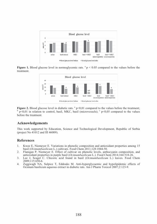
188
Figure 1. Blood glucose level in normoglycemic rats. a p < 0.05 compared to the values before the treatment.
Figure 2. Blood glucose level in diabetic rats. a p<0,01 compared to the values before the treatment; b p<0,01 in relation to control, basil, MKC, basil (microvessels); c p<0,05 compared to the values before the treatment.
Acknowledgements
This work supported by Education, Science and Technological Development, Republic of Serbia (project No 41012 and III 46009).
References1. Kwee E, Niemeyer E. Variations in phenolic composition and antioxidant properties among 15
basil (Ocimumbasilicum L.) cultivars. Food Chem 2011;128:1044-50. 2. Flanigan P, Niemeyer E. Effect of cultivar on phenolic levels, anthocyanin composition, and
antioxidant properties in purple basil (Ocimumbasilicum L.). Food Chem 2014;164:518-26.3. Lee J, Scagel C. Chicoric acid found in basil (Ocimumbasilicum L.) leaves. Food Chem
2009;115:650-6.4. Zeggwagh NA, Sulpice T, Eddouks M. Anti-hyperglycaemic and hypolipidemic effects of
Ocimum basilicum aqueous extract in diabetic rats. Am J Pharm Toxicol 2007;2:123-9.
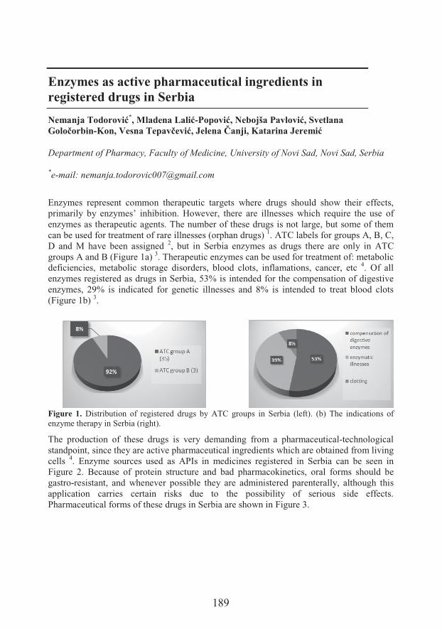
189
Enzymes as active pharmaceutical ingredients in registered drugs in Serbia
Nemanja Todorovi�*��������(���-8�&����-� � "�8���������������*���/�� �-����'����:�&��/����������7�� ��������������
Department of Pharmacy, Faculty of Medicine, University of Novi Sad, Novi Sad, Serbia
*e-mail: [email protected]
Enzymes represent common therapeutic targets where drugs should show their effects, primarily by enzymes’ inhibition. However, there are illnesses which require the use of enzymes as therapeutic agents. The number of these drugs is not large, but some of them can be used for treatment of rare illnesses (orphan drugs) 1. ATC labels for groups A, B, C, D and M have been assigned 2, but in Serbia enzymes as drugs there are only in ATC groups A and B (Figure 1a) 3. Therapeutic enzymes can be used for treatment of: metabolic deficiencies, metabolic storage disorders, blood clots, inflamations, cancer, etc 4. Of all enzymes registered as drugs in Serbia, 53% is intended for the compensation of digestive enzymes, 29% is indicated for genetic illnesses and 8% is intended to treat blood clots(Figure 1b) 3.
Figure 1. Distribution of registered drugs by ATC groups in Serbia (left). (b) The indications of enzyme therapy in Serbia (right).
The production of these drugs is very demanding from a pharmaceutical-technological standpoint, since they are active pharmaceutical ingredients which are obtained from living cells 4. Enzyme sources used as APIs in medicines registered in Serbia can be seen in Figure 2. Because of protein structure and bad pharmacokinetics, oral forms should be gastro-resistant, and whenever possible they are administered parenterally, although this application carries certain risks due to the possibility of serious side effects. Pharmaceutical forms of these drugs in Serbia are shown in Figure 3.
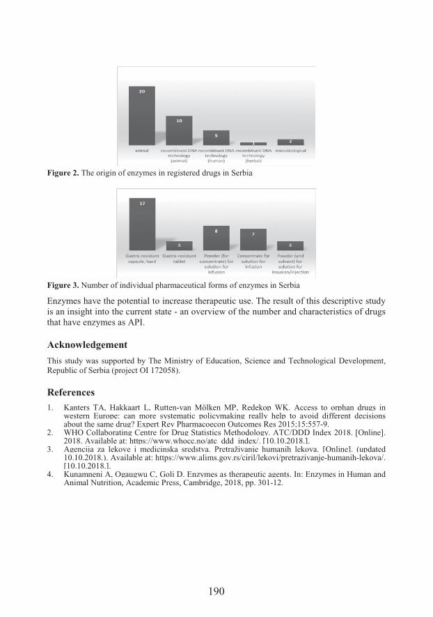
190
Figure 2. The origin of enzymes in registered drugs in Serbia
Figure 3. Number of individual pharmaceutical forms of enzymes in Serbia
Enzymes have the potential to increase therapeutic use. The result of this descriptive study is an insight into the current state - an overview of the number and characteristics of drugs that have enzymes as API.
Acknowledgement
This study was supported by The Ministry of Education, Science and Technological Development, Republic of Serbia (project OI 172058).
References
1. Kanters TA, Hakkaart L, Rutten-van Mölken MP, Redekop WK. Access to orphan drugs in western Europe: can more systematic policymaking really help to avoid different decisions about the same drug? Expert Rev Pharmacoecon Outcomes Res 2015;15:557-9.
2. WHO Collaborating Centre for Drug Statistics Methodology. ATC/DDD Index 2018. [Online]. 2018. Available at: https://www.whocc.no/atc_ddd_index/. [10.10.2018.].
3. Agencija za lekove i medicinska sredstva. Pretraživanje humanih lekova. [Online]. (updated 10.10.2018.). Available at: https://www.alims.gov.rs/ciril/lekovi/pretrazivanje-humanih-lekova/. [10.10.2018.].
4. Kunamneni A, Ogaugwu C, Goli D. Enzymes as therapeutic agents. In: Enzymes in Human and Animal Nutrition, Academic Press, Cambridge, 2018, pp. 301-12.
191
The effect of spermidine supplementation on catalase and Cu, Zn superoxide dismutase gene expression in honey bees
Iva Uzelac1, Tatjana V. Nik���1������($'�!�"����1����0���9�/�1, Željko D. 8�&���1, Ivan Pihler2, 6�� ����� �1�������8����1*
1Department of Biology and Ecology, Faculty of Sciences, University of Novi Sad, NoviSad, Serbia2Faculty of Agriculture, University of Novi Sad
*e-mail: [email protected]
Honey bee (Apis mellifera, L.) is the most important managed pollinator species in the world. During recent decades, a decrease in honey bee colony numbers has been observed. Multiple harmful factors causing chronic subletal stress contribute to this, including exposure to xenobiotics especially pesticides, malnutrition, habitat loss, environmental conditions, parasites and pathogens 1,2. Due to the highly complex and significant role bees play in the ecosystem and the progressive decline in their number, health and survival of the honey bees has been the subject of many studies in recent years 3. Research on nutritional supplements in the form of laboratory tests, with the aim of improving their health and immunity are becoming more and more actual.Spermidine administration is linked to increased survival in several animal models 4.Spermidine is a polyamine compound with various metabolic known and unknown functions within organisms. Exogenous supply of spermidine prolongs the lifespan of several model organisms including yeast (Saccharomyces cerevisiae), nematodes (Caenorhabditis elegans), and flies (Drosophila melanogaster) and significantly reduces age-related oxidative stress related protein damage in mice, indicating that this agent may act as a universal anti-aging drug 5.The aim of this study was to test the potential impact of spermidine in diet on catalase and Cu, Zn superoxide dismutase gene expression in honey bees. Chronic oral tests in laboratory conditions are standard form of testing the isolated effects of certain substances on the health and lifespan of the honey bees. This avoids the combined impact of various factors from the environment, which greatly complicate the research of natural populations and the correct interpretation of the results. The diet of the bees in a ten-day chronic test was supplemented with 0.1 mM spermidine and hydrogen peroxide (H2O2), a well known pro-oxidant. Honey bees were kept in controlled conditions at 34°C and 65% RH in dark. Two experimental groups were set up: C - fed with 50% sucrose and S - fed with 0.1 mM spermidine in 50% sucrose, 1% H2O2. In order to reduce biological variation, only healthy bees originating from the same queen and approximately the same age were used.
192
The results showed that there was no statistically significant difference in the total volume of the solution consumed by one bee in 10 days between these two experimental groups, on average 0.2 mL, indicating that the addition of hydrogen peroxide and spermidine in given concentrations was neither attractive nor repulsive for honey bees. Based on the volume of sucrose, the average mass of spermidine that one bee had consumed in 10 days 7 &�X�g?�c������� ���$� � ������H��� �9Y|��*'����&#��&�����'��D.,� analysis showed that there was no change in the expression of the catalase and Cu, Zn superoxide dismutase genes in the S group compared to C, which means that after a chronic ten days supplementation, spermidine with H2O2 did not affect the expression of these genes. The positive effect of spermidine is reflected in the fact that it acts the opposite to hydrogen peroxide, has an antioxidant effect and neutralizes its effect. This opens the possibility of its application in the treatment or conservation of bee colonies during the winter period. Spermidine could influence successful overwintering and better spring development, which is one of the biggest problems in beekeeping today. In addition, a better understanding of the mechanisms of action of spermidine, which is used as supplement in human nutrition, may increase the spectrum of its use.These are the first results and certainly additional research is necessary to get a comprehensive picture of the effects of spermidine on the bees and the mechanism of its action.
Acknowledgements
This study was supported by the Provincial Secretariat for Higher Education and Scientific Research, AP Vojvodina, Novi Sad, grant No. 142-451-2862/2018.
References1. Neumann P, Carreck NL. Honey bee colony losses. J Api Res 2010;49:1-6.2. Steinmann N, Corona M, Neumann P, Dainat B. Overwintering is associated with reduced
expression of immune genes and higher susceptibility to virus infection in honey bees. PloS One 2015;10:e0129956.
3. Potts SG, et al. Declines of managed honey bees and beekeepers in Europe. J Api Res 2010;49:15-22.
4. Kiechl S, et al. Higher spermidine intake is linked to lower mortality: a prospective population-based study. Am J Clin Nutr 2018;108:371-80.
5. Madeo F, Eisenberg T, Büttner S, Ruckenstuhl C, Kroemer G. Spermidine: a novel autophagy inducer and longevity elixir. Autophagy 2010;6:160-2.
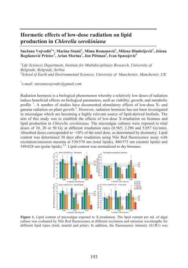
193
Hormetic effects of low-dose radiation on lipid production in Chlorella sorokiniana
���0���'� ����1?�����������1����#�������1������6��� ���1, Jelena Bogdano��8�����1, Arian Morina1, Jon Pittman2�4����&��� ���1
1Life Sciences Department, Institute for Multidisciplinary Research, University of Belgrade, Belgrade, Serbia2School of Earth and Environmental Sciences, University of Manchester, Manchester, UK
*e-mail: [email protected]
Radiation hormesis is a biological phenomenon whereby a relatively low doses of radiation induce beneficial effects on biological parameters, such as viability, growth, and metabolic profile 1. A number of studies have documented stimulatory effects of low-dose X- and gamma radiation on plant growth 2. However, radiation hormesis has not been investigated in microalgae which are becoming a highly relevant source of lipid-derived biofuels. The aim of this study was to establish the effects of low-dose X-irradiation on biomass and lipid production in Chlorella sorokiniana. The microalgae cultures were exposed to total doses of 10, 20 or 50 Gy at different irradiation rates (0.565; 2.290 and 5.057 Gy/min). Absorbed doses corresponded to ~10% of the total dose, as determined by dosimetry. Lipid content was determined 30 days after irradiation using Nile Red fluorescence assay with excitation/emission maxima at 530/570 nm (total lipids), 480/575 nm (neutral lipids) and 549/628 nm (polar lipids) 3,4. Lipid content was normalized to dry biomass.
Figure 1. Lipid content of microalgae exposed to X-irradiation. The lipid content per mL of algal culture was evaluated by Nile Red fluorescence at different excitation and emission wavelengths for different lipid types (total, neutral and polar). In addition, the fluorescence intensity (S1/R1) was
194
normalized to biomass to evaluate the content of lipids per mg of biomass. Mean values ± SE are presented. * - statistically significant compared to control. Mean control values (Ctrl; 12 replicates) are presented as full line ± SE (dashed line).
The results showed that the exposure to specific doses/rates led to increased total lipid content in microalgae culture (Figure 1). The increase could not be attributed to a particular lipid group – polar or neutral. In addition, there was no significant change in the lipid content normalized to biomass. Our results imply that X-ray irradiation exerted hormetic effects in Chlorella sorokiniana on the level of growth/proliferation which resulted in higher lipid content in the culture but not in the dry biomass. Further examination of hormetic effects of low-dose radiation using different settings, such as other growth phases and microalgal species, are warranted.
Acknowledgements
This study was supported by the NATO Science for Peace and Security Project SPS G5320 (Radiation Hormesis for Higher Microalgae Biofuels Yield)
References1. Calabrese EJ, Baldwin LA. Toxicology rethinks its central belief – hormesis demands a
reappraisal of the way risks are assessed. Nature 2003;421:691–2.2. Calabrese EJ, Blain RB. Hormesis and plant biology. Environ. Pollut 2009;157:42–8.3. Orr V, Rehmann L. Improvement of the Nile Red fluorescence assay for determination of total
lipid content in microalgae independent of chlorophyll content. J Appl Phycol 2015;27:2181–9.4. Elsey D, Jameson D, Raleigh B, Cooney MJ. Fluorescent measurement of microalgal neutral
lipids. J Microbiol Methods 2007;68:639–42.
195
Prooxidative effects of shikonin derivatives in human breast cancer cell line MDA-MB-231
�����'�!�1*�)��9 ������2����"����2�-����'�!���1
1Department of Chemistry, Faculty of Science, University of Kragujevac, Kragujevac, Serbia2Department of Biology and Ecology, Faculty of Science, University of Kragujevac
*e-mail: [email protected]
Breast cancer is one of the most common cancer types and represents the leading cause of mortality among women worldwide. Cell metabolism and survival are strongly depended on the amount of different oxidants present in cytoplasm. Shikonin and its derivatives are napthoquinone pigments known for their wide range of biological activities like wound healing, antiinflammatory, antitumor, antimicrobial and antithrombotic properties 1-4.*'�� ��� ��� �'�&� &�# $� 7 &� ��� ��������� �'�� ������&� ��� �� �H� &� �#&� ��� ;-methylbutyrylshikonin, acetylshikonin and �-hydroxyisovalerylshikon on human breast cancer cell line MDA-MB-231 as a potential mechanism of proapoptotic and antiproliferative properties of these compounds obtained in our previous study 5.The cells were treated with increasing concentrations of three shikonin derivatives (0.1 μg/mL to 100 μg/mL) during 24, 48, and 72 h, after which the evaluation of levels of superoxide anion radical, nitrites and glutathione was performed. All tested compounds expressed the significant prooxidative activity and induced dose- and time-dependent increase in level of superoxide anion radical and nitrites. Also, all examined compounds significantly increased the concentration of oxidized glutathione suggesting an activation ��� ����� ����H� ��� ���' ��&�&�� ;-methylbutyrylshikonin has exerted the strongest prooxidative effects in MDA-MB-231 cells, while �-hydroxyisovalerylshikon has shown the highest increase in nitrite production compared to non-treated cells. The levels of �H� �N� � ��#� �'����� 7���� �'�� '��'�&�� ��� �'�� ��� ������ 7��'� ;-methylbutyrylshikonin, which correspond to the highest concentration of superoxide anion radical among these three derivatives. The results obtained in this experiment indicates strong prooxidative potential of the examined compounds in MDA-MB-231 cells, which could be one of an important mechanism in ameliorating the growth rate and stimulating the levels of apoptosis of these cells. According to these data, derivatives of shikonin could have a significant beneficial role in developing a new strategies against breast cancer.
196
Acknowledgements
This work was financially supported by the Ministry of Education, Science and Technological Development of the Republic of Serbia under Grants OI172016.
References
1. Papageorgiou VP, Assimopoulou AN, Ballis AC. Alkannins and shikonins: a new class of wound healing agents. Curr Med Chem 2008;15:3248-67.
2. Papageorgiou VP, Assimopoulou AN, Couladouros EA, Hepworth D, Nicolaou KC. The chemistry and biology of alkannin, shikonin, and related naphthazarin natural products. AngewChem Int Ed 1999;38:270-300.
3. Wang L, Li F, Liu X, Chen B, Yu K, Wang MK. Meroterpenoids and a naphthoquinone fromArnebia euchroma and their cytotoxic activity. Planta Med 2015;81:320-6.
4. Naz S, Ahmad S, Ajaz Rasool S, Asad Sayeed S, Siddiqi R. Antibacterial activity directed isolation of compounds from Onosma hispidum. Microbiol Res 2006;161:43-8.
5. Vukic MD, et al. Antibacterial and cytotoxic activities of naphthoquinone pigments from Onosma visianii. Clem Excli J 2017;16:73–88.
197
The potential of biochar in improving microbial activity of soils in Vojvodina Province
Milorad Živanov1*�6��,���+ ���2, Jel�������!���2���3��@�����"�3,Branislava Tintor2, Jovica Vasin1, Bojan Vojnov3
1Laboratory for Soil and Agroecology, Institute of Field and Vegetable Crops, Novi Sad,Serbia2Soybean Department, Institute of Field and Vegetable Crops, Novi Sad3Department of Field and Vegetable Crops, Faculty of Agriculture, University of Novi Sad,Novi Sad; Serbia
*e-mail: [email protected]
Biochar enhances soil fertility by changing soil physical and chemical properties, or by improving soil biological properties 1. Microorganisms are important determinant of soil fertility, because of their role in mineralization of organic matter and involvement in biogeochemical cycles 2. Since the microbial activity in soil is controlled by numerous abiotic and biotic factors, microorganisms are very sensitive to changes in their environment including biochar amendment 3. As a constitutive enzyme of all microorganisms linked to the general microbial respiratory process, dehydrogenase activity (DHA) represents a reliable indicator of general microbial activity in soil. Therefore, the objective of this study was to examine effect of biochar soil amendment on dehydrogenase activity. A 2-year pot experiment was performed using five soil types (Mollic Gleysol: GL-mo, Calcaric Fluvisol: FL-ca, Eutric Cambisol: CM-eu, Gleyic Chernozem: CH-gl, and Haplic Chernozem: CH-ha) and five biochar doses (0, 25, 50, 100, and 150 g pot-1).Experimental crops were winter wheat (Triticum aestivum L.) in the first year, and sunflower (Helianthus annuus L.) in the second year. After harvesting sunflower at the end of the experiment, the samples were collected from all experimental pots assembled for each soil type and each biochar dose in order to examine the effect of biochar application. Dehydrogenase activity was determined spectrophotometrically by measuring the extinction of colored triphenyl-formazan (TPF) formed by reducing a colorless triphenyl-tetrazolium chloride (TTC) 4. Significant differences in DHA were observed between all types of soil. The highest enzymatic activity was found in the chernozems, and the lowest values were recorded in Mollic Gleysol (Figure 1A). DHA was significantly increased by the application of biochar in all treatments in relation to control, except with addition of 100 g pot-1. The largest increase of DHA (38%) was obtained in the 50 g treatment, which was significantly higher compared to other treatments (Figure 1B). Significant influence of both experimental factors, as well as their interaction, on the dehydrogenase activity revealed that biochar could potentially affect microbial activity of examined soils.
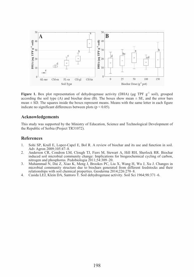
198
Figure 1. ��H� ���� ����&��� ����� ��� �'$ ����� &�� �����$� F�O+@� Fc�� *.�� �-1 soil), grouped according the soil type (A) and biochar dose (B). The boxes show mean ± SE, and the error bars mean ± SD. The squares inside the boxes represent means. Means with the same letter in each figure indicate no significant differences between plots (p < 0.05).
Acknowledgements
This study was supported by the Ministry of Education, Science and Technological Development of the Republic of Serbia (Project TR31072).
References
1. Sohi SP, Krull E, Lopez-Capel E, Bol R. A review of biochar and its use and function in soil. Adv Agron 2009;105:47–8.
2. Anderson CR, Condron LM, Clough TJ, Fiers M, Stewart A, Hill RH, Sherlock RR. Biochar induced soil microbial community change: Implications for biogeochemical cycling of carbon, nitrogen and phosphorus. Pedobiologia 2011;54:309–20.
3. Muhammad N, Dai Z, Xiao K, Meng J, Brookes PC, Liu X, Wang H, Wu J, Xu J. Changes in microbial community structure due to biochars generated from different feedstocks and their relationships with soil chemical properties. Geoderma 2014;226:270–8.
4. Casida LEJ, Klein DA, Santoro T. Soil dehydrogenase activity. Soil Sci 1964;98:371–6.

