shoulder joint complex consists of four basic articulations- 1.glenohumeral joint....
TRANSCRIPT

Shoulder Joint Complex
Consists of four basic articulations-1. Glenohumeral joint.2. Acromioclavicular
joint.3. Sternoclavicular joint.4. Scapulothoracic
articulation.

Shoulder Joint

Type of Joint
Synovial joint –•Ball-and-socket type of joint.
• Multiaxial • Simple •Typical

Articular surfaces
• Between larger spheroidal head of humerus and shallow glenoid cavity of scapula.
• Articular surface covered by hyaline articular cartilage.
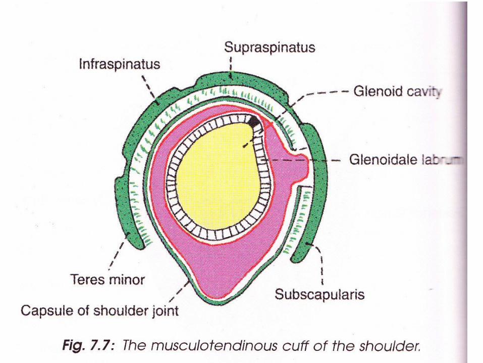
• Glenoid cavity is deepened by glenoid labrum (fibrocartilaginous rim).


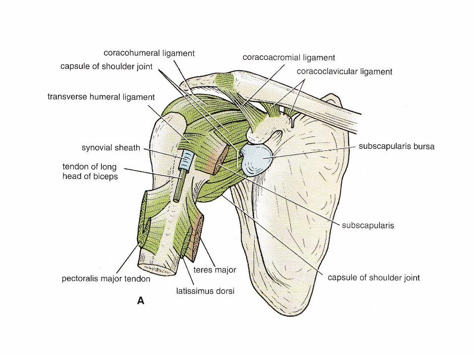
Ligaments
1. Capsular ligament• Surrounds joint and attached :- Medially to the scapula beyond the supraglenoid
tubercle and the margins of the labrum.- Laterally to the anatomical neck of humerus,except
inferiorly where it extends 1.5 cms below on to the surgical neck of humerus.
• Thin and lax, allow wide range of movement.


Synovial Membrane
• Lines capsule and is attached to the margins of the cartilage covering the articular surface.
• Communicates with subscapular and infraspinatus bursae around the joint
• Forms tubular sheath around the tendon of the long head of biceps brachii.
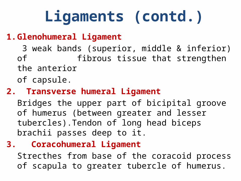
Ligaments (contd.)1. Glenohumeral Ligament
3 weak bands (superior, middle & inferior) of fibrous tissue that strengthen the anterior of capsule.
2. Transverse humeral LigamentBridges the upper part of bicipital groove of humerus (between greater and lesser tubercles).Tendon of long head biceps brachii passes deep to it.
3. Coracohumeral LigamentStrecthes from base of the coracoid process of scapula to greater tubercle of humerus.
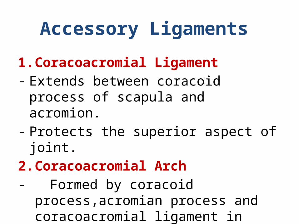
Accessory Ligaments
1. Coracoacromial Ligament- Extends between coracoid process of scapula
and acromion.- Protects the superior aspect of joint.2. Coracoacromial Arch- Formed by coracoid process,acromian process
and coracoacromial ligament in between.- Protective arch for head of humerus from
above.


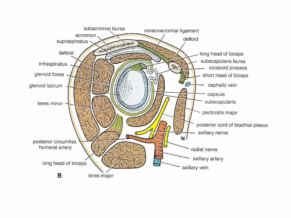
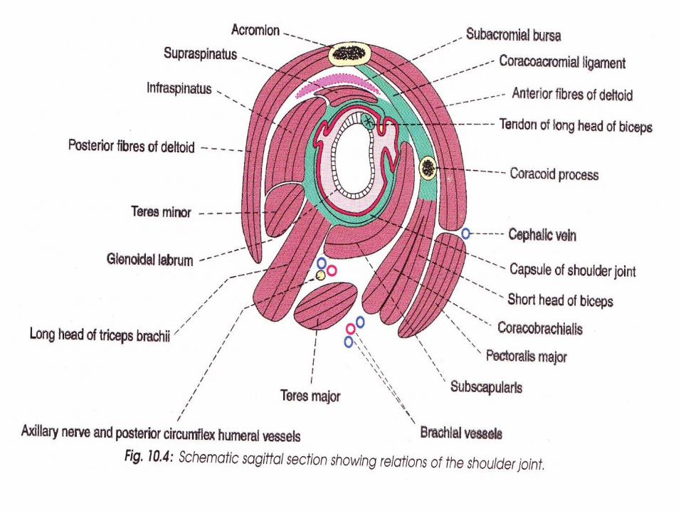
Bursae Related To The Joint
1. Subacromial (Subdeltoid) bursa lies between coracoacromial ligament &acromian process
above, and supraspinatus & joint capsule below. -Largest bursa of body2. Subscapularis bursa between tendon of subscapularis and neck of scapula.3. Infraspinatus bursa -between tendon of supraspinatus and posterolateral aspect of
joint capsule.
The bursae around the joint communicate with the cavity of joint
Opening of bursa means opening joint cavity.


Blood Supply • Anterior circumflex
humeral artery
• Posterior circumflex humeral artery
• Suprascapular artery
• Subscapular artery

Blood Supply

Nerve Supply • Axillary nerve
• Suprascapular nerve
• Musculocutaneous nerve


Relations
• Superiorly- Supraspinatus m.- Subacromial bursa- Coracoacromial ligament- Deltoid m.• Inferiorly- Long head triceps brachii m.- Axillary nerve- Post. circumflex humeral
vessels
• Anteriorly- Subscapularis m.- Coracobrachialis- Short head of biceps brachii- Deltoid• Posteriorly- Infraspinatus- Teres minor- Deltoid• Within the joint- Tendons of long head biceps
brachii
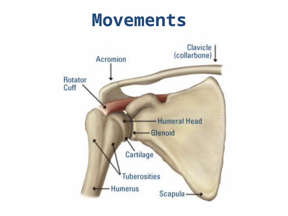
Movements

• Flexion – Arm moves forwards & medially.
• Extension – Arm moves backwards & laterally.
Flexion & Extension
MAIN ACCESSORYMOVEMENT
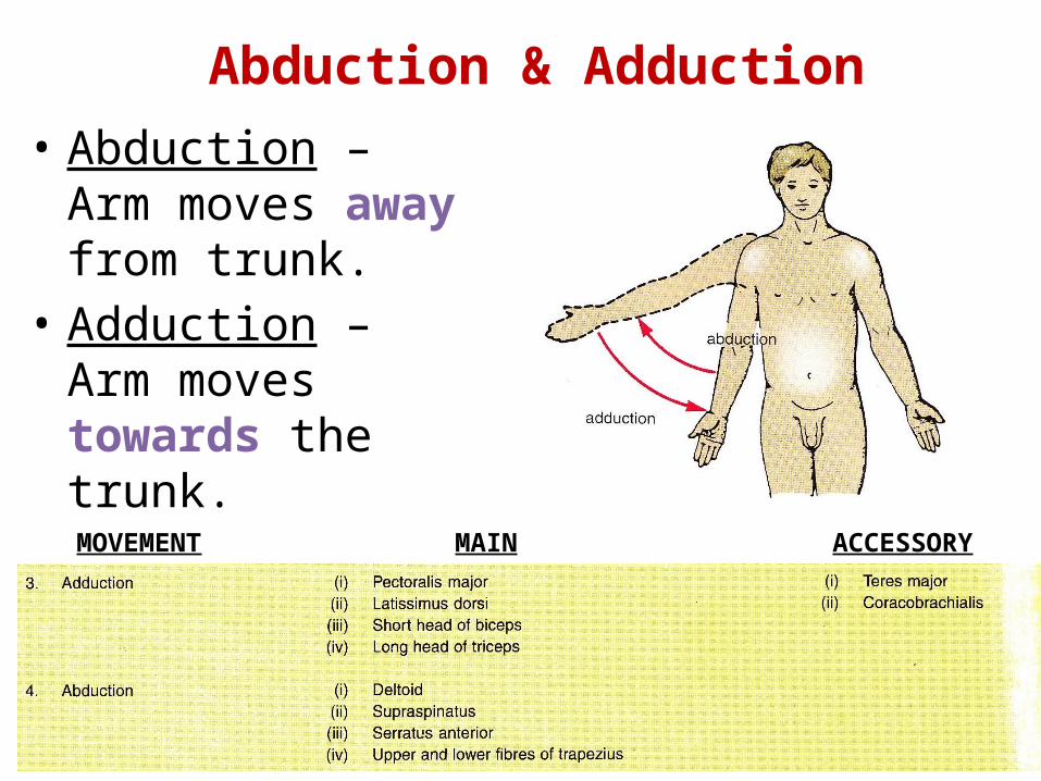
• Abduction – Arm moves away from trunk.
• Adduction – Arm moves towards the trunk.
Abduction & Adduction
MAIN ACCESSORYMOVEMENT
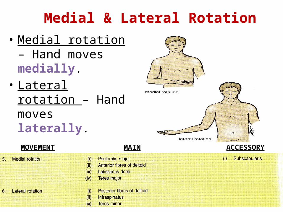
• Medial rotation – Hand moves medially.
• Lateral rotation – Hand moves laterally.
Medial & Lateral Rotation
MAIN ACCESSORYMOVEMENT

• Combination of dif. movements, results in hand moving along a circle.
Circumduction

Factors providing stability to joint
1. Rotator cuff- capsule of the joint is strengthened by slips
of tendons of subscapularis m., supraspinatus m., infraspinatus m. & teres minor (rotator cuff muscles).
• Tone of muscles grasp head of humerus and pull it medially to hold it against shallow glenoid cavity.


Factors providing stability to joint(contd.)
• Coracoacromial arch.• Long head of biceps tendon.• Glenoid labrum.

Applied Anatomy
• Dislocation of shoulder joint -mostly occurs inferiorly. -axillary nerve injured due to close proximity. -clinically described as – anterior and
posterior dislocation. -caused by excessive extension and lateral
rotation of humerus. -presents as hollow in rounded contour of
shoulder and prominent tip.


• Frozen shoulder ( Adhesive capsulitis) - pain and uniform limitation of all
movements of the joint. -no radiological changes. -due to shrinkage of joint capsule.• Rotator cuff disorders - calcific supraspinatus tendinitis. - subacromial bursitis. - painful arc syndrome.

Thank You!