sialidase and sialoglycoproteases can modulate virulence in porphyromonas gingivalis
TRANSCRIPT

INFECTION AND IMMUNITY, July 2011, p. 2779–2791 Vol. 79, No. 70019-9567/11/$12.00 doi:10.1128/IAI.00106-11Copyright © 2011, American Society for Microbiology. All Rights Reserved.
Sialidase and Sialoglycoproteases Can Modulate Virulence inPorphyromonas gingivalis�†
Wilson Aruni, Elaine Vanterpool, Devon Osbourne, Francis Roy, Arun Muthiah,Yuetan Dou, and Hansel M. Fletcher*
Division of Microbiology and Molecular Genetics, School of Medicine, Loma Linda University, Loma Linda, California 92350
Received 31 January 2011/Returned for modification 26 February 2011/Accepted 31 March 2011
The Porphyromonas gingivalis recombinant VimA can interact with the gingipains and several other proteins,including a sialidase. Sialylation can be involved in protein maturation; however, its role in virulence regu-lation in P. gingivalis is unknown. The three sialidase-related proteins in P. gingivalis showed the characteristicsialidase Asp signature motif (SXDXGXTW) and other unique domains. To evaluate the roles of the associatedgenes, randomly chosen P. gingivalis isogenic mutants created by allelic exchange and designated FLL401(PG0778::ermF), FLL402 (PG1724::ermF), and FLL403 (PG0352::ermF-ermAM) were characterized. Similar tothe wild-type strain, FLL402 and FLL403 displayed a black-pigmented phenotype in contrast to FLL401, whichwas not black pigmented. Sialidase activity in P. gingivalis FLL401 was reduced by approximately 70% incomparison to those in FLL402 and FLL403, which were reduced by approximately 42% and 5%, respectively.Although there were no changes in the expression of the gingipain genes, their activities were reduced by 60 to90% in all the isogenic mutants compared to that for the wild type. Immunoreactive bands representing thecatalytic domains for RgpA, RgpB, and Kgp were present in FLL402 and FLL403 but were missing in FLL401.While adhesion was decreased, the capacity for invasion of epithelial cells by the isogenic mutants wasincreased by 11 to 16% over that of the wild-type strain. Isogenic mutants defective in PG0778 and PG0352 weremore sensitive to hydrogen peroxide than the wild type. Taken together, these results suggest that the P.gingivalis sialidase activity may be involved in regulating gingipain activity and other virulence factors and maybe important in the pathogenesis of this organism.
Porphyromonas gingivalis, a Gram-negative anaerobic bacte-rium, is an important etiological agent of adult chronic perio-dontitis. In addition, this organism appears to be associatedwith cardiovascular diseases and an increased risk of pretermbirth and low birth weight of infants in pregnant women withinfection-induced periodontal disease (4). The ability of P.gingivalis to colonize the periodontal pocket and interact withother organisms such as Tannerella forsythia and Fusobacteriumnucleatum is a prerequisite for infection-induced periodontaldiseases (34). Several cell surface-associated virulence factors(e.g., gingipains, fimbriae, hemagglutinin, capsule, and lipo-polysaccharide) that can directly or indirectly affect the perio-dontium or facilitate interaction with other periodontopathicpathogens have been characterized in P. gingivalis (16). How-ever, the roles of sialidase and sialopeptidases as potentialvirulence factors in P. gingivalis are yet to be explored. Becauseof their importance in the breakdown of carbohydrates andother glycoprotein conjugates, these enzymes could facilitateattachment and enhance commensalism with other oral patho-gens, especially biofilm formers (44).
Sialidases (neuraminidases) are glycosylhydrolases thatcleave the glycoketosidic linkages of sialic acid (Sia) O-accep-tor substrates by an exohydrolytic reaction. Functionally simi-
lar to sialidases, the O-sialoglycoprotease hydrolyzes the SiaO-acceptor substrate through an endohydrolytic reaction (36,55). Sialidase activity has been found in viruses, bacteria, pro-tozoa, fungi, and metazoans (6, 7, 55). Bacterial sialidases havebeen considered virulence factors in many pathogenic organ-isms, such as Corynebacterium diphtheriae, Vibrio cholerae,Streptococcus pneumoniae, and group B streptococci, whichcolonize mucosal surfaces (8). They have been shown to beinvolved in infection and tissue destruction (57), peroxidescavenging during oxidative stress (22), and the modulationof host innate immunity (55). Furthermore, production ofsialidase may be a critical factor in the provision of freesialic acid, a fermentable carbohydrate source for bacterialproliferation (8, 20).
Sialic acid has been predominantly found as the terminalcarbohydrate in eukaryotes and prokaryotes. Sialic acid natu-rally occurs in prokaryotes as nine-carbon keto sugar acidsderived from N-acetylneuraminic acid (Neu5Ac) (53). Manycommensal and pathogenic bacteria use environmental (host)sialic acids as sources of carbon, nitrogen, energy, and aminosugars for cell wall synthesis (35). The breakdown of sialic acidresidues and sialoconjugates by sialidases contributes to a widerange of important biological functions such as cellular inter-action and conformational stabilization of glycoproteins in thecell membrane that could expose or mask receptors for ligandbinding and other enzymatic interactions (2). While the role ofsialidase in sialic acid metabolism has been known in other oralpathogens like Tannerella forsythia (38), it is yet to be exploredin P. gingivalis.
Glycosylation is a posttranslational regulatory mechanism
* Corresponding author. Mailing address: Division of Microbiologyand Molecular Genetics, School of Medicine, Loma Linda University,Loma Linda, CA 92350. Phone: (909) 558-4472. Fax: (909) 558-4035.E-mail: [email protected].
† Supplemental material for this article may be found at http://iai.asm.org/.
� Published ahead of print on 18 April 2011.
2779
Dow
nloa
ded
from
http
s://j
ourn
als.
asm
.org
/jour
nal/i
ai o
n 30
Jan
uary
202
2 by
87.
110.
64.1
8.

that is important in gingipain biogenesis and pathogenicity inP. gingivalis (13, 25). Further, the posttranslational addition ofcarbohydrates to the gingipains is highly variable, thus implyinga role for multiple factors in this process (10). We previouslyreported that the VimA protein, which can modulate gingipainactivity in P. gingivalis, can also interact with a putative siali-dase protein (1, 30, 50, 53). Although sialylation can beinvolved in protein maturation, its role, if any, in gingipainmaturation/activation is unknown. Here we report the charac-terization of a sialidase and two sialoglycoproteases from P.gingivalis.
MATERIALS AND METHODS
Bioinformatics analysis. The DNA and amino acid sequences were retrievedfrom the Oralgen database (Los Alamos National Laboratory; http://www.oralgen.lanl.gov) and aligned using Bioedit (http://www.mbio.ncsu.edu/bioedit/bioedit.html). The phylogenetic relationship between the oral pathogens wasanalyzed using MEGA version 4.0 (49). The distance was calculated using theKimura 2-parameter model, and for clustering the neighbor-joining method wasemployed using bootstrap values based on 1,000 replicates (39). The amino acidsequences were analyzed using ClustalW version 2.0 (http://www.ebi.ac.uk/). Thesecondary structure prediction and modeling of the protein were performedusing Modeler 9v8 (41). The models were validated using the WHATIF program(56). The transcription factor binding motifs were identified using PROMOversion 8.3 (11). The signal peptide and potential cleavage sites were predictedusing both the neural network and hidden Markov model (23).
Bacterial strains and growth condition. Bacterial strains and plasmids used inthis study are listed in Table 1. P. gingivalis strains were grown in brain heartinfusion (BHI) broth (Difco, Detroit, MI) supplemented with hemin (5 �g/ml),vitamin K (0.5 �g/ml), and cysteine (0.1%). Experiments with hydrogen peroxidewere performed in BHI without cysteine. All cultures were incubated at 37°Cunless otherwise stated. P. gingivalis strains were maintained in an anaerobicchamber (Coy Manufacturing, Ann Arbor, MI) in 10% H2, 10% CO2, and 80%N2. Growth rates for P. gingivalis strains were determined spectrophotometrically(optical density at 600 nm [OD600]). Antibiotics were used at the followingconcentrations: clindamycin, 0.5 �g/ml; erythromycin, 300 �g/ml; tetracycline, 3�g/ml; carbenicillin, 100 �g/ml.
DNA isolation and analysis. P. gingivalis chromosomal DNA was prepared aspreviously described (27). For plasmid DNA analysis, DNA extraction was per-formed by the alkaline lysis procedure previously reported (12). For large-scale
preparation, plasmids were purified using the Qiagen (Santa Clarita, CA) Plas-mid Maxi kit.
Construction of PG0778- and PG1724-defective mutants. Construction of iso-genic mutants defective in the PG0778 and PG1724 genes was carried out bylong-PCR-based fusion of several fragments using overlapping extension PCR aspreviously described (20). The primers used in the development of deletionmutants are given in Table S1 in the supplemental material. Briefly, 1-kilobaseflanking fragments upstream and downstream of the target genes were PCRamplified from the chromosomal DNA of P. gingivalis W83. The ermF cassettewas amplified from the pVA2198 plasmid (Table 1) using custom-made oligo-nucleotide primers containing overlapping nucleotides for the upstream anddownstream fragments. These three fragments were fused together using theforward primer of the upstream fragment and the reverse primer of the down-stream fragment. The fusion PCR program consisted of 1 cycle of 5 min at 94°C,followed by 30 cycles of 30 s at 94°C, 30 s at 55°C, and 4 min at 68°C, with a finalextension of 5 min at 68°C. The PCR-fused fragment was used to transform P.gingivalis W83 by electroporation as previously described (1). The cells wereplated on BHI agar containing 10 �g/ml of erythromycin and incubated at 37°Cfor 7 days. The correct gene replacement in the erythromycin-resistant mutantswas confirmed by colony PCR and DNA sequencing.
Generation of PG0352-defective mutant P. gingivalis. A 2.5-kb fragment car-rying the intact PG0352 and downstream gene PG0353 was amplified by PCRusing custom-made oligonucleotide primers (see Table S1 in the supplementalmaterial). This fragment was cloned into the pCR-2.1-TOPO plasmid vector(Invitrogen, Carlsbad, CA) and was designated pFLL403a. The ermF-ermAMcassette, which confers erythromycin/clindamycin resistance in Escherichia coliand P. gingivalis (9), was PCR amplified from pVA2198 with Pfu Turbo (Strat-agene, La Jolla, CA) and ligated into the HincII restriction site of the PG0352gene. The resultant recombinant plasmid, pFLL403b, was used as a donor inelectroporation of P. gingivalis W83.
Complementation of the PG0778-, PG1724-, and PG0352-defective isogenicmutants. DNA fragments containing an upstream regulatory region and theopen reading frame (ORF) for PG0778, PG1724, or PG0352 were amplified fromP. gingivalis W83 chromosomal DNA using the appropriate primer set (see TableS1 in the supplemental material). A BamHI restriction site was designed at the5� ends of both primers to facilitate the subcloning of the PCR fragment into theBamHI-digested pT-COW plasmid (17). The purified recombined plasmids,designated pFLL401a, pFLL402a, and pFLL403c, were used to electrotransformP. gingivalis FLL401 (PG0778::ermF), FLL402 (PG1724::ermF), and FLL403(PG0352::ermF-ermAM), respectively. The transformants were selected on BHIagar plates in the presence of erythromycin and tetracycline.
Electroporation. Electroporation of cells was performed as previously re-ported (12). Briefly, 1 ml of an actively growing culture of P. gingivalis was used
TABLE 1. Strains and plasmids used
Strain or plasmid Characteristics Referenceor source
StrainsP. gingivalis W83 Wild type 31FLL92 vimA-defective mutant 52FLL401 PG0778 defective This studyFLL402 PG1724 defective This studyFLL403 PG0352 defective This studyFLL401c FLL401 with pFLL401a This studyFLL402c FLL402 with pFLL402a This studyDH5� F� �80dlacZ�M15 �(lacZYA-argF)U169 recA1 endA1 hsdR17(rK
� mK�) phoA supE44 � thi-1
gyrA96 relA1Invitrogen
Top10 F� mcrA �(mrr-hsdRMS-mcrBC) �80dlacZ�M15 �lacX74 recA1 ara139 �(ara-leu)7697 galUgalK rpsl (Strr) endA1 nupG
Invitrogen
PlasmidspCR-2.1.TOPO Apr Kmr InvitrogenpVA2198 Spr ermF-ermAM 12pT-COW Apr tetQ 13apFLL401a Apr tetQ TC::PG0778 This studypFLL402a Apr tetQ TC::PG1724 This studypFLL403c Apr tetQ TC::PG0352 This studypFLL403a pCR-2.1-TOPO PG0352-PG0353 This studypFLL403b pFLL403a with PG0352 interrupted with ermF-ermAM This study
2780 ARUNI ET AL. INFECT. IMMUN.
Dow
nloa
ded
from
http
s://j
ourn
als.
asm
.org
/jour
nal/i
ai o
n 30
Jan
uary
202
2 by
87.
110.
64.1
8.

to inoculate 10 ml of BHI broth and incubated overnight at 37°C. Ninety milli-liters of the same prewarmed (37°C) medium was then inoculated with 3 ml ofthe overnight culture and was incubated for an additional 4 h until an OD600 of0.7 was obtained. The cells were then harvested by centrifugation at 3,000 g for15 min at 4°C and were washed twice in 50 ml of electroporation buffer (10%glycerol, 1 mM MgCl2), filter sterilized, and stored at 4°C. The final cell pelletwas resuspended in 0.5 ml of electroporation buffer. A 100-�l sample of resus-pended cells and 5 �g of DNA were placed in a sterile electrode cuvette (0.2-cmgap). The cells were then pulsed with a Gene Pulser (Bio-Rad, Hercules, CA) at2,500 V for 9.5 ms and then were incubated on ice for 5 min. The cell suspensionwas then added to 0.5 ml of BHI broth and was incubated for approximately 16 h.A 100-�l sample was plated on solid medium containing erythromycin and wasincubated anaerobically at 37°C for 5 to 10 days.
Growth analysis during oxidative stress conditions. Actively growing culturesof P. gingivalis W83, FLL401, FLL402, and FLL403 were incubated at 37°C underanaerobic conditions for 24 h. For adaptation to oxidative stress conditions, thewild type and isogenic mutants were grown in BHI broth (without cysteine) plus0.25 mM H2O2. Controls were grown in the absence of H2O2. Growth rates weredetermined spectrophotometrically (OD600).
RT-PCR analysis. Total RNA was extracted from P. gingivalis strains grown toearly stationary phase (OD600 of 1.2 to 1.3) using the RiboPure kit (Ambion,Foster City, CA) according to the manufacturer’s instructions. The primers usedfor reverse transcription-PCR (RT-PCR) analysis are listed in Table S1 in thesupplemental material. The RT-PCR mixture (50 �l) contained 1 �g of templateRNA in Superscript One-Step RT-PCR mix (Invitrogen, Carlsbad, CA). RT-PCR cycling conditions were 1 cycle of 5 min at 94°C followed by 30 cycles of 30 sat 94°C, 30 s at 54°C, and 1 min at 68°C, with final extension of 5 min at 68°C.
SDS-PAGE and immunoblot analysis. Sodium dodecyl sulfate-polyacrylamidegel electrophoresis (SDS-PAGE) was performed with a 4 to 12% Bis-Tris sep-arating gel in MOPS (morpholinepropanesulfonic acid)-SDS running buffer ac-cording to the manufacturer’s instructions (NuPAGE Novex gels; Invitrogen,Carlsbad, CA). Samples were prepared (65% sample, 25% 4 NuPAGE lithiumdodecyl sulfate [LDS] sample buffer, 10% NuPAGE reducing agent), heated at72°C for 10 min, and then electrophoresed at 200 V for 65 min in the XCellSureLock minicell system (Invitrogen, Carlsbad, CA). The protein bands werevisualized by staining with SimplyBlue SafeStain (Invitrogen, Carlsbad, CA). Theseparated proteins were then transferred to BioTrace nitrocellulose membranes(Pall Corporation, San Diego, CA) using a semidry Trans-Blot apparatus (Bio-Rad, Hercules, CA) at 15 V for 25 min. The blots were probed with gingipain-specific antibodies (45). Immunoreactive proteins were detected by the proce-dure described in the Western Lightning Plus chemiluminescence reagent kit(Perkin-Elmer Life Sciences, Fremont, CA). The secondary antibody was goatanti-rabbit or anti-chicken IgG (heavy plus light chains)–horseradish peroxidase(HRP) conjugate (Zymed Laboratories, San Francisco, CA).
Gingipain activity assay. The presence of Arg-X- and Lys-X-specific cysteineprotease activities was determined using a microplate reader (Bio-Rad) by themethod of Fletcher et al. (12) for cells grown to exponential phase (OD600 of0.8). Briefly, activity of arginine gingipains was measured with 1 mM BAPNA(N�-benzoyl-DL-arginine-p-nitroanilide) in activated protease buffer (0.2 M Tris-HCl, 0.1 M NaCl, 5 mM CaCl2, 10 mM L-cysteine, pH 7.6). Lysine gingipainactivity was measured with ALNA (acetyl-Lys-p-nitroanilide–HCl). After thesubstrate and culture were incubated, the reaction was stopped by the additionof 50 �l of glacial acetic acid. The OD405 was then measured against a blanksample containing no proteinase.
Assay for estimation of total protease activity. Total protease estimation useda fluorescence resonance energy transfer (FRET)-based method (protease assaykit; Molecular Probes, Carlsbad, CA) in accordance with the manufacturer’sinstructions. The protease substrate comprises a fluorophore and a quenchermoiety separated by an amino acid sequence. Protease cleavage of the aminoacid sequence separates the fluorophore from the quencher, resulting in theemission of a detectable fluorescent signal. The magnitude of the resultant signalis proportional to the degree of substrate cleavage and can therefore be used toquantitate the enzyme activity. The assay was performed in a 96-well plate. Thefluorescence was measured using the microplate reader FLx800 (BioTek) at490-nm excitation and 520-nm emission wavelengths.
Sialidase assay. Sialidase estimation in the P. gingivalis mutants was carriedout using the Amplex Red neuraminidase (sialidase) assay kit (MolecularProbes, Carlsbad, CA). Briefly, 50 �l of the P. gingivalis sample was mixed with50 �l of the Amplex Red reagent, 50 �l of HRP, and 50 �l of the galactoseoxidase solution. Finally, 50 �l of the substrate was added to the mixture, and themixture was incubated at 37°C for 30 min in a light-protected container. Theassay utilizes Amplex Red to detect H2O2 generated by galactose oxidase oxi-dation of desialylated galactose, the end product of neuraminidase activity. The
H2O2 in the presence of HRP reacts with Amplex Red reagent to generateresorufin, the red fluorescent oxidation product, which is detected spectropho-tometrically (OD492) using the BioTek FLx800 microplate reader. Substratesused in the sialidase assay include 3�sialyllactose, O-sialyl-D-lactose, 3N-acetyl-neuraminic acid, mucin (bovine submaxillary), feutin, and colomic acid. (Sigma-Aldrich, St. Louis, MO). A neuraminidase and a nonneuraminidase suppliedwith the kit were used as positive and negative controls, respectively.
Hemagglutination assay. Hemagglutination activity was determined usingsheep, bovine, chicken, and horse red blood cells (RBCs) as previously reported(51). Twenty-four-hour cultures of P. gingivalis W83 and mutants were harvestedby centrifugation (10,000 g for 10 min), washed twice in phosphate-bufferedsaline (PBS) buffer, and resuspended to a final OD600 of 1.2. Erythrocytes werewashed twice with 1 PBS and resuspended in 1 PBS to a final concentrationof 1%. An aliquot (100-�l volume) of the bacterial suspension was seriallydiluted 2-fold with PBS in a round-bottom 96-well microtiter plate. An equalvolume (100 �l) of 1% erythrocytes was mixed with each dilution and incubatedat 4°C for 3 h. Hemagglutination was visually assessed, and the hemagglutinationtiter was determined as the reciprocal of the highest dilution showing completehemagglutination.
Lectin binding hemagglutination assay. Sialidase production was assayed inmicrotiter plates using fresh sheep erythrocytes. The erythrocytes were collectedand suspended in Alsever’s solution (10%) and washed three times with 0.01 MPBS buffer (PBS, Na2HPO4, KH2PO4, NaCl, pH 7.4). Bacterial cells were grownin BHI broth, resuspended to 1.5 108 bacteria/ml, and then washed three timesby centrifugation (12,000 g, 10 min) in PBS. An aliquot (1 ml) of bacterial cellswas added to 10 ml of the washed erythrocytes to achieve a final concentrationof 1%, and the cells were then carefully homogenized and incubated at 37°C for4 h under anaerobic conditions. Lectins from Agaricus bisporus (ABA; 1.8 mg/ml), Erythrina cristagalli (ECA; 2.3 mg/ml), Limulus polyphemus (LPA; 2.5 mg/ml), Triticum vulgaris (WGA; 2.5 mg/ml), and Arachis hypogaea (PNA; 2.5mg/ml) and concanavalin A (ConA; 2.1 mg/ml) (Sigma-Aldrich, St. Louis, MO)were serially diluted in PBS. An aliquot (5 �l) of each dilution was then addedto the suspension of bacteria and erythrocytes (1:1), which was incubated at roomtemperature for 1 h followed by 12 h at 4°C. The presence of sialidase wasindicated by agglutination (bacteria-erythrocytes-lectin). A mixture of bacteriaand erythrocytes without lectin, which results in precipitation, was used as thenegative control. A mixture of erythrocytes with PBS was used as the RBCcontrol. The agglutination titer was defined as the reciprocal of the endpointdilution. The inhibition of the sialidase production was verified in the wild-typeW83 strain as previously reported (29). Briefly, bacteria treated with 1 ml of 1mM galactose and incubated at room temperature for 30 min were washed inPBS and harvested by centrifugation (12,000 g, 10 min). Five microliters ofdiluted lectins was mixed with 50 �l of bacterial cells (1.5 108 cells/ml), treatedwith 1 mM galactose, and added to 50 �l of erythrocytes (1% final concentra-tion). Plates were homogenized and incubated at 37°C for 1 h. Enzyme inhibitionwas indicated by the precipitation of the erythrocytes. Overnight incubation wasavoided, as the test system showed lysis of RBCs.
Electron microscopy. Transmission electron microscopy (TEM) was per-formed using the FEI Tecnai G2 TEM according to the method of Harris (17).Briefly, Formvar-carbon-coated grids were prepared; the Formvar support wasremoved by placing the grids in an atmosphere of solvent vapor. The grids werethen placed on a wire mesh in a glass petri dish, with carbon tetrachloride belowthe wire mesh. Five to 10 ml of the sample was placed under the carbon side ofa 4- by 5-mm square of mica (approximately twice the size of an EM grid). Thegrid was washed in 0.5% acetic acid and then with acetone. The carbon film wasbroken to free the specimen grid, after which the grid was placed in stainsolution-neutral 1% aqueous phosphotungstic acid for 30 s. After being blotteddry, the grid was examined using the FEI Tecnai G2 TEM.
Epithelial cell culture. HeLa cells were grown and maintained in Dulbecco’smodified Eagle’s medium supplemented with 10% fetal bovine serum, penicillin(100 IU/ml), streptomycin (100 IU/ml), and amphotericin B (2.5 mg/ml) (Invit-rogen, Carlsbad, CA) at 37°C under a 5% CO2 atmosphere. Confluent stockcultures were trypsinized, adjusted to approximately 5 103 cells/ml, seeded into12-well plates (Nunc, Rochester, NY) at 1 ml per well, and further incubated for48 h to reach semiconfluence (105 cells per well).
Adherence and standard antibiotic protection assay. Invasion was quantifiedusing the standard antibiotic protection assay (58). Briefly an isolated colonyharvested from solid agar plate was grown to exponential phase in BHI broth.The cells were then centrifuged, washed three times in PBS, and then adjustedto 107 CFU/ml of bacteria (confirmed by colony count) in Dulbecco’s modifiedEagle’s medium. Epithelial cell monolayers were washed three times with PBS,infected with bacteria at a multiplicity of infection (MOI) of 1:100 (105 epithelialcells), and then incubated at 37°C for 90 min under a 5% CO2 atmosphere.
VOL. 79, 2011 SIALIDASE ACTIVITY IN PORPHYROMONAS GINGIVALIS 2781
Dow
nloa
ded
from
http
s://j
ourn
als.
asm
.org
/jour
nal/i
ai o
n 30
Jan
uary
202
2 by
87.
110.
64.1
8.

Nonadherent bacteria were removed by washing with PBS, while cell surface-bound bacteria were killed with metronidazole (200 �g/ml, 60 min). P. gingivalisW83 and all the isogenic mutants are sensitive to 100 �g/ml of metronidazole.After removal of antibiotic, the internalized bacteria were released by osmoticlysis in sterile distilled water with scraping. Lysates were serially diluted, plated(in duplicate) on BHI agar, and incubated for 6 to 10 days. The number ofbacterial cells recovered was expressed as a percentage of the original inoculum.The number of adherent bacteria was obtained by subtracting the number ofintracellular bacteria from the total number of bacteria obtained in the absenceof metronidazole (6).
RESULTS
In silico analysis. The genome of P. gingivalis revealed thepresence of three sialidase-related genes, of which one(PG0352) is annotated as a “conserved hypothetical protein orputative sialidase” gene. The other two genes (PG0778 andPG1724) are annotated as O-sialoglycoprotease genes (gcp)(www.oralgen.lanl.gov). No upstream or downstream sialidase-related genes are associated with PG0352, PG0778, or PG1724.P. gingivalis was missing homologs similar to a large putativesialic acid utilization and transport locus present in the ge-nome of T. forsythia (oral pathogen sequence databases [www.oralgen.lanl.gov]).
The clad containing PG0352 was more closely related (68%homology) to sialidases from Actinomyces naeslundii (encodedby ANA1493), T. forsythia (encoded by TF0035 and TF2207),Clostridium perfringens, and Bacteroides fragilis (Fig. 1A).PGO352 from P. gingivalis W83 shows 85% homology toPGN1608 from P. gingivalis ATCC 33277. The variations weredue to amino acid substitutions at positions 60 (A3V), 87(L3I), 151(H3N), 152 (N3D), 159 (Q3K), 160 (R3K), 183(L3H), 185 (E3K), and 189 (H3Y). These variations werepredominantly found in the transmembrane domain of theprotein.
The PG0352 sialidase clad was more closely related to theO-sialoglycoprotease clad, which contains P. gingivalis PG0778,T. forsythia TF0020, and F. nucleatum FN0928 (Fig. 1B). It isnoteworthy that, similar to what was found for P. gingivalis, thetwo O-sialoglycoproteases (encoded by gcp) from T. forsythia(TF0020 and TF1855) segregated into different clads. How-ever, TF0020 was more closely related to the sialidases.
The second cluster containing PG1724 is bifurcated into twosubclads. PG1724 is more closely related to the O-sialoglyco-proteases from the oral pathogens (Fig. 1C).
Multiple amino acid sequence alignment of the sialidase andtwo sialidase-related proteins from P. gingivalis revealedunique domains and motifs (see Fig. S1 in the supplementalmaterial). The O-sialoglycoprotease PG0778 revealed an N-terminal metal binding domain followed by a transmembanehelix domain consisting of 22 amino acids between positions 17and 37. The end of the transmembrane domain bears a twinarginine peptide signal followed by a cleavage site (see Fig.S1A in the supplemental material). PG0352 revealed a 13-amino-acid stretch of an inner membrane domain followed bya transmembrane domain consisting of 17 amino acids. A sig-nal peptide consisting of His-Leu-Ser-Gly followed by a poten-tial cleavage site was observed at the end of the transmem-brane domain (see Fig. S1B in the supplemental material).This transmembrane domain appears to form the helix encom-passing a hydrophobic region of 17 amino acids. The presenceof a signal peptide at the N terminus suggests that the sialidase
of P. gingivalis PG0352 is membrane bound. The entire proteincontains repetitive hydrophobic aspartic acid motifs in six po-sitions and also contains a long catalytic domain between po-sitions 182 and 240. The catalytic domain encompasses theconsensus a sequence motif that is characteristic of other siali-dases (see Fig. S1B in the supplemental material). It alsocontains the characteristic SXDXGXTW domain at four posi-tions between amino acid positions 230 and 240, 290 and 300,360 and 370, 410 and 420, and 468 and 475. However, thesignature sequence motif at the fifth position, between 468 and475, was found to be partial and incomplete (see Fig. S2 in thesupplemental material). These signature motifs are well con-served in the bacterial sialidases and also in the other oralpathogens studied (7). A carbohydrate-specific lectin bindingdomain between amino acids 198 and 218 was predicted.
PG1724 revealed an N-terminal metalloprotease consensusdomain with a potential cleavage site. A transmembrane do-main was observed between positions 136 and 152. In contrastto PG0778, PG1724 contains only one transmembrane domain(see Fig. S1C in the supplemental material). Two metal bind-ing histidine domains are present, one at the 115th position
FIG. 1. Phylogenetic analysis of sialidases and O-sialoglycopro-tease from several oral and anaerobic bacteria. The P. gingivalis puta-tive sialidase (PG0352) segregates in clad A with sialidases from otherbacteria. One of the P. gingivalis O-sialoglycoproteases (PG0778) seg-regates in clad B. Clad C segregates into two subclads, one bearing thestreptococcal O-sialoglycoprotease and the second bearing the P. gin-givalis O-sialoglycoprotease (PG1724) along with those of other peri-odontal pathogens.
2782 ARUNI ET AL. INFECT. IMMUN.
Dow
nloa
ded
from
http
s://j
ourn
als.
asm
.org
/jour
nal/i
ai o
n 30
Jan
uary
202
2 by
87.
110.
64.1
8.

before the transmembrane domain and the other at the 289thposition. Both PG0778 and PG1724 contain metal bindingdomains at the N-terminal and C-terminal ends, with a cleav-age site at the N terminus. However, PG1724 lacks a signalpeptide. In PG1724 a lectin binding domain was observedbetween amino acids 277 and 282.
Multiple sequence alignment of the O-sialoglycoproteaseamino acid sequences of oral pathogens with PG0778 andPG1724 revealed a DAXG/DAXD consensus signature motif.Asp boxlike motifs (SXDXTXSV, SXAXGXXW, SXGXSXII,and SXSXGXXTX) similar to the unique Asp box sequenceSXDXGXTW of the sialidases were observed (see Fig. S3 inthe supplemental material).
In silico modeling of PG0352 confirmed that the proteincould be classified under the BNR-neuraminidase class (Pfamstructural classification CL434). PG0778 and PG1724 showedthe catalytic beta propeller structure in which PG0778 had aunique terminal cleft and a molecular groove structure (seeFig. S4 in the supplemental material).
Construction of PG0352, PG0778, and PG1724 deletion mu-tants in P. gingivalis W83. Isogenic mutants of P. gingivalis de-fective in PG0352, PG0778, and PG1724 were constructed byallelic-exchange mutagenesis. The schematic representation ofthe genetic structures of PG0352, PG0778, and PG1724 is shownin Fig. 2A. Following electroporation and plating on selectivemedium, we detected erythromycin-resistant colonies after a 5- to7-day incubation period. The mutants were confirmed by colonyPCR (Fig. 2B, C, and D) and DNA sequencing. To compare theirphenotypic properties with those of wild-type strain W83, allmutants were plated on BHI blood agar plates. Similar to thewild-type strain, the PG0352- and PG1724-defective mutantsdisplayed a black-pigmented phenotype. In contrast, thePG0778-defective isogenic mutants were not black pigmented.Mutants designated P. gingivalis FLL401 (PG0778::ermF), P.gingivalis FLL402 (PG1724::ermF), and P. gingivalis FLL403(PG0352::ermF-ermAM) were randomly chosen for further study.Inactivation of the sialidase gene (PG0352) and sialoglycopro-tease genes (PG0778 and PG1724) had no polar effects on theexpression of their downstream genes (Fig. 2E). A generationtime of approximately 3 h was determined for the wild type and P.gingivalis FLL403 compared to 10 and 13 h for P. gingivalisFLL402 and P. gingivalis FLL401, respectively. Complementationof the PG0778-, PG1724-, and PG0352-defective isogenic mutantswas confirmed by RT-PCR analysis (Fig. 2F, G, and H).
PG0778 is the major contributor to sialidase activity in P.gingivalis. Sialidase activity was determined using whole-cellcultures of the P. gingivalis isogenic mutant strains and theircomplemented derivatives. As shown in Fig. 3A, sialidase ac-tivity in P. gingivalis FLL401 was reduced by approximately70% in comparison to those in FLL402 and FLL403, whichwere reduced by approximately 42 and 5%, respectively. Com-plementation with the wild-type genes in trans restored siali-dase activity to the parent strain levels (see Fig. S5A in thesupplemental material). Most of the sialidase activity in P.gingivalis W83 and isogenic mutants was localized to the extra-cellular fraction (Fig. 3B).
Disruption of the sialidase-related genes affects gingipainactivity. The gingipains, which are both extracellular and cellmembrane associated, consist of arginine-specific protease(Arg-gingipain [Rgp]) and lysine-specific protease (Lys-gin-
gipain [Kgp]) (9). Posttranslational modification, including theaddition of sialic acid residues, is important in gingipain bio-genesis (9, 10, 53). Because the VimA protein can play a rolein the regulation of this process and can interact with otherproteins, including PG0352 (53), the effect of sialidase/sialogly-coprotease on gingipain activity was determined. Inactivationof PG0778, PG1724, and PG0352 resulted in reduced gingipainactivity in the isogenic mutants compared to the wild type (Fig.3C). It is noteworthy that P. gingivalis FLL401 (PG0778::ermF),which showed background gingipain activity, also had thegreatest reduction of sialidase activity (Fig. 3A). P. gingivalisFLL92, a vimA-defective mutant which has been previouslyshown to have reduced gingipain activity (1, 30, 53), alsoshowed reduced sialidase activity (Fig. 3A). Although therewas a dramatic reduction in gingipain activity in P. gingivalissialidase/sialoglycoprotease isogenic mutants, there were nochanges in the expression of the gingipain genes (data notshown).
Among the proteolytic enzymes in P. gingivalis, the gin-gipains are responsible for at least 85% of the proteolyticactivity (24). A FRET-based method was used to determinethe effects of sialidase activity on nongingipain proteolytic ac-tivity in P. gingivalis. Inactivation of PG0352 had no significanteffect on proteolytic activity; however, in PG0778- andPG1774-defective mutants the reductions were 35% and 52%,respectively. It is noteworthy that, although P. gingivalis FLL92showed reduced gingipain activity, consistent with previousreports (30) (Fig. 3C), nongingipain activity was reduced byonly approximately 5% compared to the wild-type W83 strain(Fig. 3A). Taken together, these results suggest that O-sialogly-coprotease and sialidase may be involved in the posttranscrip-tional regulation of gingipains although we cannot rule out itseffects on other nongingipain enzymes. Complementation ofthe sialidase/sialoglycoprotease isogenic mutant with the wild-type gene restored gingipain activity to wild-type levels (seeFig. S5B in the supplemental material).
Alteration in gingipain biogenesis in sialidase/sialoglyco-protease-defective mutants. The presence of mRNA tran-scripts for the gingipain genes in the P. gingivalis sialidase/sialoglycoprotease-defective mutants may suggest thatPG0778, PG1724, and PG0352 are involved in posttranscrip-tional regulation of gingipain maturation and activation. Toexamine any changes in extracellular or cellular proteins fromP. gingivalis FLL401, FLL402, and FLL403 in comparison tothe wild-type W83 and vimA-defective mutant FLL92, frac-tions from the exponential growth phases of all the strains wereanalyzed by SDS-PAGE. As shown in Fig. 4A, there weremultiple protein bands, in contrast to results for the wild typeand P. gingivalis FLL92, that were unique to the sialidase/sialoglycoprotease-defective mutants. The presence of the gin-gipains was evaluated with antibodies against RgpA, RgpB,and Kgp. As shown in Fig. 4B to D, the sialidase- and sialogly-coprotease-defective mutants had multiple unique immunore-active bands for RgpB, RgpA, and Kgp in contrast to thewild-type strain and P. gingivalis FLL92, the vimA-defectivemutant. There were no immunoreactive bands for RgpA in P.gingivalis FLL401. Immunoreactive bands representing the cat-alytic domains for RgpA, RgpB, and Kgp were present in P.gingivalis FLL402 and FLL403 (Fig. 4B to D). Similar bandswere missing in P. gingivalis FLL401.
VOL. 79, 2011 SIALIDASE ACTIVITY IN PORPHYROMONAS GINGIVALIS 2783
Dow
nloa
ded
from
http
s://j
ourn
als.
asm
.org
/jour
nal/i
ai o
n 30
Jan
uary
202
2 by
87.
110.
64.1
8.

Differential recognition of sialic acid subtypes by P. gingi-valis. Sialylated glycoconjugates are common binding sitesfor both bacteria and other microbes (42, 48). Variation inglycan linkages and the extent of different substitutions insialic acid are species specific (Table 2). Sialidase-sensitive
hemagglutination with RBCs from different species wasused to determine differential recognition of sialic acid sub-types by the P. gingivalis sialidase/sialoglycoprotease-defec-tive mutants. As shown in Table 3, there was no hemagglu-tination of the horse RBCs by the isogenic mutants or
FIG. 2. Construction and confirmation of P. gingivalis isogenic mutants. (A) Chromosomal structures of PG0778, PG1724, and PG0352. Hatchedarrows indicate the coding genes, and white arrows indicate genes located downstream. The direction of the arrows indicates the transcription orientation.(B) Colony PCR confirmation of the mutant FLL401.Lane 1, molecular size markers; lane 2, PCR amplification of 2.2-kb fragment from W83; lane 3,1.8-kb PCR amplicon. The primers used were 1 and 6 (see Table S2 in the supplemental material). (C) Colony PCR confirmation of the mutant FLL402.Lane 1, molecular size markers; lane 2. 1.7-kb PCR amplicon from W83; lane 3, 0.9-kb PCR amplicon from FLL402. The primers used were 7 and 12(see Table S2 in the supplemental material). (D) Colony PCR confirmation of the mutant FLL403. Lane 1, molecular size markers; lane 2, 2.5-kb PCRamplicon from W83; lane 3, 2.3-kb PCR amplicon from FLL402. The primers used were 13 and 14 (see Table S2 in the supplemental material).(E) RT-PCR confirmation showing the nonpolar effects on the inactivated sialidase (PG0352) and sialoglycoproteases (PG0778 and PG1724) in isogenicdefective mutants. Lane 1, PG0778 product using W83 cDNA as the template (472 bp); lane 2, PG0778 product using FLL401 cDNA as the template;lane 3, PG0777 product using FLL401 cDNA as the template (436 bp); lane 4, PG1724 product using W83 cDNA as the template (451 bp); lane 5,PG1724 product using FLL402 cDNA as the template; lane 6, PG1725 product using FLL402 cDNA as the template (447 bp); lane 7, PG0352 productusing W83 cDNA as the template (360 bp); lane 8, PG0352 product using FLL403 cDNA as the template; lane 9, PG0353 product using FLL403 cDNAas the template (179 bp). (F) Transcription analysis of P. gingivalis mutant FLL401 by RT-PCR. The primers used were 19 and 20 (see Table S2 in thesupplemental material). Lane 1, FLL401 mutant showing no amplification of the transcript; lane 2, molecular size markers; lane 3, complemented FLL401showing a 472-bp amplified product; lane 4, wild-type W83. (G) Transcription analysis of P. gingivalis mutant FLL402 by RT-PCR. The primers used were21 and 22 (see Table S2 in the supplemental material). Lane 1, complemented FLL401 showing 451-bp amplified product; lane 2, wild-type W83; lane3, molecular size markers; lane 4, FLL401 mutant showing no amplification of the transcript. (H) Transcription analysis of P. gingivalis mutant FLL403by RT-PCR. The primers used were 25 and 26 (see Table S2 in the supplemental material). Lane 1, FLL403 mutant showing no amplification of thetranscript; lane 2, complemented FLL403 showing 360-bp amplified product; lane 3, wild-type W83; lane 4, molecular size markers.
2784 ARUNI ET AL. INFECT. IMMUN.
Dow
nloa
ded
from
http
s://j
ourn
als.
asm
.org
/jour
nal/i
ai o
n 30
Jan
uary
202
2 by
87.
110.
64.1
8.

wild-type strain. In contrast, a differential pattern of hem-agglutination was observed for bovine, chicken, and sheepRBCs. High titers of P. gingivalis FLL401 were required toagglutinate bovine and sheep RBCs (Table 3). It is note-worthy that the wild-type strain and P. gingivalis FLL92showed the same hemagglutination profile. Complementa-
tion of the isogenic mutant with the wild-type gene restoredhemagglutination activity to wild-type levels.
Differential substrate specificity of P. gingivalis mutants.The ability of the P. gingivalis sialidase/sialoglycoprotease-defective mutants to release sialic acid from a variety ofnatural substrates with specific glycoside linkages was eval-uated (Fig. 5A). As shown in Fig. 5B, P. gingivalis FLL401,FLL402, and FLL403 had reduced activity compared to thewild-type strain against all the substrates tested. P. gingivalisFLL401 showed the lowest activity against mucin and3�sialyllactose. P. gingivalis FLL403, carrying the defectivesialidase, showed the lowest activity against 3N-acetyl-neuraminic acid. Complementation of the sialidase/sialogly-coprotease isogenic mutant with the wild-type gene restoredsubstrate specificity similar to that of the wild type (see Fig.S5C in the supplemental material).
The sialidase/sialoglycoprotease-defective mutants recog-nized different glycosidic linkages. The abilities of differentlectins (Table 4) to interact with the bacterial cell surface wereevaluated using hemagglutination of sheep RBCs. In the pres-ence of ConA, there was no hemagglutination in the wild typecompared to the sialidase/sialoglycoprotease-defective mu-tants, which showed high titers (Table 5). The wild typeshowed high (256) titers for both LPA and WGA in contrastto the isogenic sialidase/sialoglycoprotease-defective mutants,which had titers of 4 to 16. P. gingivalis FLL403 had the lowesthemagglutination titer for LPA and the highest titer for PNA.Overall the lowest hemagglutination titers were observed for P.gingivalis FLL401, FLL402, and FLL403 for ABA, PNA, andLPA, respectively.
The cell surface is altered in the sialidase/sialoglycopro-tease-defective mutants. Sialylation can play a role in cellsurface modification (28, 43). Electron microscopy was usedto evaluate the cell surface ultrastructure of the wild typecompared to the sialidase/sialoglycoprotease-defective mu-tants. While the wild type revealed an intact cell wall show-ing blebbing vesicles (Fig. 6A), P. gingivalis FLL401, theO-sialoglycoprotease-defective mutant, revealed a fuzzy andthick external layer of the membrane that was found to beuniform along the whole area of the cell (Fig. 6B). The P.gingivalis FLL402 mutant showed an intact outer coveringwith minimal change in surface morphology (Fig. 6C).FLL403 showed a diffused outer covering and a denselystained cytoplasm (Fig. 6D). It is noteworthy that the threemutants did not show vesicle formation under the samegrowth conditions.
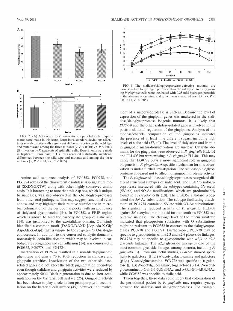
The sialidase/sialoglycoproteases can modulate the invasivecapacity of P. gingivalis. To investigate a putative role of thesialidase (PG0352) and O-sialoglycoproteases (PG0778 andPG1724) in the invasive capacity of P. gingivalis, we com-pared the wild type and isogenic mutants P. gingivalisFLL401, FLL402, and FLL403. Figure 7A shows differentialadherences to HeLa cells for all 3 mutants compared to thewild type. P. gingivalis FLL401 showed the least adherence.FLL402 showed more adherence than the other two mu-tants. P. gingivalis FLL401 showed the highest invasion ca-pacity, followed by FLL402 and FLL403 in descending order(Fig. 7B). In comparison with that of the wild-type strain,the invasion capacity was increased by 16% in FLL401, 13%in FLL402, and 11% in FLL401. Complementation of the
FIG. 3. Comparison of total protease and sialidase activities in P.gingivalis. P. gingivalis was grown to exponential phase (OD600 of 0.8)in 10 ml of BHI broth supplemented with hemin and vitamin K.(A) Total protease and sialidase activities of P. gingivalis mutantsFLL401, FLL402, FLL403, and FLL92. Total protease activity wasestimated using a FRET-based assay, and sialidase was estimated usingan enzyme-based assay method. *, P 0.001; **, P 0.01. (B) Dis-tribution of sialidase activity in P. gingivalis. Sialidase activity using theAmplex Red neuraminidase kit was tested in the total cell fraction (T),the extracellular fraction (Ex), and cell lysate (CL). *, P 0.001.(C) Gingipain activity of P. gingivalis mutants. Activity against BAPNA(Rgp) and ALNA (Kgp) was tested in whole-cell culture. The resultsshown are from three independent experiments. *, P 0.001; **, P 0.01.
VOL. 79, 2011 SIALIDASE ACTIVITY IN PORPHYROMONAS GINGIVALIS 2785
Dow
nloa
ded
from
http
s://j
ourn
als.
asm
.org
/jour
nal/i
ai o
n 30
Jan
uary
202
2 by
87.
110.
64.1
8.

isogenic mutant with the wild-type gene restored the adhe-sion and invasion capacity to the wild-type levels.
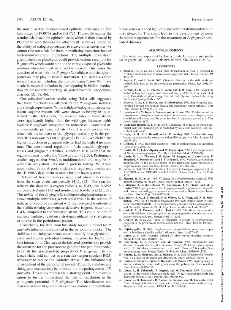
The sialidase/sialoglycoprotease-defective mutants are sen-sitive to oxidative stress. Sialic acid can play a protective role
FIG. 4. Gingipain profile of P. gingivalis sialidase mutants compared with vimA mutant (FLL92) and wild-type W83 from exponential-phasecultures. Acetone-precipitated proteins isolated from supernatants from cultures grown to an OD600 of 0.8 were analyzed by SDS-PAGE andstained with SimplyBlue SafeStain (A) or analyzed by immunoblotting using gingipain-specific antibodies (B to D). Approximately 20 to 25 �g ofprotein was loaded in each lane. Antibodies: chicken anti-RgpA (B), rabbit anti-RgpB (C), and chicken anti-Kgp (D). Ex, extracellular fraction;CL, cell lysate fraction. Arrows indicate the unprocessed, partially processed, or mature gingipains; MW, molecular weight markers (thousands).
TABLE 2. Variation in glycan linkages in various species of RBCs
Source of RBCs Substitution(s)a
Human.....................................5N-Ac (no Gc), no 9O-AcChicken....................................5N-Ac, �95%; 5N-Gc, 5%Sheep .......................................5N-Ac (no Gc), no 9O-AcBovine......................................5N-AcHorse .......................................5N-Gc, 90%; 4O-Ac, 6%; 9O-Ac, 4%
a Gc, glycolyl; Ac, acetyl.
TABLE 3. Hemagglutination titers of P. gingivalisa
StrainTiter for RBCs from:
Bovine Chicken Sheep Horse
FLL401 16 8 16 0FLL402 2 0 16 0FLL403 0 0 8 0W83 2 0 16 0FLL92 4 0 16 0
a Hemagglutination titers of P. gingivalis mutants in bovine, chicken, sheep,and horse RBCs. There was no hemagglutination in horse RBCs by isogenicmutants. FLL401 showed high titers in bovine and sheep RBCs. FLL402 andFLL403 showed highest titers in sheep RBCs. Hemagglutination titer was de-termined as the reciprocal of the highest dilution showing complete hemagglu-tination.
2786 ARUNI ET AL. INFECT. IMMUN.
Dow
nloa
ded
from
http
s://j
ourn
als.
asm
.org
/jour
nal/i
ai o
n 30
Jan
uary
202
2 by
87.
110.
64.1
8.

against oxidative damage (22). The relative significance of theP. gingivalis sialidase and O-sialoglycoprotease in oxidativestress resistance was evaluated by exposure of the isogenicdefective mutants to hydrogen peroxide. As shown in Fig. 8,
isogenic mutants defective in PG0778 and PG0352 were moresensitive to hydrogen peroxide than the wild type. P. gingivalisFLL402, carrying the PG1724 inactivated gene, showed a sen-sitivity pattern similar to that of the wild type.
FIG. 5. Substrate specificity of P. gingivalis mutants.(A) Table showing substrates used in this study with their respective glycoside linkages tothe substrate. (B) Sialidase activity of P. gingivalis W83 and isogenic mutants FLL401, FLL402, and FLL403. The assay was performed using theAmplex Red neuraminidase (sialidase) assay kit (Molecular Probes). The three P. gingivalis mutants showed significant reduction in activitycompared with the wild type (*, P 0.001; **, P 0.01).
TABLE 4. Lectins and their sugar-specific linkages
Lectin (source) Sugar specificity
ABA (Agaricus bisporus)............................Galactose (� 1,3)N-acetylgalactosamine
ConA (Canavalia ensiformis) ....................�-Mannose; �-glucoseECA (Erythrina cristagalli).........................Galactose (� 1,4)
N-acetylglucosamineLPA (Limulus polyphemus) .......................Sialic acid
(N-acetylneuraminic acid)WGA (Triticum vulgaris) ...........................N-Acetyl-�-D-glucosaminePNA (Arachis hypogaea) ............................D-Galactose (� 1,3)
N-acetylglucosamineD-Galactose (� 1,4)
N-acetylglucosamine
TABLE 5. Summary of lectin hemagglutination titersa
StrainTiter in the presence of:
ABA ECA LPA ConA WGA PNA
W83 32 64 256 0 256 16FLL401 0 2 4 128 4 8FLL402 8 32 16 256 16 0FLL403 8 4 0 256 8 128
a Hemagglutination titer is the reciprocal of the highest dilution showingagglutination. P. gingivalis mutants FLL401 and FLL402 showed very low hem-agglutination titers in the presence of ECA and LPA.
VOL. 79, 2011 SIALIDASE ACTIVITY IN PORPHYROMONAS GINGIVALIS 2787
Dow
nloa
ded
from
http
s://j
ourn
als.
asm
.org
/jour
nal/i
ai o
n 30
Jan
uary
202
2 by
87.
110.
64.1
8.

DISCUSSION
The microenvironment of the periodontal pocket containsan abundance of sialylated glycoproteins such as fibronectin(33), transferrin, and immunoglobulins (15), found in salivaand gingival crevicular fluid (48). While a number of periodon-tal bacteria are known to exploit host sialylated glycoproteinsas a nutrient source, sialidase activity also plays an importantrole in their pathogenicity. Sialidase treatment of immunoglob-ulins can make them more susceptible to proteolytic degrada-tion; sialidases may also reveal cryptic receptors and/or adhe-sion sites for bacterial binding/interaction and hence tissue andhost tropism (5, 19). Our studies have identified three siali-dase-related genes in P. gingivalis which have shown a specificpattern of clustering with other related genes from the bacte-ria. Our in silico analysis and gene knockout studies revealedPG0352 to be a putative sialidase and an ortholog of theproducts of nanH of T. forsythia (TF0035) and ANA1493 and
ANA2709 of A. naeslundii. PG1724 showed similarity to glyco-proteases from Bacillus subtilis, Haemophilus influenzae, and E.coli. Further, based on domain similarity, PG0778 and PG1724were grouped with other metal-dependent proteases and alsomay have chaperone activity (oral pathogen sequence data-bases [www.oralgen.lanl.gov]). In our study, the sialoglycopro-tease appears to be important for the growth of P. gingivalis. Itis likely that these enzymes are important in the breakdown ofglycoprotein conjugate to satisfy the asaccharolytic require-ments of P. gingivalis. However, we cannot rule out the involve-ment of these enzymes in other important metabolic activitiesof the cell, including the generation of sialic acid from host-derived substrates. This sialic acid could be involved in stabilityof proteins, including the gingipains. Thus, it is likely thatimportant proteins involved in both the metabolic and patho-genic capacity of the organism are modulated by the presenceor absence of sialic acid. This is under further investigation.
FIG. 6. Electron micrograph showing variation in the surface morphology of P. gingivalis W83 and isogenic mutants FLL401, FLL402, andFLL403. The samples were processed from log-phase cultures of the mutants (OD600 0.8) negatively stained on 400 mesh copper grids using1% phosphotungstic acid and viewed under the transmission electron microscope (FEI Technai G2). (A) W83 showing blebbing of membranevesicles (V); (B) FLL401 showing fuzzy and thick external layer (E); (C) FLL402 showing intact outer covering; (D) FLL403 showing diffused outercovering with dense cytoplasm. Images represent a consistent view of several fields.
2788 ARUNI ET AL. INFECT. IMMUN.
Dow
nloa
ded
from
http
s://j
ourn
als.
asm
.org
/jour
nal/i
ai o
n 30
Jan
uary
202
2 by
87.
110.
64.1
8.
Amino acid sequence analysis of PG0352, PG0778, andPG1724 revealed the characteristic sialidase Asp signature mo-tif (SXDXGXTW) along with other highly conserved aminoacids. It is interesting to note that this Asp box, which is uniqueto sialidases, was also observed in the O-sialoglycoproteasesfrom other oral pathogens. This may suggest functional relat-edness and may highlight their relative significance in micro-bial colonization of the periodontal pocket with an abundanceof sialylated glycoproteins (54). In PG0352, a FRIP region,which is known to bind the carboxylate group of sialic acid(14), was juxtaposed to the exosialidase domain. Our studyidentified a common motif (DAXG/DAXD [Asp-Ala-X-Gly/Asp-Ala-X-Asp]) that is unique to the P. gingivalis O-sialogly-coproteases. In addition to the conserved catalytic domain, anoncatalytic lectin-like domain, which may be involved in car-bohydrate recognition and cell adhesion (14), was conserved inPG0352, PG0778, and PG1724.
Inactivation of PG0778 resulted in a non-black-pigmentedphenotype and also a 70 to 90% reduction in sialidase andgingipain activities. Inactivation of the two other sialidase-related genes did not affect the black pigmentation phenotypeeven though sialidase and gingipain activities were reduced byapproximately 50%. Black pigmentation is due to iron accu-mulation on the bacterial cell surface (26). Gingipain activityhas been shown to play a role in iron protoporphyrin accumu-lation on the bacterial cell surface (45); however, the involve-
ment of a sialoglycoprotease is unclear. Because the level ofexpression of the gingipain genes was unaltered in the siali-dase/sialoglycoprotease isogenic mutants, it is likely thatPG0778 and the other sialidase-related gene is involved in theposttranslational regulation of the gingipains. Analysis of themonosaccharide composition of the gingipains indicatesthe presence of at least nine different sugars, including highlevels of sialic acid (37, 40). The level of sialylation and its rolein gingipain maturation/activation are unclear. Catalytic do-mains for the gingipains were observed in P. gingivalis FLL402and FLL403 but were missing in P. gingivalis FLL401. This mayimply that PG0778 plays a more significant role in gingipainbiogenesis in P. gingivalis. A specific mechanism for this obser-vation is under further investigation. The sialidase/sialoglyco-protease appeared not to affect nongingipain protease activity.
The P. gingivalis sialidase/sialoglycoproteases recognized dif-ferent structural subtypes of sialic acid. The PG0778 sialogly-coprotease interacted with the subtypes containing 5N-acetyl(5N-Ac) and 9O-Ac modifications, which are predominantlyfound in eukaryotic cells (18). The PG0352 sialidase recog-nized the 5N-Ac substitution. The subtype facilitating attach-ment of PG1774 contained 5N-Ac with 9O-Ac substitutions.The significantly reduced activity of P. gingivalis FLL403against 3N-acetylneuraminic acid further confirms PG0352 as aputative sialidase. The cleavage level of the mucin substrateindicated that glycoprotein substrates with O substitutionsmight be resistant to PG0352 in contrast to the sialoglycopro-teases PG0778 and PG1724. Furthermore, PG0778 may bespecific to glycoproteins with �2,3 and �2,6 glyco-side linkages.PG1724 may be specific to glycoproteins with �2,3 or �2,8glycoside linkages. The �2,3 glycoside linkage is one of themost common glycoside linkages among bacteria, including P.gingivalis (3). From our lectin studies, PG0778 showed speci-ficity to galactose (� 1,3) N-acetylgalactosamine and galactose(�1,4) N-acetylglucosamine. PG1724 was specific to D-galac-tose (� 1,3) N-acetylglucosamine, D-galactose (� 1,4) N-acetyl-glucosamine, D-Gal-�-1-3dGalNAc, and D-Gal-�-1-4dGlcNAc,while PG0352 was specific to sialic acid.
Taken together, these data could imply that colonization ofthe periodontal pocket by P. gingivalis may require synergybetween the sialidase and sialoglycoproteases. For example,
FIG. 8. The sialidase/sialoglycoprotease-defective mutants aremore sensitive to hydrogen peroxide than the wild type. Actively grow-ing P. gingivalis cells were incubated with 0.25 mM hydrogen peroxidein the absence of cysteine, and growth was measured over 25 h (*, P 0.001; **, P 0.05).
FIG. 7. (A) Adherence by P. gingivalis to epithelial cells. Experi-ments were made in triplicate. Error bars, standard deviations (SD). ttests revealed statistically significant differences between the wild typeand mutants and among the three mutants (*, P 0.001; **, P 0.01).(B) Invasion by P. gingivalis of epithelial cells. Experiments were madein triplicate. Error bars, SD. t tests revealed statistically significantdifferences between the wild type and mutants and among the threemutants (*, P 0.01; **, P 0.05).
VOL. 79, 2011 SIALIDASE ACTIVITY IN PORPHYROMONAS GINGIVALIS 2789
Dow
nloa
ded
from
http
s://j
ourn
als.
asm
.org
/jour
nal/i
ai o
n 30
Jan
uary
202
2 by
87.
110.
64.1
8.
the mucin on the mucin-covered epithelial cells may be firsthydrolyzed by PG0778 and/or PG1724. This would expose theterminal sialic acid on epithelial cells, which is then cleaved byPG0352 to mediate/enhance attachment. However, based onthe ability of sialoglycoproteases to cleave other substrates, wecannot rule out a role for them in mediating bacterium-host orbacterium-bacterium interactions. The multiple desialylatedglycoproteins or glycolipids could provide various receptors forP. gingivalis which would bind to the various exposed glycosidicresidues when terminal sialic acid is cleaved. This raises thequestion of what role the P. gingivalis sialidase and sialoglyco-proteases may play in biofilm formation. The sialidases fromseveral bacteria, including the oral pathogen T. forsythia, havea role in mucosal infection by participating in biofilm produc-tion by presumably targeting sialylated bacterial exopolysac-charides (32, 38, 46).
Our adhesion/invasion studies using HeLa cells suggestedthat these functions are affected by the P. gingivalis sialidaseand sialoglycoproteases. While sialidase/sialoglycoprotease-de-ficient isogenic mutant cells were shown to be physically at-tached to the HeLa cells, the invasion rates of these strainswere significantly higher than the wild type. Because highlyinvasive P. gingivalis subtypes have reduced cell-associated ar-ginine-specific protease activity (47), it is still unclear whatdirect role the sialidase or sialoglycoproteases play in this pro-cess. It is noteworthy that P. gingivalis FLL401, which had thehighest reduction in gingipain activity, had the highest invasionrate. The coordinated regulation of sialidase/sialoglycopro-teases and gingipain activities is unclear. It is likely that theregulation may involve the VimA protein (52, 53). Preliminarystudies suggest that VimA is multifunctional and may be in-volved in acetylation (31) and in protein sorting (W. Aruni,unpublished data). A posttranslational regulatory mechanismthat is VimA dependent is under further investigation.
Release of free monomeric sialic acid when it is cleavedfrom the sugar chain can detoxify H2O2 (22). This reactionreduces the dangerous oxygen radicals, as H2O2 and NANAare converted into H2O and nontoxic carboxylic acid (21, 22).The ability of the P. gingivalis sialidase/sialoglycoproteases tocleave multiple substrates, which could result in the release ofsialic acid, would be consistent with the increased sensitivity ofthe sialidase/sialoglycoprotease-defective isogenic mutants toH2O2 compared to the wild-type strain. This could be one ofmultiple oxidative resistance strategies utilized by P. gingivalisto survive in the periodontal pocket.
Collectively, the data from this study suggest a model for P.gingivalis infection and survival in the periodontal pocket. Thesialidase and sialoglycoproteases can modify host glycoconju-gates and expose potential binding receptors for bacterium-host interactions. Cleavage of desialylated proteins can providethe substrate for the proteases to generate the peptides neededto satisfy the asaccharolytic property of P. gingivalis. The re-leased sialic acid can act as a reactive oxygen species (ROS)scavenger to reduce the oxidative stress in the inflammatoryenvironment of the periodontal pocket. Thus, the sialidase andsialoglycoproteases may be important in the pathogenesis of P.gingivalis. This study represents a starting point in our explo-ration to further understand the role of sialylation in thepathogenic potential of P. gingivalis. The identification andcharacterization of genes such as trans-sialidase and sialyltrans-
ferase genes will shed light on sialic acid metabolism/utilizationin P. gingivalis. This could lead to the development of noveltherapeutic approaches for the treatment of P. gingivalis-asso-ciated diseases.
ACKNOWLEDGMENTS
This work was supported by Loma Linda University and publichealth grants DE-13664 and DE-019730 from NIDER (to H.M.F.).
REFERENCES
1. Abaibou, H., et al. 2001. vimA gene downstream of recA is involved invirulence modulation in Porphyromonas gingivalis W83. Infect. Immun. 69:325–335.
2. Angata, T., and A. Varki. 2002. Chemical diversity in the sialic acids andrelated alpha-keto acids: an evolutionary perspective. Chem. Rev. 102:439–469.
3. Bertozzi, C. R., H. H. Freeze, A. Varki, and J. D. Esko. 2009. Glycans inbiotechnology and the pharmaceutical industry, p. 769–784. In A. Varki et al.(ed.), Essentials of glycobiology, 2nd ed. Cold Spring Harbor LaboratoryPress, Cold Spring Harbor, NY.
4. Bobetsis, Y. A., S. P. Barros, and S. Offenbacher. 2006. Exploring the rela-tionship between periodontal disease and pregnancy complications. J. Am.Dent. Assoc. 137(Suppl.):7S–13S.
5. Cacalano, G., M. Kays, L. Saiman, and A. Prince. 1992. Production of thePseudomonas aeruginosa neuraminidase is increased under hyperosmolarconditions and is regulated by genes involved in alginate expression. J. Clin.Invest. 89:1866–1874.
6. Castaneda-Roldan, E. I., et al. 2004. Adherence of brucella to human epi-thelial cells and macrophages is mediated by sialic acid residues. Cell. Mi-crobiol. 6:435–445.
7. Copley, R. R., R. B. Russell, and C. P. Ponting. 2001. Sialidase-like Asp-boxes: sequence-similar structures within different protein folds. Protein Sci.10:285–292.
8. Corfield, T. 1992. Bacterial sialidases—roles in pathogenicity and nutrition.Glycobiology 2:509–521.
9. Curtis, M. A., J. duse-Opoku, and M. Rangarajan. 2001. Cysteine proteasesof Porphyromonas gingivalis. Crit. Rev. Oral Biol. Med. 12:192–216.
10. Curtis, M. A., A. Thickett, J. M. Slaney, M. Rangarajan, J. duse-Opoku, P.Shepherd, N. Paramonov, and E. F. Hounsell. 1999. Variable carbohydratemodifications to the catalytic chains of the RgpA and RgpB proteases ofPorphyromonas gingivalis W50. Infect. Immun. 67:3816–3823.
11. Farre, D., et al. 2003. Identification of patterns in biological sequences at theALGGEN server: PROMO and MALGEN. Nucleic Acids Res. 31:3651–3653.
12. Fletcher, H. M., et al. 1995. Virulence of a Porphyromonas gingivalis W83mutant defective in the prtH gene. Infect. Immun. 63:1521–1528.
13. Gallagher, A., J. duse-Opoku, M. Rangarajan, J. M. Slaney, and M. A.Curtis. 2003. Glycosylation of the Arg-gingipains of Porphyromonas gingivalisand comparison with glycoconjugate structure and synthesis in other bacte-ria. Curr. Protein Pept. Sci. 4:427–441.
13a.Gardner, R. G., J. B. Russell, D. B. Wilson, G. R. Wang, and N. B. Shoe-maker. 1996. Use of a modified Bacteroides-Prevotella shuttle vector to trans-fer a reconstructed beta-1,4-D-endoglucanase gene into Bacteroides uniformisand Prevotella ruminicola B(1)4. Appl. Environ. Microbiol. 62:196–202.
14. Gaskell, A., S. Crennell, and G. Taylor. 1995. The three domains of abacterial sialidase: a beta-propeller, an immunoglobulin module and a ga-lactose-binding jelly-roll. Structure 3:1197–1205.
15. Grenier, D., et al. 2001. Role of gingipains in growth of Porphyromonasgingivalis in the presence of human serum albumin. Infect. Immun. 69:5166–5172.
16. Hajishengallis, G. 2009. Porphyromonas gingivalis-host interactions: openwar or intelligent guerilla tactics? Microbes Infect. 11:637–645.
17. Harris, J. R. 2007. Negative staining of thinly spread biological samples.Methods Mol. Biol. 369:107–142.
18. Haverkamp, J., R. Schauer, and M. Wember. 1976. Neuraminic acidderivatives newly discovered in humans: N-acetyl-9-O-L-lactoylneuraminicacid, N, 9-O-diacetylneuraminic acid and N-acetyl-2,3-dehydro-2-de-oxyneuraminic acid. Hoppe-Seylers Z. Physiol. Chem. 357:1699–1705.
19. Honma, K., E. Mishima, and A. Sharma. 2011. Role of Tannerella forsythiaNanH sialidase in epithelial cell attachment. Infect. Immun. 79:393–401.
20. Horton, R. M., Z. L. Cai, S. N. Ho, and L. R. Pease. 1990. Gene splicing byoverlap extension: tailor-made genes using the polymerase chain reaction.Biotechniques 8:528–535.
21. Iijima, R., H. Takahashi, S. Ikegami, and M. Yamazaki. 2007. Character-ization of the reaction between sialic acid (N-acetylneuraminic acid) andhydrogen peroxide. Biol. Pharm. Bull. 30:580–582.
22. Iijima, R., H. Takahashi, R. Namme, S. Ikegami, and M. Yamazaki. 2004.Novel biological function of sialic acid (N-acetylneuraminic acid) as a hy-drogen peroxide scavenger. FEBS Lett. 561:163–166.
2790 ARUNI ET AL. INFECT. IMMUN.
Dow
nloa
ded
from
http
s://j
ourn
als.
asm
.org
/jour
nal/i
ai o
n 30
Jan
uary
202
2 by
87.
110.
64.1
8.

23. Johnson, L. S., S. R. Eddy, and E. Portugaly. 2010. Hidden Markov modelspeed heuristic and iterative HMM search procedure. BMC Bioinformatics11:431.
24. Kadowaki, T., et al. 1998. Arg-gingipain acts as a major processing enzymefor various cell surface proteins in Porphyromonas gingivalis. J. Biol. Chem.273:29072–29076.
25. Lamont, R. J., and H. F. Jenkinson. 1998. Life below the gum line: patho-genic mechanisms of Porphyromonas gingivalis. Microbiol. Mol. Biol. Rev.62:1244–1263.
26. Lewis, J. P., J. A. Dawson, J. C. Hannis, D. Muddiman, and F. L. Macrina.1999. Hemoglobinase activity of the lysine gingipain protease (Kgp) of Por-phyromonas gingivalis W83. J. Bacteriol. 181:4905–4913.
27. Marmur, J. 1961. A procedure for the isolation of deoxyribonucleic acidfrom micro-organisms. J. Mol. Biol. 3:585–594.
28. Nabi, I. R., and A. Raz. 1987. Cell shape modulation alters glycosylation ofa metastatic melanoma cell-surface antigen. Int. J. Cancer 40:396–402.
29. Nakano, V., R. M. Fontes Piazza, and M. J. vila-Campos. 2006. A rapid assayof the sialidase activity in species of the Bacteroides fragilis group by usingpeanut lectin hemagglutination. Anaerobe 12:238–241.
30. Olango, G. J., F. Roy, S. M. Sheets, M. K. Young, and H. M. Fletcher. 2003.Gingipain RgpB is excreted as a proenzyme in the vimA-defective mutantPorphyromonas gingivalis FLL92. Infect. Immun. 71:3740–3747.
31. Osbourne, D. O., et al. 2010. The role of vimA in cell surface biogenesis inPorphyromonas gingivalis. Microbiology 156:2180–2193.
32. Parker, D., et al. 2009. The NanA neuraminidase of Streptococcus pneu-moniae is involved in biofilm formation. Infect. Immun. 77:3722–3730.
33. Pham, T. K., et al. 2010. A quantitative proteomic analysis of biofilm adap-tation by the periodontal pathogen Tannerella forsythia. Proteomics 10:3130–3141.
34. Pihlstrom, B. L., B. S. Michalowicz, and N. W. Johnson. 2005. Periodontaldiseases. Lancet 366:1809–1820.
35. Plumbridge, J., and E. Vimr. 1999. Convergent pathways for utilization ofthe amino sugars N-acetylglucosamine, N-acetylmannosamine, and N-acetyl-neuraminic acid by Escherichia coli. J. Bacteriol. 181:47–54.
36. Powell, L. D., and A. P. Varki. 2001. Sialidases. Curr. Protoc. Mol. Biol.Chapter 17:Unit17.
37. Rangarajan, M., A. Hashim, J. duse-Opoku, N. Paramonov, E. F. Hounsell,and M. A. Curtis. 2005. Expression of Arg-gingipain RgpB is required forcorrect glycosylation and stability of monomeric Arg-gingipain RgpA fromPorphyromonas gingivalis W50. Infect. Immun. 73:4864–4878.
38. Roy, S., C. W. Douglas, and G. P. Stafford. 2010. A novel sialic acid utiliza-tion and uptake system in the periodontal pathogen Tannerella forsythia. J.Bacteriol. 192:2285–2293.
39. Saitou, N., and M. Nei. 1987. The neighbor-joining method: a new methodfor reconstructing phylogenetic trees. Mol. Biol. Evol. 4:406–425.
40. Sakai, E., et al. 2007. Construction of recombinant hemagglutinin derivedfrom the gingipain-encoding gene of Porphyromonas gingivalis, identificationof its target protein on erythrocytes, and inhibition of hemagglutination byan interdomain regional peptide. J. Bacteriol. 189:3977–3986.
41. Sali, A., and T. L. Blundell. 1993. Comparative protein modelling by satis-faction of spatial restraints. J. Mol. Biol. 234:779–815.
42. Schauer, R. 2009. Sialic acids as regulators of molecular and cellular inter-actions. Curr. Opin. Struct. Biol. 19:507–514.
43. Severi, E., D. W. Hood, and G. H. Thomas. 2007. Sialic acid utilization bybacterial pathogens. Microbiology 153:2817–2822.
44. Sharma, A., S. Inagaki, W. Sigurdson, and H. K. Kuramitsu. 2005. Synergybetween Tannerella forsythia and Fusobacterium nucleatum in biofilm forma-tion. Oral Microbiol. Immunol. 20:39–42.
45. Smalley, J. W., M. F. Thomas, A. J. Birss, R. Withnall, and J. Silver. 2004.A combination of both arginine- and lysine-specific gingipain activity ofPorphyromonas gingivalis is necessary for the generation of the micro-oxobishaem-containing pigment from haemoglobin. Biochem. J. 379:833–840.
46. Soong, G., et al. 2006. Bacterial neuraminidase facilitates mucosal infectionby participating in biofilm production. J. Clin. Invest. 116:2297–2305.
47. Suwannakul, S., G. P. Stafford, S. A. Whawell, and C. W. Douglas. 2010.Identification of bistable populations of Porphyromonas gingivalis that differin epithelial cell invasion. Microbiology 156:3052–3064.
48. Takamatsu, D., B. A. Bensing, A. Prakobphol, S. J. Fisher, and P. M. Sullam.2006. Binding of the streptococcal surface glycoproteins GspB and Hsa tohuman salivary proteins. Infect. Immun. 74:1933–1940.
49. Tamura, K., J. Dudley, M. Nei, and S. Kumar. 2007. MEGA4: MolecularEvolutionary Genetics Analysis (MEGA) software version 4.0. Mol. Biol.Evol. 24:1596–1599.
50. Vanterpool, E., A. W. Aruni, F. Roy, and H. M. Fletcher. 2010. regT canmodulate gingipain activity and response to oxidative stress in Porphyromo-nas gingivalis. Microbiology 156:3065–3072.
51. Vanterpool, E., F. Roy, and H. M. Fletcher. 2005. Inactivation of vimF, aputative glycosyltransferase gene downstream of vimE, alters glycosylationand activation of the gingipains in Porphyromonas gingivalis W83. Infect.Immun. 73:3971–3982.
52. Vanterpool, E., F. Roy, L. Sandberg, and H. M. Fletcher. 2005. Alteredgingipain maturation in vimA- and vimE-defective isogenic mutants of Por-phyromonas gingivalis. Infect. Immun. 73:1357–1366.
53. Vanterpool, E., et al. 2006. VimA is part of the maturation pathway for themajor gingipains of Porphyromonas gingivalis W83. Microbiology 152:3383–3389.
54. Varki, A. 2009. Multiple changes in sialic acid biology during human evolu-tion. Glycoconj. J. 26:231–245.
55. Vimr, E. R. 1994. Microbial sialidases: does bigger always mean better?Trends Microbiol. 2:271–277.
56. Vriend, G. 1990. WHAT IF: a molecular modeling and drug design program.J. Mol. Graph. 8:52–56, 29.
57. Wang, Q., B. J. Chang, and T. V. Riley. 2010. Erysipelothrix rhusiopathiae.Vet. Microbiol. 140:405–417.
58. Yilmaz, O., P. A. Young, R. J. Lamont, and G. E. Kenny. 2003. Gingivalepithelial cell signalling and cytoskeletal responses to Porphyromonas gingi-valis invasion. Microbiology 149:2417–2426.
Editor: A. J. Baumler
VOL. 79, 2011 SIALIDASE ACTIVITY IN PORPHYROMONAS GINGIVALIS 2791
Dow
nloa
ded
from
http
s://j
ourn
als.
asm
.org
/jour
nal/i
ai o
n 30
Jan
uary
202
2 by
87.
110.
64.1
8.