signal transmission and signal transduction xia qiang, md & phd department of physiology room...
TRANSCRIPT

Signal transmission and
signal transduction
XIA Qiang, MD & PhDDepartment of Physiology
Room 518, Block C, Research Building
School of Medicine, Zijingang Campus
Email: [email protected]
Tel: 88206417 (Undergraduate school),
88208252 (Medical school)
OUTLINE
Intercellular signal transmissionChemical transmissionElectrical transmission
Signal transduction pathwayPathways initiated by intracellular receptorsPathways initiated by plasma membrane receptors

Intercellular signal transmission
Chemical transmissionChemical signals
Neurotransmitters:

Intercellular signal transmission
Chemical transmissionChemical signals
Neurotransmitters: Humoral factors:
HormonesCytokinesBioactivators
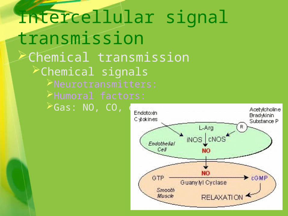
Intercellular signal transmission
Chemical transmissionChemical signals
Neurotransmitters: Humoral factors: Gas: NO, CO, etc.

Intercellular signal transmission
Chemical transmissionChemical signalsReceptors
Membrane receptorsIntracellular receptors

Receptors on the surface of a cell are typically proteins that span the membrane

Cells B & C lack the matching receptorsTherefore are not directly affected by the signal
Only Cell A has thematching receptorsfor this chemical messenger, so it is the only one that responds


Intercellular signal transmission
Electrical transmissionGap junction
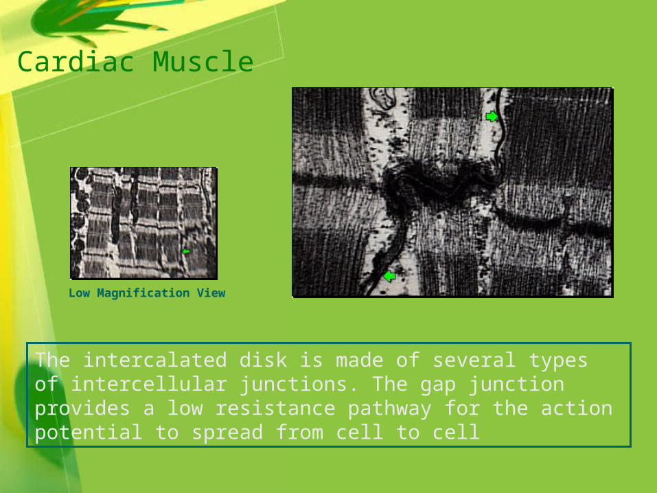
Low Magnification View
The intercalated disk is made of several types of intercellular junctions. The gap junction provides a low resistance pathway for the action potential to spread from cell to cell
Cardiac Muscle
Signal transduction pathway
Pathways initiated by intracellular receptorsPathways initiated by plasma membrane
receptors

… but at the target cellthe signal moves easily through the membraneand binds to its receptor
This hydrophobic signal requires a carrier protein while in the plasma …

Signal transduction pathway
Pathways initiated by intracellular receptorsPathways initiated by plasma membrane
receptors (transmembrane signal transduction)
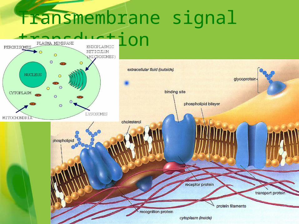
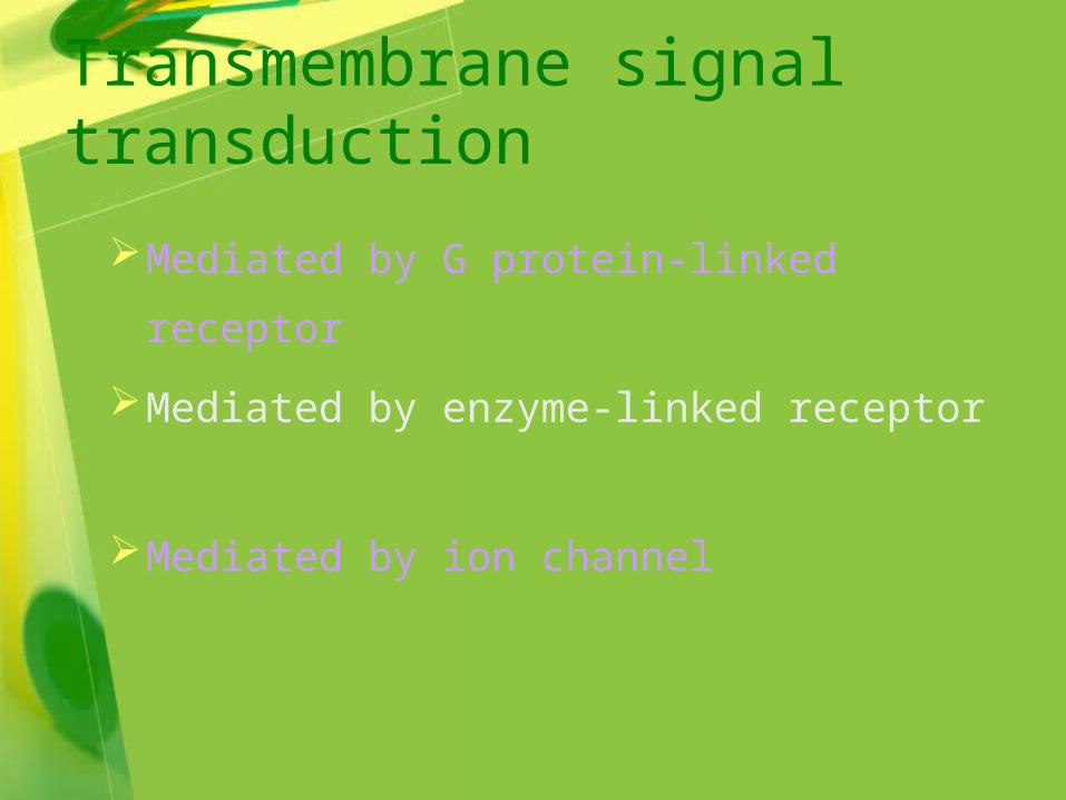
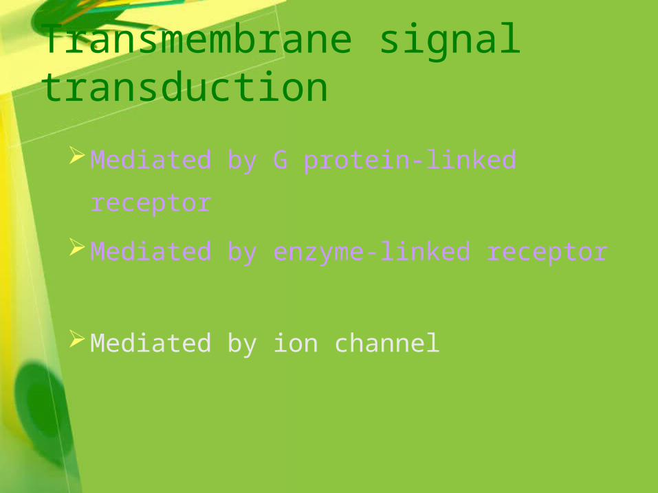
Transmembrane signal transduction

Mediated by G protein-linked receptor
Mediated by enzyme-linked receptor
Mediated by ion channel
Transmembrane signal transduction

Binding of ligands to membrane-spanning receptorsactivates diverse response mechanisms
Mediated by G protein-linked receptor
Mediated by enzyme-linked receptor
Mediated by ion channel
Transmembrane signal transduction

The Nobel Prize in Physiology or Medicine 1994
"G-proteins and the role of these proteins in signal transduction in cells"
Alfred G. Gilman Martin Rodbell

The Discovery of G Proteins
Normal Lymphoma Cell Mutated Lymphoma Cell

Activation and Inactivation of the G Protein
1. The G protein, composed of alpha-, beta- and gamma-subunits, in its resting state with bound GDP.
2. The receptor with bound hormone activates the G protein and replaces GDP...
3. ...with GTP and the G proteins is activated. The subunits separate.
4. Some seconds later the GTP, bound to the alpha-subunit, is hydrolysed to GDP. The subunits recombine.

G-protein-coupled Receptors

Signaling molecules involved
Second messenger
G protein effector
Protein kinase


(1) cAMP-PKA pathway
(2) IP3-Ca2+ pathway
(3) DG-PKC pathway
(4) G protein-ion channel pathway
Main signaling pathwaysMain signaling pathways

(1) cAMP-PKA pathway
Gs, Gi

The cyclic AMP second messenger system

Adenylyl cyclase forms cAMP,a “second messenger” that activates enzymes used in cellular responses
The phosphodiesterase enzymes “terminate” thesecond messenger cAMP

The cAMP system rapidly amplifies the responsecapacity of cells: here, one “first messenger” ledto the formation of one million product molecules
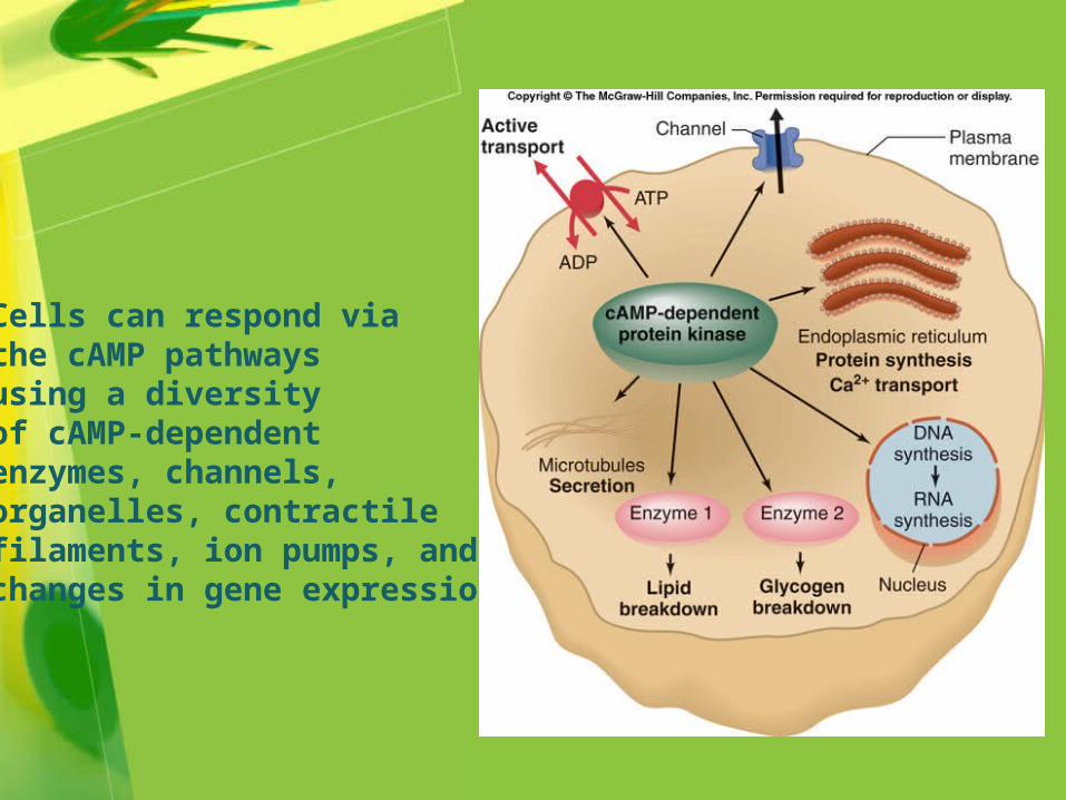
Cells can respond via the cAMP pathwaysusing a diversity of cAMP-dependentenzymes, channels,organelles, contractile filaments, ion pumps, and changes in gene expression
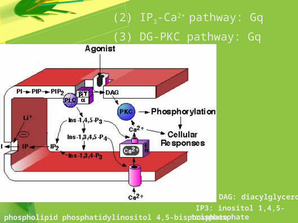
(2) IP3-Ca2+ pathway: Gq
(3) DG-PKC pathway: Gq
PIP2: phospholipid phosphatidylinositol 4,5-bisphosphate
DAG: diacylglycerol
IP3: inositol 1,4,5-trisphosphate

This receptor-G-protein complex is linked to and activates phospholipase C, leading to an increase in IP3 and DAG, which work together to activate enzymes and to increase intracellular calcium levels
Click here to play theMembrane Bound Receptors,
G Proteins,and Calcium Channels
Flash Animation

(4) G protein-ion channel pathway(4) G protein-ion channel pathway

Binding of the ligand to the receptor alters the receptor’s shape, which activates an associated G-protein, which then activates effector proteins,i.e., enzyme functions or ion channels
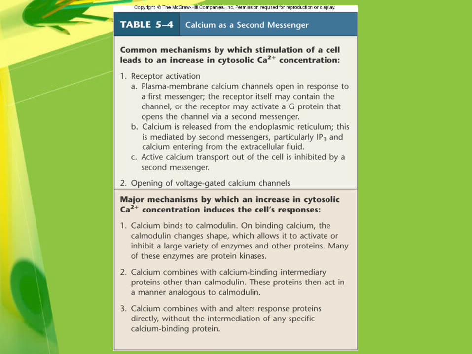

The calcium-calmodulinsystem is similar to some of the cAMP pathways, because it results in the activation of protein kinases that can phosphorylate key proteins required for cellular responses

The “arachidonic acid cascade” is activated in inflammation responses; “cox inhibitors” block cyclooxygenase

Not all responses to hydrophilic signals are immediate:
Increases in gene expression can occur, and the resulting proteins can increase the target cells’ response

Eicosanoid: A lipid mediator of inflammation derived from the 20-carbon atom arachidonic acid (20 in Greek is "eicosa") or a similar fatty acid. The eicosanoids include the prostaglandins, prostacyclin, thromboxane, and leukotrienes.
Mediated by G protein-linked receptor
Mediated by enzyme-linked receptor
Mediated by ion channel
Transmembrane signal transduction

Binding of the ligand to the receptor alters the receptor’s shape, which activates its enzyme function, phosphorylating an intracellular protein

(1) Tyrosine Kinase Receptor
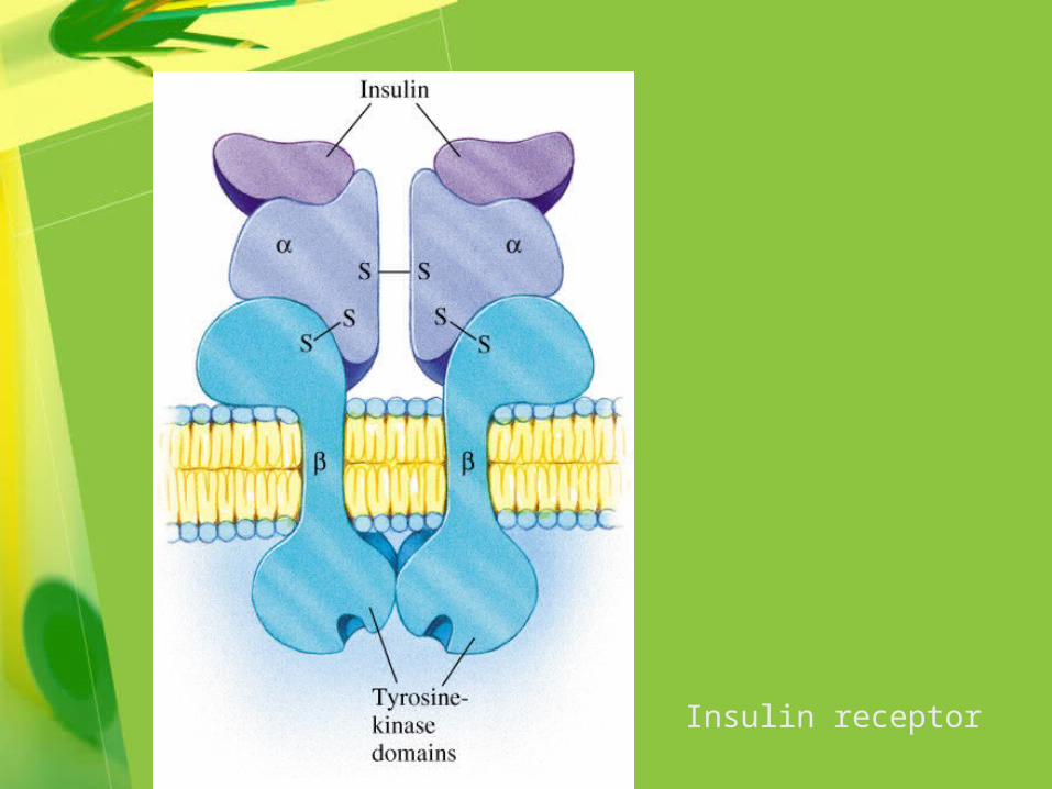
Insulin receptor

Growth factor receptor

Mitogen-activated protein kinase (MAPK) pathway
•PTK: protein tyrosine kinasePTK: protein tyrosine kinase
•Ras: G protein. It consists of an Ras: G protein. It consists of an subunit, subunit, subunit, and subunit, and subunit subunit
•MAPKKK: MAPK kinase kinaseMAPKKK: MAPK kinase kinase
•MAPKK: MAPK kinaseMAPKK: MAPK kinase
•MKP: MAPK phosphatasesMKP: MAPK phosphatases
•TF: tissue factor TF: tissue factor

(2) Receptor-associated tyrosine kinase
JAK stands for Janus kinase or Just Another Kinase
JAK2=Tyrosine protein kinase 2
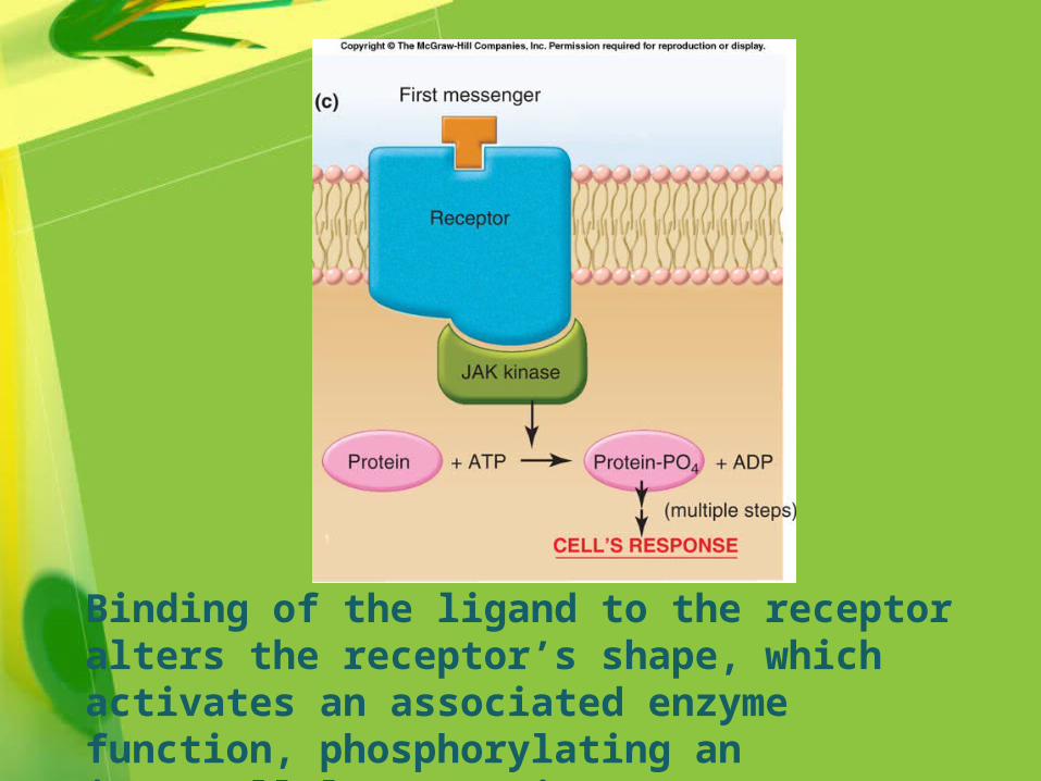
Binding of the ligand to the receptor alters the receptor’s shape, which activates an associated enzyme function, phosphorylating an intracellular protein

(3) Receptor guanylyl cyclase
Soluble GC
Mediated by G protein-linked receptor
Mediated by enzyme-linked receptor
Mediated by ion channel
Transmembrane signal transduction

Binding of the ligand to the receptoralters the receptor’s shape, which then opens (or closes) an ion channel

NN22-ACh receptor channel-ACh receptor channel


Any other pathway else???

Pheromone
Pheromones are chemicals emitted by living organisms to send messages to individuals of the same species.

The male silk moth bombyx mori (Fig. 1, left) can detect very low quantities of the sex pheromone bombykol emitted by the female, and at the same time distinguishes bombykol from many other, often similar, volatile compounds in the air. A first step of olfaction is the tranfer of bombykol to the pheromone receptor at the neuronal membrane via the pheromone-binding protein (BmorPBP). Highly efficient uptake and release kinetics of bombykol at the pheromone binding protein is essential for olfactory function. However, BmorPBP features a central hydrophobic binding cavity for bombykol, completely encapsulating the ligand (Fig. 1, right) and the exit/entrance gate for bombykol is not known. Yet, the ligand has to enter and exit the cavity fast and reversibly.

THANK YOU FOR YOUR ATTENTION!