skull base and calvarial deformities: association with ... · pdf fileherniation of the...
TRANSCRIPT

Skull Base and Calvarial Deformities: Association with IntracranialChanges in Craniofacial Syndromes
Aya M. Tokumaru, A. James Barkovich, Samuel F. Ciricillo, and Michael S. B. Edwards
PURPOSE: To analyze the skull and brain malformations in patients with craniofacial syndromes.METHODS: A retrospective analysis of imaging studies of 21 children with craniofacial anomalies(8 with Apert syndrome, 6 with Pfeiffer syndrome, 4 with Crouzon syndrome, 1 with Robertsyndrome, 1 with Coffin-Lowry-syndrome, and 1 with Saethre-Chotzen syndrome) was carried outusing CT (21 patients), MR imaging (9 patients), and MR venography (2 patients). A series ofqualitative and quantitative assessments of the skull base and intracranial structures was per-formed. RESULTS: Skull base abnormalities were present in all patients. Intracranial abnormalitiesincluded ventriculomegaly, frank hydrocephalus, callosal anomalies, hypoplasia/absence of theseptum pellucidum, hypoplasia/dysplasia of the hippocampus, dysplasias or distortions of thecerebral cortex, and parenchymal hemorrhage. The anomalies of the corpus callosum, septumpellucidum, and hippocampus appeared primary, whereas the others may have been the result ofbrain distortion by the calvarial anomaly. MR imaging was more useful than CT for evaluating brainabnormalities. In the two patients in whom it was performed, MR venography showed anomalies ofthe venous system, indicating that venous anomalies, possibly related to the skull base hypoplasia,may contribute to the intracranial abnormalities. CONCLUSION: A wide range of neuroimagingabnormalities are present in the craniofacial syndromes. Some of these are clearly primary,whereas others appear to be related to the small skull base and sutural synostoses. MR venographymay prove useful in defining the cause of some of the associated anomalies.
Index terms: Brain, abnormalities and anomalies; Children, diseases; Skull, abnormalities andanomalies
AJNR Am J Neuroradiol 17:619–630, April 1996
Craniofacial anomalies are developmentalabnormalities of the face and skull that are fre-quently associated with malformations of thecentral nervous system. From an embryologicperspective, most of these disorders are be-lieved to result from insufficient formation, orinadequate migration, of mesenchyme to theskull base and the face (1, 2). Anomalies of thebrain are particularly important, as studies have
Received May 25, 1995; accepted after revision October 18.From the Departments of Radiology (A.M.T., A.J.B.) and Neurosurgery
(A.J.B., S.F.C., M.S.B.E.), University of California, San Francisco; and theDepartment of Radiology, Kameda Medical Center, Kamogawa City, Japan(A.M.T.).
Address reprint requests to A. James Barkovich, MD, Department ofNeuroradiology, Room L-371, University of California, San Francisco, 505Parnassus Ave, San Francisco, CA 94143.
AJNR 17:619–630, Apr 1996 0195-6108/96/1704–0619
q American Society of Neuroradiology
61
shown that those patients with brain anomalieshave poorer clinical outcomes (3, 4). The pur-pose of this study was to analyze the skull base,calvaria, and intracranial contents of patientswith these disorders using computed tomogra-phy (CT), magnetic resonance (MR) imaging,and MR venography. We briefly discuss the clin-ical manifestations and theories of the patho-physiology of these disorders.
Materials and MethodsA review of records in the pediatric neurosurgery divi-
sion and radiology file room at our institution revealed thatneuroimaging studies were available for 21 children whohad undergone craniofacial surgery over the past 10 years.A retrospective analysis of imaging studies of these chil-dren (8 with Apert syndrome, 4 with Crouzon syndrome, 6with Pfeiffer syndrome, 1 with Robert syndrome, 1 withCoffin-Lowry syndrome, and 1 with Saethre-Chotzen syn-drome) was carried out. Patients ranged from 1 day to 9years old at the time of the study. Standard axial CT
9

examinations were performed using 3- to 5-mm-thick sec-tions and processed with both bone and soft-tissue algo-rithms in all of the 21 patients. Two patients, both withbilateral external auditory canal altresia, had temporalbone CT studies with 1.5-mm axial and coronal sectionsthrough the petrous portions of the temporal bones using abone algorithm. MR examinations were performed in 9patients, all at 1.5 T. MR imaging parameters included a256 3 128 or 192 matrix; 3- to 7-mm section thickness(with an intersection gap of 1 to 3 mm). Axial 4-mmspin-echo sequences at 2500–3000/30–60,80–120/1(repetition time/echo time/excitations), and sagittal spin-echo sequences at 600/11/1 were obtained in all 9 pa-tients. Coronal spin-echo sequences at 600/11/1 wereobtained in 5 patients. A three-dimensional gradient-echosequence with gradient spoilers was obtained in 2 patientsat 36/8, theta 5 358, and 1.5-mm partition size. Two-dimensional time-of-flight MR venography (45/8, theta 5408, 1.5-mm partition size, matrix 5 256 3 128) wasperformed in 2 patients. The initial MR venogram wasobtained to ascertain a possible venous cause for a tem-poral lobe hemorrhage. The second venographic studywas done as part of a new protocol for examining patientswith craniofacial syndromes.
The following qualitative features and quantitative mea-surements were assessed: (a) The number and location offused sutures. Sutural fusion was identified by careful ex-amination of the entirety of the suture on the bone algo-rithm thin-section images. Continuity of bone across thesuture line on any image was considered diagnostic offusion. (b) The appearance of the orbit, in particular thedegree of proptosis in relation to the interzygomatic lineand lateral orbital angle. (c) The appearance of the skullbase (appearance of skull base synchondroses, size of theclivus and posterior fossa, craniozygomatic measure-ments (5), relationship of normal skull base structures).The craniozygomatic measurements were made on thepreoperative CT scans obtained in the patients who wereless than 1 year old; the values obtained were comparedwith normal control values listed in Carr et al (5). (d) Theappearance of the calvaria, in particular thinning or irreg-ularity of bone and presence of pseudoencephaloceles. (e)The appearance of the brain, in particular anomalies of thecorpus callosum, septum pellucidum, and cerebral cortex,ventricular size, configuration of the hippocampus, opticnerve size, tonsillar or transtentorial herniation, and evi-dence of parenchymal damage.
All of these features impact on the severity of thecraniofacial deformity or on neurologic psychological de-velopment (3, 6, 7). Qualitative assessments, such asclival hypoplasia and optic nerve atrophy, were deter-mined by consensus of two of the authors, both experi-enced neuroradiologists. Only those structures that wereagreed upon by both authors ad definitely abnormal weregraded as abnormal (hypoplastic, small, and so on).Questionable cases were considered normal. The findingsare summarized in Table 1.
620 TOKUMARU
Results
A single suture was involved in 2 patients andmultiple sutures in 19 patients. CT evidence ofbilateral coronal synostosis was recognized in 8of 8 patients with Apert syndrome, in 2 of 4patients with Crouzon syndrome, and in 1 of 6patients with Pfeiffer syndrome. CT findings inpatients with bilateral coronal synostosis in-cluded duplication of the sphenoid ridge, thick-ening of the bone around the frontozygomaticsuture, foreshortened anteroposterior length ofthe anterior cranial fossa, shallow orbits, appar-ent anterior and superior displacement of thesphenoid bones, and anterior displacement ofthe petrous bones.Patients with Apert, Crouzon, Pfeiffer, and
Saethre-Chotzen syndrome all had widening ofthe lateral orbital angle, more protrusive globes,and shorter medial and lateral orbital walls thanthe others in this series.Deformity of the skull base was recognized in
almost all the patients. Craniozygomatic mea-surements were outside the 95% confidencelimits established from the control group re-ported by Carr et al (5) in 178 (87%) of 205 totalmeasurements. The anterior orbital distancewas outside the 95% confidence limits in 12 of13 patients in whom it was measured, themidinterorbital distance in 11 of 13, the lateralorbital angle in 9 of 13, the intertemporal dis-tance in 14 of 14, globe protrusion in 10 of 14,medial orbital wall protrusion in 13 of 13, lateralorbital wall angle in 14 of 14, medial orbital walllength in 14 of 14, lateral orbital wall length in10 of 14, interzygomatic arch distance in 12 of13, interzygomatic buttress distance in 9 of 13,zygomatic arch length in 8 of 12, intercoronallength in 14 of 15, skull length in 13 of 15, andinterparietal width in 15 of 15. Seven patientswith Apert syndrome had expansion of the mid-dle cranial fossa with elevation and forwardbowing of the sphenoid ridge and lateral bowingof the temporal squamosa (Figs 1 and 2). Onepatient with Apert syndrome and one patientwith Crouzon syndrome had bilateral atresia ofthe external auditory canals and distortion of themiddle ear structures (Fig 3). Four patients hadhypoplasia of the clivus, especially the basioc-ciput, and an apparently small posterior fossamanifest by tonsillar or transtentorial herniationof the cerebellum (Figs 2 and 4); this was mostsevere in patients with Crouzon syndrome (Fig5). Widening of the orbital angle (Fig 2) was
AJNR: 17, April 1996

TABLE 1: Imaging findings in 21 children with craniofacial anomalies
Patient Syndrome Suture Synostoses
OrbitSkullBase
Deformity
Calvaria
NasalSkew
ParanasalSinuses
VentriclesProptosis
Wide LateralOrbital Angle
Thinning,Irregularity
Psuedo-encephal-
ocele
1 Apert Bilateral coronal Y Y Y N N N Hypoplastic Large2 Apert Bilateral coronal Y Y Y N N N Hypoplastic Large3 Apert Bilateral coronal, L
lambdoidY Y Y N N N Hypoplastic Large
4 Apert Bilateral coronal, Lsphenooccipital
Y Y Y Y N N Hypoplastic Large
5 Apert Bilateral coronal Y Y Y Y N N Hypoplastic Large6 Apert Sagittal, metopic, L
coronalY Y Y Y Y Y Equivocal Large
7 Apert Bilateral coronal Y Y Y Y N N Hypoplastic Large8 Apert Cloverleaf Y Y Y Y Y Y Hypoplastic Large9 Crouzon Bilateral coronal Y Y N N N N Normal Normal10 Crouzon Metopic, R lambdoid N N Y Y N N Hypoplastic Normal11 Crouzon Cloverleaf Y Y Y Y Y Y Hypoplastic Large12 Crouzon Sagittal Y Y Y N N N Hypoplastic Normal13 Pfeiffer Bilateral lambdoid Y Y Y N N Y Hypoplastic Large14 Pfeiffer Bilateral coronal,
metopicN N Y N N Y Equivocal Large
15 Pfeiffer Cloverleaf N N Y Y Y Y Hypoplastic Large16 Pfeiffer Metopic, bilateral
coronal, lambdoidY Y Y Y Y Y Hypoplastic Large
17 Pfeiffer Metopic, sagittal, Lcoronal
Y Y Y Y Y N Hypoplastic Large
18 Pfeiffer Coronal, lambdoid,metopic, Lspenofrontal
Y Y Y Y Y Y Hypoplastic Large
19 Robert L lambdoid N N N N N N Normal Large20 Coffin-
LowryMetopic, sagittal N N N N N N Normal Normal
21 Saethre-Chotzen
R coronal Y Y Y Y N N Normal Normal
Note.—Y indicates yes; N, no.
AJNR: 17, April 1996 CRANIOFACIAL SYNDROMES 621
identified in 16 patients, all of whom had asso-ciated deformity of the superior orbital fissure.Fourteen patients had thinning and irregular-
ity of the calvaria. This consisted of apparentspicules of bone extending centrally from theinner table, perpendicular to the calvarial sur-face (Figs 4 and 5). Thinning of the bone, cre-ating the appearance of a calvarial defect, wasnoted in 3 of these patients; frank herniation ofintracranial contents through the defect wasseen in 2 (Figs 4B and 5B). Seven of 14 patientswith thinning and irregularity of the calvaria hadassociated tonsillar and/or transtentorial herni-ation (Figs 4 and 5).The frequency of ventricular dilatation was
high, identified in 16 patients. However, pro-gressive ventricular enlargement, leading to adiagnosis of hydrocephalus and ventriculoperi-toneal shunt placement (Fig 4), was detected inonly 4 of the 15; enlarged anterior third ventric-
ular recesses (Fig 2B) were identified in thesepatients. MR venography in one patient withhydrocephalus showed obliteration of bilateraltransverse and sigmoid sinuses. Five patientswith ventricular enlargement did not have en-largement of the anterior recess of the thirdventricle, which suggested that these patientshad “distortion” ventriculomegaly (see “Discus-sion”) rather than hydrocephalus. Six patientswith Apert syndrome had a characteristic con-figuration of ventriculomegaly in which the fron-tal horns of the lateral ventricles were dispro-portionately dilated (Fig 1).Brain anomalies were frequently identified.
Callosal anomalies (Fig 6) were identified inthree patients and defects of the septum pellu-cidum (Fig 1) were recognized in six patients.These anomalies were especially common inthe patients with Apert syndrome. An anomalyof the temporal lobe, including the hippocam-

Fig 1. Patient 4. Apert syndrome.A, Axial spin-echo (3000/120) MR im-
age shows expansion of the middle cranialfossae (arrows) with elevation and forwardbowing of the sphenoid ridges and lateralbowing of the temporal squamosa.
B and C, Coronal spoiled gradient-echo(36/8) images show abnormality of thehippocampus and partial absence of theseptum pellucidum. The right hippocam-pus (curved arrow) is abnormally elevatedand vertical, whereas the left hippocampus(straight arrow) appears hypoplastic. Thegyral patterns of both temporal lobes areabnormal. Both leaves of the septum pel-lucidum appear absent in B but present inC.
D, Axial spin-echo (3000/120) imageshows disproportionate enlargement of thefrontal horns (arrows) of the lateral ventri-cles and absence of the right leaf of theseptum pellucidum.
622 TOKUMARU AJNR: 17, April 1996
pus and adjacent temporal horn (Figs 1 and 6),was identified on thin-section coronal and sag-ittal MR images in four patients; three of thesepatients had associated septal defects (Fig 1)and one of them had associated callosal agen-esis (Fig 6). In addition, patient 4 had an atyp-ical gyral pattern in the temporal lobes bilater-ally (Fig 1) without an obvious neocorticalanomaly. We were unable to detect abnormali-ties of the olfactory system in any of the pa-tients.Optic nerve atrophy was detected in five
cases. In all, our findings were verified by fun-doscopic examination. Two of these five hadmarked hydrocephalus (Fig 2), suggesting thepresence of high intracranial pressure. Sagittaland coronal MR images revealed obviouslystretched optic nerves and chiasm in three pa-tients. The optic canals appeared normal in sizein all of these patients.
Low-density areas in the cerebral white mat-ter were detected on CT scans in seven patients.Small high-density areas were recognized in theimmediate periventricular region in one patientwith Apert syndrome. Another patient with Ap-ert syndrome and a cloverleaf skull had hemor-rhage within the lateral temporal lobe, consid-ered to be the result of venous infarction. MRvenography in this patient (Fig 2C) did not showeither the transverse or the sigmoid sinus, sug-gesting obliteration of venous flow.
Discussion
The number of identified craniofacial syn-dromes is large and growing (8–11). A sum-mary of several of the most common inheritedforms and their essential features is presented inTable 2 (12). Most affected patients may be

Fig 2. Patient 8: Apert syndrome.A, Axial CT scan shows expansion of the middle cranial fossae with elevation and forward bowing of the sphenoid ridges and lateral
bowing of the temporal squamosa. The orbital angle is widened and the optic nerves are small. Low attenuation (arrows) is from a venousinfarct in the right temporal lobe.
B, Sagittal spin-echo (600/11) MR image shows oxycephaly caused by bilateral coronal synostosis and hydrocephalus, the lattermanifest as enlargement of the anterior recesses of the third ventricle (curved black arrow). The posterior fossa is small, in part becauseof the small clivus (small arrows).
C, Maximum intensity projection from a two-dimensional time-of-flight MR venogram shows absence of flow in both transverse andsigmoid sinuses.
Fig 3. Patient 10: Crouzon syndrome.A, Axial and B, coronal CT scans show
atresia of the external auditory canal(curved arrow). The middle ear is filledwith fluid. The configuration of the epitym-panum (straight arrows) is unusual, as itcurves anteriorly and medially with respectto the vestibule.
AJNR: 17, April 1996 CRANIOFACIAL SYNDROMES 623

Fig 4. Patient 17: Pfeiffer syndrome.A, Sagittal spin-echo (600/11) MR im-
age shows tonsillar herniation through theforamen magnum (large curved arrow).The basiocciput (small arrows) appearsvery small.
B, Axial bone window CT scan showsspicules of bones extending from the irreg-ular calvarial surface. A calvarial defect isnoted in the right occipital region, withfrank herniation of intracranial contents(arrows) through the defect.
Fig 5. Patient 11: Crouzon syndrome.A, Axial CT scan shows spicules of bone extending inward from irregular calvarial surface. Lateral ventricles (arrows) are enlarged.
This patient had progressive hydrocephalus that required ventriculoperitoneal shunting.B, Sagittal (600/11) MR image shows a vary hypoplastic clivus, especially the basiocciput (small white arrows). The posterior fossa
is very small, with consequent herniation of the cerebellum (black arrows) into the cervical subarachnoid space. Herniation (curvedwhite arrow) of cerebellum through a calvarial defect is seen posteriorly. The abnormality of the corpus callosum was attributed to theshunted hydrocephalus.
C, Coronal spin-echo (600/11) MR image shows upward herniation of the cerebellum (arrows) through the tentorial incisura and adiffusely abnormal gyral pattern; however, the cortical thickness appears normal. It cannot be determined from this imaging appearancewhether the gyral pattern is distorted from a combination of the abnormal calvarial shape and hydrocephalus or whether the cortex isinherently abnormal.
624 TOKUMARU AJNR: 17, April 1996
categorized according to syndromes on the ba-sis of characteristics of associated anomalies ofthe hands and feet, cardiovascular system, orpedigrees in which relatives have been identi-fied as having specific syndromes (8–11, 13).However, as the genetic basis of these syn-dromes are uncovered, the separation of thesepatients by specific syndromes remains tenta-
tive, difficult, and, at times, confusing (14, 15).In patients with craniofacial disorders, it is im-portant to know the number and location ofsutural synostoses precisely; according to Co-hen, a clear description of which suture or su-tures are involved and the extent of involvementis more important anatomically and therapeuti-cally than is the genetic classification (8, 9).
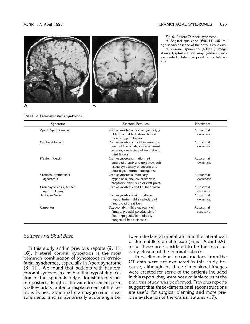
Fig 6. Patient 7: Apert syndrome.A, Sagittal spin-echo (600/11) MR im-
age shows absence of the corpus callosum.B, Coronal spin-echo (600/11) image
shows dysplastic hippocampi (arrows), withassociated dilated temporal horns bilater-ally.
TABLE 2: Craniosynostosis syndromes
Syndrome Essential Features Inheritance
Apert, Apert-Crouzon Craniosynostosis, severe syndactylyof hands and feet, down-turnedmouth, hypertelorism
Autosomaldominant
Saethre-Chotzen Craniosynostosis, facial asymmetry,low-hairline ptosis, deviated nasalseptum, syndactyly of second andthird fingers
Autosomaldominant
Pfeiffer, Noack Craniosynostosis, malformedenlarged thumb and great toe, soft-tissue syndactyly of second andthird digits, normal intelligence
Autosomaldominant
Crouzon, craniofacialdysostosis
Craniosynostosis, maxillaryhypoplasia, shallow orbits withproptosis, bifid uvula or cleft palate
Autosomaldominant
Craniosynostosis, fibularaplasia, Lowry
Craniosynostosis and fibular aplasia Autosomalrecessive
Jackson-Weiss Craniosynostosis with midfacehypooplasia, mild syndactyly offeet, broad great toes
Autosomaldominant
Carpenter Oxycephaly, mild syndactyly offingers, preaxial polydactyly offeet, hypogenitalism, obesity,congenital heart disease
Autosomalrecessive
AJNR: 17, April 1996 CRANIOFACIAL SYNDROMES 625
Sutures and Skull Base
In this study and in previous reports (9, 11,16), bilateral coronal synostosis is the mostcommon combination of synostoses in cranio-facial syndromes, especially in Apert syndrome(3, 11). We found that patients with bilateralcoronal synostosis also had findings of duplica-tion of the sphenoid ridge, foreshortened an-teroposterior length of the anterior cranial fossa,shallow orbits, anterior displacement of the pe-trous bones, abnormal craniozygomatic mea-surements, and an abnormally acute angle be-
tween the lateral orbital wall and the lateral wallof the middle cranial fossae (Figs 1A and 2A);all of these are considered to be the result ofearly closure of the coronal sutures.Three-dimensional reconstructions from the
CT data were not evaluated in this study be-cause, although the three-dimensional imageswere created for some of the patients includedin this report, they were not available to us at thetime this study was performed. Previous reportssuggest that three-dimensional reconstructionsare useful for surgical planning and more pre-cise evaluation of the cranial sutures (17).

Bilateral coronal synostosis is a characteristicfinding in Apert syndrome and may result fromspecific developmental derangements of theskull base. Kreiborg and Cohen (4) andKreiborg et al (17) have reported that coronalsutures are completely fused at birth in Apertsyndrome, despite widely patent sagittal andmetopic sutures. They suggested that this su-tural fusion results from the close approxima-tion of the frontal, sphenoid, and parietal bones,which permits rapid ossification of the cornalarea. In contrast, the gap across the sagittal andmetopic sutures in patients with Apert syn-drome is so wide that it takes a long time forbony islands to form within the gap, coalesce,and, eventually, completely bridge the gap.This difference in sutural closure may resultfrom known anatomic and physiological differ-ences between the midline (sagittal andmetopic) sutures and the off-midline (coronaland lambdoid) sutures (11, 17, 18). Closure ofthe midline sutures is end-to-end, whereas thenonmidline sutures close by overlapping (bev-eled type). In addition, the attachment of thefalx and the dura of the superior sagittal sinus tothe skull vault immediately beneath the sagittalsuture may protect it from the direct remodelinginfluence of elevated intracranial pressure, incontrast to the coronal suture, which directlyoverlies the dura and pulsating brain.The importance of the skull base in the gen-
esis of the calvarial deformities of craniosynos-tosis was first stressed by Moss (19), who pos-tulated that spatial malpositioning of the basalpoints of dural attachment is the primary under-lying anomaly in craniosynostosis; this malpo-sitioning theoretically results in the transmis-sion of aberrant tensile forces upward throughthe dura to produce synostosis of the overlyingsuture. Evidence is accumulating to indicatethat the cartilaginous cranial base is primarilyinvolved in craniofacial anomalies; for example,the synchondroses of the skull base are com-pletely fused at birth in patients with Crouzonsyndrome and show early progressive fusion inApert syndrome (17). However, other reportssuggest that the cranial base malformations are,at least in some part, the result of calvarialsynostosis (20, 21). Whether primary or sec-ondary, it is of interest to note that deformities ofthe skull base were commonly recognized in ourpatients. These anomalies are of clinical impor-tance because of the many vital structures thatpass through the skull base. Cranial neuropa-
626 TOKUMARU
thies can result from hypoplasia of skull baseforamina. As had been described with achon-droplasia, a small skull base can cause hydro-cephalus in infants and increased intracranialpressure in older children and adults (22–25). Itis tempting to postulate that the small skull basewas a factor in the hydrocephalus seen in sev-eral of the patients in this series. Both upwardherniation of the cerebellum through the inci-sura (Fig 5) and downward herniation throughthe foramen magnum (Figs 2, 4, and 5) canresult from a normal-sized cerebellum in a hy-poplastic posterior fossa (26, 27). Deformity ofthe optic canals and superior orbital fissuresmay impair visual acuity by compression of theoptic nerve (as seen in patient 9) or ocularmotility by compression of cranial nerves III, IV,and VI.Only two patients in our series were noted to
have anomalies of the middle ear. However,these two patients had bilateral external audi-tory canal atresia and were the only two patientsin the study in whom dedicated temporal bonestudies were performed. Hearing difficulties arecommon in Apert syndrome and in the othercraniofacial disorders (8), but they are usuallyattributed to the frequent bouts of otitis mediathat result from the high frequency of cleft pal-ate and bifid uvula and the high arching palate(28, 29). Congenital conductive hearing loss,however, is not rare in these disorders (29, 30).The possibility of congenital hearing loss shouldnot be overlooked because of preoccupationwith other more obvious problems in these in-fants. The radiologist can play a key role in thisregard by carefully scrutinizing the middle ear inaffected children.Eight of the patients described in this article
had plagiocephaly, with asymmetric fusion ofthe sutures (see Table 1). This observation con-firms once again that patients with the samesyndrome may have abnormal fusion of differ-ent sutures and that sutural fusion may beasymmetric in the craniofacial syndromes. Thisasymmetry is most apparent in Saethre-Chot-zen syndrome, in which unilateral coronal syn-ostosis is frequently seen (8). However, the factthat asymmetric synostosis is possible in othersyndromes, as well, should be kept in mindduring the examination of these children. It isessential, therefore, to specifically identifywhich suture or sutures are involved in eachpatient.
AJNR: 17, April 1996

AJNR: 17, April 1996 CRANIOFACIAL SYNDROMES 627
Ventricular Enlargement and Hydrocephalus
Ventricular enlargement in craniofacialanomalies is a common finding; however, themechanism of this phenomenon varies and insome cases the pathophysiology is poorly un-derstood (6, 22, 24). Some authors have pos-tulated that hydrocephalus can result from ob-struction of cerebrospinal fluid (CSF) pathwaysat the level of the basal cisterns owing to a smallskull base (31, 32); in the present study, fourpatients had findings of a small skull base withcompression of the basal cisterns. Noetzel et al(33) have suggested that hydrocephalus occursas a result of an intrinsic abnormality in theembryology of the brain related to the defectiveformation of the cranium. Other authors havesuggested that increased resistance to venousoutflow may be an important cause of the en-larged ventricles (22–25). Although we did notdirectly measure venous pressures in this study,the MR venograms dramatically showed dys-plasia or occlusion of the transverse and sig-moid sinuses in the two patients in whom theywere obtained. This finding suggests that ve-nous anomalies, primary venous occlusions orvenous outflow obstruction with secondary ve-nous occlusion, may be a part of these syn-dromes. The differentiation is significant for un-derstanding the venous flow abnormality andinitiating the appropriate management. Verifi-cation of these findings and determination of thecause is an important area for future research.Differentiating benign ventriculomegaly from
hydrocephalus in craniofacial syndromes canbe difficult (6, 24, 34). Progressive hydroceph-alus appears to be more common in Crouzonand Pfeiffer syndromes than in Apert syndromeor other craniofacial anomalies (34, 35). Thefrequent presence of ventriculomegaly and thedifficulty in differentiating it from frank hydro-cephalus is another area in which craniofacialsyndromes are similar to achondroplasia, an-other condition in which the skull base is hypo-plastic. In both disorders, separate tables of“normal” head circumferences have been cre-ated to adjust for the atypical shapes and sizesof infants’ heads (7). In craniofacial syndromes,the diagnosis of hydrocephalus is made bydocumentating progressive enlargement of thelateral ventricles (3, 24). In five of our patients,ventriculomegaly was recognized without asso-ciated enlargement of the anterior recesses ofthe third ventricle and without progression, thus
indicating that frank hydrocephalus was notpresent. The mechanism of the ventriculo-megaly in these children is unknown. It may berelated to the abnormal cranial configuration, acondition that has been called distortion ven-triculomegaly (3, 6). In those patients in whomfrank hydrocephalus is present, it is possiblethat restricted venous outflow through the smallskull base, as has been demonstrated in achon-droplasia (23, 25), plays a role. The restrictedvenous outflow results in increased dural sinusvenous pressure. Because the resorption of CSFinto the dural venous sinuses is believed to relyon a CSF-to-venous sinus pressure gradient,elevated venous pressure reduces the gradientand, consequently, impairs CSF resorption.Our observation of disproportionate enlarge-
ment of the frontal horns of patients with Apertsyndrome supports the theory of distortion ven-triculomegaly. In Apert syndrome, the metopicand sagittal sutures are widely patent at birth; ahuge bony defect is present from the glabella tothe posterior fontanel (4, 17). As discussed inthe previous section, this defect gradually fills inby the formation of islands of membranousbone within it (4, 17). We postulate that thefrontal horns of patients with Apert syndromeare disproportionately large because the ante-rior midline bone defect allows greater expan-sion of the grain in the anterior cranial fossa;consequently, the ventricles in the anterior re-gion can enlarge more than the posterior partsof the ventricles where the smaller calvaria re-stricts growth. This hypothesis is in accordancewith the observations of Hochwald et al (36)and the calculations of Shapiro et al (37) thatventricular size increases if the “container” ofthe brain (the skull or dura) is removed. In thecase of Apert syndrome, localized expansion ofthe skull and dura in the frontal area allowslocalized ventricular enlargement.
Cerebral Abnormalities
Significant cerebral abnormalities—for ex-ample, agenesis of the corpus callosum, hip-pocampal hypoplasia, and septal defects—have been noted in pathologic studies of somepatients with craniofacial anomalies (6, 8, 10,16) and were confirmed by MR imaging in thepatients described in this article. On the basis ofthe limbic system abnormalities identified intheir pathologic studies, Maksem and Roess-mann (38) suggested that the abnormalities of

the brain parenchyma in Apert syndrome areprimary, occurring as early as the sixth week ofembryonic life. Others have postulated that thehippocampal abnormalities are related to dis-tortion of the brain by the calvarial deformity (3,6). Our observation that the septum pellucidumand corpus callosum are commonly anomalousin patients with hippocampal anomalies sup-ports the postulate that the hippocampal anom-aly is a primary abnormality related to malde-velopment of the limbic system. The cause ofthe distortions of the cerebral cortex is less ob-vious. We found that the most severe gyral ab-normalities were in the patients with the mostsevere cranial malformations (Fig 5). Polymi-crogyria has been reported, albeit rarely, incraniofacial anomalies (6); however, mostcases of reported gyral anomalies (6, 39, 40)appear to be primarily the result of distortion ofthe cerebral cortex by the calvarial anomalies,consistent with our observations.Nonprogressive mental retardation is com-
mon in Apert syndrome (3, 24, 41), but thecause is uncertain. Although ventricular dilata-tion is found frequently in Apert syndrome, clin-ical signs suggesting a significant degree of hy-drocephalus are rarely documented, and itseems unlikely that hydrocephalus is responsi-ble for the retardation (3, 41). Mental retarda-tion is often present in patients (without cranio-facial syndromes) with agenesis of the corpuscallosum, probably because of the high fre-quency of associated cerebral malformations(42–44); nevertheless, some patients with cal-losal agenesis are neurologically and intellectu-ally normal (45). Both agenesis of the corpuscallosum (46) and Apert syndrome (6) are of-ten associated with malformations involvinglimbic structures. Because some structures ofthe limbic system, in particular the hippocam-pus, are important in memory, it is tempting tosuggest that the limbic system abnormalitiesare an important component of the intellectualimpairment in both conditions.It is difficult to attribute defects of the spetum
pellucidum and corpus callosum to secondaryevents. Septal thinning and necrosis are knownto be associated with long-standing hydroceph-alus (47); however, as discussed earlier, frankhydrocephalus is uncommon in Apert syn-drome, whereas septal absence is rather com-mon (6). Moreover, callosal agenesis is unques-tionably a primary anomaly. Therefore, ourresults seem to confirm an increased frequency
628 TOKUMARU
of primary brain anomalies, at least in Apert andPfeiffer syndromes.The parenchymal hemorrhage in patient 8
was an interesting finding because it occurred inareas that most commonly hemorrhage in neo-nates as a result of hemorrhagic infarction fromvein of Labbe occlusion (48). MR venographyshowed occlusion of both transverse sinuses. Itis of interest that both infants who had MRvenography in this study had venous anomaliesor occlusions. If, in fact, the large ventricularsize and, when present, hydrocephalus in thesepatients are the result of venous outflow restric-tion through the small vascular foramina of thehypoplastic skull base, venous anomalies andocclusions are not surprising and, perhaps, thevenous sinuses should be examined routinely inaffected patients. MR venography could be auseful method for evaluating venous flow non-invasively both in patients with ventriculo-megaly and in those with parenchymal injury.
Abnormalities of the Orbit
Visual loss is the most serious ophthalmo-logic problem in patients with craniosynostosis.The most frequent causes of visual loss areacquired types of ocular disease, such as opticatrophy and corneal damage, which most com-monly result from abnormalities in the bonyorbit or skull, and amblyopia resulting fromstrabismus or refractive differences between thetwo eyes (3, 10, 16). Thus, it is important toassess the optic nerves, cranial nerves III, IV,and VI, the extraocular muscles, and the orbitalfissures precisely. In our study, optic nerve at-rophy (Fig 2) was commonly detected. Askewed appearance of the superior orbital fis-sure was recognized in one patient on CT scans.In patients with hydrocephalus, MR imaging re-vealed stretching of the optic nerves and chi-asm associated with enlarged anterior recessesof the third ventricle (Fig 2B). Other patientshad evidence of small optic canals, which canresult in pressure atrophy of the nerves. Com-pression of the vascular supply to the opticnerve has been thought to occur in some situ-ations from sudden changes in intracranialpressures (49). Kinking of the optic nerve as ittraverses the distorted skull base may also be afactor, as may prolonged papilledema from in-creased intracranial pressure (16, 50).It is important to analyze the extraocular
muscles themselves in patients with craniofa-
AJNR: 17, April 1996

AJNR: 17, April 1996 CRANIOFACIAL SYNDROMES 629
cial anomalies because ocular motility distur-bances of differing causes frequently occur (8).Deformity of the orbital fissure may result incranial nerve impairment, whereas dysplasia ofthe orbit contributes to disturbed function of themuscles themselves. Gobin (51) and Morax(52) have shown that the hypoplastic maxilla inpatients with craniosynostosis can cause an al-teration of mechanical forces of the inferior ob-lique muscle, enhancing its effect and corre-spondingly diminishing action of the superioroblique muscle. Hypoplasia of the maxilla,which was a common occurrence in this study,is therefore an important finding to report andrepair.Proptosis (Fig 2) is frequently recognized in
craniofacial anomalies and was present in 16 ofthe patients in this series. Proptosis can be pro-duced by many factors in these patients, includ-ing arrested growth of the maxilla, shortenedanterior cranial base, depressed planum sphe-noidale, and forward displacement of thegreater wing of the sphenoid bone. Imagingdoes not significantly contribute to the diagno-sis or management of proptosis.
Summary
To summarize, we have reviewed the cranialimaging findings of 21 patients with a variety ofcraniofacial syndromes. We have analyzed anddiscussed a number of the features of this dis-order and the imaging manifestations of boththe skull and brain. Radiologic assessment is animportant component of the clinical evaluationof affected patients. An understanding of thesedisorders and their basic pathophysiology is es-sential for appropriate management and correctinterpretation of the imaging studies.
References1. Mazzola HK. Congenital malformations in the frontonasal area:
their pathogenesis and classification. Clin Plastic Surg 1976;3:573–609
2. Kawamoto HK, Jr. The kaleidoscopic world of rare craniofacialclefts: order out of chaos (Tessier classification). Clin Plast Surg1976;3:529–572
3. Cohen MM Jr, Kreiborg S. An updated pediatric perspective on theApert syndrome. Am J Dis Child 1993;147:989–993
4. Kreiborg S, Cohen MM Jr. Characteristics of the infant Apert skulland its subsequent development. J Craniofac Genet Dev Biol1990;10:399–410
5. Carr M, Posnick K, Pron G, Armstrong D. Cranio-orbito-zygomaticmeasurements from standard CT scans in unoperated Crouzonand Apert infants: comparison with normal controls. Cleft PalateCraniofac J 1992;29:129–136
6. Cohen MM Jr, Kreiborg S. The central nervous system in the Apertsyndrome. Am J Med Genet 1990;35:36–45
7. Cohen MM Jr, Kreiborg S. Cranial size and configuration in theApert syndrome. J Craniofac Genet Dev Biol 1994;14:153–162
8. Cohen MM Jr. Craniosynostosis: Diagnosis, Evaluation, and Man-agement. New York: Raven, 1986
9. Cohen MM Jr. Craniosynostosis update 1987. Am J Med Genet1988;4(suppl):99–148
10. Bixler D, Ward RE. Craniosynostosis. In: Myrianthopoulos NC, ed.Malformations. New York: Elsevier, 1987:113–128
11. Cohen MM Jr. Sutural biology and the correlates of craniosynos-tosis. Am J Med Genet 1993;47:581–616
12. Barkovich AJ. Congenital malformation: In: Pediatric Neuroimag-ing., 2nd ed. New York: Raven Press 1995:261–267
13. Cohen MM Jr. Pfeiffer syndrome update, clinical subtypes, andguidelines for differential diagnosis. Am J Med Genet 1993;45:300–307
14. Rutland P, Pulleyn L, Reardon W, et al. Identical mutations in theFGFR2 gene cause both Pfeiffer and Crouzon syndrome pheno-types. Nature Genetics 1995;9:173–176
15. Wilkie A, Slaney S, Oldridge M, et al. Apert syndrome results fromlocalized mutations of FGFR2 and is allelic with Crouzon syn-drome. Nature Genetics 1995;9:165–172
16. Warkany J, Lemire RJ, Cohen Jr. MM. Mental Retardation andCongenital Malformations of the Central Nervous System. Chi-cago: Yearbook; 1981:224–243
17. Kreiborg S, Marsh J, Cohen MM Jr, et al. Comparative threedimensional analysis of CT-scans of the calvaria and cranial basein Apert and Crouzon syndromes. J Craniomaxillofac Surg 1993;21:181–188
18. Kokich VG. Biology of sutures. In: Cohen MM Jr, ed. Craniosyn-ostosis: Diagnosis, Evaluation, and management. New York:Raven, 1986:81–103
19. Moss ML. The pathogenesis of premature cranial synostosis inman. Acta Anat (Basel) 1959;37:351–370
20. Persson M, Magnusson B, Thilander B. Sutural closure in rabbitand man: a morphological and histochemical study. J Anat 1978;125:313–321
21. Albright SL, Byrd RP. Suture pathology in craniosynostosis. JNeurosurg 1981;54:384–387
22. Sainte-Rose C, LaCombe J, Peirre-Kahn A, et al. Intracranialvenous sinus hypertension: cause or consequence of hydroceph-alus in infants? J Neurosurg 1984;60:727–736
23. Pierre-Kahn A, Hirsh JF, Renier D, Metzger J, Maroteaux P. Hy-drocephalus and achondroplasia: a study of 25 observations.Childs Brain 1980;7:205–219
24. Murovic JA, Posnick JC, Drake JM, Humphreys RP, Hoffman HJ,Hendrick EB. Hydrocephalus in Apert syndrome: a retrospectivereview. Pediatr Neurosurg 1993;19:151–155
25. Steinbok P, Hall J, Flodmark O. Hydrocephalus in achondropla-sia: the possible role of intracranial venous hypertension. J Neu-rosurg 1989;71:42–48.
26. Emery JL, MacKenzie N. Medullo-cervical dislocation deformity(Chiari II deformity) related to neurospinal dysraphism (myelo-meningocele). Brain 1973;96:155–162
27. McLone DG, Knepper PA. The cause of Chiari II malformation: aunified theory. Pediatr Neurosci 1989;15:1–12
28. Bergstrom L, Neblett L, Hemenway W. Otologic manifestations ofacrocephalosyndactyly. Arch Otolaryngol 1972;96:117–123

29. Gould HJ, Caldarelli DD. Hearing and otopathology in Apert syn-drome. Arch Otolaryngol 1982;108:347–349
30. Phillips S, Miyamoto R. Congenital conductive hearing loss inApert syndrome. Otolaryngol Head Neck Surg 1986;95:429–433
31. Russell DS. Observations on the Pathology of Hydrocephalus.London: His Majesty’s Stationery Office; 1949:1–138
32. Barkovich AJ, Edwards MSB. Applications of neuroimaging inhydrocephalus. Pediatr Neurosurg 1992;18:65–83
33. Noetzel M, Marsh J, Palkes H, Gado M. Hydrocephalus and mentalretardation in craniosynostosis. J Pediatr 1985;107:885–892
34. Golabi M, Edwards MSB, Ousterhout DK. Craniosynostosis andhydrocephalus. Neurosurgery 1987;21:63–67
35. Collman H, Sorensen N, Krauss J, Muhling J. Hydrocephalus incraniosynostosis. Childs Nerv Syst 1988;4:279–285
36. Hochwald GM, Epstein F, Malhan C, et al. The role of skull anddural in experimental feline hydrocephalus. Dev Med Child Neurol1972;14(Suppl 27):65–69
37. Shapiro K, Fried A, Takei F, Kohn I. Effect of the skull and dura onneural axis pressure-volume relationships and CSF hydrodynam-ics. J Neurosurg 1985;63:76–81
38. Maksem JA, Roessmann U. Apert’s syndrome with central ner-vous system anomalies. Acta Neuropathol 1979;48:59–61
39. de Leon GA, de Leon G, Frover WS, Zaeri N, Alburger P. Agenesisof the corpus callosum and limbic malformation in Apert syn-drome (type I acrocephalosyndactyly. Arch Neurol 1987;4:979–982
40. Kreiborg S, Prydsoe U, Dahl E, Fogh-Andersen F. Calvarium andcranial base in Apert’s syndrome: an autopsy report. Cleft PalateCraniofac 1976;13:296–303
41. Lefebvre A, Travis F, Arndt E, Munro IR. A psychiatric profilebefore and after reconstructive surgery in children with Apert’ssyndrome. Br J Plast Surg 1986;39:510–513
630 TOKUMARU
42. Gross H, Jellinger K, Kaltenback E, et al. Zur Morphologie undPathogenese des Balkenmangels. In: Jellinger K, ed. AktuelleProbleme der Neuropathologie. Vienna: Facultas; 1973:227–249
43. Parrish ML, Roessmann U, Levinsohn MW. Agenesis of the corpuscallosum: a study of the frequency of associated malformations.Ann Neurol 1979;6:349–354
44. Barkovich AJ, Norman D. Anomalies of the corpus callosum:correlation with further anomalies of the brain. AJNR Am J Neu-roradiol 1988;9:493–501
45. Loeser JD, Alvord EC. Clinicopathological correlations in agene-sis of the corpus callosum. Neurology 1968;18:745–756
46. Atlas SW, Zimmerman RA, Bilaniuk LT, et al. Corpus callosumand limbic system: neuroanatomic MR evaluation of developmen-tal anomalies. Radiology 1986;160:355–362
47. Barkovich AJ, Norman D. Absence of septum pellucidum: a use-ful sign in the diagnosis of congenital brain malformations. AJNRAm J Neuroradiol 1988;9:1107–1114
48. Barkovich AJ. Destructive brain disorders of childhood. In: Bark-ovich AJ, ed. Pediatric Neuroimaging. 2nd Ed. New York: RavenPress; 1995:107–175
49. Cohen MM Jr. An etiologic and nosologic overview of craniosyn-ostosis syndromes. Birth Defects 1975;11:137–189
50. Gupta S, Ghose S, Rohatgi M, Kumar A, Das A. The optic nerve inchildren with craniosynostosis: a pre and post-surgical evaluation.Doc Ophthalmol 1993;83:271–278
51. Gobin MH. New viewpoints on the pathogenesis and treatment ofstrabismus. Part 1: pathogenesis of strabismus. J Fr Ophthalmol1980;3:541–546
52. Morax S. Oculomotor disorders in craniofacial malformations. JCraniomaxillofac Surg 1984;12:1–10
AJNR: 17, April 1996